32
ASSESSING THE PATIENT WITH VESTIBULAR SYMPTOMS www.otometrics.com

ASSESSING THE PATIENT WITH VESTIBULAR SYMPTOMS
www.otometrics.com

www.otometrics.com
This educational booklet was developed in collaboration with:
Jill Craig M.A. GN Otometrics Business Manager/Audiologist for the Evoked Potential product line
Wendy Crumley-Welsh M.S., CCC-A GN Otometrics Business Manager/Audiologist for the Vestibular product line
Lee Dibble, PT, PhD, ATC Associate Professor Department of Physical Therapy University of UtahSalt Lake City, UT, USA
Dr Jorge Kattah, MD, Neuro-OpthalmologistIllinois Neurological Institute Balance CenterPeoria, IL, USA
Dr Leonardo ManzariSapienza Università di RomaCassino, Italy
Dr. Nicolás PérezConsultor ClínicoDirector del Departamento de Otorrinolaringología CLINICA UNIVERSITARIA DE NAVARRAPamplona, Spain
Priv.-Doz. Dr. med. Holger RamboldHead of the Vestibular and Oculomotor laboratoryCommunity Hospital Altötting, Dept. of NeurologyAltötting, Germany
Prof. Dr. med. Frank SchmälDirector Balance DepartmentENT Center MünsterlandGreven, Germany
Dr David Szmulewicz, MDNeurologist & Neuro-otologistHead of Balance Disorders & Ataxia ServiceRoyal Victorian Eye & Ear HospitalMelbourne, Australia
Dr. med. Konrad P. WeberInterdisciplinary Center for Vertigo and Balance DisordersUniversity Hospital ZürichZürich, Switzerland

3
TABLE OF CONTENTS
INTRODUCTION 4
PERIPHERAL VESTIBULAR DISORDERS 9
BENIGN PAROXYSMAL POSITIONAL VERTIGO (BPPV) 10
VESTIBULAR NEURITIS 11
MENIÈRE’S DISEASE 15
SUPERIOR CANAL DEHISCENCE 18
CENTRAL VESTIBULAR DISORDERS 19
VESTIBULAR MIGRAINE 20
VERTEBROBASILAR ISCHEMIC STROKE 22
VESTIBULAR SCHWANNOMA 24
WERNICKE’S ENCEPHALOPATHY 26
MULTIPLE SCLEROSIS PLAQUES 28
VESTIBULO-CEREBELLAR DISEASE 30
www.otometrics.com
INTRODUCTIONICS Impulse has evolved into more than only a vHIT system. ICS Impulse offers a modular solution allowing you to determine which modules are needed to assess the patients in your clinic. Choose from the Monocular Video Frenzel, Oculomotor module, vHIT Module (Lateral only or Lateral/LARP/RALP) and Positional Module. ICS Impulse with the ability to record eye movement with vision and with vision denied allows you to assess the vestibular system and assists you in determining if the disorder is peripheral or central. ICS Impulse not only records eye and head velocity to assess the VOR system but also records eye position and measures the slow phase velocity of the nystagmus in both real time and in analysis of the data. With Head Position Feedback, ICS Impulse assists in properly positioning the patient during test set-up and data collection. ICS Impulse provides the ability to playback the eye trace, eye video, SPV graph, head position feedback or synchronized room video together synchronously. This allows you to review the data collected at any time and know exactly how your patient was positioned and what occurred during testing.
Understanding the benefits of vHITHead Impulse is a side of lesion specific test that detects a deficiency of the vestibulo-ocular reflex and identifies which ear and which semicircular canal is affected in cases of peripheral vestibulopathy.
How does vHIT compare to caloric testing?HIT• Side of Lesion specific • Detects abnormalities in all six semicircular canals in cases with peripheral vestibular loss
(Lateral, Anterior and Posterior)• Ability to test patients even if they have middle ear disorders• Ability to test patients who do not tolerate calorics (young children, elderly, or patients with
severe hearing loss)• Tests with stimuli replicating the patient’s everyday situations (physiological stimulus) • Stimulus does not persist between tests
Note: A head impulse should not be performed on patients with a neck injury, or on patients who have been toldby their physicians to limit or avoid neck movement activity.
Caloric• Ear-specific • Detects cases of peripheral vestibular loss in Lateral semicircular canal• Tests at Low Frequencies (~0.025 Hz)• Stimulus can persist between irrigations especially if not performed properly• External and middle ear disorder may prohibit performing the test• Some patients will not tolerate caloric testing or will not allow the caloric test to be completed

5
How does Impulse benefit the patient?Patient comfort is greatly enhanced by the lightest goggles in the industry. ICS Impulse allows you to assess the patient for peripheral and central disorders. When performing vHIT, the sophisticated cameras allow for smaller amplitude head impulses of only 15 to 20 degrees. This makes the video head impulse test (vHIT) more pleasant for the patient even in patients with acute vertigo. Unlike caloric testing, vHIT does not result in adverse reactions; therefore, making it easy to perform vHIT on multiple patient visits.
How does Impulse benefit the physician?Easily assess and treat patients who have BPPV. With the full test battery determine if the patient has a peripheral or central disorder. ICS Impulse vHIT assesses all 6 semicircular canals and is the only test to assess the anterior and posterior canals. Impulse is small and portable allowing for patients to be assessed in a clinic and at the bed-side. It is easy to assess anyone of any age who can wear the goggles. Impulse vHIT detects more abnormalities than visual observation and reduces false negatives. Due to the fact that Impulse vHIT does not result in adverse reactions, one can easily assess the patient multiple times (e.g. after vestibular rehabilitation, during drug therapy, etc). If you are not the one collecting the data increase your confidence in the data with the ability to synchronously playback all components of data collection and review at normal speed or in slow motion.
Time savings What time savings can be expected in the clinic with ICS Impulse in the test battery? 2.5 days a month can be saved. Assumption: 10 patients seen per day. Impulse test time = 10 mins; Caloric test time = 30 mins.
Abnormality % of patients with disorder
Patients pr. month Number of calorics saved
Time saved due to fewer calorics
BPPV 40% 80 Not needed
Vestibular neuritis 10% 20 20 7 hours
Menière’s 15% 30 May perform caloric
Vestibular migraine 20% 40 40 13 hours
Other (SCD, Central, perilyph fistula, etc)
15% 30
Total 100% 200 60 20 hours/month
www.otometrics.com
DETERMINING IF THE PATIENT HAS A DISORDER AFFECTING THE VESTIBULAR SYSTEMThis short guide to assessing the patient with a central or peripheral vestibular disorder is intended to be used as a quick reference of the commonly diagnosed disorders and is no way an extensive list. The guide represents the global trend for assessing the patient with vestibular disorders in the most efficient and effective manner. And it emphasizes the diagnostic tools which are most valuable for determining the presence or absence of particular vestibular disorders. This is only a quick guide and is in no way a substitute for your medical training or clinical judgment.
Case History: This is one of the most important steps of assessing the patient. A thorough case history will assist in determining the diagnosis of the patient.
The important information is:• Onset of symptoms: spontaneous, head or visual motion provoked• Temporal course: Is dizziness intermittent or continuous? Does it last seconds,
minutes, hours, days or weeks?• Type of dizziness: objects in room spinning, feeling of spinning in the head,
imbalance, light headedness, disorientation, falls, unsteadiness• Does the patient have signs of a central disorder (e.g. double vision, dysarthria, disturbances
of sensation)?• How does the patient walk? Can they walk? Is their gait normal?• Other symptoms: nausea, vomiting, headaches, motion sickness, intolerance of light,
oscillopsia, heart palpitations, feeling of panic, drop attacks• Hearing: aural fullness, tinnitus (low or high frequency), progressive loss of hearing,
fluctuating hearing loss, sensitive to noise, intolerance of sound• Past medical history: head trauma, back surgery, ototoxic drugs (e.g. gentamicin),
diabetes, perilymphatic fistula
Note: It is necessary to rule out central causes of dizziness (e.g. stroke, traumatic brain injury, cardiovascular disease, neurological disorders (Multiple Sclerosis), anxiety, and side effects from medications or street drugs.)
Test Descriptions and Purpose: These are the common tests for assessing the patient with vestibular disorders. Which tests are performed depends on the results of the case history and the physical exam. It may also depend on your facility’s protocol.
7
Physical Exam (e.g. one minute eye exam): During the one minute eye exam you can have a general idea if the disorder is central or peripheral. Watch for nystagmus and pathological eye oscillations. A physician assesses the patient by asking them to watch their finger as it is moved to assess gaze, smooth pursuit and saccadic eye movement. It should also be determined if the patient has Strabismus (cover test and alternating cover test) or Internuclear opthalmoplegia.
Standard Neurological Exam: During the neurological screening you can have a general idea if the disorder is central or peripheral. This exam is an assessment of the sensory neurons and motor responses, especially reflexes, to determine whether the nervous system is impaired.
Hearing Exam: The assessment of hearing is a major step for the differential diagnosis of peripheral and central vestibulopathies and for the planning of treatment. A pure tone audiogram (with air and bone conducted stimuli) along with tympanometry and acoustic reflexes are minimally needed. The hearing assessment may also include speech testing and auditory evoked potentials. It is essential when determining if a patient has superior canal dehiscence, Menière’s disease, vestibular schwannoma or perilymph fistula.
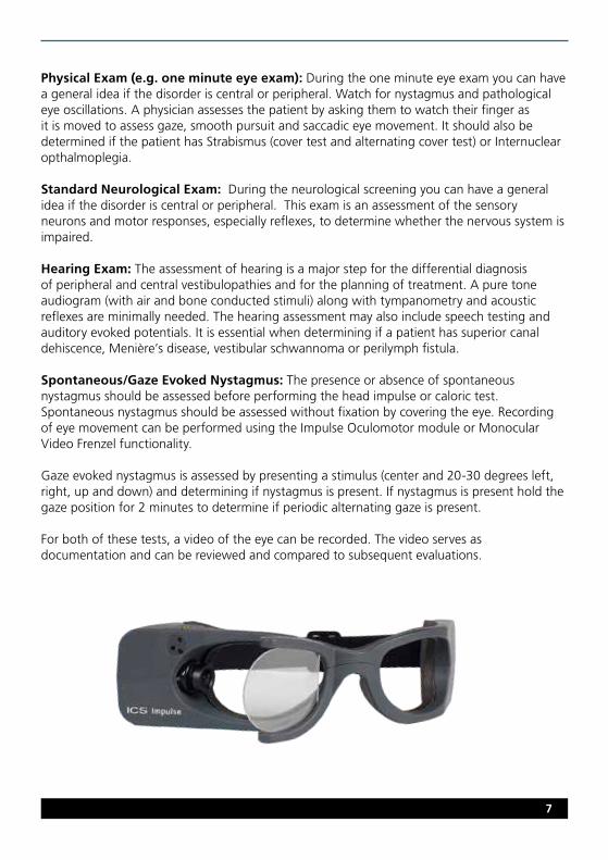
Spontaneous/Gaze Evoked Nystagmus: The presence or absence of spontaneous nystagmus should be assessed before performing the head impulse or caloric test. Spontaneous nystagmus should be assessed without fixation by covering the eye. Recording of eye movement can be performed using the Impulse Oculomotor module or Monocular Video Frenzel functionality.
Gaze evoked nystagmus is assessed by presenting a stimulus (center and 20-30 degrees left, right, up and down) and determining if nystagmus is present. If nystagmus is present hold the gaze position for 2 minutes to determine if periodic alternating gaze is present.
For both of these tests, a video of the eye can be recorded. The video serves as documentation and can be reviewed and compared to subsequent evaluations.

www.otometrics.com
VOR (VVOR & VORS): Visual VOR (vestibulo-ocular reflex) and VOR Suppression identify the presence or absence of saccadic eye movement in order to simultaneously test for the co-existence of vestibular and cerebellar pathology and thus diagnose vestibulo-cerebellar disease. VVOR assesses the patient’s vestibulo-ocular reflex with visual enhancement. VORS assesses the patient’s vestibulo-ocular reflex without visual enhancement.
Skew Deviation: Skew Deviation assesses the patient’s ocular alignment using a cover-uncover test. The purpose of this test is to identify if vertical ocular misalignment occurs as a result of covering and uncovering the eye. A vertical misalignment of the eyes is caused by a right–left imbalance of vestibular neural firing due to damage to the prenuclear vestibular input to the ocular motor nuclei affecting particularly otolithic inputs to the oculomotor system.
Impulse (i.e. head impulse or vHIT): The vHIT test is the only test that assesses all six semicircular canals independently and with a physiological stimulus, similar to how the patient uses the vestibular ocular reflex system in daily life. The test is essential in determining if the peripheral vestibulopathy affects the superior or inferior branch of the nerve, if the loss is unilateral vs bilateral, or only affecting the anterior, posterior or lateral canals. In combination with VEMPs, all 5 end organs for both ears can be assessed.
cVEMP: Cervical Vestibular Evoked Myogenic Potential assesses the saccule using air or bone conduction stimuli. The only test used to easily assess the saccule.
oVEMP: Ocular Vestibular Evoked Myogenic Potential assesses predominantly the utricle using air or bone conduction stimuli. The completely objective test used to easily assess the utricle.
Dix-Hallpike (i.e Hallpike – Stenger) and Lateral positioning: A dynamic positional test that positions the sitting patient with their head turned 45 degrees to the left or right and then quickly moved into a supine position with the head tilted back and slightly lower than the shoulders. The purpose of the maneuver is to provoke the canaloliths to move and stimulate the canal or cupula. This is the only test that can clearly diagnose the presence ofposterior canal or anterior canal BPPV (benign paroxysmal positional vertigo). Other canals must be evaluated with the head hanging and with the head lateral positions. BPPV typically exhibits a crescendo-decrescendo nystagmus observed with a delay of about 10 seconds, with a torsional component to the undermost ear and vertical upbeat component. To diagnose lateral canal BPPV, the patient must lie on their back and turn the head to the left and then to the right.

9
EcochG: Electrocochleography is an electrophysiology test that assists in diagnosing cochlear hydrops by comparing the ratio of the summating potential and the action potential.
Caloric: Bithermal caloric test stimulates the left or right ear with warm and cool air or water causing a fluid density change in the lateral canal. By comparing the response of the left and right ear to warm and cool stimuli one can determine if there is a unilateral or bilateral weakness. Caloric testing is non-physiological stimulus and only assesses the lateral semicircular canal.
Case History Physical Exam Suspect BPPV?
YES
NO
&
Dix-Hallpike
Caloric EcochG
Patient with Peripheral Vestibular Symptoms:
Additional Diagnostic tests: Use dependent on results of the above tests
Hearing ExamSpontaneous/
Gaze Impulse VEMP
PERIPHERAL VESTIBULAR DISORDERS

www.otometrics.com
BENIGN PAROXYSMAL POSITIONAL VERTIGO (BPPV) What: Most common cause of vertigo as a result of canalithiasis (cupulolithiasis is rare). Otoconia detach from the utricle and enter the posterior canal (~80%), the lateral canal (~18%) or the anterior canal (<2 %).
Symptoms: Brief episodes (less than 1 minute) of vertigo caused by rapid changes of head position. Typically caused by bending the head up or down, or rolling over in bed. Vertigo lasts 30 seconds to 2 minutes. May complain of mild postural instability between attacks.
Workflow:
If Dix-Hallpike identifies burst of nystagmus (typically torsional) then subsides when testing is complete this indicates the presence of posterior canal BPPV. If Dix-Hallpike is normal (no nystagmus present), then the patient must lie on their back and turn the head to the left and then to the right. If a horizontal nystagmus in direction of the lower ear occurs (geotrophic) this is a sign for a typical lateral canal BPPV. If a horizontal nystagmus in direction of the upper ear occurs (ageotrophic) this is a sign for an atypical lateral canal BPPV. If no nystagmus occurs, then continue to investigate to see if the patient may have another diagnosis.
Results:Dix Hallpike:BPPV is a typical crescendo-decrescendo nystagmus observed with a delay of about 10 seconds and with a torsional component to the undermost ear and vertical upbeat component.
Impulse: Normal vHIT results confirms a diagnosis of primary BPPV. Abnormal vHIT results suggests that further tests are needed to disclose other disorders and that this is secondary BPPV.
References:
Aw ST, Todd MJ, Aw GE et al (2005) Benign positional nystagmus: A study of its three-dimensional spatio-temporal characteristics. Neurology 64:1897-1905.
Baloh RW, Honrubia V, Jacobson K (1987) Benign positional vertigo: clinical and oculographic features in 240 cases. Neurology 37:371-8.
Baloh RW, Jacobson K, Honrubia V (1993) Horizontal semicircular canal variant of benign positional vertigo. Neurology 43:2542-9.
Baloh RW, Yue Q, Jacobson KM et al (1995) Persistent direction? Changing positional nystagmus: another variant of benign positional nystagmus? Neurology 45:1297-1301.
Perez-Fernandez N, Martinez-Lopez M & Manrique-Huarte R (2014) Vestibulo-ocular reflex in patients with superior semicircular canal benign paroxysmal positional vertigo (BPPV) Acta Oto-Laryngologica. 2014; 134: 485–490.
Case History Physical Exam Dix-Hallpike Repositioning Maneuver

11
VESTIBULAR NEURITISWhat: Acute vestibulopathy caused by inflammation of the inner ear or vestibular nerves. This inflammation disrupts the transmission of the information from the ear to the brain. This is typically viral or degenerative. Can affect the superior or the inferior branch of the vestibular nerve.
Symptoms: Prolonged severe rotational vertigo, head movement worsens the symptoms, postural imbalance to the side of the lesion, nausea, and spontaneous horizontal/torsional nystagmus beating toward the good ear.
Workflow:
If Impulse identifies catch-up saccades and cVEMP or oVEMP is abnormal then test is complete. Catch-up saccades in the lateral or anterior canals and abnormal oVEMP indicate superior vestibular neuritis. Catch-up saccades in the posterior canals and abnormal cVEMP indicate inferior vestibular neuritis.
Additional test to confirm but not necessary:
If Impulse is normal, then continue to investigate to see if the patient may have another diagnosis.
Results:
Spontaneous Nystagmus: Horizontal/torsional nystagmus beating toward the good ear.
Case History
Caloric
Physical Exam Spontaneous Impulse VEMP

www.otometrics.com
oVEMP: Absent response contralateral to the lesion side while stimulating ipsilesional.(It should be noted that in the fields of neurology and neurophysiology convention is to have N1 (e.g. n10) as a upwards deflection i.e. the reverse of what is shown below).
Caloric: Unilateral Weakness.
Impulse: Presence of Catch-up Saccades (covert or overt)and reduced VOR gain.
cVEMP: Reduction of amplitude on affected side.(It should be noted that in the fields of neurology and neurophysiology convention is to have P1 (e.g. p13) as a downward deflection and N1 (i.e. n23) as an upwards deflection i.e. the reverse of what is shown below).

13
Superior Vestibular Neuritis (affects the lateral and anterior canal)
Differentiating Superior and Inferior Vestibular Neuritis
I I I I I I I I I I
I
I
Clinical Test * Healthy Subjects
Superior Vestibular Neuritis
Inferior VestibularNeuritis
Unilateral Vestibular
Loss
Horizontal head turn to ipsilateral horizontal canal ✖ ✖
Pitch head impulse test in the plane of the ipsilateral anterior canal, head turn nose down - tests ipsilateral canal
✖ ✖
oVEMP n10 beneath the contralater-al eye to bone conducted vibration at FZ, or air-conducted sound of one ear - tests utricular macula of the ear opposite to the eye
✖ ✖
cVEMP p13-n23 over ipsilateral sternocleidomastoid (SCM) muscle to bone conducted vibration at Fz, or air-conducted sound of one ear - tests saccular macula of the ear on the same side
✖ ✖
Pitch head impulse in the plane of the ipsilateral posterior canal, head turn nose up - tests ipsilateral posterior canal
✖ ✖
= Normal Response
Horizontal canal ampulla
VestibularDivisionVIII
nerve
Inferior
Superior
CochlearDivision
Anteriorcanal
ampulla
cochlea
Posterior canal
ampulla
Saccular macula
“shank”
“hook”
Utricular macula
= Abnormal Response✖
*) Ian S. Curthoys, PhDThe Interpretation of Clinical Tests of Peripheral Vestibular FunctionThe Laryngoscope: Volume 122, Issue 6, pages 1342–1352, June 2012

www.otometrics.com
References:
Akin FW, Murnane OD, Panus PC, Caruthers SK, Wilikinson AE & Proffitt TM (2004) The influence of voluntary tonic EMG level on the vestibular evoked myogenic potential. J Rehab Res Dev 41(3B):473-480.
Aw ST, Fetter M, Cremer PD, Karlberg M, Halmagyi GM. Individual semicircular canal function in superior and inferior vestibular neuritis. Neurology 2001;57:768–774.
Curthoys IS, Iwasaki S, Chihara Y, Ushio M, McGarvie LA & Burgess A. The ocular vestibular-evoked myogenic potential to air-conducted sound; probable superior vestibular nerve origin. Clinical Neurophysiology 122 (2011) 611–6.
Govender S, Rosengren SM, Colebatch JG. Vestibular neuritis has selective effects on air- and bone-conducted cervical and ocular vestibular-evoked myogenic potentials. Clin Neurophysiol 2011;122:1246–1253.
Halmagyi GM, Weber KP, Curthoys IS. Vestibular function after acute vestibular neuritis. Restor Neurol Neurosci 2010;28:37–46.
MacDougall HG, Weber KP, McGarvie LA, Halmagyi GM, Curthoys IS. The video head impulse test: diagnostic accuracy in peripheral vestibulopathy. Neurology 2009;73:1134–1141.
Manzari L, Tedesco AR, Burgess AM, Curthoys IS. Ocular vestibular evoked myogenic potentials to bone conducted vibration in superior vestibular neuritis show utricular function. Otolaryngol Head Neck Surg 2010;143:274–280.
Manzari L, Burgess AM, MacDougall HG, Curthoys IS. Objective verification of full recovery of dynamic vestibular function after superior vestibular neuritis. Laryngoscope 2011;121:2496–2500.
Manzari L, Burgess AM, Curthoys IS. Ocular and cervical vestibular evoked myogenic potentials to bone conducted vibration in patients with probable inferior vestibular neuritis. J Laryngol Otol In press.
Manzari L, MacDougall HG, Burgess AM, Curthoys IS. New, fast, clinical vestibular tests identify whether a vertigo attack is due to early Meniere’s disease or vestibular neuritis. Laryngoscope 2009: DOI: 10.1002/lary.23479
Pedro Luiz Mangabeira Albernaz & Francisco Carlos Zuma E Maia The video head impulse test. Acta Oto-Laryngologica. 2014; 134: 1245–1250
Pedro L Mangabeira Albernaz & Flavia Salvaterra Cusin The Video Head Impulse Test in a Case of Suspected Bilateral Loss of Vestibular Function Int Arch Otorhinolaryngol Case Report DOI http://dx.doi.org/10.1055/s-0034-1395999
Monstad P, Okstad S, Mygland A. Inferior vestibular neuritis: 3 cases with clinical features of acute vestibular neuritis, normal calorics but indications of saccular failure. BMC Neurol 2006;6:45.
Shin B-S, Oh S-Y, Kim JS, et al. Cervical and ocular vestibular-evoked myogenic potentials in acute vestibular neuritis. Clin Neurophysiol 2012;123:369–375.
Todd NPM, Rosengren SM, Aw ST & Colebatch JG (2007) Ocular vestibular evoked myogenic potentials (oVEMPs) produced by air- and bone-conducted sound. Clin Neurophys 118:381-390.
Weber KP, Aw ST, Todd MJ, McGarvie LA, Curthoys IS, Halmagyi GM. Head impulse test in unilateral vestibular loss: vestibulo-ocular reflex and catch-up saccades. Neurology 2008;70:454–463.
Weber KP, MacDougall HG, Halmagyi GM, Curthoys IS. Impulsive testing of semicircular canal function using video-oculography. Ann NY Acad Sci 2009;1164:486–491.
Zhou G & Cox LC (2004) Vestibular evoked myogenic potentials: history and overview 13(2):135-43.
Inferior Vestibular Neuritis (affects the posterior canal)

15
MENIÈRE’S DISEASEWhat: Due to the presence of inner ear hydrops. Can be due to cochlear hydrops (causing hearing loss) or vestibular hydrops (causing vestibular symptoms).
Symptoms: According to the American Academy of Otolaryngology, Menière’s disease describes a set of episodic symptoms including vertigo (attacks of a spinning sensation), hearing loss, tinnitus (a roaring, buzzing, or ringing sound in the ear), and a sensation of fullness in the affected ear. Episodes typically last from 20 minutes up to 4 hours. Hearing loss is often intermittent, occurring mainly at the time of the attacks of vertigo. Loud sounds may seem distorted and cause discomfort. Usually, the hearing loss involves mainly the lower pitches, but over time this often affects tones of all pitches. After months or years of the disease, hearing loss often becomes permanent. Tinnitus and fullness of the ear may come and go with changes in hearing, occur during or just before attacks, or be constant.
Workflow:
If Impulse is abnormal with increased gain and cVEMP has reduced amplitude then test complete.
Additional tests to confirm:
If Impulse is normal, then continue to investigate to see if the patient may have another diagnosis.
Case History
EcochG
Physical Exam
Caloric
Hearing Exam Impulse VEMP

www.otometrics.com
Results:
Audiogram: Ipsilateral sensorineural hearing loss with involvement in the low frequencies.
Caloric: Unilateral Weakness
Impulse: Increased gain during attack.
Electocochleography: SP/AP ratio abnormal
cVEMP: reduction of amplitude on affected side.
oVEMP: Increased amplitude as compared to normals (about 5–6 uV).
17
References:
Cal R & Bahmad Jr F. Vestibular Evoked Myogenic Potentials: an overview. Braz J Otorhinolaryngol. 2009:75(3):456-462.
De Waele C, Huy PT, Diard JP et al. Saccular dysfunction in Menière’s disease. Am J Otol. 1999;2 0(2):223-32.
Manzari L, Burgess AM, MacDougall HG, Bradshaw AP & Curthoys IS. Rapid fluctuations in dynamic semicircular canal function in early Menière’s disease. Eur Arch Otolaryngol 2010:DOI 10.1007/s00405-010-1442-5.
Manzari L, MacDougall HG, Burgess AM, Curthoys IS. New, fast, clinical vestibular tests identify whether a vertigo attack is due to early Menière’s disease or vestibular neuritis. Laryngoscope 2009: DOI: 10.1002/lary.23479
Pedro Luiz Mangabeira Albernaz & Francisco Carlos Zuma E Maia The video head impulse test. Acta Oto-Laryngologica. 2014; 134: 1245–1250
Rauch SD, Zhou G, Kujawa SG, Guinan JJ, Herrmann BS. Vestibular evoked myogenic potentials show altered tuning in patients with Menière’s disease. Otol Neurotol. 2004; 25(3):333-8.
Wen M-H, Cheng P-W, Young Y-H. Augmentation of ocular vestibular evoked myogenic potentials via bone-conducted vibration stimuli in Menière’s Disease. Otolaryngol Head Neck Surg. DOI: 10.1177/ 0194599811433982.

www.otometrics.com
oVEMP: Increased amplitude as compared to normals and reduced threshold response contralateral to the lesion side while stimulating ipsilesional.
SUPERIOR CANAL DEHISCENCE (THIRD WINDOW PATHOLOGY)
What: Sound and/or pressure-induced vertigo due to dehiscence (thinning) of bone overlying the superior semicircular canal.
Symptoms: Chronic dysequilibrium, oscillopsia (sensation that objects are moving), conductive hyperacusis, sound and/or pressure induced eye movements.
Workflow:
If cVEMP has abnormally low threshold in involved ear (< 80 dB), if oVEMP has increased amplitude and abnormally low threshold in the involved ear then test is complete and a CAT scan should be performed to confirm existence of superior canal dehiscence. If VEMP is normal, then continue to investigate to see if the patient may have another diagnosis.
Results:
Hearing Exam: Bone thresholds better than air thresholds and acoustic reflexes are present.
cVEMP: Abnormally low threshold in involved ear (< 80 dB).
Case History Physical Exam Hearing Exam VEMP

19
References:
Chien WW, Carey JP & Minor LB Canal dehiscence 2011, 24:25-31.
Manzari L, Burgess AM, McGarvie LA, Curthoys IS. Ocular and cervical vestibular-evoked myogenic potentials to 500Hz Fz bone conducted vibration in superior semicircular canal dehiscence. Ear Hear In press.
Manzari L, Burgess AM, MacDougall HG, Curthoys IS. Enhanced otolithic function in semicircular canal dehiscence. Acta Otolaryngol 2011;131:107–112.
Minor LB, Cremer PD, Carey JP, Della Santina CC, Streubel S-O, Weg N. Symptoms and signs in superior canal dehiscence syndrome. Ann NY Acad Sci 2001;942:259–273.
Rosengren SM, Aw ST, Halmagyi GM, Todd NP, Colebatch JG. Ocular vestibular evoked myogenic potentials in superior canal dehiscence. J Neurol Neurosurg Psychiatry 2008;79:559–568.
Streubel SO, Cremer PD, Carey JP, et al. Vestibular-evoked myogenic potentials in the diagnosis of superior canal dehiscence syndrome. Acta Otolaryngol Suppl 2001;545:41–49.
CENTRAL VESTIBULAR DISORDERS
Case History
&Hearing Exam VEMP
Additional Diagnostic tests: Use dependent on results of the above tests
Patient with Central Vestibular Symptoms:
ImpulseSkew Deviation
Physical Exam Gaze/Spontaneous
VOR (VVOR/VORS)
Neurological Exam
www.otometrics.com
VESTIBULAR MIGRAINE What: Episodic vertigo or dizziness with or without migraineous headaches or migraineous symptoms in patients with a diagnosed migraine (according to the criteria of the International Headache Society). Exclusion of other disease. Common in children and adults.
Symptoms: Vertigo (often positional) or dizziness associated with migraineous symptoms as phonophobia (intolerance of sound), photophobia (intolerance of light), migraineous aura (scintillating scotoma, aphasia, etc.), motion sensitivity, tinnitus (high pitched). Migraineous headache might be present (in about 30% of patients no headaches). High comorbidity of anxiety and depression. Sometimes central ocular motor or vestibular signs. Workflow: This is an exclusion diagnosis
If all other diagnoses have been ruled out, spontaneous nystagmus or gaze nystagmus exists with and without fixation, Impulse may be normal or exhibit peripheral vestibular deficits. VEMP will typically be normal. If Gaze is normal or VEMP is abnormal, then continue to investigate to see if the patient may have another diagnosis.
Results:
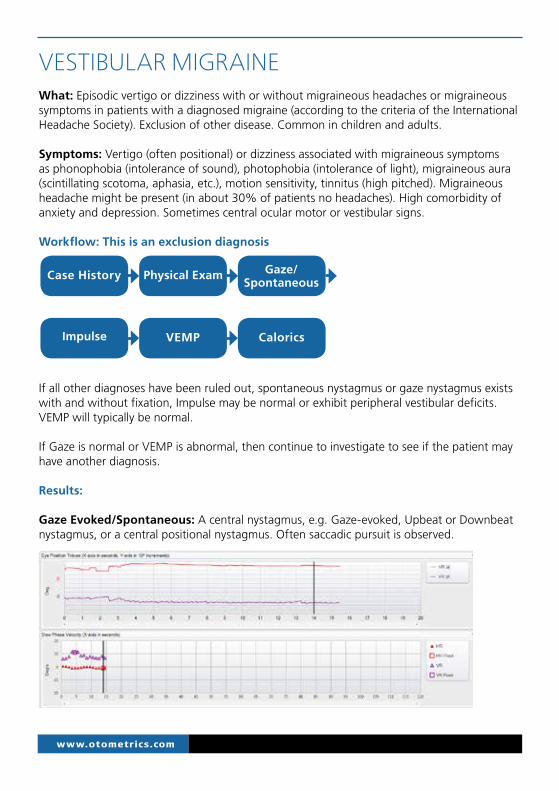
Gaze Evoked/Spontaneous: A central nystagmus, e.g. Gaze-evoked, Upbeat or Downbeat nystagmus, or a central positional nystagmus. Often saccadic pursuit is observed.
Case History Physical Exam Gaze/Spontaneous
Impulse VEMP Calorics

21
VEMP: within normal limits (some literature has reported a reduction in amplitude).
cVEMP
oVEMP
References:
Baloh RW. Neuro-otology of migraine. Headache 1997;37(10):615–621.
Brantberg K, Trees N, Baloh RW. Migraine associated vertigo. Acta Otolaryngol 2005;125:276–279.
Cohen JM, Bigal ME, & Newman LC Migraine and Vestibular Symptoms—Identifying clinical features that predict “Vestibular Migraine”. Headache: J Head Face Pain 51(9):1393-7.
Pedro Luiz Mangabeira Albernaz & Francisco Carlos Zuma E Maia The video head impulse test. Acta Oto-Laryngologica. 2014; 134: 1245–1250
Von Brevern M, Radtke A, Clarke AH, Lempert T. Migrainous vertigo presenting as episodic positional vertigo. Neurology 2004; 62:469–472.
Impulse: Typically the response will be normal but peripheral vestibular deficits may be observed and result in the presence of Catch-up Saccades (covert or overt).

www.otometrics.com
VERTEBROBASILAR ISCHEMIC STROKE What: Reduction of blood flow to the brainstem, cerebellum and inner ear.
Symptoms: Symptoms of vertebrobasilar ischemic stroke are variable and depend on which branch of the artery is affected. Posterior & superior inferior cerebellar artery can result in Wallenberg’s syndrome resulting in dysphagia, difficulty swallowing, spontaneous vertigo, nystagmus, trouble walking, gait disturbance, ipsilateral limb ataxia and facial pain or numb-ness, contralateral body numbness, hoarseness, constricted pupil, droopy eyelid, decreased sweating. Anterior inferior cerebellar artery symptoms similar to the above mentioned but with facial nerve paralysis, tinnitus and recent onset of hearing loss.
Sign of Stroke: INFARCT: Impulse Normal, Fast phase Alternating nystagmus (spontaneous nystagmus changing direction with gaze), Refixation on Cover Test (skew deviation).
Workflow:
Results:Audiogram: Ipsilateral sensorineural hearing loss.
Gaze Evoked/Spontaneous: A central nystagmus. e.g. Gaze Evoked, typically direction changing nystagmus.
Case History
Impulse
Skew Deviation Physical Exam Gaze/
Spontaneous Neurological
Exam
Additional Diagnostic tests: Use dependent on results of the above tests.
VOR (VVOR/VORS)
Hearing Exam

23
VOR: If cerebellar damage exists there may be the presence of catch-up saccades to head movement at slow velocities.
Skew Deviation: Average Eye Position Shift of greater than 1 degree when eye is covered and uncovered denoting ocular misalignment may occur depending on where the damage resides.
Impulse: Typically the response will be normal but some research has shown low gain and catch-up saccades can occur.
References:
Brodsky MC, Donahue SP, Vaphiades M, Brandt T. Skew deviation revisited. Surv Ophthalmol. 2006;51:105–128.
Chen L, Todd M, Halmagyi GM et.al. Head impulse gain and saccade analysis in pontine cerebellar stroke and vestibular neuritis. Neurol-ogy 2014; 83:1-10
Mantokoudis G, Saber Tehrani AS, Wozniak A et.al. VOR gain by head impulse video-oculography differentiates acoute vestibular neuritis from stroke. Otol & Neurotol 2014
Newman-Toker DE, Kattah JC, Alvernia JE & Wang DZ. Normal head impulse test differentiates acute cerebellar stroke from vestibular neuritis. Neurology 2008;70:2378-2385.
Newman-Toker DE, Saber Tehrani AS, Mantokoudis G et.al. Quantitative video-oculography to help diagnose stroke in acute vertigo and dizziness : Toward an ECG for the eye. Stoke 2013; April DOI:10.1161/StrokeAHA.111.000014.
Vibert D, Hausler R, Safran AB, et al. Diplopia from skew deviation in unilateral peripheral vestibular lesions. Acta Otolaryngol 1996;116:170-6.
www.otometrics.com
VESTIBULAR SCHWANNOMA What: Benign, slow-growing tumor that originates from the Schwann cells of the vestibular portion of VIII cranial nerve.
Symptoms: Symptoms are episodic or positional vertigo, disequilibrium, tinnitus, and usually asymmetric hearing loss. If the tumor is pressing on the facial nerve then facial weakness may occur.
Workflow:
Results:
Audiogram: Ipsilateral sensorineural hearing loss.
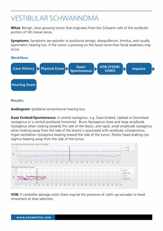
Gaze Evoked/Spontaneous: A central nystagmus. e.g. Gaze Evoked, Upbeat or Downbeat nystagmus or a central positional horizontal. Bruns Nystagmus (slow and large amplitude nystagmus when looking towards the side of the lesion, and rapid, small amplitude nystagmus when looking away from the side of the lesion) is associated with vestibular schwannoma. Hyper-ventilation nystagmus beating toward the side of the tumor. Paretic head-shaking nys-tagmus beating away from the side of the tumor.
VOR: If cerebellar damage exists there may be the presence of catch-up saccades to head movement at slow velocities.
Hearing Exam
Case History Physical Exam Gaze/Spontaneous
VOR (VVOR/VORS) Impulse

25
Impulse: Catch-up saccades present during vHIT and may occur on the ipsilesional side and the contralesional side may also exhibit an abnormal response.
References:
Batuecas-Caletrio A, Santa Cruz-Ruiz S, Munoz-Herrera A, & Perez-Fernandez N (2015) The Map of Dizziness in Vestibular Schwannoma Laryngoscope. 2015 Jun 18. doi: 10.1002/lary.25402.
Batuecas-Caletrio A, Santacruz-Ruiz S, Munoz-Herrera A, Perez-Fernandez N. The vestibulo-ocular reflex and subjective balance after vestibular schwannoma surgery. Laryngoscope 2013.
Mantokoudis G, Schubert MC, Saber Tehrani AS, et.al. Early adaptation and compensation of clinical vestibular responses after unilateral vestibular deaffrentation surgery. Otol & Neurotol 2013
Minor LB, Haslwanter T, Straumann D, Zee DS. Hyperventilation-induced nystagmus in patients with vestibular schwannoma. Neurology 1999:53:2158-2168

www.otometrics.com
WERNICKE’S ENCEPHALOPATHY What: Reduction of vitamin-B reserves in particular thiamine (B1) resulting in biochemical lesions of the central nervous system. It can be associated with alcohol misuse or malnutrition.
Symptoms: The classic triad is oculomotor abnormalities, cerebellar dysfunction and confu-sion but some patients may not express all 3 symptoms. Other signs of thiamine deficiency: weight loss in past year, poor nutrition with high carbohydrate intake, recurrent episodes of vomiting in past month, loss of appetite, fatigue, weakness, double vision, giddiness, insom-nia, anxiety, difficulty concentrating, and memory loss.
Workflow:
Results:
Gaze Evoked/Spontaneous: A central nystagmus. e.g. Gaze Evoked, typically central positional horizontal nystagmus bilaterally sometimes vertical nystagmus.
VOR: If cerebellar damage exists there may be the presence of catch-up saccades to head movement at slow velocities and failure of VOR suppression.
ImpulseCase History Physical Exam Gaze/Spontaneous
VOR (VVOR/VORS)

27
Impulse: Catch-up saccades present during vHIT and may affect all 6 canals. Abnormal vHIT recovers after high dose of intravenous thiamine was administered. *Figure contained in the below article.
References:
*Kattah JC, Dhanani SS, Pula JH et.al. Vestibular signs of thiamine deficiency during the early phase of suspected Wernicke encephalopathy. Neurology Clinical Practice Dec 2014:460-467.
Szmulewicz DJ, Waterson JA, MacDougall et al. Cerebellar ataxia, neuropathy, vestibular areflexia syndrome (CANVAS): a review of the clinical features and video-oculographic diagnosis. Ann NY Acad Sci 2011; 1233:139.
www.otometrics.com
MULTIPLE SCLEROSIS PLAQUES What: an autoimmune disorder of the central nervous system caused by plaques or lesions in the white matter of the brain and spinal cord. Demyelinating plaques in and around the 8th nerve fascicle or vestibular nuclei have been identified as causing vestibular symptoms.
Symptoms: Varies depending on where the plaques occur. Typically loss of sensitivity or changes in sensation such as tingling, pins and needles or numbness, muscle weakness, pronounced reflexes, muscle spasms, or difficulty in moving; difficulties with coordination and balance (ataxia); problems with speech or swallowing, nystagmus and double vision.
Workflow:
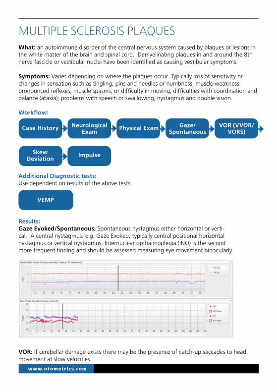
Results:Gaze Evoked/Spontaneous: Spontaneous nystagmus either horizontal or verti-cal. A central nystagmus. e.g. Gaze Evoked, typically central positional horizontal nystagmus or vertical nystagmus. Internuclear opthalmoplegia (INO) is the second more frequent finding and should be assessed measuring eye movement binocularly.
VOR: If cerebellar damage exists there may be the presence of catch-up saccades to head movement at slow velocities.
Case History
ImpulseSkew Deviation
Physical Exam Gaze/Spontaneous
VOR (VVOR/VORS)
Neurological Exam
Additional Diagnostic tests: Use dependent on results of the above tests.
VEMP

29
Skew Deviation: Acute INO can result in skew deviation. Average Eye Position Shift of greater than 1 degree when eye is covered and uncovered denoting ocular misalignment.
Impulse: Catch-up saccades present during vHIT and may affect all 6 canals. Note this data collected on a patient was key to the diagnosis of MS because the right lateral gain was bet-ter than the anterior and posterior gain.
VEMP: Absent or prolonged latency if there is brainstem involvement. May complement or clarify other findings. Ocular VEMP has been reported to show a high frequency of abnormality.
References:
Barona-Lleo L, Zulueta-Santos C, Murie-Fernandez M, Pérez-Fernández N. Recent onset disequilibrium mimicking acute vestibulopathy in early multiple sclerosis Am J Otolaryngol. 2014 Jul-Aug;35(4):529-34.
Gabelić T , Krbot Skorić M, Adamec I, Barun B, Zadro I, Habek M. The vestibular evoked myogenic potentials (VEMP) score: a promising tool for evaluation of brainstem involvement in multiple sclerosis. Eur J Neurol. 2015 Feb;22(2):261-e21. doi: 10.1111/ene.12557. Epub 2014 Sep 8.
Gazioglu S, Boz C Ocular and cervical vestibular evoked myogenic potentials in multiple sclerosis patients. Clin Neurophysiol. 2012 Sep;123(9):1872-9. doi: 10.1016/j.clinph.2012.01.022. Epub 2012 Mar 12.
Güven H, Bayır O, Aytaç E, Ozdek A, Comoğlu SS, Korkmaz H. Vestibular-evoked myogenic potentials, clinical evaluation, and imaging findings in multiple sclerosis. Neurol Sci. 2014 Feb;35(2):221-6. doi: 10.1007/s10072-013-1483-9. Epub 2013 Jun 27.
Ivanković A, Nesek Mađarić V, Starčević K, Krbot Skorić M, Gabelić T, Adamec I, Habek M. Auditory evoked potentials and vestibu-lar evoked myogenic potentials in evaluation of brainstem lesions in multiple sclerosis. J Neurol Sci. 2013 May 15;328(1-2):24-7. doi: 10.1016/j.jns.2013.02.005. Epub 2013 Mar 13.
Pula JH, Newman-TokerDE, Kattah JC. Multiple sclerosis as a cause of the acute vestibular syndrome. J Neurol 2013;260:1649–54.

www.otometrics.com
VESTIBULO-CEREBELLAR DISEASE What: Conditions with the compound deficit of vestibular and cerebellar pathology:
• Spinocerebellar ataxia 3 & 6• Friedreich’s ataxia• Cerebellar ataxia with neuropathy and vestibular areflexia syndrome• Multiple system atrophy predominantly of the cerebellar subtype• Idiopathic cerebellar ataxia with bilateral vestibulopathy
Symptoms: Gait imbalance, dysesthesia, oscillopsia, dizziness and intrinsic falls.
Workflow:
Results:
Gaze Evoked/Spontaneous: A central nystagmus. e.g. Gaze Evoked, typically central posi-tional horizontal nystagmus and downbeat nystagmus. Often saccadic pursuit is observed.
VOR: Presence of catchup-saccades to head movement at slow velocities and failure of VOR suppression.
Case History
ImpulseVOR (VVOR/VORS)
Physical Exam Gaze/Spontaneous
Neurological Exam

31
Impulse: Catch-up saccades present during vHIT and may affect all 6 canals.
References:
Szmulewicz DJ, Waterson JA, MacDougall et al. Cerebellar ataxia, neuropathy, vestibular areflexia syndrome (CANVAS): a review of the clinical features and video-oculographic diagnosis. Ann NY Acad Sci 2011; 1233:139.
Migliaccio A, Halmagyi G, McGarvie L et al. Cerebellar ataxia with bilateral vestibulopathy: description of a syndrome and its characteristic clinical sign. Brain 2004 127;280.

GN Otometrics, Headquarters. +45 45 75 55 55. [email protected] GN Otometrics, North America. 1-800-289-2150. [email protected]
www.otometrics.com
Meet us online to learn more about our thinking, ideas, solutions and the
way in which we support you in your endeavours. We’re always ready for
and welcome a dialogue.
www.icsimpulse.com
www.headimpulse.com
facebook.com/otometrics
twitter.com/otometrics
ICS - the leader in vestibular testing
ICS is a leading global provider of diagnostic devices for balance dis-
orders. Founded in 1981, the company has a history of developing
ground-breaking products that provide pinpoint accuracy for balance
testing. ICS is an expert brand of GN Otometrics.
Sp
eci
fica
tio
ns
are
su
bje
ct t
o c
ha
ng
e w
ith
ou
t n
oti
ce.
Co
pyr
igh
t ©
GN
Oto
me
tric
s. 2
01
5/1
1.
7-2
6-2
09
0-E
N/0
7.
Pa
rt n
o.
7-2
6-2
09
00
-EN
.


