+ All Categories
Home > Documents > Assessment of Disintegration and Dissolution of Dosage Forms In Vivo Using Gamma Scintigraphy

Assessment of Disintegration and Dissolution of Dosage Forms In Vivo Using Gamma Scintigraphy

Date post: 08-Dec-2016
Category:
Upload: neena
View: 217 times
Download: 0 times
Share this document with a friend
71
DRUG DEVELOPMENT AND INDUSTRIAL PHARMACY, 14(2&3), 211-281 (1988) ASSESSMENT OF DISINTEGRATION AND DISSOLUTION OF DOSAGE FORMS IN VNO USING GAMMA SCINTIGRAPHY Clive G. Wilson and Neena Washington Department of Physiology and Pharmacology, Queen's Medical Centre, Nottingham, NG7 2UH. U.K. The measurements of the in uitro rate of disintegration and dissolution of dosage forms are considered to be the most available predictors of the behaviour of dosage forms and the plasma concentration - time profile. However, the interaction of the formulation with physiological processes has shown that prediction of bioavailability by such simple tests is inadequate and has highlighted the need to establish methodology which would enable the determination of in uiuo rates of dissolution and disintegration. Over the past ten years, the technique of gamma scintigraphy has made a significant contribution to the understanding of the behaviour of formulations in the body. This review provides an overview of the technique and its advantages and limitations in 211 Copyright 0 1988 by Marcel Dekker, Inc. Drug Development and Industrial Pharmacy Downloaded from informahealthcare.com by University of Notre Dame Australia on 05/14/13 For personal use only.
Transcript

DRUG DEVELOPMENT AND INDUSTRIAL PHARMACY, 14(2&3), 211-281 (1988)

ASSESSMENT OF DISINTEGRATION AND DISSOLUTION OF

DOSAGE FORMS IN VNO USING GAMMA SCINTIGRAPHY

Clive G. Wilson and Neena Washington

Department of Physiology and Pharmacology, Queen's Medical

Centre, Nottingham, NG7 2UH. U.K.

The measurements of the in uitro rate of disintegration and

dissolution of dosage forms are considered to be the most available

predictors of the behaviour of dosage forms and the plasma

concentration - time profile. However, the interaction of the

formulation with physiological processes has shown that prediction

of bioavailability by such simple tests is inadequate and has

highlighted the need t o establish methodology which would enable

the determination of in uiuo rates of dissolution and disintegration.

Over the past ten years, the technique of gamma scintigraphy has

made a significant contribution t o the understanding of the

behaviour of formulations in the body. This review provides an

overview of the technique and its advantages and limitations in

211

Copyright 0 1988 by Marcel Dekker, Inc.

Dru

g D

evel

opm

ent a

nd I

ndus

tria

l Pha

rmac

y D

ownl

oade

d fr

om in

form

ahea

lthca

re.c

om b

y U

nive

rsity

of

Not

re D

ame

Aus

tral

ia o

n 05

/14/

13Fo

r pe

rson

al u

se o

nly.

2 12 WILSON AND WASHINGTON

pharmaceutical research, together with illustrations showing some

of the applications in the measurement of disintegration and

dissolution of dosage forms.

The bioavailability of a drug from a formulation is influenced by a

complex interplay of physiological and physicochemical factors;

however it is accepted that the primary determinant of absorption is

the rate at which drug is released from the formulation into solution.

This, in turn, is determined by the rate of disintegration of the dosage

form, which increases the surface area and hence the amount of

drug exposed t o the medium. The drug must dissolve in the

gastrointestinal fluids to be absorbed and hence the the absorption of

many drugs, especially those with poor water solubility, is

dissolution rate-limited.

The ability to control of the rate of presentation of a drug and

achieve a desired in uiuo behaviour, by manipulation of excipients in

the formulation, became a major tool in formulation development

and generated the need for in uitro tests which would allow the

effects of manufacturing variables to be studied. The knowledge that

the pH of body fluids changed along the gastrointestinal tract from

stomach to colon increased the need for sophistication of the tests and

attempts were made to begin t o simulate in uiuo conditions. The

majority of drugs are weak acids or bases and the dissolution is

therefore dependent upon the pH of the gut fluid. There is

considerable variation in the pH within the gastrointestinal tract,

Dru

g D

evel

opm

ent a

nd I

ndus

tria

l Pha

rmac

y D

ownl

oade

d fr

om in

form

ahea

lthca

re.c

om b

y U

nive

rsity

of

Not

re D

ame

Aus

tral

ia o

n 05

/14/

13Fo

r pe

rson

al u

se o

nly.

DISINTEGRATION AND DISSOLUTION USING GAMMA SCINTIGRAPHY 213

and most physiology texts state that the gastric pH is in the range 1 - 3, with a pH of 5 - 6 in the duodenum, increasing to 7 - 8 in the

proximal jejunum and 8 in the large intestine. However, there is

some evidence that the pH of the fasting stomach in man may be

much higher (Kuna, 1964)

The muscular actions of the gastrointestinal tract stir and

agitate the preparation during its transit, thus a paddle was

incorporated into the dissolution apparatus to break-up the stagnant

diffusion layers of fluid. Levy (1963) found that agitation of tablets in

the stomach as observed by x-rays was mild and his observations

were used to decide stirring conditions for in uitro dissolution tests.

Further refinements include conducting the test at body temperature

and the addition of digestive enzymes and surfactants such as

pepsin, bile salts and lecithins, since these have been shown to affect

in uitro dissolution (Mayersohn, 1979).

The ability t o determine drug levels in body fluids enabled

researchers to examine the effect of formulation variables on

bioavailability. It soon became clear that the application of simple in uitro tests was inadequate to explain the behaviour of some

preparations. The dissolution of a dose form and the release of a

drug in some instances does not correlate with the absorption of the

drug into the systemic circulation (Toothaker and Welling, 1980).

The application of various designs of in vitro apparatus to simulate

absorption was largely unsuccessful and investigators turned to

other methods of trying to explain the relationship between the

release characteristics of a formulation and the plasma

concentration-time profile.

Dru

g D

evel

opm

ent a

nd I

ndus

tria

l Pha

rmac

y D

ownl

oade

d fr

om in

form

ahea

lthca

re.c

om b

y U

nive

rsity

of

Not

re D

ame

Aus

tral

ia o

n 05

/14/

13Fo

r pe

rson

al u

se o

nly.

214 WILSON AND WASHINGTON

In addition, demand arose for more sophisticated formulations,

especially sustained o r controlled release preparations. This caused

further problems in the establishment of the appropriate in vitro

test. Interest in controlled release preparations was fuelled by three

main objectives. Firstly, there were an increasing number of

observations that certain drugs were irregularly absorbed from the

gastrointestinal tract. This led to the concept of absorption windows,

in which the intestinal contents or nature of the epithelium of

specific areas of the gastrointestinal tract optimised absorption, and

it became important to the pharmacist to take advantage of this

phenomenon t o increase bioavailability. Secondly, there was

increasing attention paid to the application of enteric coatings and

slow release products to avoid local toxicity. Thirdly, there was an

attempt t o improve patient compliance in multiple daily dosing

regimens and the reduction of the minimization of 'peaks and

troughs' in the plasma concentration time profile. This led to the

development of new systems which attempted to reduce the number

of daily doses of a drug, releasing the drug slowly within the

gastrointestinal tract over a period of hours. These sustained release

preparations can be formulated either as single or multiple unit dose

forms. A major concern with sustained release devices is that since

they contain up t o a whole day's dose of drug in a single unit, they

may "dose-dump'' with serious consequences for the patient. Thus

visualisation of the behaviour of the dosage form within the

gastrointestinal tract became an important research goal to aid in

the development of new technology systems.

Direct observation of the rate of disintegration for a solid dosage

form in uiuo have involved uncomfortable procedures for the subject.

Dru

g D

evel

opm

ent a

nd I

ndus

tria

l Pha

rmac

y D

ownl

oade

d fr

om in

form

ahea

lthca

re.c

om b

y U

nive

rsity

of

Not

re D

ame

Aus

tral

ia o

n 05

/14/

13Fo

r pe

rson

al u

se o

nly.

DISINTEGRATION AND DISSOLUTION USING GAMMA SCINTIGRAPHY 215

Early measurements of rates of disintegration were carried out by

attaching a string t o the tablet, which was then swallowed and

periodically recovered and weighed. Alternatively the tablet was

recovered by inducing emesis (Steinberg et al., 1965). It is possible to

directly observe the behaviour of tablets during endoscopy, but the

patient has to be sedated. Dimethicone also has to be administered to

prevent frothing of the stomach contents which would obscure the

behaviour the the preparation. Such procedures are so invasive that

they cannot be regarded as satisfactory as the basis of routine

investigative techniques.

X-ray techniques have been widely applied to the study of the

physiology of the gastrointestinal tract and the behaviour of tablets

containing contrast materials. Roentgenography o r fluoroscopy

allows the dose form to be followed throughout the gastrointestinal

tract; however, the radiation hazard to the subject is too high to

permit the position of the dose form to be established with repeated

images. The technique has been used t o follow the oesophageal

transit of dosage forms (Channer and Virjee, 1986) and the

dispersion of multiparticulate systems (Galeone et al., 1981 1. X-ray

techniques can be used to establish the time of disintegration of a

formulation, but further quantification of the image is not possible.

A further consideration is that the high density of the contrast

materials e.g. barium sulphate (4.5 x kg m-3), is very different to the

density of most drugs and excipients (1.0 to 1.5 x kg m-3 1; however,

studies at Nottingham have shown that density in the range 0.9 to

2.0 x kg m-3 does not affect gastrointestinal transit (Bechgaard et

al., 1985).

Dru

g D

evel

opm

ent a

nd I

ndus

tria

l Pha

rmac

y D

ownl

oade

d fr

om in

form

ahea

lthca

re.c

om b

y U

nive

rsity

of

Not

re D

ame

Aus

tral

ia o

n 05

/14/

13Fo

r pe

rson

al u

se o

nly.

216 WILSON AND WASHINGTON

The technique of gamma scintigraphy is well established within

the field of nuclear medicine to monitor pathological conditions.

Within the last ten years, it is increasingly being used to measure the

in uiuo behaviour of pharmaceutical dosage forms. Gamma

scintigraphy allows the passage of the formulation throughout the

gastrointestinal tract to be monitored and stasis of a formulation can

easily be detected. The position of a formulation and the degree of

dispersion within the gastrointestinal tract can be related to the

simultaneous plasma concentration for the drug. Simultaneous

pharmacokinetic and scintigraphic profiles for a formulation have

facilitated the design of suitable dosage forms for drugs with poor

bioavailability. The majority of drugs are absorbed from the intestine

and factors affecting the delivery to this region, e.g. food, can be

studied using a dual isotope technique.

The gamma camera has a large field of view, which can be split

up into the equivalent of a matrix of several thousand finely

collimated gamma detectors. The principle of operation may be

described with reference to Figure 1.

The gamma camera consists of a detector linked to a computer.

The radiolabelled formulation is administered to the subject who is

positioned in front of the collimator. The gamma rays emitted from

the formulation pass through the body and form an image on a 40 cm

diameter thallium-doped sodium iodide crystal. A lead collimator is

used to absorb the gamma rays which fall obliquely to the crystal.

The gamma rays cause the emission of photons within the crystal

Dru

g D

evel

opm

ent a

nd I

ndus

tria

l Pha

rmac

y D

ownl

oade

d fr

om in

form

ahea

lthca

re.c

om b

y U

nive

rsity

of

Not

re D

ame

Aus

tral

ia o

n 05

/14/

13Fo

r pe

rson

al u

se o

nly.

DISINTEGRATION AND DISSOLUTION USING GAMMA SCINTIGRAPHY 21 7

I Activity

cintillator Crystal

Photomultiplier Array

Anal y si ng Circuits

Computer

Figure 1 - Schematic of the gamma camera

and a hexagonal array of 37 or 74 photomultipliers mounted behind

the crystal converts the light emitted into electrical signals, which

are processed to obtain the x and y co-ordinates of the emission. The

photomultiplier signal amplitude is related to the energy of the

detected gamma photon, thus the photons from different isotopes can

be distinguished. Information concerning distribution of the energy

is stored as a pixel matrix on a minicomputer for later analysis.

Gamma camera imaging can be carried out using two

alternative methods, static imaging in which single acquisitions are

stored, and dynamic imaging in which a sequence of data of varying

frame time can be obtained. The latter technique is used to follow

rapid processes, such as drainage of an aqueous formulation from

the eye. Acquisition of data can also be triggered by external events.

Dru

g D

evel

opm

ent a

nd I

ndus

tria

l Pha

rmac

y D

ownl

oade

d fr

om in

form

ahea

lthca

re.c

om b

y U

nive

rsity

of

Not

re D

ame

Aus

tral

ia o

n 05

/14/

13Fo

r pe

rson

al u

se o

nly.

218 WILSON AND WASHINGTON

The most common applications are following wall motions of the

heart, in which a set point in the ECG is used as the start point for a

rapid series of short frames or the deposition of an aerosol in the lung

using the point between inhalation and expiration as the start of the

imaging cycle. It is necessary to add the frames from the same point

in the cycle to obtain sufficient counts to form an image. Normally,

gastrointestinal transit is sufficiently slow to be resolved by static

imaging, however, dynamic imaging is required to study

oesophageal transit.

An important advantage of this technique is that the field of view

can be arbitrarily divided up into areas and the amount of isotope

within these areas can be accurately quantified, and hence the

movement and distribution can be followed. The division of an image

into regions of interest is illustrated in Figure 2. To facilitate

alignment of the images, anatomical markers consisting of small

sealed sources are taped to the abdomen opposite the stomach both

anteriorly and posteriorly to act as a reference points.

A limitation of the technique of gamma scintigraphy is that very

little anatomical information is gained, unless the formulation

outlines easily recognised organs such as the stomach and large

bowel. When non-disintegrating matrix systems are studied,

identification of the position of the object becomes difficult and it is

necessary t o administer a second radiopharmaceutical to outline the

gastrointestinal tract. A radionuclide with a different energy is

chosen and it is usually better to use a lower energy than that used to

label the preparation, for example a solution of technetium-99m

diethylenetriaminepentaacetic acid (DTPA) administered with a

Dru

g D

evel

opm

ent a

nd I

ndus

tria

l Pha

rmac

y D

ownl

oade

d fr

om in

form

ahea

lthca

re.c

om b

y U

nive

rsity

of

Not

re D

ame

Aus

tral

ia o

n 05

/14/

13Fo

r pe

rson

al u

se o

nly.

DISINTEGRATION AND DISSOLUTION USING GAMMA SCINTIGRAPHY 219

Anatomical Marker

Stomach Region of Interest

Tablet Region of Interest

Released Activity in Ascending and Transverse Colon

- ~~ ~~ ~

Figure 2 - Division of the image into regions of interest.

tablet labelled with indium-111. These two radionuclides have

energies which can be discriminated by the gamma camera and two

channels are used to acquire the simultaneous images from each

marker. When the "softer" isotope is used to mark the tablet together

with the indium as a liquid marker, there is a "scatter-down" of

energy from the indium into the technetium channel which has to be

corrected. The correction is made by subtracting a fixed proportion

of one channel from the other. This correction factor is a fixed

calculable function of the isotopes and will not vary within the course

of the study.

Attenuation is a problem with "soft" gamma-emitters such as

technetium-99m. Air does not attenuate gamma rays, but tissues

Dru

g D

evel

opm

ent a

nd I

ndus

tria

l Pha

rmac

y D

ownl

oade

d fr

om in

form

ahea

lthca

re.c

om b

y U

nive

rsity

of

Not

re D

ame

Aus

tral

ia o

n 05

/14/

13Fo

r pe

rson

al u

se o

nly.

220 WILSON AND WASHINGTON

attenuate to a variable degree. The combination of attenuation and

movement of the isotope in the anterior-posterior plane within the

body produces a significant error, The fundus of the stomach lies

more posteriorly than the antrum and thus as the material moves

from the fundus to the antrum, the count rate in the anterior view

rises. The counts in the stomach are greater in the anterior scans

than the posterior, but as the tracer moves to the small intestine, the

counts from posterior scans increase. Tha calculation of the

geometric mean of anterior and posterior counts allows a partial

correction for this error (Hardy and Perkins, 1985; Tothill et al.,

1978). Hard gamma emitters such as indium-113m do not have the

problem of attenuation, but the counting efficiency is lower.

The stomach and large bowel have a characteristic appearance

in the gamma camera image hence the exact position of the

formulation can be visualised directly within these areas. The small

intestine is more convoluted, folding back on itself and hence the

position of a single unit cannot be accurately identified by gamma

scintigraphy with anterior- posterior imaging. This limitation was

overcome for a single non-disintegrating unit in the study by Kaus

and coworkers (1984a) who imaged from the front and the side, and

used three dimensional coordinate geometry to calculate the position

of the dose form. Images were aligned by placing a square array of

markers visible in each image.

The small intestinal transit time (SIT") for single objects is more

commonly calculated as the time from the object leaving the stomach

to its arrival at the ileocaecal junction. For diffuse sources such as

pellets, suspensions or a meal, the SITT is usually defined as the

Dru

g D

evel

opm

ent a

nd I

ndus

tria

l Pha

rmac

y D

ownl

oade

d fr

om in

form

ahea

lthca

re.c

om b

y U

nive

rsity

of

Not

re D

ame

Aus

tral

ia o

n 05

/14/

13Fo

r pe

rson

al u

se o

nly.

DISINTEGRATION AND DISSOLUTION USING GAMMA SCINTIGRAPHY 221

time difference between 50% of the material leaving the stomach and

50% arrival at the ascending colon. The major disadvantage of this

method is the loss of the majority of the information contained in the

gastric emptying and colon arrival curves, since only a single point

on each is used. An alternative technique used at Nottingham

employs the entire data set. When the stomach contains, for

example, 90% of its initial contents, 10% of the contents will have

entered the small intestine. Consequently, the time at which 10% of

the material has arrived at the ileocaecal junction marks the transit

of this portion of the activity. Generally, the time difference between

x% of the material being in the stomach, and (lOO-x)% arriving at the

ileocaecal junction is a measure of the transit time (Figure 3). If this

transit time is measured at intervals (conveniently lo%), the mean

SITT can be defined as the average of the set of values obtained. In

addition it is possible to detect drug induced changes in the rate of

small intestinal transit time occurring over the time course of the

experiment which would not be evident using the simple 50%

met hod.

There are a number of techniques related to gamma scintigraphy

which require different instrumentation. The most familiar of these

is tomography, in which the gamma camera is moved around the

subject taking images every 10' to 15O of rotation. The subject is

supported on a couch inside the yoke of the camera and the detector

takes approximately 12 minutes to acquire an image. The data can

be processed to show transverse slices through the body at various

Dru

g D

evel

opm

ent a

nd I

ndus

tria

l Pha

rmac

y D

ownl

oade

d fr

om in

form

ahea

lthca

re.c

om b

y U

nive

rsity

of

Not

re D

ame

Aus

tral

ia o

n 05

/14/

13Fo

r pe

rson

al u

se o

nly.

222 WILSON AND WASHINGTON

Metoclopramide I Placebo

6

5

4

Time (h) 3

2

1

0 90%- 80%- 70%- 60% - 50%- 40% - 30%- 20%- 10% - 10% 20% 30% 40% 50% 60% 70% 80% 90%

% Emptying t o % Arrival

Figure 3 - Deconvolution of the small intestinal transit data

levels and can be used to discriminate overlapping structures, which

cannot be resolved by planar scintigraphy. If a flood source emitting

gamma rays is mounted opposite the detector with the subject

between the two, a transmission tomogram can be obtained. This shows areas of low attenuation e.g. the lung space, since air does not

attenuate gamma rays to the same extent as tissues. This technique

has been used to study the deposition of aerosols; the total lung space

can be seen and if the image of the distribution of the aerosol is

superimposed, the efficiency of lung deposition can be assessed

(Phipps et al., 1987).

Positron emission tomography (PET) is another scintigraphic

method which differs fundamentally from the techniques described

Dru

g D

evel

opm

ent a

nd I

ndus

tria

l Pha

rmac

y D

ownl

oade

d fr

om in

form

ahea

lthca

re.c

om b

y U

nive

rsity

of

Not

re D

ame

Aus

tral

ia o

n 05

/14/

13Fo

r pe

rson

al u

se o

nly.

DISINTEGRATION AND DISSOLUTION USING GAMMA SCINTIGRAPHY 223

so far and involves the detection of gamma rays emitted by positron

emitters. The positron has the same mass but opposite charge to the

electron and is sometimes known as an anti-electron. In tissue, the

particle rapidly loses its energy and is annihilated on combination

with an electron, which results in the simultaneous emission of two

0.51 MeV photons in diametrically opposite directions. This feature

is the basis of the positron emission tomography technique described

by Ter-Pogossian et al., (1980). As explained below, PET requires the

use of cyclotron produced radionuclides and facilities for the rapid

synthesis of the radiopharmaceuticals.

None of the imaging techniques described so far are capable of

differentiating between label that has been finely dispersed and that

which is in solution, i.e. they cannot detect the dissolution process

itself. This can be performed by using the perturbed angular

correlation technique, which was first applied to this problem by

Beihn and Digenis (1981). This is not an imaging method but can

usefully be performed concurrently with an imaging study.

The technique is based on the gamma decay cascade of indium-

111. This isotope emits a 173 keV photon to form an unstable

intermediate nuclear state, which decays with a half-life of 850

nanoseconds and the emission of a 247 keV photon to the ground

state. Due to interactions with the nuclear magnetic moment, the

two photons are emitted with an angular correlation. If the emitting

nucleus is fixed, i.e. is in a rigid or viscous matrix, the correlation

between the two photons is preserved. However, if the nucleus is free

to rotate, which occurs on a timescale similar to that of the decay of

Dru

g D

evel

opm

ent a

nd I

ndus

tria

l Pha

rmac

y D

ownl

oade

d fr

om in

form

ahea

lthca

re.c

om b

y U

nive

rsity

of

Not

re D

ame

Aus

tral

ia o

n 05

/14/

13Fo

r pe

rson

al u

se o

nly.

2 24 WILSON AND WASHINGTON

-~

Indium-11 1

VISCOSITY

\

NO CORRELATION

GROUND STATE

Figure 4 - The principle of perturbed angular correlation

the intermediate state, the nucleus loses its "memory" of its position

when the first photon was emitted, and the correlation of the second

photon is lost (Figure 4). The correlation can be measured using a

suitable arrangement of three gamma detectors and coincidence

techniques (Beihn and Digenis, 1981). Thus the dissolution of the

isotope from the formulation can be monitored in vivo or in vitro.

Dru

g D

evel

opm

ent a

nd I

ndus

tria

l Pha

rmac

y D

ownl

oade

d fr

om in

form

ahea

lthca

re.c

om b

y U

nive

rsity

of

Not

re D

ame

Aus

tral

ia o

n 05

/14/

13Fo

r pe

rson

al u

se o

nly.

DISINTEGRATION AND DISSOLUTION USING GAMMA SCINTIGRAPHY 225

Table 1 - Isotopes used in radionuclide imaging studies.

Nuclide

Positron Emitters Carbon 11 Nitrogen 13 Oxygen 15 Fluorine 18

Gamma Emitters Gallium 69 Selenium 75 Krypton 81m Technetium 99m Indium 111 Indium 113m Iodine 123 Iodine 131 Xenon 133 Thallium 201

~~

Half-Life

20.5 min 10.0 min 2.0 min

110.0 min

78 hr 118.5 days 13 sec 6.0 hr 2.8 days 1.7 hr 13 hr 8.05 days 5.3 days 3.0 days

Principle Energies (kev)

51 1 (13+) 51 1 (13+) 51 1 (13+) 511(13+)

93,184,296 136,265 190 140 171,245 393 159 360(13-) 81 69,83 -

Direct isotopic labelling describes the process by which a stable

atom in a compound is replaced by a radioactive atom of the same

element. The majority of drugs contain the elements C, H, N, 0, P or

S. Hydrogen, phosphorus and sulphur do not have suitable gamma

emitting isotopes and the best available isotopes of carbon, nitrogen

and oxygen are positron emitters with very short half lives (See Table

1).

Despite the difficulty associated with the rapid synthesis and

purification of compounds labelled with these emitters, molecules of

Dru

g D

evel

opm

ent a

nd I

ndus

tria

l Pha

rmac

y D

ownl

oade

d fr

om in

form

ahea

lthca

re.c

om b

y U

nive

rsity

of

Not

re D

ame

Aus

tral

ia o

n 05

/14/

13Fo

r pe

rson

al u

se o

nly.

226 WILSON AND WASHINGTON

considerable complexity have been produced including C-

amphetamine and l1 C-phenytoin. Fluorine-1 8 is also a positron

emitter with a somewhat longer half-life than l lC . The stability of the

C-F bond and the steric similarity of fluorine and hydrogen means

that fluorine-18 is an extremely attractive alternative to native

molecules since many of the biological features of the tagged

molecule are likely to be retained. Fluorine-1 8 labelled substrates

include 8F- labelled 6-fluoro-dopamine and 8F-labelled 2-deoxy-2-

fluoro-D-glucose. However production of positron emitting isotopes is

only available to those centres possessing a cyclotron and i t is

unlikely that these isotopes will be used for studies of the behaviour of

dosage forms. Since native labels are ruled out, the researcher is left

with a choice of "foreign" covalent or metal ion nuclide markers. - A covalent label is an atom which has chemically reacted with

the drug molecule, usually by addition o r exchange processes. The

most common label used is iodine which is sufficiently reactive to be

easily incorporated in many molecules e.g. by addition across double

bonds, iodination of benzene rings or catalysed exchange with

existing iodine. Iodine-131 has been used for many years both in

radiotherapy and as a diagnostic radiopharmaceutical. However,

the &radiation from iodine-1 31 yields high radiation dosimetry and

the safer isotope iodine-1 23 has superceded iodine-1 31 in imaging

studies. Iodine-131 does have the advantage of a longer half-life and

therefore is still of value in experiments where the behaviour of a

formulation, such as an intra-muscular depot, is to be followed over

many days. A fuller review of the subject is covered by Kelly (1984).

Dru

g D

evel

opm

ent a

nd I

ndus

tria

l Pha

rmac

y D

ownl

oade

d fr

om in

form

ahea

lthca

re.c

om b

y U

nive

rsity

of

Not

re D

ame

Aus

tral

ia o

n 05

/14/

13Fo

r pe

rson

al u

se o

nly.

DISINTEGRATION AND DISSOLUTION USING GAMMA SCINTIGRAPHY 227 - A number of metal nuclides are suitable for use in human

studies. The most useful are those which can be obtained from self-

contained generators. Technetium-99m is the most commonly used

radionuclide with a monochromatic 140 keV peak, no beta or alpha radiation and a half life of 6.03 hours. The generator contains a

molybdenum-99 source (ammonium molybdate adsorbed anto

alumina) within a lead-shield. The technetium-99m is eluted as the

pertechnetate ion Tc04- which is relatively unreactive, but reduction

of the Tc7+ ion in an acidic medium yields the more reactive Tc4+

which can be combined with a wide variety of chelating compounds,

colloids and lipophilic complexes.

Another generator-produced radionuclide, indium-1 13m, has

great importance in gamma camera studies since its energy can be

discriminated from technetium-99m allowing a double-labelling

experiment to be performed. The generator contains tin-11 3 (half-life

118 days) and therefore has a long working life. The half-life of

indium-l13m is relatively short (1.7 hours) and for many applications

the longer-lived isotope indium-11 1 (half-life 2.8 days) has replaced

indium-1 13m in our studies of drug-formulations, however, indium-

111 is produced by a cyclotron and thus cannot be produced on site.

The use of gamma emitters for clinical or research purposes is

an area in which the relative risks due t o radiation are poorly

understood outside of the hospital or research laboratory. The use of

short-lived gamma emitters such as technetium-99m, iodine-1 23 and

Dru

g D

evel

opm

ent a

nd I

ndus

tria

l Pha

rmac

y D

ownl

oade

d fr

om in

form

ahea

lthca

re.c

om b

y U

nive

rsity

of

Not

re D

ame

Aus

tral

ia o

n 05

/14/

13Fo

r pe

rson

al u

se o

nly.

228 WILSON AND WASHINGTON

the indium isotopes are associated with a low dosimetry. In the

United Kingdom, the dosimetry is calculated as an "effective dose

equivalent" and the calculated annual dose is divided into three

categories (i) within the natural variations of background radiation

(c 0.5 mSv) , (ii) within the dose band for members of the public not

involved in handling radioisotopes or x-ray sources (0.5 mSv - 5 mSv)

and (iii) the acceptable range for radiation workers (5 mSv - 50 mSv).

The effective dose equivalent of technetium used in a typical

gastrointestinal study is approximately 0.01 7 mSv per megabequerel,

which can be seen to not significantly increase the radiation burden

of a volunteer.

One of three strategies can be followed to incorporate a radiolabel

as a marker in a formulation. Firstly, it is possible to label the drug

directly by substitution of a radioactive atom for a native atom in the

molecule, for example, replacing iodine-127 by iodine-123 or iodine-

131 into iodinated compounds. Rao and co-workers (1983) in our

laboratories, have used antimony-1 25 to prepare radioactive sodium

stibogluconate for incorporation into liposomal preparations. A

related approach is to use radioisotopes whose chemical and physical

propeyties are similar t o the test atom. This approach has been used

to radiolabel aluminium containing antacids with a radioisotope of

indium since both atoms occur in group IIIb of the periodic table

(Washington et al., 1985).

The second method is t o radiolabel an inert marker whose

Often the materials physical behaviour mimics that of the drug.

Dru

g D

evel

opm

ent a

nd I

ndus

tria

l Pha

rmac

y D

ownl

oade

d fr

om in

form

ahea

lthca

re.c

om b

y U

nive

rsity

of

Not

re D

ame

Aus

tral

ia o

n 05

/14/

13Fo

r pe

rson

al u

se o

nly.

DISINTEGRATION AND DISSOLUTION USING GAMMA SCINTIGRAPHY 229

used are ion-exchange resins, particles or solutions which are not

absorbed, for example, chelates of diethylenetriaminepentaacetic

acid (DTPA) with technetium-99m or indium. 'Amberlite' resins, into which radioisotopes can be incorporated by ion exchange, also

provide useful radiopharmaceuticals for the study of drug

formulations, particularly suspensions and pellets. The final

approach is more applicable to the study of pharmacodynamic

properties of drugs. Food can be radiolabelled by incorporation of

technetium-99m sulphur colloid into egg, liver, mushrooms, bran or

other foods. For gastrointestinal transit studies, it is important that

the label is not absorbed into the blood pool. Biological systems

themselves can be labelled, such as erythrocytes or leucocytes, and

the changes in distribution following drug treatment can be

monitored (Hardy and Wilson, 1981 1.

A major disadvantage with the previously discussed techniques

of labelling, in which an active isotope is incorporated into a dosage

form, is that the active material must be added prior t o any

manufacturing steps and hence the production apparatus must be

located within a radioisotope laboratory. This can be avoided by using

the technique of neutron activation of the dosage form. A non-

radioactive (stable) isotope of a suitable element is incorporated into

the formulation which can then be processed in the normal manner.

The formulation is then irradiated with neutrons from an atomic

reactor. The stable isotope absorbs the neutrons to produce an

unstable isotope whose gamma emission can be detected in the

normal way. The factors governing the selection of suitable isotopes

for activation have been described by Parr and co-workers (1986).

They are a) stability and absence of toxicity, b) low dosimetry of the

Dru

g D

evel

opm

ent a

nd I

ndus

tria

l Pha

rmac

y D

ownl

oade

d fr

om in

form

ahea

lthca

re.c

om b

y U

nive

rsity

of

Not

re D

ame

Aus

tral

ia o

n 05

/14/

13Fo

r pe

rson

al u

se o

nly.

2 30 WILSON AND WASHINGTON

radionuclide produced, c ) high natural abundance and d) a large

neutron capture cross-section. Suitable precursors are barium-1 38,

erbium-1 70 and samarium-1 52, although other isotopes have been

used (Christensen et al., 1984; Parr et al., 1986). A problem with the

technique is that, t o ensure predictable dosimetry, only the desired

isotope should be activated; radionuclide purity can be tested by

gamma ray spectroscopy. Traces of sodium-23 and potassium-41 are

strongly activated and are absorbed by the body and hence

contamination of the formulations with these elements should be

avoided.

Theodorakis and coworkers (1980) described a method of labelling

intact tablets with iodine-131 for administration to dogs. The tablet

was exposed to vapours of 13112 in carbon tetrachloride for 5 hours to

allow the iodine to adsorb onto the tablet surface. The tablets were

then administered t o anaeasthetised dogs and their behaviour

followed by gamma scintigraphy for 1 hour. The dosimetry

associated with iodine-131 precludes the use of this technique in man

and the absorption of the halide into the bloodstream eventually

masks the position of the tablet.

OF OF J3-

Gamma scintigraphy was first used to study the behaviour of

capsules in uiuo by Casey and coworkers in 1976. To date, gamma

scintigraphy has been used to investigate the behaviour of a wide

variety of dosage forms including tablets, capsules, suspensions,

Dru

g D

evel

opm

ent a

nd I

ndus

tria

l Pha

rmac

y D

ownl

oade

d fr

om in

form

ahea

lthca

re.c

om b

y U

nive

rsity

of

Not

re D

ame

Aus

tral

ia o

n 05

/14/

13Fo

r pe

rson

al u

se o

nly.

DISINTEGRATION AND DISSOLUTION USING GAMMA SCINTIGRAPHY 231

multiparticulates, aerosols, rectal foams, suppositories, osmotic

pumps and ocular inserts.

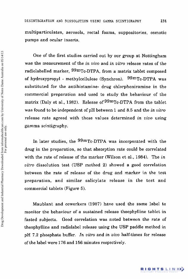
One of the first studies carried out by our group a t Nottingham

was the measurement of the in uiuo and in uitro release rates of the

radiolabelled marker, ggmTc-DTPA, from a matrix tablet composed

of hydroxypropyl - methylcellulose (Synchron). 99mTc-DTPA was

substituted for the antihistamine- drug chlorpheniramine in the

commercial preparation and used t o study the behaviour of the

matrix (Daly et al., 1982). Release of99mTc-DTPA from the tablet

was found to be independent of pH between 1 and 8.5 and the in uitro

release rate agreed with those values determined in uiuo using

gamma s cin ti gr ap h y .

In later studies, the 99mTc-DTPA was incorporated with the

drug in the preparation, so that absorption rate could be correlated

with the rate of release of the marker (Wilson et al., 1984). The in

uitro dissolution test (USP method 2) showed a good correlation

between the rate of release of the drug and marker in the test

preparation, and similar salicylate release in the test and

commercial tablets (Figure 5).

Maublant and coworkers (1987) have used the same label t o

monitor the behaviour of a sustained release theophylline tablet in

fasted subjects. Good correlation was noted between the rate of

theophylline and radiolabel release using the USP paddle method in

pH 7.2 phosphate buffer. In uitro and in uiuo half-times for release

of the label were 176 and 156 minutes respectively.

Dru

g D

evel

opm

ent a

nd I

ndus

tria

l Pha

rmac

y D

ownl

oade

d fr

om in

form

ahea

lthca

re.c

om b

y U

nive

rsity

of

Not

re D

ame

Aus

tral

ia o

n 05

/14/

13Fo

r pe

rson

al u

se o

nly.

2 32 WILSON AND WASHINGTON

0 0 1 2 3 4

I Time (hours)

Figure 5 - The rate of release of drug and radiolabel marker from the test preparation, compared to the release characteristics of the drug from the commercial preparation.

A finding which has been confirmed in several studies, is that

the rate of release of the marker in uiuo is significantly different to

that observed in uitro (Figure 6).

This is probably due to the differences in pH and the stirring

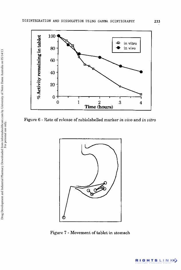
conditions in the gastrointestinal tract. Although Levy (1 963) found

that agitation of tablets in the stomach as observed by x-rays was

mild, studies by our group of a 800 mg naproxen tablet (Figure 7)

demonstrated considerable movement. in the pylorus for several

hours in fed subjects as the tablet was pushed to the duodenum then

retropulsed to the antrum since i t was too big to be emptied (Davis et

al., 1986a; Wilson et al., 1987a).

Dru

g D

evel

opm

ent a

nd I

ndus

tria

l Pha

rmac

y D

ownl

oade

d fr

om in

form

ahea

lthca

re.c

om b

y U

nive

rsity

of

Not

re D

ame

Aus

tral

ia o

n 05

/14/

13Fo

r pe

rson

al u

se o

nly.

DISINTEGRATION AND DISSOLUTION USING GAMMA SCINTIGRAPHY 233

5 ' 5 20 3 c l8 0

0 1 2 3 4 Time (hours)

Figure 6 - Rate of release of rabiolabelled marker in vivo and in vitro

Figure 7 - Movement of tablet in stomach

Dru

g D

evel

opm

ent a

nd I

ndus

tria

l Pha

rmac

y D

ownl

oade

d fr

om in

form

ahea

lthca

re.c

om b

y U

nive

rsity

of

Not

re D

ame

Aus

tral

ia o

n 05

/14/

13Fo

r pe

rson

al u

se o

nly.

2 34 WILSON AND WASHINGTON

l i Y b h a d The major complication when studying the dissolution of dosage

forms in uivo is the presence of food within the gut. Food affects the

rate a t which dose forms travel through the gastrointestinal tract

and the degree of spread of the formulation. The time for which a

dose form remains in the stomach can vary enormously depending

upon it's size and shape and the amount of food present at the time of

administration. Food influences gastric pH and there may be

chemical o r physical interactions between the food and drug. In

addition the food also changes the viscosity of the gastrointestinal

fluid in which the drug is presented to the absorbing mucosa.

Some research workers have found that the basal gastric pH can

be surprisingly high. Kuna (1964) measured the fasting pH of gastric

contents in dogs and man. In 403 tests in dogs, 77% had a gastric pH

of 6 or above, compared to 35% in 1556 human tests. Less than 2% of

the human subjects had a resting pH below 1.5. In our studies we

have found that the basal gastric pH in normal healthy students to be

around 1.8. The rate of secretion is approximately 1 t o 1.5 ml per

minute rising to a maximum rate of 2 to 4 ml after stimulation.

Meals markedly alter the pH, which can increase to 3 - 5 after eating,

particularly if the meal contains large amounts of easily digested

protein. A typical pH trace is shown in Figure 8.

The changes in pH will be especially important when developing

products designed to be gastro-resistant, e.g. for acid-labile or

potentially irritant drugs and gamma scintigraphy may be combined

with in uivo pH measurement to investigate the efficiency of enteric-

coating. In a recent study reported by Hardy and co-workers (1987a),

Dru

g D

evel

opm

ent a

nd I

ndus

tria

l Pha

rmac

y D

ownl

oade

d fr

om in

form

ahea

lthca

re.c

om b

y U

nive

rsity

of

Not

re D

ame

Aus

tral

ia o

n 05

/14/

13Fo

r pe

rson

al u

se o

nly.

DISINTEGRATION AND DISSOLUTION USING GAMMA SCINTIGRAPHY 235

-20 0 20 40 60 80 100 120 140 160 180 200 Time (minutes)

Figure 8 - Typical gastric pH changes observed after an scrambled egg meal

both pH radiotelemetry capsules and enteric coated naproxen tablets

were radiolabelled and administered to fed subjects. The local pH

and rate of disintegration were monitored simultaneously. The pH

remained below 2 within the stomach, except for a transient rise

after food. Five tablets disintegrated in the small intestine

approximately 1.2 h after gastric emptying, 1 disintegrated in the

stomach at pH 1.1 and 1 tablet remained intact in the stomach for 9 h.

The median gastric emptying time for the tablets and telemetry

capsules were 3.3 h and 4.2 h respectively.

The therapeutic efficacy of glyceryl trinitrate in the treatment of

anginal pain is limited by the short half-life of the drug and high

Dru

g D

evel

opm

ent a

nd I

ndus

tria

l Pha

rmac

y D

ownl

oade

d fr

om in

form

ahea

lthca

re.c

om b

y U

nive

rsity

of

Not

re D

ame

Aus

tral

ia o

n 05

/14/

13Fo

r pe

rson

al u

se o

nly.

236 WILSON AND WASHINGTON

hepatic clearance. Over the past few years there have been several

initiatives t o develop sustained release formulations to enable the

drug to be used prophylactically. One of the newer formulations is a

buccal or transmucosal tablet of glyceryl trinitrate which is placed

between the teeth and the inside of the lips. The surface of the tablet

quickly gels and serves both to anchor the tablet in position and to

control the rate of diffusion of the drug. The tablet is based on a

matrix of modified hydroxypropylmethylcellulose (Schor, 1980). The

tablets are friable and the gel layer breaks on removal, and the

advantage of gamma scintigraphy is that the in situ dissolution can

be measured without disturbing the tablet. Gamma scintigraphy

was used t o study the inter- and intra-subject variation, the effect of

position in the buccal cavity and of chewing and drinking on the rate

of release of 99mTc - DTPA from the tablet. With the tablet placed in

the upper buccal pouch it was noted that between subjects there were

marked differences in the rates of release, whereas within an

individual measured on four occasions the variation was quite small.

This did not appear to be due to differences in saliva flow rate and the

rate of dissolution probably correlates best with the extent to which

the subject talked during the experiment. Articulation of the cheek

surfaces during speech increases the erosion of the tablet surface

releasing the marker o r drug into the buccal cavity. However, the

rate of release of marker did not increase when the subject drank hot

coffee or chewed gum.

Chewable formulations are used for the delivery of antacids

where the flavouring agents give the sensation of relief and such a

system may be preferred by the patient who has difficulty in

swallowing tablets or capsules. The most important physiological

Dru

g D

evel

opm

ent a

nd I

ndus

tria

l Pha

rmac

y D

ownl

oade

d fr

om in

form

ahea

lthca

re.c

om b

y U

nive

rsity

of

Not

re D

ame

Aus

tral

ia o

n 05

/14/

13Fo

r pe

rson

al u

se o

nly.

DISINTEGRATION AND DISSOLUTION USING GAMMA SCINTIGRAPHY 23 7

% remaining

100 90 80 70 60 50 40 30 20 10 0

0 5 10 15 20 25 Time (minutes)

Figure 9 - Effect of chewing and sucking a formulation on the dissolution of the marker.

variable is likely to be whether the subject sucks or chews the

formulation, a type of dissolution test which is hard to recreate in

uitro. The rate of release of 99mTc - DTPA from such formulations

has been monitored in uiuo in a group of volunteers who either

sucked or chewed capsules containing various excipients. The

results are shown in Figure 9 and illustrate the marked effect on

dissolution of chewing the capsule.

An alternative strategy for the patient who has difficulty in

taking an intact formulation is t o use a dose form designed to

disintegrate in the buccal cavity such as an Expidet (American Home

Products Corporation).

Dru

g D

evel

opm

ent a

nd I

ndus

tria

l Pha

rmac

y D

ownl

oade

d fr

om in

form

ahea

lthca

re.c

om b

y U

nive

rsity

of

Not

re D

ame

Aus

tral

ia o

n 05

/14/

13Fo

r pe

rson

al u

se o

nly.

238 WILSON AND WASHINGTON

ExDideos Recently a new type of dosage form based on a freeze-dried

mixture of drug and fast-dissolving excipients has been introduced to

deliver sedative drugs such as benzodiazepines. Expidets are solid

dose forms which do not have to be taken with water and are useful

where swallowing is difficult o r oesophageal clearance is impaired.

Incorporation of technetium-99m labelled micronised "Amberlite"

CG400 resin during manufacture enabled the deposition and

clearance of these formulations t o be followed by gamma

scintigraphy (Wilson et al., 1987b). The micronised resin was chosen

as a marker since the units are intended for benzodiazepine delivery

and the two candidate drugs, lorazepam and oxazepam have low

aqueous solubility at the pHs likely to be encountered in the buccal

cavity. Two marker loading were used, 2.5 mg and 10 mg, and the

effect of incorporating salivary stimulants talidsaccharin and

citrate investigated. At the end of each experiment, the head was

outlined with a cobalt-57 source. The buccal cavity, glottis and upper

oesophagus could then be clearly discriminated (Figure 10).

It was noted that the buccal clearance of the formulation

containing the 10 mg resin was significantly faster (50 k 20 s) than

that containing 2.5 mg resin (190 * 70 6); however, calculation of the

total activity remaining after dissolution showed that the amount

remaining on the tongue was approximately 1 mg in each case. This

probably represents the amount of resin trapped within the papillae

of the tongue. Incorporation of salivary stimulants made little

difference to the rate of dissolution of the formulation. This was not

unexpected since salivary stimulants increase the output of the

Dru

g D

evel

opm

ent a

nd I

ndus

tria

l Pha

rmac

y D

ownl

oade

d fr

om in

form

ahea

lthca

re.c

om b

y U

nive

rsity

of

Not

re D

ame

Aus

tral

ia o

n 05

/14/

13Fo

r pe

rson

al u

se o

nly.

DISINTEGRATION AND DISSOLUTION USING GAMMA SCINTIGRAPHY

Profile Drawn with Cobalt Pen

.:I<:.:.:::.. , . .\. :.:.. . .

..:$;,..

'.. . .. . . . . _ . .::!.:: . .

239

I

Figure 10 - The clearance of the activity release from the Expidet in the buccal cavity.

submandibular and sublingual salivary glands, which discharge

watery secretions onto the floor of the mouth, wetting the side of the

tongue and cheek surfaces. The posterior third of the tongue surface

contains mucus glands, but the quantity of secretion is relatively

small. Thus increased saliva flow may not result in a more aqueous

phase available for dissolution of the dosage form from the tongue

surface.

sltuuub Hard gelatin capsules have found a variety of applications in

drug formulation. The capsule can be used as a container for

powdered drug, multiparticulate systems, a liquid-fill matrix or oily

vehicle. The nature of the interior of the fill of the capsule is known

to affect the rate of disintegration. A hydrophobic interior, reduces

the rate of disintegration compared t o that of a water soluble

Dru

g D

evel

opm

ent a

nd I

ndus

tria

l Pha

rmac

y D

ownl

oade

d fr

om in

form

ahea

lthca

re.c

om b

y U

nive

rsity

of

Not

re D

ame

Aus

tral

ia o

n 05

/14/

13Fo

r pe

rson

al u

se o

nly.

2 40 WILSON AND WASHINGTON

material. The particle size can also be important as illustrated by the experiments of Hunter and co-workers (1980).

In their experiments, they used Tc-99m labelled 'Amberlite'

resin which was graded to three sizes, with geometric means of a)

25 pm, b) 9 pm c) 150 - 210 pm. Sample b had been milled down to

obtain the appropriate size. 'Amberlite' resin was chosen as it has a

similar density to most pharmaceutical materials. The in uitro tests

showed that capsules containing the powder batches a and c had

shorter disintegration times than b. The milled resin was found to

be more hydrophobic, decreasing the wettability of the powder and

increasing the time to disintegration of the capsule (8 minutes for b

compared to 2-3 minutes for a and c). Scintigraphic evidence

confirmed the results of the in uitro tests. Formulation b showed

little dispersion suggesting that the gastric emptying of the capsule

fill took place with the turnover of gastric mucus. In later

experiments (Hunter et al., 1983), the researchers compared

formulations a and b with a third soluble formulation which

consisted of [113mInl indium chloride recrystallised with sodium

chloride. The three formulations were administered t o subjects

either fasted or with a light breakfast and in both cases, the capsules

were administered with 100 ml water. Despite the good in uitro

disintegration characteristics of formulation a, the dispersion in the

fasted state was limited and the capsule emptied from the stomach

largely undisintegrated. When taken after a meal, the dispersion

was improved. Formulation b in earlier trials had been

demonstrated to be less dispersible. This was confirmed and the

activity was observed to leave the stomach in both fasted and fed

states as a bolus. For formulation c there was no differences in the

Dru

g D

evel

opm

ent a

nd I

ndus

tria

l Pha

rmac

y D

ownl

oade

d fr

om in

form

ahea

lthca

re.c

om b

y U

nive

rsity

of

Not

re D

ame

Aus

tral

ia o

n 05

/14/

13Fo

r pe

rson

al u

se o

nly.

DISINTEGRATION AND DISSOLUTION USING GAMMA SCINTIGRAPHY 241

behaviour observed in fasted and fed subjects; in both cases the

capsule dissolved rapidly and the activity emptied from the stomach

in a mono-exponential pattern.

From our own observations and the experiments of Hunter and

coworkers (1980, 1982 and 1983) i t has been established that the

dispersion of the capsule fill is limited in fasted subjects and the

material empties from the stomach as a bolus. The dispersion is

increased if the capsule is taken with a meal, particularly if the meal

has high liquid content. This is of importance since patients are

often instructed to take medications with a meal, but it is unclear

whether this means before, during o r after food.

A recent study in our laboratory has examined the effects of the

time of dosing relative to a standard meal (O'Reilley et al., 1987). The

behaviour of a multiparticulate dosage form has been followed in six

healthy volunteers who received a capsule containing radiolabelled

'Amberlite' beads 10 minutes prior, during and 10 minutes after a

meal of a total energy content of 3800 kJ. The particles were released

from all capsules within a few minutes. After dosing with the

capsule during or after a meal, the pellets tended to remain in the

upper half of the stomach. In these cases, the gastric emptying

pattern was approximately linear with time. The gastric emptying

half-times (T50) were similar for the experiments between 3 - 4

hours; however, over the initial 100 minutes, the particles taken

before the meal emptied fastest and the emptying followed an

exponential pattern with time.

Dru

g D

evel

opm

ent a

nd I

ndus

tria

l Pha

rmac

y D

ownl

oade

d fr

om in

form

ahea

lthca

re.c

om b

y U

nive

rsity

of

Not

re D

ame

Aus

tral

ia o

n 05

/14/

13Fo

r pe

rson

al u

se o

nly.

242 WILSON AND WASHINGTON

In a second experiment, the gastric emptying of pellets

predispersed in a meal was compared to that of a capsule containing

the same number of pellets. This system was analogous to the

"sprinkle" formulations which have been suggested for theophylline

administration. Although the distribution in the stomach of the

predispersed pellets was more even, the gastric emptying following

both manoeuvres was similar with no significant differences between

the emptying rates (O'Reilly et al., 1987). Two important points were

determined in addition t o our main findings. First, "sprinkle"

systems have to be dispersed into a high viscosity medium e.g. jam or

mashed potatoes, otherwise they may fall through the meal prior to

eating, with the consequent risk of under-dosing. Particles as large

as 800 pm are probably unsuitable as the subjects complained of the

sensation of "grittiness", when eating their meal. This increases the

desire to masticate and for sustained release formulations would

increase the risk of dose-dumping. Thus it is a prerequisite of such

systems that they be relatively small:- under 500 pm, for example.

There have been relatively fewer studies of the behaviour of soft

gelatin capsules in man. From our pilot studies, we have observed

that the time of disintegration of soft gelatin capsule formulations is

highly variable, particularly if the formulations are given without

food. The emptying tends to follow the break up of the capsule in the

pylorus. A group at the Welsh School of Pharmacy has compared the

dispersion of oils from soft gelatin capsules in man and rabbits

(Armstrong et al., 1983) using x-ray techniques and gamma

scintigraphy. Soft gelatin capsules were filled with iodinated cotton

Dru

g D

evel

opm

ent a

nd I

ndus

tria

l Pha

rmac

y D

ownl

oade

d fr

om in

form

ahea

lthca

re.c

om b

y U

nive

rsity

of

Not

re D

ame

Aus

tral

ia o

n 05

/14/

13Fo

r pe

rson

al u

se o

nly.

DISINTEGRATION AND DISSOLUTION USING GAMMA SCINTIGRAPHY 243

seed oil (Lipiodol) for x-ray studies or iodine-123 labelled ethyl oleate

for gamma camera studies in humans. The effects of various

surfactants was also investigated.

In the rabbit (x-ray) studies, disintegration of the capsule began

after 2-3 minutes, swelling into a more isometric shape. This

behaviour was observable in uitro and was associated with the

breakdown of the capsule a t the sealing line. Subsequently it was

difficult to assess whether the shell had dissolved with the oil as one

discrete globule, or whether the oil had emerged from the shell before

it had completely dissolved. When 1% polysorbate 80 was added to the

formulation, mean disintegration time of the soft gelatin capsule

decreased markedly, supporting the findings of Hunter et al., (1980)

and Casey e t al., (1976). Analysis of variance showed that the

presence of surfactant in the formulation to be most important factor

influencing dispersion.

From the gamma camera studies in man, the authors defined

the disintegration time as the time at which the regular shape of the

oil droplet was lost. Three different strategies for calculation of the

degree of dispersion were used. First, they used a fixed area of 5 x 4

pixels on the whole field of view; an approach that proved to be invalid

since as capsule moved around the stomach, it moved out of the fixed

area. The second approach was to use a moveable area of 5 x 4 pixels;

however, problems were experienced when the oil divided to give

multiple areas of high count rate and it proved difficult to select the

area of maximum activity. The third technique involved the

automatic generation of contours and is based on the calculation of

number of pixels which exhibit activity of greater than 5% of the

Dru

g D

evel

opm

ent a

nd I

ndus

tria

l Pha

rmac

y D

ownl

oade

d fr

om in

form

ahea

lthca

re.c

om b

y U

nive

rsity

of

Not

re D

ame

Aus

tral

ia o

n 05

/14/

13Fo

r pe

rson

al u

se o

nly.

2 44 W I L S O N AND WASHINGTON

maximum activity in the frame. The technique is not subjective and

shows a rapid rise a t the presumed point of capsule disintegration

and liberation of its contents. Spreading was defined as commencing

when the area covered by the 5% contour doubled in magnitude.

Using the latter technique they determined that the mean time to

disintegration was 12.3 k 6.7 minutes and to spreading 14.3 k 10.2

minutes,

The spreading area of the suppository determines the release

area of the drug from the delivery form, Furthermore, the position of

the formulation in the rectum determines how much of the released

drug avoids hepatic first pass metabolism since the drainage

territories of the inferior haemmorhodial and middle rectal vein

differ. Animal species, particularly the rat and dog, have been

widely used to measure the dissolution of suppository formulations,

usually by the incorporation of a fluorescent dye or coloured marker.

Tukker (1983) first described an elegant use of gamma scintigraphy

to quantify the spreading of suppositories in recumbent dogs. The

author constructed a series of activity profiles, measuring the activity

in each of the pixels along the centre line of the image. The

subsequent images were then stacked to yield an impression of the

way that the suppositories spread with time. The results show that

the addition of surfactants markedly affected in uiuo spreading.

Similarly preadministration of neostigmine which increases colonic

motility markedly increased the spreading of the Witepsol H15

suppository.

Dru

g D

evel

opm

ent a

nd I

ndus

tria

l Pha

rmac

y D

ownl

oade

d fr

om in

form

ahea

lthca

re.c

om b

y U

nive

rsity

of

Not

re D

ame

Aus

tral

ia o

n 05

/14/

13Fo

r pe

rson

al u

se o

nly.

DISINTEGRATION AND DISSOLUTION USING GAMMA SCINTIGRAPHY 245

Hardy and coworkers (1 987b) have described the spreading

behaviour of suppository bases and incorporated suspension. The

bases, Witepsol H15 and Labrafil WL2514, were labelled by the

incorporation of small amounts of iodine-1 23 labelled unsaturated

markers (arachis oil and Labrafil WL2700 respectively). The

suspension consisted of micronised cationic exchange resin

incorporated throughout the base at a disperse phase loading of 10%

w/v.

The limits of spreading were defined as the edge of the 20%

contours, defining 20% of the maximum activity in each frame.

Analysis of the data showed that little spreading occurred and both

base and suspension tended to move together. Most spread occurred

within the first hour after dosing and reached a maximum of 8 to 10

cm. In a few subjects, separation of base and resin occurred

particularly in the suppositories composed of the surfactant material

WL2514.

Treatment of the proximal bowel is clearly not achieved by use of

suppositories and the strategy most commonly employed is the

delivery of the drug as a rectal enema. Penetration into the

transverse colon is however poor and Hardy e t al. (1986) have

commented that the optimum enema volume is about 100 ml.

Increasing the volume to 200 ml did not enhance dispersion and 50

ml doses showed less spreading. Although administration of the

enema or intake of food caused increased motility, neither

manoeuvre increased the spreading of the enema.

Dru

g D

evel

opm

ent a

nd I

ndus

tria

l Pha

rmac

y D

ownl

oade

d fr

om in

form

ahea

lthca

re.c

om b

y U

nive

rsity

of

Not

re D

ame

Aus

tral

ia o

n 05

/14/

13Fo

r pe

rson

al u

se o

nly.

246 WILSON AND WASHINGTON

The development of a small osmotically-driven device, consisting

of an osmotic core containing drug surrounded by a semi-permeable

membrane was first described by Theeuwes (1975). In the operation

of an 'Osmet' device developed by Alza Corporation, water is

osmotically imbibed across the semi-permeable membrane, swelling

the osmotic compartment and squeezing the drug reservoir

uniformly along the axis. Since water is incompressible and the

semi-permeable membrane is relatively rigid, there is a

corresponding amount of drug solution from the reservoir squeezed

through the delivery orifice.

It is expected that delivery from such a system should be

relatively independent of pH and agitation conditions and this has

been tested by gamma scintigraphy (Davis et al., 1984a). The release

of In-111 DTPA from the 2 0 0 ~ 1 capacity 'Osmet' with a nominal

steady-state delivery of 15 p1 h-1 was defined in vitro. One of these

units, together with a capsule containing a number of technetium-

99m labelled 'Amberlite' beads was administered t o each of six

volunteers with or without food. The release of the radiolabelled

marker was unaffected by the presence of food and was similar to

that found in vitro (Figure 11) confirming the original hypothesis.

An osmotic tablet system ('OROS', Alza Corporation) for delivery

of a number of drug candidates has been developed and one such

system was marketed for the delivery of indomethacin. Since the

interior of the unit is solid, an alternative strategy was needed to

identify the site of initial release. In order to follow the behaviour in

man, we developed a method to label the position of the tablet and to

Dru

g D

evel

opm

ent a

nd I

ndus

tria

l Pha

rmac

y D

ownl

oade

d fr

om in

form

ahea

lthca

re.c

om b

y U

nive

rsity

of

Not

re D

ame

Aus

tral

ia o

n 05

/14/

13Fo

r pe

rson

al u

se o

nly.

DISINTEGRATION AND DISSOLUTION USING GAMMA SCINTIGRAPHY 247

100 LJ

2o 1

* in vivo (114.2) h + in vitrn In-lnl

0 100 200 300 400 500 600 700 Time (min)

Figure 11 - The release of the radiolabel marker from an "Osmet" in

uiuo and in uitro

depth of 7 mm. This was packed with dried indium-111 labelled

'Amberlite' CG 120 cation-exchange resin and sealed with a small

blob of 'Araldite' resin containing technetium-99m labelled

'Amberlite' CG 400 anion exchange resin (Figure 12).

The release of indium-111 from the device was observed to follow

zero-order kinetics for at least 6 hours in the USP test (method 2) and

this method of labelling was used to follow the gastrointestinal

transit of the unit. The onset of release of label from the delivery

orifice defined the time at which drug was pumped out and helped to

establish the position of the unit in uiuo.

Dru

g D

evel

opm

ent a

nd I

ndus

tria

l Pha

rmac

y D

ownl

oade

d fr

om in

form

ahea

lthca

re.c

om b

y U

nive

rsity

of

Not

re D

ame

Aus

tral

ia o

n 05

/14/

13Fo

r pe

rson

al u

se o

nly.

248 WILSON AND WASHINGTON

I r Delivery orifice

Tc-99m labelled

,,,,,,,,,,,,,I,,,,,,,,,,,,,, I,I,,,,,,II,II,,,,,C,,,,,,,, ............................

Semi-permeable indium- 1 1 1 membrane

Osmotic Core labelled ion exchange resin

Figure 12 - Labelling of the interior core and exterior surface of an osmotic tablet

ODht-c Fo- For drugs administered topically in the eye, the sites of intended

activity can be intra-ocular as in the treatment of glaucoma with

transcorneal penetration the predominant requirement o r extra-

ocular, for the treatment of conjunctivitis, blepharitis o r keratitis

sicca. The dosage form most commonly used is the eyedrop,

although it has the disadvantage that the majority of the instilled

drug is lost due to drainage via the nasolacrimal duct in the first 15 to

30 seconds (Shell, 1982). Much of the present research has been

directed towards perfecting sustained release devices to deliver drugs

continuously. Three major approaches have been investigated :

presoaked matrices typically based on soft-contact lens material

soaked in the drug; diffusional devices containing a central reservoir

of drug enclosed between rate-controlling polymeric membranes and

erodible systems which release drugs at a rate proportional to the

dissolution rate of the matrix.

Dru

g D

evel

opm

ent a

nd I

ndus

tria

l Pha

rmac

y D

ownl

oade

d fr

om in

form

ahea

lthca

re.c

om b

y U

nive

rsity

of

Not

re D

ame

Aus

tral

ia o

n 05

/14/

13Fo

r pe

rson

al u

se o

nly.

DISINTEGRATION AND DISSOLUTION USING GAMMA SCINTIGRAPHY 249

We have developed a gamma scintigraphic technique to

investigate the behaviour of polyvinyl alcohol films inserts in man

(Fitzgerald et al., 1986; Olejnik and Wilson, 1987). In these

experiments we have tried to model the presentation of a suspension

of drug which would be released as the matrix erodes. Gohensol GH-

17, average molecular weight 98,000 with 87 - 89% hydrolysis was

used as the base for the matrix. A sterile solution of the polymer in

water was prepared. Technetium-99m labelled sulphur colloid o r

[ggmTc] sodium pertechnetate was added to the concentrated solution

and the preparation spread onto a melinex backing sheet. The film

was dried under aseptic conditions a t 70° C and cut into 25 mm2

sections with a scalpel. In vitro dissolution tests were carried out in

distilled water, with the film supported on a wire mesh. A paddle

stirrer was positioned 2 cm above the mesh and rotated a t 60 r.p.m. to

reduce stagnant layer formation. Samples were removed a t intervals

and the technetium-99m content of the fluid monitored.

Release of the soluble pertechnetate label occurred rapidly before

the matrix had dissolved whereas the sulphur colloid label was

released as a function of the square root of time. This indicated that

the sulphur colloid was a more appropriate marker for entrapped

drug and this radiopharmaceutical was used for the volunteer

studies .

The volunteers were positioned 5 cm away from the pinhole

collimator with the head supported by an ophthalmic table. A 5 x 5

mm square piece of film was placed under the lower eyelid and

images recorded over a period of thirty minutes. The plot of activity

versus time followed a monoexponential curve with a mean half time

Dru

g D

evel

opm

ent a

nd I

ndus

tria

l Pha

rmac

y D

ownl

oade

d fr

om in

form

ahea

lthca

re.c

om b

y U

nive

rsity

of

Not

re D

ame

Aus

tral

ia o

n 05

/14/

13Fo

r pe

rson

al u

se o

nly.

2 50 WILSON AND WASHINGTON

%Remaining

20 - - l o - - 04 I

0 5 10 15 20 25 30 Time (min)

Figure 13 - The precorneal clearance of activity from a polyvinyl alcohol film in man.

of eight minutes (Figure 13), although the correlation coefficient was

equally as good for the relationship between activity remaining and

and the square root of time (Higuchi plot).

There was great individual variation in rates of clearance with the

slowest half time of 23 minutes and the fastest of 3 minutes. The

presence of irritant material in the eye causes reflex blinking and

increased tear flow so there is a need for the surface to hydrate quickly

t o minimise corneal sensation. Blinking is probably the most

important physiological factor influencing precorneal clearance and

increased rates of blinking are associated with increased precorneal

turnover.

Dru

g D

evel

opm

ent a

nd I

ndus

tria

l Pha

rmac

y D

ownl

oade

d fr

om in

form

ahea

lthca

re.c

om b

y U

nive

rsity

of

Not

re D

ame

Aus

tral

ia o

n 05

/14/

13Fo

r pe

rson

al u

se o

nly.

DISINTEGRATION AND DISSOLUTION USING GAMMA SCINTIGRAPHY 251

The period for which a dose form remains in the environment of

each region of the gastrointestinal tract is determined by gut motility.

Transit through the small intestine is fairly uniform and difficult t o

alter, however, the residence of a formulation in the stomach can be

extremely variable and this can affect the rate of presentation of the

drug to the site of absorption.

The oesophageal transit of dose forms is extremely rapid, usually

in the order of 10 to 14 seconds. It is well recognised that tablets o r

capsules taken by patients in the supine position may lodge in the

oesophagus, causing damage and irritation (D'Arcy, 1984; Channer

and Virjee, 1985). If tablets are taken without water, the risk is greatly

increased and the units may remain lodged in the lower oesophagus

until they disintegrate (Hey e t al., 1982). The problem can be

aggravated in patients who have cardiac pathologies in which the left

side of the heart is enlarged o r who are elderly and have oesophageal

dysfunction. Retention of the dosage form in the oesophagus has been

demonstrated to delay drug absorption, as drugs cannot easily pass

through the stratified squamous epithelium of the oesophageal mucosa

(Channer and Roberts, 1985).

The hydration of a sticky material against the mucosal epithelium

greatly increases the chance of adhesion and has been recognised as a

hazard of formulations containing gelatin or cellulose derivatives

Dru

g D

evel

opm

ent a

nd I

ndus

tria

l Pha

rmac

y D

ownl

oade

d fr

om in

form

ahea

lthca

re.c

om b

y U

nive

rsity

of

Not

re D

ame

Aus

tral

ia o

n 05

/14/

13Fo

r pe

rson

al u

se o

nly.

252 WILSON AND WASHINGTON

(Swisher e t al., 1984). The tendency of hydroxymethylcellulose to

adhere can be adjusted by incorporation of sucrose which reduces

surface stickiness; conversely, addition of lactose or titanium oxide and

talc increases the tendency to adhere (Marvola et al., 1983). The

interior surface of the oesophagus is moist rather than wet and a

dosage form in contact with the mucosa will cause partial dehydration

at the site of contact as the unit hydrates, resulting in formation of a gel

between the formulation and the mucosa. The unit then disintegrates

from its non-contact side. Disintegration of the lodged formulation is

slow, first because the amount of dissolution fluid available is low,

being dependent on the volume of swallowed saliva and secondly due to

the reduced surface area available for dissolution.

Fell (1983) has challenged the belief that gelatin capsules are more

likely to stick than tablets, and concludes that the evidence suggests

that the two dosage forms should be regarded as having equal potential

to adhere. Out of a total of 200 people dosed a t Nottingham with various

preparations contained in hard gelatin capsules, we have found little

evidence of oesophageal lodging or adhesion of the units elsewhere in

the gastrointestinal tract.

The most dramatic effect of food is that it produces significant

changes in the gastric motility patterns and a clear discrimination can

occur between the gastric emptying of single units and

multiparticulates. Food can increase, decrease or delay the absorption

of a drug. The absorption of most drugs is slower from the stomach

than from the small intestine (Levine, 1970; Heading et al., 1973) and

Dru

g D

evel

opm

ent a

nd I

ndus

tria

l Pha

rmac

y D

ownl

oade

d fr

om in

form

ahea

lthca

re.c

om b

y U

nive

rsity

of

Not

re D

ame

Aus

tral

ia o

n 05

/14/

13Fo

r pe

rson

al u

se o

nly.

DISINTEGRATION AND DISSOLUTION USING GAMMA SCINTIGRAPHY 253

the rate a t which gastric emptying occurs can be a controlling factor

in the onset of drug absorption (Heading et al., 1973). Gamma

scintigraphy has been used to investigate the gastric emptying time

of liquid formulations. It has been demonstrated that 10 to 20 ml of a

liquid antacid or anti-reflux agent administered to fasted subjects

empties from the stomach within 30 minutes (Jenkins et al., 1983;

Washington et al., 1986). Gastric residence of the same formulation

can be increased to more than 2 hours by the ingestion of a meal 30

minutes prior t o administration of the formulation (May et al., 1984).

The function of the stomach is to provide a reservoir of ingested

food and regulate emptying into the intestine to provide a constant

calorific input. Digestion and absorption is facilitated by enzymatic

action and the milling and grinding movements of the pyloric antrum

which triturates food to fine particles. Pressures of up to 60 cm H,O (43

mm Hg) have been recorded in the antral mill (Quigley and Brody,

1950). Emptying of the pylorus occurs in discrete episodes of 2 - 5

seconds duration and the majority occur as the terminal antrum,

pylorus and duodenum relax at the end of each peristaltic cycle (King et

al., 1984). The liquid component of a meal empties exponentially, but

the emptying of solids is linear after a variable lag time. A transverse

mid-gastric band was first noted by William Beaumont in 1833

(republished in 1955) and this has subsequently been found to separate

the function of proximal and distal stomach. The distribution of food

across the mid-gastric band is believed to be a major component of the

lag phase in solid emptying (Moore et al., 1986; Collins et al., 1987). The

lag phase is dependent upon the size of the food particles in the

stomach, the larger the particles, the longer the stomach requires to

Dru

g D

evel

opm

ent a

nd I

ndus

tria

l Pha

rmac

y D

ownl

oade

d fr

om in

form

ahea

lthca

re.c

om b

y U

nive

rsity

of

Not

re D

ame

Aus

tral

ia o

n 05

/14/

13Fo

r pe

rson

al u

se o

nly.

2 54 WILSON AND WASHINGTON

break them down into a size suitable t o exit through the pylorus.

Eventually, all the digestible material is emptied from the stomach,

leaving a residue of mucus and undigested solids. Large tablets or

capsules, whether intact or in large fragments, will also be treated by

the stomach as an indigestible material since they do not possess a

significant calorific value. The migrating myoelectric potential or

'housekeeper wave' serves to remove the debris from the stomach by

strong contractions against an open pylorus during the fasted mode.

This will sweep undisintegrated tablets and capsules into the

intestine. The 'housekeeper sequence' occurs a t approximately two

to three hourly intervals. If food is given a t any time while the

stomach is in the fasted mode, it reverts to the fed mode and the

'housekeeper sequence' is suppressed until the stomach is again

empty (Figure 14).

A study by Park and co-workers (1984) examined the effect of size

and shape of tablets on the rate of their gastric emptying in fasted

volunteers. The largest tablet studies was 17.6 x 9.5 mm. It was

reported that the physical properties of the tablets did not affect the

gastric emptying time and 80% of the dose forms emptied by 2 h.

However, the gastric emptying of large single units from fasted

volunteers is extremely erratic and can vary from a few minutes t o

three hours (Kaus et al., 1984a; Wilson et al., 1984). This can explain

the variability observed in drug-plasma profiles when large

tablets,enteric coated units or sustained release matrix tablets are

administered to fasted volunteers. The rationale for using fasted

volunteers in clinical trials has been to decrease variability in the onset

Dru

g D

evel

opm

ent a

nd I

ndus

tria

l Pha

rmac

y D

ownl

oade

d fr

om in

form

ahea

lthca

re.c

om b

y U

nive

rsity

of

Not

re D

ame

Aus

tral

ia o

n 05

/14/

13Fo

r pe

rson

al u

se o

nly.

DISINTEGRATION AND DISSOLUTION USING GAMMA SCINTIGRAPHY 255

~ ~~

PHASE 3 STOMACE Emptied of indigestible food

by Housekeeper Contractions ~~'I'ESTINE: Distally Migrating Peristalsis

Stomach empty of digestible food

PHASE 2

Mixing Contractions in Stomach

and Small

Intestine

Stomach Empty of

Digestible Food

\

PHASE 1

No Activity Stomach Empty of Digestible Food

Figure 14 - Motility patterns in the stomach

of drug absorption, but the fasting dosing schedule actually

introduces a large source of variation due t o unpredictable gastric

emptying. This calls in to question the requirement of regulatory

authorities of using fasted volunteers in clinical trials. It would be

far better to administer single units with a light meal of energy value

Dru

g D

evel

opm

ent a

nd I

ndus

tria

l Pha

rmac

y D

ownl

oade

d fr

om in

form

ahea

lthca

re.c

om b

y U

nive

rsity

of

Not

re D

ame

Aus

tral

ia o

n 05

/14/

13Fo

r pe

rson

al u

se o

nly.

2 56 WILSON AND WASHINGTON

no greater than 1500 kJ, which would have the effect of bringing

motility patterns into phase. A further point is that the kinetics of

gastric emptying and drug absorption are markedly altered by the

size of the meal and these effects may be much larger than small

changes in bioavailability induced by different formulations.

A popular method of delivering sustained release oral

preparations is the multiparticulate or pelleted system contained

within a hard gelatin capsule. Davis and coworkers (1985) has

described the emptying of pellets from fasted subjects as a random

event, with the particles tending t o empty as a series of boluses. This,

however, is dependent upon nature of capsule and how quickly it

disperses, since the volume of fluid available for dissolution is low in

the stomach of a fasted individual. The pellets empty more slowly in

the presence of food, as the calorific load t o the duodenum is

controlled which causes the spread of the pellets to be greater in the

gastrointestinal tract.

The differences in behaviour between a single unit and a pelleted

formulation is illustrated by the study of Davis and coworkers (1984b),

who described the simultaneous administration of both formulations.

In general, the pellets emptied as a series of boluses from the

stomach, ahead of the tablets which were expelled with the onset of

the housekeeper sequence. In some cases the pellet formulations

failed to disintegrate, and they too were emptied as a single bolus.

There have been several studies which have demonstrated that large

non-disintegrating tablets can remain in the stomach for up to 12

hours if they are administered with a large breakfast (3600 kJ) and

the subject is fed at regular intervals throughout the day (Davis et al.,

Dru

g D

evel

opm

ent a

nd I

ndus

tria

l Pha

rmac

y D

ownl

oade

d fr

om in

form

ahea

lthca

re.c

om b

y U

nive

rsity

of

Not

re D

ame

Aus

tral

ia o

n 05

/14/

13Fo

r pe

rson

al u

se o

nly.

DISINTEGRATION AND DISSOLUTION USING GAMMA SCINTIGRAPHY 257

1984a; Wilson et al., 1987b). If the tablet is enteric coated, or the drug

is not acid soluble, the appearance of the drug in the plasma can be

greatly delayed in fed subjects.

One method of prolonging exposure of the upper small intestine

to high concentrations of drug is to retain the drug delivery system in

the stomach. This also advantageous for drugs which are acid

soluble. Muller-Lissner and coworkers (1 981 described a floating

capsule for sustained delivery of diazepam which has been evaluated

by gamma scintigraphy. The capsule contained 10 mg of diazepam-

Ni-methy1-14C and drug absorption was calculated by collection of

14CO2 in expired air. The matrix was labelled by the inclusion of

51Cr and 57Co labelled microspheres. These systems have no

intrinsic property of gastric retention, and rely on flotation on

ingested food. Muller-Lissner and Blum (1981) have described a

study to investigate the effect of food on the gastric emptying times of

non-disintegrating floating and sinking capsules. Both types of

capsules were administered simultaneously to each subject. In

fasted subjects, both capsules left the stomach within 2.5 h. A high

fat meal consisting of 200 ml cream and milk, delayed the emptying

of the capsules. Two of the six sinking capsules were evacuated

within 2.5 h, with the remainder being emptied by 5 h. The majority

of the floating capsules were emptied from the stomach between 2.5

and 5 hours, but in one subject both capsules were emptied after 12

hours and in another, one after 12 h and the second after 24 h. This

demonstrates that although the specific gravity of the capsules has

little effect on the gastric residence time in fasted subjects, in

agreement with the studies by Christensen and coworkers (19841,

food increases the effect of capsule density to a variable degree.

Dru

g D

evel

opm

ent a

nd I

ndus

tria

l Pha

rmac

y D

ownl

oade

d fr

om in

form

ahea

lthca

re.c

om b

y U

nive

rsity

of

Not

re D

ame

Aus

tral

ia o

n 05

/14/

13Fo

r pe

rson

al u

se o

nly.

258 WILSON AND WASHINGTON

There are well documented differences in the rate of gastric

emptying between normal subjects and patients and gastrointestinal

transit may be either faster o r slower than normal. The most

extreme example is seen in patients who have had vagotomy and

pyloroplasty in which 80% of the meal may be emptied in the first 10

minutes of onset of eating (Holt et al., 1982). In the elderly, the

differential between solid and liquid emptying is less evident and

liquids are emptied more slowly than in younger subjects. Evans

(1981) measured the mean gastric emptying half-time (T50) for liquids

as 123 minutes in a group average age 77 years, compared to 50

minutes in a younger group of average age 26 years.

There is a statistically significant difference in the gastric

emptying times for males and females. A recent study by Datz and

coworkers (1987) demonstrated that the T50 for the solid phase of a

meal was 59.8 & 3.7 minutes for males and 92.4 k 7.5 minutes for

females whereas the T50 for the liquid phase was 30.3 f 2.3 and 53.8 * 4.9 minutes for males and females respectively. The authors could

not fully account for their findings but suspected that the effect is due

to sex hormones especially progesterone and oestradiol on

gastrointestinal motility .

Drugs which modify motility may be expected to alter the plasma

concentration-time profile both of themselves and of coadministered

drug, but the effects are sometimes subtle in healthy subjects.

Metoclopramide is used to accelerate gastric emptying in

pharmacokinetic studies. Kaus and coworkers (1 984c) described the

transit of a radiolabelled solid perspex capsule after i.v.

Dru

g D

evel

opm

ent a

nd I

ndus

tria

l Pha

rmac

y D

ownl

oade

d fr

om in

form

ahea

lthca

re.c

om b

y U

nive

rsity

of

Not

re D

ame

Aus

tral

ia o

n 05

/14/

13Fo

r pe

rson

al u

se o

nly.

DISINTEGRATION AND DISSOLUTION USING GAMMA SCINTIGRAPHY 259

administration of metoclopramide (10 mg). The drug had no effect on

gastric emptying of the unit but had a variable effect on increasing

the transit of the capsule through the first part of the small intestine.

The authors conclude that the effect of metoclopramide on gastric

emptying may only be important when gastric emptying is

abnormally slow, an opinion which our studies tend to support. An

explanation for the observed effect of metoclopramide on drug

absorption is that the intestinal transit may be altered, decreasing

the contact time.

t of J3osgge Forrgs

The function of the small intestine is to optimise the digestion

and absorption of nutrients. It is often overlooked that there are two

distinct patterns of small intestinal motility. During the fed phase,

the contractions serve to mix food with enzymes and digestive

secretions, circulate the contents t o facilitate contact with the

intestinal mucosa and finally propel the contents towards the large

bowel. The contractions which serve t o mix the food are called

segmental contractions and locally squeeze the food t o enable

spreading and contact with the intestinal villi. Coordinated

muscular contraction over a length of intestine produces the

peristaltic wave which propels the food in a aboral direction. Pellets

administered with a meal are emptied more slowly from the

stomach, and are more widely distributed within the small intestine,

with an average small intestinal transit time of approximately 200

minutes (Davis et al., 1984b, 1987).

The small intestinal transit time for pharmaceutical dosage forms

has been reviewed by Davis and coworkers (198613). The mean transit

Dru

g D

evel

opm

ent a

nd I

ndus

tria

l Pha

rmac

y D

ownl

oade

d fr

om in

form

ahea

lthca

re.c

om b

y U

nive

rsity

of

Not

re D

ame

Aus

tral

ia o

n 05

/14/

13Fo

r pe

rson

al u

se o

nly.

2 60 WILSON AND WASHINGTON

Solution Pellets n=23 n=81

T

Single unit n=84

Figure 15 - The small intestinal transit of pharmaceutical dosage forms in man (after Davis et al., 1986b).

time for the formulations studied was between three and four hours

(Figure 15). In the past, figures of 5 to 8 hours have been quoted in

physiology texts, which have led to overestimates of the time available

for drug absorption from the small intestine when formulating

sustained release preparations. The data from 201 studies revealed

that small intestinal transit time in healthy subjects is not influenced

by the physical state, o r the size of the dosage form nor by the

presence of food; however, transit may be slightly slowed by high

calorific loads. Exercise has also been demonstrated not to affect

small intestinal transit time (Ollerenshaw et al., 1987). Most

physical factors, such as density, appear to be unimportant but the

effects of viscosity have not been hlly investigated.

Dru

g D

evel

opm

ent a

nd I

ndus

tria

l Pha

rmac

y D

ownl

oade

d fr

om in

form

ahea

lthca

re.c

om b

y U

nive

rsity

of

Not

re D

ame

Aus

tral

ia o

n 05

/14/

13Fo

r pe

rson

al u

se o

nly.

DISINTEGRATION AND DISSOLUTION USING GAMMA SCINTIGRAPHY 261

The passage of a single non-disintegrating perspex capsule,

similar in shape and size t o a conventional No. 1 hard gelatin

capsule, was measured through the small intestine as described

earlier (Kaus et al., 1984a). It was found that the passage of the unit

through the duodenum was too rapid to be measured, but the mean

transit rate through the small intestine was 4.2 - 5.6 cm min-1;

gastric emptying of the capsule was erratic and ranged from 15 to 197

min. It is interesting to note, that although drugs are best absorbed

from the duodenum, the passage through this area is usually too

rapid to allow significant transfer to occur.

M o v T h r o u e h the

Stasis of material a t the ileocaecal junction is a normal

phenomenon as propulsive peristaltic waves become weaker towards

the end of the small intestine. This causes the materials such as

suspensions or pellets to bunch at the junction before being swept

through into the ascending colon or is seen as a period of stasis of

intact tablets. Patients who take non-steroidal anti-inflammatory

drugs have an increased incidence of gastric bleeding and peptic

ulceration, and there have been attempts t o reduce this by enteric

coating the formulation or the use of controlled delivery devices. This

may only be a partial therapeutic advantage as there is evidence to

suggest that non-steroidal anti-inflammatory drugs may cause

inflammation of the ileocaecal junction due to local irritant effects.

Day (1983) reported two cases in which indomethacin delivered in an

osmotic pump was associated with intestinal perforation. In one

case it seemed probable that the capsule, being rigid and of the right

size to become trapped, lodged in a diverticulum. There has been

some confusion in the literature that this behaviour may be attributed

Dru

g D

evel

opm

ent a

nd I

ndus

tria

l Pha

rmac

y D

ownl

oade

d fr

om in

form

ahea

lthca

re.c

om b

y U

nive

rsity

of

Not

re D

ame

Aus

tral

ia o

n 05

/14/

13Fo

r pe

rson

al u

se o

nly.

262 WILSON AND WASHINGTON

t o the film coat but mucoadhesion is only seen in the oesophagus and

should be discriminated from stasis.

The ideal system for delivery of a drug to the proximal colon

would avoid release of the active compound whilst in the stomach

and small intestine, but allow dispersion on reaching the caecum.

Dew and coworkers (1982) described a Eudragit-S coated capsule

preparation which delivers the encapsulated drug to the ascending

colon. Candidate drugs may be 5-aminosalicyclic acid or steroids for

the management of ulcerative colitis.

Within the colon, dispersible systems such as pellets become

widely distributed (Hardy and Perkins, 1985) but large single units or

fragments of tablets travel rapidly through the colon ahead of the

smaller pellets (Figure 16) (Hardy et al., 1985, Davis et al., 1984b).

This phenomenon is related to the observation that batches of

markers of increasing sizes given with successive meals become

interdispersed within the large intestine (Halls, 1965). This would be

in accordance with the larger particles moving fastest.

The results from the scintigraphic study provide the data upon

which to base the design of systems for the delivery of drugs to the

proximal colon. The drug should be retained within the preparation

for approximately the first 5 hours after administration to the fasted

patient, t o allow time for gastric emptying and transit through the

small intestine. The drug preparation should then disperse into

small fragments allowing release of the material over the 10 to 1 2

hours and dispersion through the ascending and transverse colon.

Dru

g D

evel

opm

ent a

nd I

ndus

tria

l Pha

rmac

y D

ownl

oade

d fr

om in

form

ahea

lthca

re.c

om b

y U

nive

rsity

of

Not

re D

ame

Aus

tral

ia o

n 05

/14/

13Fo

r pe

rson

al u

se o

nly.

DISINTEGRATION AND DISSOLUTION USING GAMMA SCINTIGRAPHY 263

Figure 16 - The movement of pellets and a capsule in the colon. Large intact units travel ahead of the pellets.

It is not reliable to extend the release profile over longer times

because of the variability of excretion patterns and the slower

diffusion through consolidating faecal material.

DRUG -ON AND PO- - One of the most important applications of gamma scintigraphy is

the correlation of the plasma concentration-time profile with the

position of the formulation since it allows the identification of the

'absorption window', the region of the gastrointestinal tract from

which the drug is well absorbed. A further use of the technique is to

examine possible sources of variability observed in the plasma

Dru

g D

evel

opm

ent a

nd I

ndus

tria

l Pha

rmac

y D

ownl

oade

d fr

om in

form

ahea

lthca

re.c

om b

y U

nive

rsity

of

Not

re D

ame

Aus

tral

ia o

n 05

/14/

13Fo

r pe

rson

al u

se o

nly.

264 WILSON AND WASHINGTON

profile, for example erratic gastric emptying of the formulation. The

accurate determination of the absorptive capacity, may be carried out

by the use of a zero-order release device such as an osmotic pump

filled with the drug. The exterior of the unit can be radiolabelled and

administered with a non-absorbed radionuclide labelled marker to

outline the gastrointestinal tract. This approach has been used to

follow the absorption of oxprenolol as shown in Figures 17a and 17b

Two extreme cases are shown in which the units had vastly

differing transit times. Figure 17a shows that the drug is well

absorbed in the colon for this subject, since blood levels establish a

plateau during the transit through the ascending, transverse and

descending loops. For subject 4 (Figure 17b), the area under the

plasma concentration time profile is considerably less due t o the

reduced residence time of the unit in the colon. These data

emphasize the importance of drug absorption in the large bowel since

in this region of the gastrointestinal tract, a ccnsiderable portion of

the dose has to be absorbed from a sustained release formulation.

A related approach has been used to explain the absorption

kinetics of a sustained release aspirin tablet which showed zero-

order release characteristics in uitro. Incorporation of the non-

absorbed marker Tc-99m DTPA into the formulation allowed the

dissolution to be followed in uiuo. As can be seen from Figure 18, the

cumulative in uiuo dissolution profile approximated to zero-order

release and correlated well with the absorption phase of the drug.

For sustained release dosage forms with first order release

profiles, the plasma concentration time curve is deconvoluted t o

Dru

g D

evel

opm

ent a

nd I

ndus

tria

l Pha

rmac

y D

ownl

oade

d fr

om in

form

ahea

lthca

re.c

om b

y U

nive

rsity

of

Not

re D

ame

Aus

tral

ia o

n 05

/14/

13Fo

r pe

rson

al u

se o

nly.

DISINTEGRATION AND DISSOLUTION USING GAMMA SCINTIGRAPHY 265

I 4501 400

350 300 250

200

150 100

50

0

(a ) subject3

Stomach Small Intes

Unit voided Colon

0 10 20 30 40 I Time (h)

,tine

250

200

150

100

50

n - 0 5 10 15 20 25

Time (h)

Figure 17 - Relationship between transit of the oxprenolol loaded osmotic pump and the plasma concentration profile for 2 volunteers.

Dru

g D

evel

opm

ent a

nd I

ndus

tria

l Pha

rmac

y D

ownl

oade

d fr

om in

form

ahea

lthca

re.c

om b

y U

nive

rsity

of

Not

re D

ame

Aus

tral

ia o

n 05

/14/

13Fo

r pe

rson

al u

se o

nly.

W I L S O N AND W A S H I N G T O N 2 66

-100

- 80

-60

-40

- 20

I ‘ U

0 10 20 30 Time (hours)

Figure 18 - Comparison of the mean salicylate concentration time profile

with the dissolution rate of the tablet.

estimate the amount of drug remaining to be absorbed (Figures 19a &

19b). However, when exploring the relationship between the gastric

emptying and absorption, i t is important that due allowance for the

bioavailability of the drug is made. Low bioavailability alters the

correlation between the absorption percentages derived from

deconvolution (percentage of drug absorbed) whereas dissolution

figures based on gamma scintigraphy data relate to the total amount

of material administered. When absorption data is corrected for low

bioavailability, the correlation is improved (Ganley et al., 1984).

This technique works satisfactorily with drugs which are well

absorbed such as ibuprofen delivered in a sustained release system

Dru

g D

evel

opm

ent a

nd I

ndus

tria

l Pha

rmac

y D

ownl

oade

d fr

om in

form

ahea

lthca

re.c

om b

y U

nive

rsity

of

Not

re D

ame

Aus

tral

ia o

n 05

/14/

13Fo

r pe

rson

al u

se o

nly.

DISINTEGRATION AND DISSOLUTION USING GAMMA SCINTIGRAPHY 267

0 5 10 15 20 25

Time (h)

Figure 19 a - Relationship between cumulative dissolution of a sustained release ibuprofen tablet and the plasma concentration profile

0 10 20 30

Time (h)

Figure 19 b - Deconvoluted plasma curve and dissolution with time

Dru

g D

evel

opm

ent a

nd I

ndus

tria

l Pha

rmac

y D

ownl

oade

d fr

om in

form

ahea

lthca

re.c

om b

y U

nive

rsity

of

Not

re D

ame

Aus

tral

ia o

n 05

/14/

13Fo

r pe

rson

al u

se o

nly.

WILSON AND WASHINGTON 2 68

I Indium-111 labelled 'Amberlite' resin (420 pm, 10 mg)

Drug in matrix (800 mg ibuprofen)

Figure 20 - Indium-111 labelled sustained release ibuprofen tablet.

using micronised indium-labelled 'Amberlite' resin t o follow the

dissolution of the matrix (Figure 20).

Although in many studies there have been good correlations

between the gamma scintigraphic data and the plasma

concentration profile, there have been examples in the literature

where the results have been completely inexplicable. Bogentoft and

coworkers (1 984) studied the absorption of acetylsalicylic acid from

enteric-coated tablets in relation to gastric emptying and in uiuo

disintegration. Tablets were labelled with 51 Cr and transit followed

in six healthy individuals in fasting and fed conditions by external

scintigraphy. In eight of the 12 experiments, the time of onset of

absorption correlated well with the time of disintegration. In four

other experiments, three in post-prandial state and one under fasting

conditions, the absorption of acetylsalicylic acid was delayed more

than 10 hours in spite of the fact that complete disintegration and

gastric emptying of the tablet seemed to have occurred.

Dru

g D

evel

opm

ent a

nd I

ndus

tria

l Pha

rmac

y D

ownl

oade

d fr

om in

form

ahea

lthca

re.c

om b

y U

nive

rsity

of

Not

re D

ame

Aus

tral

ia o

n 05

/14/

13Fo

r pe

rson

al u

se o

nly.

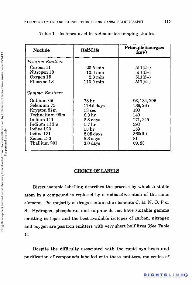
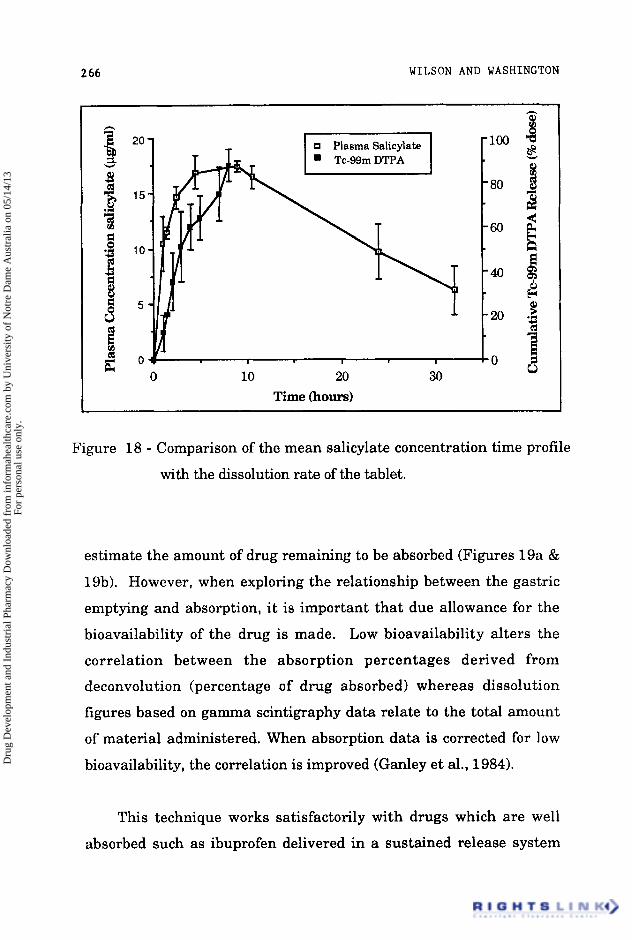
DISINTEGRATION AND DISSOLUTION USING GAMMA SCINTIGRAPHY 269

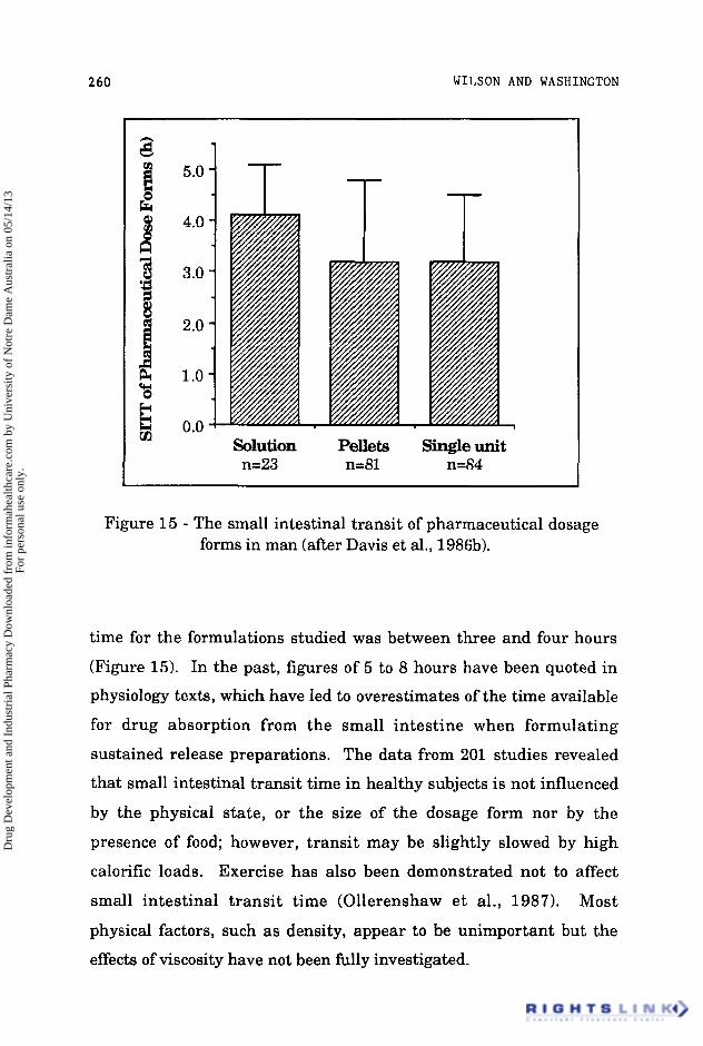
For many drugs, the absorption is dependent upon the rate of

disintegration of the dosage form and subsequent emptying into the

small intestine. The relationship between the in uiuo dispersion and

gastric emptying on the appearance of glibenclamide in the blood

after administration of a rapidly-dissolving liquid-filled capsule

formulation has been described by Ganley and coworkers (1984). In

the fasting state, the beginning of drug absorption indicated by the

first appearance of the drug in the plasma correlated well with the

start of in uiuo disintegration. Food markedly affected the dispersion

of the dosage form and delayed the appearance of the drug for an

hour, which correlated with the lag time for gastric emptying.

Inspection of the images after administration of food indicated that

the chief effect of food was t o inhibit the dispersion of the dosage form

within the stomach.

In order to study the absorption of drugs along the small

intestine, Ho, Merkle and Higuchi (1 983) modelled the absorption

process using a simple first-order model, which led to the prediction

of an exponential decrease in drug concentration with length of

small intestine. The authors then defined the intestinal reserve

length as the distance from the point at which 95% of drug had been

absorbed to the distal end of the small intestine. Although intestinal

reserve length produces a useful guideline, it makes a number of

assumptions, primarily that there is no variation in the absorptive

capacity of the small intestine along its length. This assumption

may be true for some materials, but in other cases absorption may be

carrier mediated or occur a t specific places e.g. thiouracil o r

griseofulvin. Additionally, the selection of 95% absorption as an

indicator of "complete" absorption is arbitrary; the authors present

Dru

g D

evel

opm

ent a

nd I

ndus

tria

l Pha

rmac

y D

ownl

oade

d fr

om in

form

ahea

lthca

re.c

om b

y U

nive

rsity

of

Not

re D

ame

Aus

tral

ia o

n 05

/14/

13Fo

r pe

rson

al u

se o

nly.

270 WILSON AND WASHINGTON

no data to allow such a point to be determined experimentally nor is

the available data sufficiently precise to allow extrapolation.

In spite of these shortcomings, the model does provide an

explanation of a number of phenomema, notably the variation in

absorption with transit velocity, however, the model is only semi-

quantitative a t best and must be evaluated with its physiological

limitations in mind.

A delay in gastric emptying can provide a prolonged period for

dissolution which would be expected to increase the availability of a

drug such as acyclovir, whose solubility in acidic media is relatively

high. As has been discussed, food is the major determinant affecting

gastric emptying and therefore the rate of presentation of a

suspension of the drug to the small intestine can be controlled by

administration with a light or heavy breakfast. Acyclovir (400 mg

suspension in 20 ml water), was labelled by inclusion of technetium-

99m labelled anion exchange resin and administered to healthy

volunteers with either a full English breakfast (3600kJ) or a light

continental breakfast (1 500kJ). Venous blood samples were collected

over a 24 hour period and the subjects imaged for the first 10 hours

after dosing. The heavy meal significantly decreased the rate of

gastric emptying and caused an increase in the small intestinal

transit time; however, the peak plasma concentration and the area

under the plasma-concentration-time profile were reduced. The time

to peak concentration was not significantly different with the two

meals, and occurred within two hours of dosing, suggesting that the

site of maximum absorption is situated in the proximal small

intestine. These data suggest, that the simplistic approach of the

Dru

g D

evel

opm

ent a

nd I

ndus

tria

l Pha

rmac

y D

ownl

oade

d fr

om in

form

ahea

lthca

re.c

om b

y U

nive

rsity

of

Not

re D

ame

Aus

tral

ia o

n 05

/14/

13Fo

r pe

rson

al u

se o

nly.

DISINTEGRATION AND DISSOLUTION USING GAMMA SCINTIGRAPHY 271

intestinal reserve length theory may be inadequate to predict the

behaviour of drugs which show a marked decrease in solubility when

transfered from an acidic t o a more neutral medium (Wilson et al.,

1987~).

It is widely appreciated that there is, as yet, no universal

dissolution test which in every instance would correlate in uitro

performance and in uiuo bioavailability. In view of the information

gained from scintigraphic investigations, it is probably unrealistic to

expect that a single in uitro apparatus will ever be able to model the

complex interplay between the formulation and the biological factors.

Gamma scintigraphy is a technique which has greatly advanced our

understanding of the behaviour of dosage forms and will continue to

do so, particularly in combination with pharmacokinetic and

telemetric techniques. Ultimately, it should be possible to explain all

the factors in the sequence between the release of drug from the

formulation t o the expression of the pharmacodynamic response.

The authors would like t o express their thanks to Dr Clive

Washington and Miss Jane Greaves with their advise and assistance

with the preparation with this manuscript.

Armstrong N A, James K C, Girardin H, Burch A, Davies R L & Mitchell G M. (1983).

Dru

g D

evel

opm

ent a

nd I

ndus

tria

l Pha

rmac

y D

ownl

oade

d fr

om in

form

ahea

lthca

re.c

om b

y U

nive

rsity

of

Not

re D

ame

Aus

tral

ia o

n 05

/14/

13Fo

r pe

rson

al u

se o

nly.

2 72 WILSON AND WASHINGTON

"The dispersion of oils from soft gelatin capsules 11. In uivo experiments." Int. J. Pharm. Tech. Prod. Mfr. 4:lO-13.

Beaumont W.(1955). "Experiments and observations on the gastric juice and the physiology of digestion." Dover Publishers Inc. (New York).

Bechgaard H, Christensen F N, Davis S S, Hardy J G , Taylor M J , Whalley D R & Wilson C G. (1985). "Gastrointestinal transit of pellet systems in ileostomy subjects and the effect of density." J. Pharm. Pharmacol. 37: 718-721.

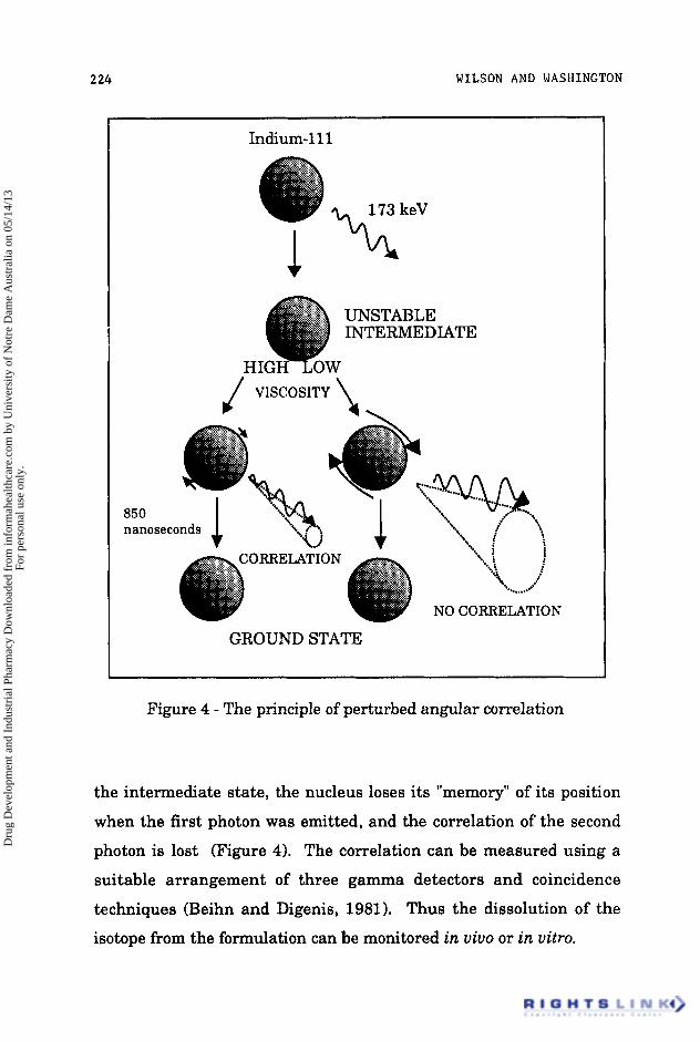
Beihn R M & Digenis G A. (1981 1. "Non-invasive dissolution measurement using perturbed angular correlation." J. Pharm. Sci. 70: 1325-1328.

Bogentoft C, Alpsten M, Ekenved G, & Hassle A B. (1984) "Absorption of acetylsalicylic acid from enteric-coated tablets in relation to gastric emptying and in-uiuo disintegration." J. Pharm. Pharmacol. 36: 350-351.

Casey D L, Beihn R M, Digenis G A & Shambhu M B. (1976). "Method for monitoring hard gelatin capsule disintegration times in humans using external scintigraphy." J. Pharm. Sci. 65: 1412-1413.

Charmer K S & Roberts C J C. (1985). "Effect of delayed oesophageal transit on acetaminophen absorption." Clin. Pharmacol. Ther. 37: 72-76.

Charmer K S & Virjee J P. (1985). "The effect of formulation on oesophageal transit." J. Pharm. Pharmacol. 37: 126-129.

Charmer K S & Virjee J P. (1986). "The effect of size and shape of tablets on their oesophageal transit." J. Clin. Pharmacol. 26: 141-146.

Dru

g D

evel

opm

ent a

nd I

ndus

tria

l Pha

rmac

y D

ownl

oade

d fr

om in

form

ahea

lthca

re.c

om b

y U

nive

rsity

of

Not

re D

ame

Aus

tral

ia o

n 05

/14/

13Fo

r pe

rson

al u

se o

nly.

DISINTEGRATION AND DISSOLUTION USING GAMMA SCINTIGRAPHY 273

Christensen J M, Ghannam M & Ayres J W. (1984). "Neutron activation of iron tablets to evaluate the effects of glycine on iron absorption." J. Pharm. Sci.. 73: 1529-1531.

Collins P J, Chatterton B E & Horowitz M. (1987). "Differential emptying rates of proximal and distal stomach in normal volunteers." J. Nucl. Med. 28: 605.

Daly P B, Davis S S, Frier M, Hardy J G, Kennerley J W & Wilson C G. (1982) "Scintigraphic assessment of the in uiuo dissolution rate of a sustained release tablet." Int. J. Pharm. 10: 17-24.

D'Arcy P F. (1984). "Oesophageal problems with tablets and capsules." Pharmacy International 5(5): 109.

Datz F L, Christian P E & Moore J. (1987). "Gender- related differences in gastric emptying." J. Nucl. Med. 28: 1204-1207.

Davis S S, Hardy J G, Stockwell A, Taylor M J, Whalley D R & Wilson C G. (1984a). "The effect of food on the gastrointestinal transit of pellets and an osmotic device (Osmet)." Int . J. Pharm. 21: 331-340.

Davis S S, Hardy J G, Taylor M J, Whalley D R & Wilson C G.(1984b). " A comparative study of the gastrointestinal transit of a pellet and tablet formulation." Int. J. Pharm. 21: 167-177

Davis S S, Hardy J G & Wilson C G. (1985). Letter to Lancet Lancet 1 : 2nd March p582.

Dru

g D

evel

opm

ent a

nd I

ndus

tria

l Pha

rmac

y D

ownl

oade

d fr

om in

form

ahea

lthca

re.c

om b

y U

nive

rsity

of

Not

re D

ame

Aus

tral

ia o

n 05

/14/

13Fo

r pe

rson

al u

se o

nly.

274 WILSON AND WASHINGTON

Davis S S, Hardy J G, Wilson C G, Feely L C & Palin K J. (1986a). "Gastrointestinal transit of a controlled release naproxen tablet formulation." Int. J. Pharm. 32: 85-90.

Davis S S, Hardy J G & Fara J W. (198613). "Transit of pharmaceutical dosage forms through the small intestine." Gut. 8: 886-892.

Davis S S, Khosla R, Wilson C G & Washington N. (1987). "The gastrointestinal transit of a controlled release pellet formulation of tiaprofenic acid." Int. J. Pharm. 34: 253-258.

Day T K, (1983). "Intestinal perforation associated with osmotic slow release indomethacin capsules." Br. Med. J. 287: 1671-1672.

Dew M J, Hughes P J, Lee M G, Evans B K & Rhodes J.(1982). "An oral preparation to release drugs in the human colon." Br. J. Clin. Pharmac. 14: 405-408.

Evans M A, Triggs E J, Cheung M, Broe G A & Creasey H. (1981). "Gastric emptying rate in the elderly, implications in drug therapy." J. Am. Geriat. SOC. 29: 201-205.

Feldman M, Smith H J & Simon T R. (1984). "Gastric emptying of solid radiopaque markers: studies in healthy subjects and diabetic patients." Gastroenterology 87: 895-902.

Fell J T. (1983). "Esophageal transit of tablets and capsules." Am. J. Hosp. Pharm. 40: 946-948.

Fitzgerald P, Hollingsbee D A, Gilbert 13 & Wilson C G. (1986). "The precorneal clearance of PVA film in man." J. Pharm. Pharmacol. 38(Suppl): 7P.

Dru

g D

evel

opm

ent a

nd I

ndus

tria

l Pha

rmac

y D

ownl

oade

d fr

om in

form

ahea

lthca

re.c

om b

y U

nive

rsity

of

Not

re D

ame

Aus

tral

ia o

n 05

/14/

13Fo

r pe

rson

al u

se o

nly.

DISINTEGRATION AND DISSOLUTION USING GAMMA SCINTIGRAPHY 215

Galeone M, Nizzola L, Cacioli D & Moise G. (1981). "In uivo demonstration of delivery mechanisms from sustained- re1 ease pellets ." Curr Ther Res. 29: 217-234.

Ganley J A, McEwen J, Calvert R T & Barker M C J. (1984). "The effect of in-uiuo dispersion and gastric emptying on glibenclamide absorption from a novel, rapidly dissolving capsule formulation." J. Pharm. Pharmacol. 36: 734-739.

Halls J.(1965). "Bowel content shift during normal defaecation." Proc. R. SOC. Med. 58: 859-860.

Hardy J G & Wilson C G. (1 981 1. "Radionuclide imaging in pharmaceutical, physiological and pharmacological research." Clinical Physics and Physiological Measurement. V01.2(2): 72-1 21.

Hardy J G & Perkins A C. (1985) "Validity of the geometric mean correction in the quantification of whole bowel transit." Nucl. Med. Comm. 6: 217-224.

Hardy J G, Wilson C G & Wood E. (1985). "Drug delivery to the proximal colon." J. Pharm. Pharmacol. 37: 874-877.

Hardy J G, Lee S W, Clark A G & Reynolds J R. (1986). "Enema volume and spreading." Int. J. Pharm. 32: 85-90.

Hardy J G, Evans D F, Zaki I, Clark A G, Tprnnesen H H & Gamst 0 N. (1987a). "Evaluation of an enteric coated naproxen tablet using gamma scintigraphy and pH monitoring." Int. J. Pharm. 37: 245-250.

Dru

g D

evel

opm

ent a

nd I

ndus

tria

l Pha

rmac

y D

ownl

oade

d fr

om in

form

ahea

lthca

re.c

om b

y U

nive

rsity

of

Not

re D

ame

Aus

tral

ia o

n 05

/14/

13Fo

r pe

rson

al u

se o

nly.

2 76 WILSON AND WASHINGTON

Hardy J G, Feely L C, Wood E & Davis S S. (198713). "The application of gamma-scintigraphy for the evaluation of the relative spreading of suppository bases in rectal hard gelatin capsules." Int. J. Pharm. 38: 103-108.

Heading R C, Nimmo J, Prescott L F & Tothill P. (1973). "The dependence of paracetamol absorption on the rate of gastric emptying." Br. J. Pharmacol. 47: 415-421.

Hey H, J~rgensen F, Sgrensen K, Hasselbalch H & Wamberg T. (1 982). "Oesophageal transit of six commonly used tablets and capsules." Br. Med. J. 285: 1717-1719.

Ho N F H, Merkle H P & Higuchi W I. (1983) "Quantitative, mechanistic and physiologically realistic approach to the biopharmaceutical design of oral drug delivery systems." Drug Devel. Ind. Pharm.9(7): 1111-1184.

Holt S, Reid J, Taylor T V,Tothill P & Heading R C. (1982). "Gastric emptying of solids in man." Gut. 23: 292-296.

Hunter E, Fell J T, Calvert R T & Sharma H. (1980). "In uiuo disintegration of hard gelatin capsules in fasting and non- fasting subjects." Int. J . Pharm. 4: 175-183.

Hunter E, Fell J T & Sharma H. (1982). "The gastric emptying of pellets contained in hard gelatin capsules." Drug Devel. Ind. Pharm. 8: 751-757.

Hunter E, Fell J T & Sharma H. (1983). "The gastric emptying of hard gelatin capsules." Int. J. Pharm. 17: 59-64.

Dru

g D

evel

opm

ent a

nd I

ndus

tria

l Pha

rmac

y D

ownl

oade

d fr

om in

form

ahea

lthca

re.c

om b

y U

nive

rsity

of

Not

re D

ame

Aus

tral

ia o

n 05

/14/

13Fo

r pe

rson

al u

se o

nly.

DISINTEGRATION AND DISSOLUTION USING GAMMA SCINTIGRAPHY 271

Jenkins J R F, Hardy J G & Wilson C G. (1983). "Monitoring antacid preparations in the stomach using gamma scintigraphy. Int. J. Pharm. 14: 143-148.

Kaus L C, Fell J T, Sharma H & Taylor D C'(1984a). "On the intestinal transit of a single non-disintegrating object." Int. J. Pharm. 20: 315-323.

Kaus L C , Sharma H & Fell J T. (198413). "Simultaneous measurement of gastric emptying of the soluble and insoluble components of a formulation using a dual isotope, gamma scintigraphic techmique." J. Pharm. Pharmacol. 36: 136-138.

Kaus L C, Fell J T, Sharma H & Taylor D C. (1984~). "Gastric emptying and intestinal transit of non-disintegrating capsules - the influence of metoclopramide." Int. J. Pharm. 22: 99-103.

Kelly J D.0984) "Choice of radionuclides for scintigraphy." in "Radionuclide Imaging in Drug Research", C G Wilson, J G Hardy, M Frier, S S Davis (Eds.). Published by Croom Helm Ltd., London. pp 39-59.

King P M, Adam R D, Pryde A, McDicken W N & Heading R C. (1 984). "Relationships of human antroduodenal motility and transpyloric movement; non-invasive observation with real-time ultrasound." Gut. 25: 1384-1391.

Kuna S. (1964). "The pH of gastric juice in the normal resting stomach." Arch. Int. Pharmacodyn. 151:79.

Levine R R. (1 970). "Factors affecting gastrointestinal absorption of drugs." Am. J.Dig. Dis. 15: 171-188.

Dru

g D

evel

opm

ent a

nd I

ndus

tria

l Pha

rmac

y D

ownl

oade

d fr

om in

form

ahea

lthca

re.c

om b

y U

nive

rsity

of

Not

re D

ame

Aus

tral

ia o

n 05

/14/

13Fo

r pe

rson

al u

se o

nly.

2 1a WILSON AND WASHINGTON

Levy G. (1 963). "Effect of particle size on dissolution and gastrointestinal absorption rates of pharmaceuticals." J. Pharm. Sci. 52: 1039.

Marvola M, Rajaniemvuo M, Marttila E, Vahervuo K & Sothmann A. (1983). "Effect of dosage form and formulation factors on the adherence of drugs to the esophagus." J. Pharm. Sci.72: 1034-1036.

Maublant J C, Sournac M, Aiache J M & Veyre A. (1987). "Dissolution rate and transit times of technetium-99m DTPA-labelled tablets." J. Nucl. Med. 28: 1199-1203.

May H A, Wilson C G & Hardy J G. (1984). "Monitoring radiolabelled antacid preparations in the stomach." Int. J. Pharm. 19: 169-176.

Mayersohn M. "Physiological factors that modify systemic drug availability and pharmacologic response in clinical practice." in Principles and Perspective in Drug Bioavailability pp.211-273 (Karger, Base1 1979).

Moore J G , Dubois A, Christian P E, Elgin D & Alazraki N. (1986). "Evidence for a midgastric transverse bands in humans." Gastroenterology .91: 540-545.

Muller-Lissner S A, Muller-Duysing W, Heinzel F & Blum A LA1 981 ). "Floating capsules with slow release of active agents." Dtsch. Med. Wschr. 106: 1143-1147.

Muller-Lissner S A & Blum A L. (1981). "The effects of specific gravity and eating on gastric emptying of slow release capsules." New Eng. J. Med. 304: 1365-1366.

Dru

g D

evel

opm

ent a

nd I

ndus

tria

l Pha

rmac

y D

ownl

oade

d fr

om in

form

ahea

lthca

re.c

om b

y U

nive

rsity

of

Not

re D

ame

Aus

tral

ia o

n 05

/14/

13Fo

r pe

rson

al u

se o

nly.

DISINTEGRATION AND DISSOLUTION USING GAMMA SCINTIGRAPHY 279

Olejnik 0 & Wilson C G. (1987). "Gamma scintigraphy update: Use of gamma scintigraphy in the development of ophthalmic formulations." Published by Excerpta Medica, Elsevier, USA.

Ollerenshaw K, Norman S, Hardy J G & Wilson C G. (1987) "Exercise and small intestinal transit." Nucl. Med. Commun.8: 105-110.

O'Reilly S, Wilson C G & Hardy J G. (1987). "The influence of food on gastric emptying of multiparticulate dosage forms . I' Int. J. Pharm. 34: 213-216.

Park H M, Chernish S M, Rosenek B D, Brunelle R L, Hargrove B & Wellman H N. (1984). "Gastric empting of enteric coated tablets." Dig. Dis. Sci. 29: 207-212.

Parr A, Beihn R M & Jay M. (1986). "Radiolabelling of an enteric coated tablet by (n, gamma) radioactivation of erbium-1 70 for scintigraphic imaging." Int. J. Pharm. 32: 251-256.

Phipps P, Borham P, Gonda I, Bailey D, Bautovich G & Anderson S. (1 987). "A rapid method for the evaluation of diagnostic radioaerosol delivery systems." Eur. J. Nucl. Med. (in press).

Quigley J P & Brody D A. (1950). "Digestive tract: intraluminal pressures gastrointestinal propulsion, gastric evacuation, pressure-wall tension relationship." in, Medical Physics Year Book, Glasser 0. (Ed.), Medical Publishers, Chicago. pp. 280-292.

Rao L S, Hardy J G & Wilson C G. (1983). "Tissue distribution and fate of free and liposome-encapsulated [125Sb] sodium stibogluconate by gamma scintigraphy." Int. J. Pharm. 17:283-290.

Dru

g D

evel

opm

ent a

nd I

ndus

tria

l Pha

rmac

y D

ownl

oade

d fr

om in

form

ahea

lthca

re.c

om b

y U

nive

rsity

of

Not

re D

ame

Aus

tral

ia o

n 05

/14/

13Fo

r pe

rson

al u

se o

nly.

280 WILSON AND WASHINGTON

Schor J M. (1980). "Sustained release therapeutic compositions." US. Patent 4226849.

Shell D H. (1982). "Pharmacokinetics of topically applied ophthalmic drugs." Sum. Ophthalmol. 26: 207-218.

Steinberg W H, Hutchins H H, Pick P G & Lazar J S. (1965). "Automated technique for determining dissolution and reaction rate of antacids." J. Pharm. Sci. 54: 625-633.

Swisher D A, Sendelbeck S L & Fara J W. (1984). "Adherence of various oral dosage forms to the oesophagus." Int. J. Pharm. 22: 219-228.

Theeuwes F.(1975). "Alimentary osmotic pump." J. Pharm. Sci. 64: 1987-1991.

Ter-Pogossin M M, Raichle M E & Sobel B E. (1980). "Positron-emission tomography." Sci. Am. 243: 141-155.

Theodorakis M C, Devous M D & Simpson D R. (1980). "Monitoring in uiuo disintegration time of tablets by external s cin tigr a p h y . J. Pharm. Sci. 69: 1107-1108.

Toothaker R D & Welling P G. (1980). "The effect of food on drug bioavailability" Ann. Rev. Pharmac. Toxic 20: 173-199.

Tothill P, McLoughlin G P & Heading R C.(1978). "Techniques and errors in scintigraphic measurements of gastric emptying. J, Nucl. Med. 19: 256-261.

Dru

g D

evel

opm

ent a

nd I

ndus

tria

l Pha

rmac

y D

ownl

oade

d fr

om in

form

ahea

lthca

re.c

om b

y U

nive

rsity

of

Not

re D

ame

Aus

tral

ia o

n 05

/14/

13Fo

r pe

rson

al u

se o

nly.

DISINTEGRATION AND DISSOLUTION USING GAMMA SCINTIGRAPHY 281

Tukker Jd1983). "Biopharmaceutics of fatty suspension suppositories: The influence of physiological and physical parameters on spreading and bioavailability in dog and man." Ph. D. thesis, University of Leiden, The Netherlands.

Washington N, Wilson C G & Davis S S. (1985). "Evaluation of 'raft-forming' antacid neutralising capacity: in uitro and in uiuo correlations." Int. J. Pharm. 27: 279-286.

Washington N, Washington C & Wilson C G. (1986). "The effect of food on the gastric emptying of a floating alginate raft." Society for Drug Research, Cambridge.

Wilson C G, Parr G D, Kennerley J W, Taylor M J, Davis S S, Hardy J G & Rees J G. (1984) "Pharmacokinetics and in uivo scintigraphic monitoring of a sustained release acetylsalicylic acid formulation." Int. J. Pharm. 18: 1-8.

Wilson C G & Hardy J G. (1985). "Gastrointestinal transit of an osmotic tablet drug delivery system." J. Pharm. Pharmacol. 37: 573-575.

Wilson C G, Washington N, Peach J, Murray G R & Kennerley J. (1987a). "The behaviour of a fast-dissolving dosage form (Expidet) followed by gamma scintigraphy." Int. J. Phann. (in press).

Wilson C G, Kamali F, Washington N, Rees J A & Sempik A K. (1 987b). "A scintigraphic and pharmacokinetic open study of an ibuprofen formulation in healthy volunteers." Int. J. Pharm. (in press).

Wilson C G, Washington N, Hardy J G & Bond S W. (1987~). "The influence of food on the absorption of acyclovir: a pharmacokinetic and scintigraphic assessment." Int. J. Pharm. 38: 221-225.

Dru

g D

evel

opm

ent a

nd I

ndus

tria

l Pha

rmac

y D

ownl

oade

d fr

om in

form

ahea

lthca

re.c

om b

y U

nive

rsity

of

Not

re D

ame

Aus

tral

ia o

n 05

/14/

13Fo

r pe

rson

al u

se o

nly.


Recommended