Background: The circle of Willis is a major collateral pathway important in ischemic conditions. The aim of our study was to assess the structural characteristics of the circle of Willis within the Turkish adult population, along with vari-ations and arteries involved in the measurement of diameters and lengths on cranial computed tomography angiography (CTA).
Material/Methods: One hundred adult patients who underwent CTA images were evaluated retrospectively. Results: Results of the study revealed 82% adult, 17% fetal, and 1% transitional configurations. A complete polygonal
structure was observed in 28% of cases. Variations of the circle of Willis were more common in the posterior portion. Hypoplasia was found to be the most common variation and was observed as a maximum in the pos-terior communicating artery (AComP).
Conclusions: The patency and size of arteries in the circle of Willis are important in occlusive cerebrovascular diseases and cerebrovascular surgery. Although CTA is an easily accessible non-invasive clinical method for demonstrating the vascular structure, CTA should be evaluated taking into account image resolution quality and difficulties in the identification of small vessels.
MeSH Keywords: Cerebral Angiography • Cerebral Arteries • Circle of Willis • Tomography Scanners, X-Ray Computed
Data Collection B Statistical Analysis CData Interpretation D
Manuscript Preparation E Literature Search FFunds Collection G
1 Department of Neurosurgery, Izmir Katip Celebi University Ataturk Education and Research Hospital, Izmir, Turkey
2 Department of Neurosurgery, Bozyaka Education and Research Hospital, Izmir, Turkey
3 Department of Radiology, Ege University School of Medicine, Izmir, Turkey4 Department of Anatomy, Ankara University School of Medicine, Ankara, Turkey
This work is licensed under a Creative CommonsAttribution-NonCommercial-NoDerivs 3.0 Unported License
Background
The circle of Willis, whose function is to protect the brain from ischemia, is the main structure that provides a constant and regular blood flow to the brain. The circle of Willis is an arterial polygon and is a vascular ring composed of branches of arteria (a) carotis interna and a. basilaris. This circle is composed of bilateral A1 segments of anterior cerebral arteries (ACAs), ante-rior communicating artery (AComA) that links these 2 arteries, bilateral P1 segments of posterior cerebral arteries (ACPs), and posterior communicating arteries (AComP) which links a. caro-tis interna (ACI) to ACP. This communicating pathway allows equalization of the blood flow between the 2 sides of the brain and permits anastomotic circulation, if a part of the circulation becomes occluded. In recent years, detailed knowledge about the circle of Willis has generally been based on cadaver stud-ies [1–5]. The Willis polygon and its principal arteries can be imaged angiographically using a non-invasive, easily accessi-ble method such as computed tomography angiography (CTA). Obtaining a 3-dimensional data set became possible with the introduction of multi-detector CT devices, and, when used in conjunction with recently developed image processing tech-niques, it has become possible to obtain angiographic imag-es of the Willis polygon and its principal arteries with better resolution at each layout [6–8]. The aim of this study was as-sessment of the structural characteristics and variations, and to measure the diameter and length of the arteries of the cir-cle of Willis on CTA in the Turkish population.
Material and Methods
Patient population
In the present study, the Willis polygon was retrospectively eval-uated among adult patients who had reported CTA normally at Ege University School of Medicine Department of Radiology. This study was approved by the Ege University ethical com-mittee. One hundred patients (57 male, 43 female, age range 16–83 years, median age 60.06) were selected. Patients who were excluded from the study were those with cerebrovascu-lar malformations such as aneurysm, arteriovenous malforma-tion, arteriovenous fistula, and acute ischemic, hemorrhagic cerebrovascular strokes.
Image technique
In Multi Detector CTA examination, a CT scan with 64 detectors was used (Somatom Sensation; Siemens, Erlangen, Germany). The examination was performed by using scans with contrast administration, so as to cover the supra-aortic and cranial area. Application of contrast was performed by giving intrave-nous (4–5 ml/s) 80 ml non-ionic contrast material iopromide
(Ultravist 370; Bayer group, Berlin, Germany) and, immediate-ly after that, 25 ml serum physiologic.
Data analysis
The data were transferred to a workstation for post-process-ing. The diameter and length of the arteries which constituted the circle of Willis were measured after proper magnification. Variations were evaluated (an invisible artery was evaluated as aplasia, while the diameter of an artery smaller than 1 mm was evaluated as hypoplasia). The structures of the circle of Willis were evaluated by being complete or incomplete imag-es (complete image means all of the arteries forming the circle without hypoplasia, and incomplete image means with apla-sia) and according to the diameter of AComP-P1 relationship. There are 3 configurations according to diameters of AComP and ACP: Adult configuration, with diameter of AComP small-er than P1; fetal configuration, with diameter of AComP larg-er than P1; and the main vascularization of the occipital lobe are provided by the ACI. The transitional configuration has a diameter of AComP the same as with P1 [9]. Data were trans-ferred to SPSS 20 (Statistical Packages for Social Sciences) program for statistical analysis. Wilcoxon test was used for comparing right and left sides. Statistical significance was cal-culated with to corrected nominal alpha values according to the Bonferroni method.
Results
The study group consisted of 43 females (43%) and 57 males (57%) and the mean age was 60.06 years (range 16–83). All of the arteries forming the circle of Willis were found in 71 samples (71%). Complete polygonal structure included all ar-teries forming the circle, however, hypoplastic arteries were not evaluated – this was obtained in just 28 samples (28%). Incomplete circles, which included invisible arteries, were found in 29 samples (6 in the anterior, 22 in the posterior, and 1 in both) (29%). Adult configuration was detected in 82 samples (82%). Fetal configuration was detected in 17 (17%) and tran-sitional configuration was detected in 1 (1%) (Tables 1, 2). Variations were found more in the posterior part. Hypoplasia was the most common variation and was found to be most common in AComP (38%). Aplasia was the second leading vari-ation and was also detected in AComP (23%). AComA hypo-plasia is the most common variation seen in the anterior part (23%). Hypoplasia was found to be most common in AComA following AComP. The most common multiple variation was the coexistence of AComA and AComP hypoplasia, which was found in 12 samples (12%) (Figures 1–4).
No statistically significant difference was noted between the groups in terms of age, sex, or bilaterally measured values.
This work is licensed under a Creative CommonsAttribution-NonCommercial-NoDerivs 3.0 Unported License
Arteria cerebri anterior
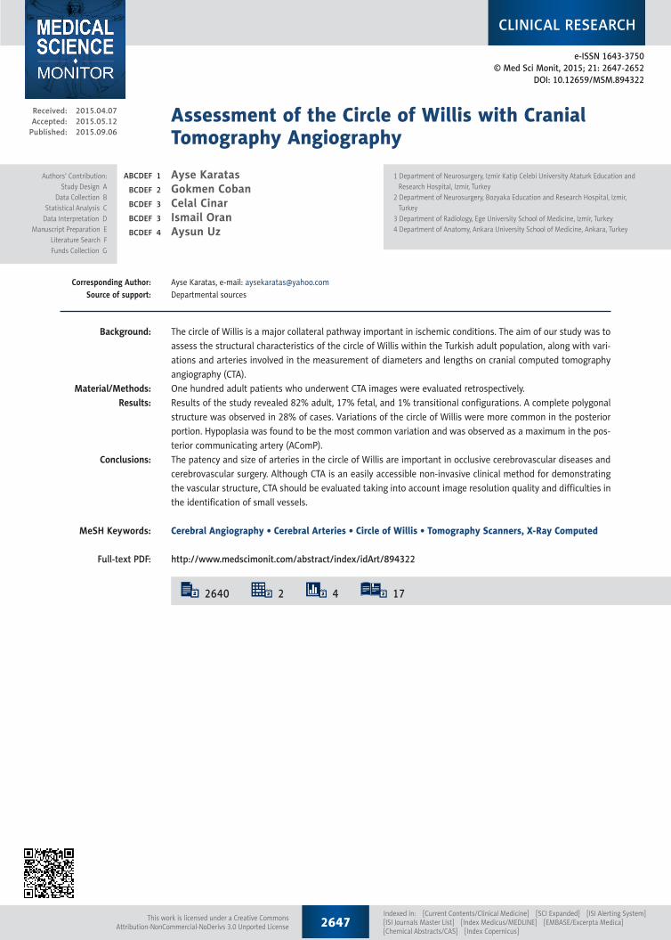
A1 length was measured as 15.61±2.79 mm in the right and 15.13±2.54 mm in the left. A1 diameter was measured as 2.15±0.63 mm in the right and 2.26±0.61 mm in the left. A1 hypoplasia was detected 3 (2 single, 1 combined; together with other variations) in the right, and 2 (combined) in the left. Aplasia was seen 5 (3 single and 2 combined) in the right, and 2 (1 single, 1 combined) in the left.
Arteria communicans anterior
The mean length of the AComA was 1.48±1.45 mm and the di-ameter was 1.39±0.83 mm. Twenty-three (7 single, 16 combined variations) hypoplasia and 1 combined aplasia were detected.
Arteria communicans posterior
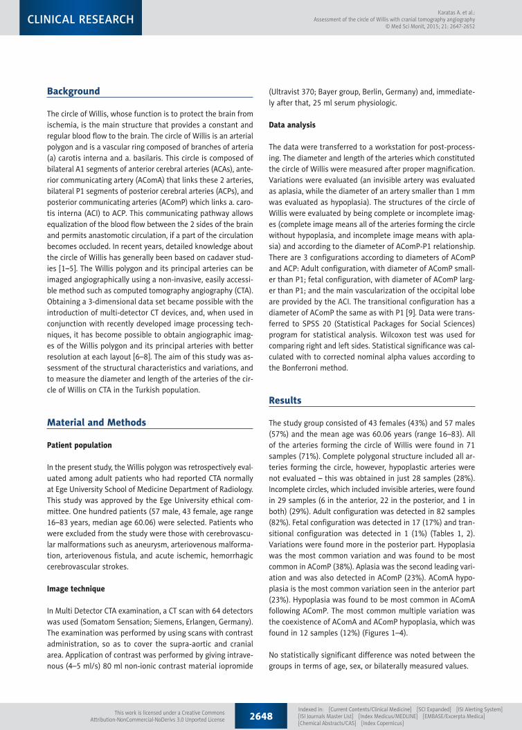
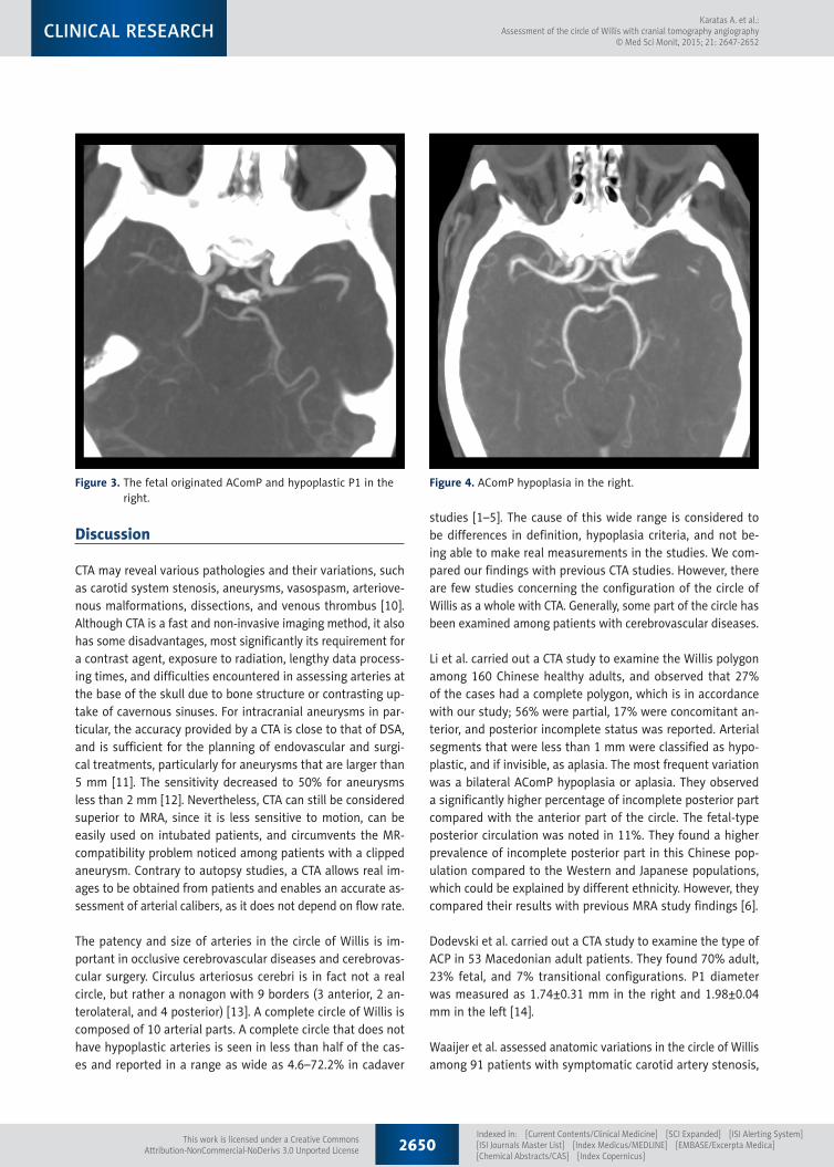
The length of the AComP was 10.99±3.07 mm in the right and 9.87 ±3.32 mm in the left. AComP diameter was 1.30±0.50 mm in the right and 1.27±0.55 mm in the left. A total of 38 hypo-plasia (3 right, 12 left, 5 bilateral, and 18 combined) and 23 aplasia (8 right, 7 left, 1 bilateral, and 7 combined) were de-tected in AComP.
Arteria cerebri posterior
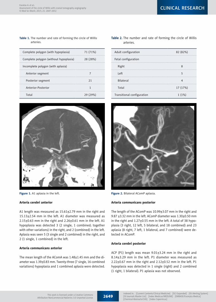
ACP (P1) length was mean 9.01±3.24 mm in the right and 8.14±3.29 mm in the left. P1 diameter was measured as 2.22±0.67 mm in the right and 2.12±0.52 mm in the left. P1 hypoplasia was detected in 1 single (right) and 2 combined (1 right, 1 bilateral). P1 aplasia was not observed.
Complete polygon (with hypoplasia) 71 (71%)
Complete polygon (without hypoplasia) 28 (28%)
Incomplete polygon (with aplasia)
Anterior segment 7
Posterior segment 21
Anterior-Posterior 1
Total 29 (29%)
Table 1. The number and rate of forming the circle of Willis arteries.
Adult configuration 82 (82%)
Fetal configuration
Right 8
Left 5
Bilateral 4
Total 17 (17%)
Transitional configuration 1 (1%)
Table 2. The number and rate of forming the circle of Willis arteries.
Figure 1. A1 aplasia in the left. Figure 2. Bilateral AComP aplasia.
This work is licensed under a Creative CommonsAttribution-NonCommercial-NoDerivs 3.0 Unported License
Discussion
CTA may reveal various pathologies and their variations, such as carotid system stenosis, aneurysms, vasospasm, arteriove-nous malformations, dissections, and venous thrombus [10]. Although CTA is a fast and non-invasive imaging method, it also has some disadvantages, most significantly its requirement for a contrast agent, exposure to radiation, lengthy data process-ing times, and difficulties encountered in assessing arteries at the base of the skull due to bone structure or contrasting up-take of cavernous sinuses. For intracranial aneurysms in par-ticular, the accuracy provided by a CTA is close to that of DSA, and is sufficient for the planning of endovascular and surgi-cal treatments, particularly for aneurysms that are larger than 5 mm [11]. The sensitivity decreased to 50% for aneurysms less than 2 mm [12]. Nevertheless, CTA can still be considered superior to MRA, since it is less sensitive to motion, can be easily used on intubated patients, and circumvents the MR-compatibility problem noticed among patients with a clipped aneurysm. Contrary to autopsy studies, a CTA allows real im-ages to be obtained from patients and enables an accurate as-sessment of arterial calibers, as it does not depend on flow rate.
The patency and size of arteries in the circle of Willis is im-portant in occlusive cerebrovascular diseases and cerebrovas-cular surgery. Circulus arteriosus cerebri is in fact not a real circle, but rather a nonagon with 9 borders (3 anterior, 2 an-terolateral, and 4 posterior) [13]. A complete circle of Willis is composed of 10 arterial parts. A complete circle that does not have hypoplastic arteries is seen in less than half of the cas-es and reported in a range as wide as 4.6–72.2% in cadaver
studies [1–5]. The cause of this wide range is considered to be differences in definition, hypoplasia criteria, and not be-ing able to make real measurements in the studies. We com-pared our findings with previous CTA studies. However, there are few studies concerning the configuration of the circle of Willis as a whole with CTA. Generally, some part of the circle has been examined among patients with cerebrovascular diseases.
Li et al. carried out a CTA study to examine the Willis polygon among 160 Chinese healthy adults, and observed that 27% of the cases had a complete polygon, which is in accordance with our study; 56% were partial, 17% were concomitant an-terior, and posterior incomplete status was reported. Arterial segments that were less than 1 mm were classified as hypo-plastic, and if invisible, as aplasia. The most frequent variation was a bilateral AComP hypoplasia or aplasia. They observed a significantly higher percentage of incomplete posterior part compared with the anterior part of the circle. The fetal-type posterior circulation was noted in 11%. They found a higher prevalence of incomplete posterior part in this Chinese pop-ulation compared to the Western and Japanese populations, which could be explained by different ethnicity. However, they compared their results with previous MRA study findings [6].
Dodevski et al. carried out a CTA study to examine the type of ACP in 53 Macedonian adult patients. They found 70% adult, 23% fetal, and 7% transitional configurations. P1 diameter was measured as 1.74±0.31 mm in the right and 1.98±0.04 mm in the left [14].
Waaijer et al. assessed anatomic variations in the circle of Willis among 91 patients with symptomatic carotid artery stenosis,
Figure 3. The fetal originated AComP and hypoplastic P1 in the right.
This work is licensed under a Creative CommonsAttribution-NonCommercial-NoDerivs 3.0 Unported License
and compared this to 91 control adult subjects with multislice CTA. The study was performed as follows for each arterial seg-ment: invisible, hypoplastic (<1 mm), or normal (³1 mm). In the control group, 1% aplasia was detected in AComA; 1% aplasia and 4% hypoplasia was detected in A1; and 27% aplasia and 40% hypoplasia was detected in AComP or P1. The study re-sults revealed 85% adult, 14% fetal, and 1% transitional con-figurations in the control group. These results were similar to our study. They concluded there was a relationship between symptomatic carotid stenosis and incomplete circle of Willis [8].
Urbanski et al. performed a study using CTA for the pre-opera-tive neurovascular assessment of 99 adult patients who were to undergo aortic surgery, and found that 59 patients had a complete polygon, 18 patients had a single abnormality (hypo-plasia or aplasia within anterior or posterior part), 13 patients had a bilateral AComP abnormality (hypoplasia or aplasia), and 9 patients had coexisting AComA and AComP abnormalities. However, despite these abnormalities, the patients developed no neurological deficits after the surgical procedure, which in-volved carotid clamping [7].
Han et al. analyzed the arterial segments of Willis among 117 subarachnoid hemorrhage patients CTA and DSA images. In the study, no flow was defined as “aplasia”, arteries luminal narrowing more than 50% were accepted as “hypoplasia”. In the AComA, 30% aplasia and 3% hypoplasia was detected. In the A1, 8% aplasia and 7% hypoplasia was detected. In the AComP, 65% aplasia and 5% hypoplasia was detected. In the P1, 9% aplasia and 8% hypoplasia was detected. The CTA results were compared with findings on the corresponding DSA imag-es. They concluded that CTA is highly accurate in the assess-ment of anatomical variations of the circle of Willis. However, CTA has limited sensitivity in depicting hypoplastic segments, although it is quite specific [10].
In the present study, variations in the Willis polygon were more common in the posterior part of the polygon, consistent with the literature, the most common of which was hypoplasia, be-ing most frequently observed in AComP (38%) [6,8]. Hypoplasia was found to be concomitant at 15% with unilateral, 5% with bilateral, and 18% with other abnormalities. The second most common variation was aplasia, which was also most frequent-ly observed in AComP (23%). In the AComA, hypoplasia was the second most common variation (23%), although AComA hypoplasia was the most common variation observed in the anterior region. The most common multiple variation was the coexistence of the AComA and AComP hypoplasia, which was encountered in 12% of the cases. A1 aplasia was observed in 7% of the cases, while AComA aplasia was observed in 1%. Aplasia was not detected in P1. A1 hypoplasia was present in 5% of cases, while P1 hypoplasia was present in 3%.
In the present study, 71% of the patients had all arteries con-stituting the Willis polygon. A complete polygonal structure without the evaluation of hypoplasic arteries was observed in just 28% of cases. An incomplete polygon with invisible arter-ies was encountered in 29 cases (29%). In our previous cadav-er study among the adult Turkish population, 91% the patients had all arteries constituting the Willis polygon. The complete and incomplete polygonal ratios were 8% and 9%, respectively, in that study [15]. The increased frequency of aplasia and de-creased frequency of hypoplasia encountered in the CTA group has been associated with the technical limitations of the CTA in the measurement of arteries with smaller diameters, which pro-hibits clear detection of hypoplasia and aplasia, as in an autopsy.
Our study results revealed 82% adult, 17% fetal, and 1% tran-sitional configurations. When we compared these results with our cadaveric study, fetal configurations were detected more frequently with CTA [15]. The higher percentage of fetal con-figurations can be explained by the infrequent occurrence of hypoplasic AComP and the significantly higher AComP diame-ters observed in the group. This is a result of the technical dif-ficulties of CTA in the assessment of hypoplasic arteries with diameters smaller than 1 mm.
Some arteries of the polygon may be so thin that interven-tion by radiological means is impossible, or they may not be present at all, and this is important in the treatment of oc-clusive cerebrovascular diseases and cerebrovascular surgery. Knowing the variations and understanding any abnormalities prior to a cerebrovascular (aneurysm, endartectomy, bypass) operation is important in ensuring safe surgery. The presence of hypoplastic or aplastic AComP is an independent risk fac-tor for ischemic cerebral infarction when the ACI is occluded [13]. However, Chuang et al. demonstrated that AComP hy-poplasia appears to be a contributor to the risk of ischemic stroke, even in the absence of ACI occlusion [16]. Miyazawa et al. also found a higher frequency of lacunes in the basal ganglia in patients with hypoplasia or aplasia in the anterior part of the circle of Willis [17]. CTA can be used practically in the evaluation of cerebral vascular structures, particularly for screening and pre-operative examination purposes in asymp-tomatic or normal individuals. CTAs should be evaluated con-sidering the image quality, venous contamination, insufficient contrast dosage, and limitations in the determination of arter-ies with small diameters. The structure of the Willis polygon, as well as the status and sufficiency of AComA and AComP, should be assessed. Aside from studies of the healthy popu-lation, the clinical implications of variations of the Willis poly-gon should be examined through studies of patients with ce-rebral arterial diseases.
The present study shows that although CTA is a valuable imag-ing method for clinical examination of the vascular structure,
This work is licensed under a Creative CommonsAttribution-NonCommercial-NoDerivs 3.0 Unported License
studies performed on cadavers are still important in this area, since CTA faces technical limitations in the imaging of some arteries. On the other hand, the abundance of variations ob-served in the Willis polygon among individuals implies that the utility of CTA as a guide will be significant.
Conclusions
Among patients due to undergo surgery, because of common variations of the Willis polygon, a pre-operative examination of
References:
1. Alpers BJ, Berry RG, Paddison RM: Anatomical studies of the circle of Willis in normal brain. Arch Neurol Psychiat, 1959; 81: 409–18
2. Baptista AG: Studies on the arteries of the brain. Acta Neurol Scand, 1964; 40: 398–414
3. Battacharji SK, Hutchinson EC, Mc Call AJ: The circle of Willis the incidance of developmental abnormalities in normal and infarcted brains. Brain, 1967; 90: 747–58
4. De Silva KRD, Silva R, Gunasekera WSL, Jayesekera RW: Prevelence of typi-cal circle of Willis and the variation in the anterior communicating artery: A study of a Sri Lankan population. Ann Indian Acad Neurol, 2009; 12: 157–61
5. Fisher CM: The circle of Willis: anatomical variations. Vasc Dis, 1965; 2: 99–105
6. Li Q, Li J, Lv F et al: A multidetector CT angiography study of variations in the circle of Willis in a Chinese population. J Clin Neurosci, 2011; 18: 379–83
7. Urbanski PP, Lenos A, Blume JC et al: Does anatomical completeness of the circle of Willis correlate with sufficient cross-perfusion during unilat-eral cerebral perfusion? Eur J Cardiothorac Surg, 2008; 33: 402–8
8. Waaijer A, van Leeuwen MS, van der Worp HB et al: Anatomic variations in the circle of Willis in patients with symptomatic carotid artery stenosis assessed with multidedector row CT Angiography. Cerebrovasc Dis, 2007; 23: 267–74
9. Padget DH: The circle of Willis. Its embriyology and anatomy. In: Dandy WE (ed.), Intracranial arterial aneurysms. Comstock publishing Co, NY, 1947; 67–90
the structure and variations of the Willis polygon through easily accessible and non-invasive techniques with CTA will decrease the potentially significant neurological complications and as-sociated secondary risks of morbidity and mortality. However, it should be noted that diagnosis of hypoplasia and aplasia during the CTA will not be sufficiently reliable, since the CTA has technical limitations in the assessment of small arteries.
Declaration of interest
The authors declare that they have no conflicts of interest.
10. Han A, Yoon DY, Chang SK et al: Accuracy of CT angiography in the assess-ment of the circle of Willis: comparison of volume-rendered images and digital subtraction angiography. Acta Radiol, 2011; 52: 889–93
11. White P, Teasdale E, Wardlaw J, Easton V: Intracranial aneurysms: CT angi-ography and MR angiography for detection-prospective blinded compari-son in a large patient cohort. Radiology, 2001; 219: 739–49
12. Wintermark M, Uske A, Chalaron M et al: Multislice computerized tomog-raphy angiography in the evaluation of intracranial aneurysms; a compar-ison with intraarterial digital subtraction angiography. J Neurosurg, 2003; 98: 828–36
14. Dodevski A, Tosovska Lazarova D, Mitreska N et al: Posterior cerebral artery – variation in the origin and clinical significance. Prilozi, 2014; 35: 163–68
15. Karatas A, Yilmaz H, Coban G et al: The anatomy of circulus arteriosus cerebri (circle of Willis). Paper presented at: 2014 European Congress of Neurosurgery, 2014; Prague, Czech Republic
16. Chuang YM, Liu CY, Pan PJ, Lin CP: Posterior communicating artery hypo-plasia as a risk factor for acute ischemic stroke in the absence of carotid artery occlusion. J Clin Neurosci, 2008; 15: 1376–81
17. Miyazawa N, Shinohara T, Yamagata Z: Association of incompleteness of the anterior part of the circle of Willis with the occurrence of lacunes in the basal ganglia. Eur J Neurol, 2011; 18: 1358–60