RESEARCH ARTICLE Association Between Cytoplasmic CRABP2, Altered Retinoic Acid Signaling, and Poor Prognosis in Glioblastoma Rong-Zong Liu, 1 Shuai Li, 1 Elizabeth Garcia, 1 Darryl D. Glubrecht, 1 Ho Yin Poon, 1 Jacob C. Easaw, 2 and Roseline Godbout 1 ABSTRACT: Retinoic acid (RA), a metabolite of vitamin A, is required for the regulation of growth and development. Aberrant expression of molecules involved in RA signaling has been reported in various cancer types including glioblastoma multiforme (GBM). Cellular retinoic acid-binding protein 2 (CRABP2) has previously been shown to play a key role in the transport of RA to retinoic acid receptors (RARs) to activate their transcription regulatory activity. Here, we demonstrate that CRABP2 is predominantly located in the cytoplasm of GBM tumors. Cytoplasmic, but not nuclear, CRABP2 levels in GBM tumors are associated with poor patient survival. Treatment of malignant glioma cell lines with RA results in a dose-dependent increase in accumulation of CRABP2 in the cytoplasm. CRABP2 knockdown reduces proliferation rates of malignant glioma cells, and enhances RA-induced RAR activation. Levels of CRYAB, a small heat shock protein with anti-apoptotic activity, and GFAP, an astrocyte-specific intermediate filament protein, are greatly reduced in CRABP2-depleted cells. Restoration of CRYAB expres- sion partially but significantly reversed the effect of CRABP2 depletion on RAR activation. Our combined in vivo and in vitro data indicate that: (i) CRABP2 is an important determinant of clinical outcome in GBM patients, and (ii) the mechanism of action of CRABP2 in GBM involves sequestration of RA in the cytoplasm and activation of an anti-apoptotic pathway, thereby enhancing proliferation and preventing RA-mediated cell death and differentiation. We propose that reducing CRABP2 levels may enhance the therapeutic index of RA in GBM patients. GLIA 2016;64:963–976 Key words: brain tumor, retinoic acid receptor, retinoic acid resistance, alpha-crystallin B Introduction A strocytomas are the most common brain malignancies, accounting for 75% of all primary gliomas (Ostrom et al., 2015). The World Health Organization (WHO) grad- ing system classifies astrocytomas as Grades I to IV. Grade IV astrocytoma (also known as glioblastoma multiforme or GBM), makes up 73.5% of all astrocytomas (Ostrom et al., 2015) and is the most aggressive of all primary brain tumors with a median survival of 12–15 months (Johnson and O’Neill, 2012). Because of their highly infiltrative nature, GBM tumors invaria- bly recur after initial surgery, radiation therapy and chemother- apy. GBM tumors are characterized by extensive heterogeneity at the cellular and molecular levels (Sottoriva et al., 2013), which poses a major challenge to the design of effective therapies. To this day, there is no curative intervention for GBM despite decades of intense research effort. Retinoic acid (RA) is an essential signaling molecule that regulates multiple biological processes, including cell prolifera- tion, differentiation, and death (Duester, 2008; Gudas, 1994; Means and Gudas, 1995; Napoli, 1996; Noy, 2010). RA has long been believed to hold great promise for cancer chemopre- vention and chemotherapy, as: (i) in vitro studies have shown that RA and its derivatives, collectively called retinoids, inhibit growth and induce apoptosis in a variety of epithelial cancer cells (Gudas, 1992; Lotan, 1996; Mongan and Gudas, 2007; Tanaka and De Luca, 2009), (ii) undifferentiated stem-like cells, thought to underlie tumor self-renewal and resistance to therapeutic agents, appear to be particularly susceptible to the View this article online at wileyonlinelibrary.com. DOI: 10.1002/glia.22976 Published online February 19, 2016 in Wiley Online Library (wileyonlinelibrary.com). Received July 31, 2015, Accepted for publication Jan 25, 2016. Address correspondence to Roseline Godbout, Department of Oncology, Cross Cancer Institute, 11560 University Avenue, Edmonton, Alberta, Canada T6G 1Z2, E-mail: [email protected]From the 1 Department of Oncology, University of Alberta, Cross Cancer Institute, 11560 University Avenue, Edmonton, Alberta, T6G 1Z2, Canada; 2 Division of Medical Oncology, University of Calgary, Calgary, Alberta, T2N 4N2, Canada Additional Supporting Information may be found in the online version of this article. V C 2016 Wiley Periodicals, Inc. 963
Transcript
RESEARCH ARTICLE
Association Between Cytoplasmic CRABP2,Altered Retinoic Acid Signaling, and Poor
Prognosis in Glioblastoma
Rong-Zong Liu,1 Shuai Li,1 Elizabeth Garcia,1 Darryl D. Glubrecht,1 Ho Yin Poon,1
Jacob C. Easaw,2 and Roseline Godbout1
ABSTRACT: Retinoic acid (RA), a metabolite of vitamin A, is required for the regulation of growth and development. Aberrantexpression of molecules involved in RA signaling has been reported in various cancer types including glioblastoma multiforme(GBM). Cellular retinoic acid-binding protein 2 (CRABP2) has previously been shown to play a key role in the transport of RAto retinoic acid receptors (RARs) to activate their transcription regulatory activity. Here, we demonstrate that CRABP2 ispredominantly located in the cytoplasm of GBM tumors. Cytoplasmic, but not nuclear, CRABP2 levels in GBM tumors areassociated with poor patient survival. Treatment of malignant glioma cell lines with RA results in a dose-dependent increasein accumulation of CRABP2 in the cytoplasm. CRABP2 knockdown reduces proliferation rates of malignant glioma cells, andenhances RA-induced RAR activation. Levels of CRYAB, a small heat shock protein with anti-apoptotic activity, and GFAP, anastrocyte-specific intermediate filament protein, are greatly reduced in CRABP2-depleted cells. Restoration of CRYAB expres-sion partially but significantly reversed the effect of CRABP2 depletion on RAR activation. Our combined in vivo and in vitrodata indicate that: (i) CRABP2 is an important determinant of clinical outcome in GBM patients, and (ii) the mechanism ofaction of CRABP2 in GBM involves sequestration of RA in the cytoplasm and activation of an anti-apoptotic pathway, therebyenhancing proliferation and preventing RA-mediated cell death and differentiation. We propose that reducing CRABP2 levelsmay enhance the therapeutic index of RA in GBM patients.
Astrocytomas are the most common brain malignancies,
accounting for �75% of all primary gliomas (Ostrom
et al., 2015). The World Health Organization (WHO) grad-
ing system classifies astrocytomas as Grades I to IV. Grade IV
astrocytoma (also known as glioblastoma multiforme or GBM),
makes up 73.5% of all astrocytomas (Ostrom et al., 2015) and
is the most aggressive of all primary brain tumors with a median
survival of 12–15 months (Johnson and O’Neill, 2012).
Because of their highly infiltrative nature, GBM tumors invaria-
bly recur after initial surgery, radiation therapy and chemother-
apy. GBM tumors are characterized by extensive heterogeneity
at the cellular and molecular levels (Sottoriva et al., 2013),
which poses a major challenge to the design of effective
therapies. To this day, there is no curative intervention for
GBM despite decades of intense research effort.
Retinoic acid (RA) is an essential signaling molecule that
regulates multiple biological processes, including cell prolifera-
tion, differentiation, and death (Duester, 2008; Gudas, 1994;
Means and Gudas, 1995; Napoli, 1996; Noy, 2010). RA has
long been believed to hold great promise for cancer chemopre-
vention and chemotherapy, as: (i) in vitro studies have shown
that RA and its derivatives, collectively called retinoids, inhibit
growth and induce apoptosis in a variety of epithelial cancer
cells (Gudas, 1992; Lotan, 1996; Mongan and Gudas, 2007;
Tanaka and De Luca, 2009), (ii) undifferentiated stem-like
cells, thought to underlie tumor self-renewal and resistance to
therapeutic agents, appear to be particularly susceptible to the
View this article online at wileyonlinelibrary.com. DOI: 10.1002/glia.22976
Published online February 19, 2016 in Wiley Online Library (wileyonlinelibrary.com). Received July 31, 2015, Accepted for publication Jan 25, 2016.
Address correspondence to Roseline Godbout, Department of Oncology, Cross Cancer Institute, 11560 University Avenue, Edmonton, Alberta, Canada T6G 1Z2,
From the 1Department of Oncology, University of Alberta, Cross Cancer Institute, 11560 University Avenue, Edmonton, Alberta, T6G 1Z2, Canada; 2Division of
Medical Oncology, University of Calgary, Calgary, Alberta, T2N 4N2, Canada
Additional Supporting Information may be found in the online version of this article.
VC 2016 Wiley Periodicals, Inc. 963
differentiation properties of retinoids (Campos et al., 2010;
Gudas and Wagner, 2011), and (iii) retinoids have been success-
fully used in the treatment of acute promyelocytic leukemia
(APL) (Huang et al., 1988; Petrie et al., 2009; Warrell et al.,
1991). However, clinical trials to test the efficacy of retinoids
for the treatment of solid cancers have for the most part pro-
duced disappointing results due to toxic side effects and devel-
opment of RA resistance (Boorjian et al., 2007; Recchia et al.,
2001; Shin et al., 2002; Singletary et al., 2002). Notably, rather
than inhibit growth, RA treatment has been shown to enhance
proliferation in some cancers (Garattini et al., 2007; Schug
et al., 2007; Verma et al., 1982).
Over the last 20 years, significant progress has been made
in our understanding of the mechanisms of action of RA. As
RA is hydrophobic, its cellular trafficking is facilitated by bind-
ing to members of the intracellular lipid-binding protein
(iLBP) family, known as cellular retinoic acid-binding protein 1
(CRABP1) and 2 (CRABP2) (Donovan et al., 1995), and fatty
acid binding protein 5 (FABP5) (Tan et al., 2002). Once bound
by these proteins, RA is transported to different parts of the cell
to either actualize its biological effects or be metabolized
(Budhu and Noy, 2002; Chambon, 1996; Dong et al., 1999;
Fiorella and Napoli, 1994; Napoli, 1996). Alternatively, RA
can be sequestered in the cytoplasm when bound to its binding
proteins, thereby modulating RA’s bioavailability and toxicity
(Fiorella and Napoli, 1994; Napoli, 1996). A key component
of RA action is its mobilization to the nucleus where it then
acts as an activator of nuclear receptors that in turn regulate the
transcription of target genes involved in growth, development
and differentiation (Budhu and Noy, 2002; Dong et al., 1999).
(1:1,000; Thermo Fisher Scientific; Cat. # MA3-1000) and FABP7
(1:1,000) (Godbout et al., 1998). Primary antibodies were detected with
horseradish peroxidase-conjugated secondary antibodies and visualized
using the ECL detection system (GE healthcare) or the Immobilon
Western detection system (Millipore, Billerica, MA).
RT-PCRTotal RNA was isolated from human MG cell lines using TRIzol
reagent (Invitrogen). First strand cDNAs were synthesized using
SuperScript II reverse transcriptase (Invitrogen) according to the
protocol provided by manufacturer. The following oligonucleotide
primers were used for PCR amplification: CRABP2, sense 5’-TGCT
GAGGAAGATTGCTGTG-3’, antisense 5’- TCTTTGTTGGTGT
AGGGGAG-3; CRYAB, sense 5’-CTTCGGAGAGCACCTGTTG-
3’, antisense 5’-TTCACGGGTGATGGGAATG-3’; GFAP, sense 5’-
TCAAGAGGAACATCGTGGTG 23’, antisense 5’-AGGAATCAG
GGATGTGGAG-3’; b-actin, sense 5’-CTGGCACCACACCTTC
TAC-3’, antisense 5’-CATACTCCTGCTTGCTGATC-3’. PCR
reactions and agarose gel electrophoresis were carried out as previ-
ously described (Liu et al., 2011).
Cell Proliferation and Cell CycleProgression AnalysisAfter siRNA depletion, U251 cells were seeded in 12-well plates at
10,000 cells per well. Cells from triplicate wells were counted at the
indicated time points with a Coulter Particle and Size Analyzer
(Coulter Corporation). Cell counts were averaged and differences
were analyzed with the two-sided student’s t-test. The MTS cell pro-
liferation assay was carried out using the CellTiter 96 Aqueous Non-
Radioactive Cell Proliferation Assay kit (Promega). Three thousand
cells were seeded in each well (5 duplicate wells per treatment) of a
96-well plate and cultured in DMEM supplemented with 10% FBS
for 72 h after siRNA knockdown. Cell viability was measured based
on absorbance as per the manufacturer’s protocol. For cell cycle anal-
ysis, cells were transfected with either scrambled or CRABP2 siRNAs
and cultured in DMEM medium supplemented with 10% FBS.
Subconfluent cells were harvested by trypsinization and fixed in ice-
cold 70% ethanol for 2 h. Cells were then incubated in a propidium
iodide (PI, SIGMA) staining buffer (1X PBS containing 3.8 mM
sodium citrate, 50 lg/mL PI and 100 lg/mL RNase A) at 48C for
3 h. Cell cycle distribution was analyzed by flow cytometry using
the FACSCanto II system (BD Biosciences). Three independent
experiments were carried out using CRABP2 siRNA1 and 2, with
two additional siRNAs (3 and 4) used to confirm the data obtained
with siRNAs 1 and 2. Each experiment was performed using tripli-
cate plates for each siRNA.
Luciferase Reporter AnalysisFollowing CRABP2 depletion with siRNA, U251 or M049 MG cells
were seeded in 12-well plates and cultured in DMEM supplemented
with 10% FBS for 24 h, and each well transfected with 0.5 lg plasmid
DNA (pGL3-RARE-luciferase) harboring a luciferase reporter gene
driven by a retinoic acid receptor response element (RARE; Addgene)
using polyethyleneimine (PEI; Polysciences). To test overexpression of
CRABP2 on RAR activation, T98 MG cells were co-transfected with
pGL3-RARE-luciferase and pcDNA3-CRABP2. Cells were cultured
in transfection medium overnight, at which time the medium was
replaced and cells cultured for an additional 24 h. For RA treatment,
cells were cultured in serum-free medium in the absence or presence of
RA for 6 h. Cell lysates were prepared using the 1X Luciferase Cell
Lysis Reagent (Promega) and luciferase activity was measured using the
Luciferase Assay System (Promega) and quantitated using a FLUOstar
OPTIMA microplate reader (BMG Labtech) following the manufac-
turer’s instructions. Each experiment was carried out in triplicate, with
readings averaged for each experiment.
Statistical AnalysisAll statistical analyses were performed using MedCalc version
12.4.0.0 (MedCalc Software). The two-sided student t-test was
employed to compute the significance level of the difference in: (i)
cell growth rates, (ii) luciferase activity among control and RA-
treated cells and control and CRABP2-depleted cells, and (iii) per-
centage of cell counts in specified cell cycle phases. Recurrence-free
survival curves were generated by the Kaplan-Meier method and
the survival probabilities between patient groups with high/positive
or low/negative subcellular CRABP2 immunoreactivity or RNA
levels were compared by logrank test. Receiver operating character-
istic (ROC) curve analysis was employed to determine the cut-off
point of mRNA levels (z-scores) of CRABP2 for overall and
disease-free survival analysis. The significance of the association
between CRYAB and GFAP RNA levels was tested with Pearson’s
correlation analysis.
Results
Aberrant Subcellular Distribution of CRABP2in GBM Tissues and Its Association withClinical OutcomesAs an intracellular chaperone for RA, CRABP2 can be found in
both the cytoplasm and nucleus. In the presence of RA,
CRABP2 translocates to the nucleus, where it interacts with
RAR thereby activating its transcription regulation activity.
However, CRABP2 can also be retained in the cytoplasm where
Liu et al.: CRABP2 in Glioblastoma
June 2016 965
it sequesters RA, or, as shown by recent studies, where it inter-
acts with the RNA-binding and stabilizing protein HuR to con-
trol RNA stability (Vreeland et al., 2014a,b). To this day, the
cytoplasmic roles of CRABP2 remain poorly understood.
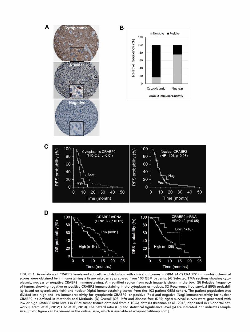
To examine the subcellular distribution of CRABP2 in
GBM, we immunostained a tissue microarray consisting of
116 human GBM tumor tissues with an antibody that specifi-
cally recognizes CRABP2 (Liu et al., 2015). Immunoreactiv-
ity data from 103 GBM tissues were of sufficient quality to
allow scoring: 0 (negative), 1 (weak), 2 (moderate) and 3
(strong). Of the 103 GBM tissues, 86 (83.5%) tumors were
positive for cytoplasmic CRABP2, with scores of 1 to 3.
However, only 25 (24.3%) tumors were positive for nuclear
CRABP2 (Fig. 1A,B). Moderate to strong immunostaining
(scores of 2 to 3) was observed in the cytoplasm of 28/86
(32.6%) GBM tissues (or 27.2% of 103 GBM tissues) and in
the nucleus of 7/25 (28%) (or 6.8% of 103 GBM tissues).
CRABP2 was detected exclusively in the cytoplasm of sixty-
two tumors, and exclusively in the nucleus in only one tumor.
These data indicate that CRABP2 is predominantly cytoplas-
mic in GBM tumors.
To determine the potential effect of subcellular CRABP2
levels on patient prognosis, we performed Kaplan-Meier univari-
ate survival analysis of the GBM patient cohort with 5 years of
clinical follow-up data. To this end, we divided the patient cohort
into subpopulations with high (moderate to strong immunoreac-
tivity; n 5 28) and low (weak or negative immunoreactivity;
n 5 75) cytoplasmic CRABP2 levels, or subpopulations with
Patients with high cytoplasmic CRABP2 levels had significantly
lower recurrence-free survival probability compared to those with
low cytoplasmic levels (P 5 0.01; Fig. 1C, left panel). In contrast,
nuclear CRABP2 levels had no effect on recurrence-free survival
(P 5 0.98; Fig. 1C, right panel). Neither cytoplasmic nor nuclear
CRABP2 levels showed significant prognostic value on overall
survival for GBM patients (data not shown).
Next, we tested the impact of CRABP2 RNA levels on
clinical outcomes using a 401 GBM patient TCGA microar-
ray dataset (with recorded recurrence/progression and vital
status) (Brennan et al., 2013) retrieved from cBioportal for
Cancer Genomics (Cerami et al., 2012; Gao et al., 2013) on
November 2015. Elevated CRABP2 RNA levels were signifi-
cantly associated with poorer overall (HR 5 1.88, P< 0.01;
left panel) and disease free prognosis (HR 5 2.42, P< 0.05;
right panel) (Fig. 1D) within 24 months of follow-up time.
These results suggest that CRABP2 levels in GBM tumors
may represent a risk factor for early recurrence and death,
and that the adverse effect of elevated CRABP2 levels may be
attributed to the aberrant accumulation of CRABP2 in the
cytoplasm of GBM cells.
CRABP2 Knockdown Represses CellGrowth in U251 CellsOne of the major biological activities of RA is to induce inhibi-
tion of cell growth by binding and activating RARs in the
nucleus, a function that is mediated by CRABP2. To determine
whether CRABP2 is expressed in MG cell lines (i.e., cell lines
derived from high grade astrocytomas), we carried out western
blot analysis of 10 MG cell lines. CRABP2 was easily detected
in 5 cell lines (M021, U87, M049, M103, U251) and barely
detectable in one cell line (U373) (Fig. 2A).
To study the effect of CRABP2 levels on cell proliferation,
we transfected U251 cells with siRNAs that specifically target
CRABP2. CRABP2 knockdown was confirmed by both RT-
PCR and western blot analysis (>90%; Fig. 2B,C). We used
cell counts to measure the growth rate of control and CRABP2-
depleted U251 cells. The growth rate of CRABP2-depleted cells
was significantly reduced compared to cells transfected with
scrambled siRNAs (Figure 2D). The difference between the
two cell populations was statistically significant starting from
96 h in culture (P< 0.01). The experiment was repeated using
a second siRNA targeting a distinct region of the CRABP2
mRNA, with similar results (data not shown). To confirm the
anti-proliferative effect of CRABP2 depletion, we measured cell
proliferation in control and CRABP2-depleted cells using the
MTS Cell Proliferation Assay system, a colorimetric-based assay
for the quantification of live cells. Compared to control siRNA,
depletion of CRABP2 with siRNA1 or siRNA2 resulted in
35% and 62% reduction, respectively, in absorbance (OD490).
These results indicate that CRABP2 may play a role in enhanc-
ing cell proliferation/survival in GBM cells.
To explore the mechanism by which CRABP2 promotes
cell growth, we examined the changes in cell cycle progression
in control versus CRABP2-depleted U251 cells. Four siRNAs
targeting distinct regions of CRABP2 were used for this experi-
ment. We observed >90% reduction in CRABP2 protein levels
in U251 cells transfected with each of the four CRABP2
siRNAs relative to scrambled siRNA control (Fig. 3A).
CRABP2 knockdown resulted in changes in the cell cycle distri-
bution, with a significant reduction in G1-phase cells (72.6%
for control cells vs an average of 54.3% for CRABP2-depleted
cells) and a significant increase in S-phase (9.8% vs. 21.2%)
and G2-phase (16% vs. 23.5%) cells (Fig. 3B–G). These data
suggest that CRABP2 promotes G1/S transition in U251 cells,
resulting in a more rapid progression through the cell cycle.
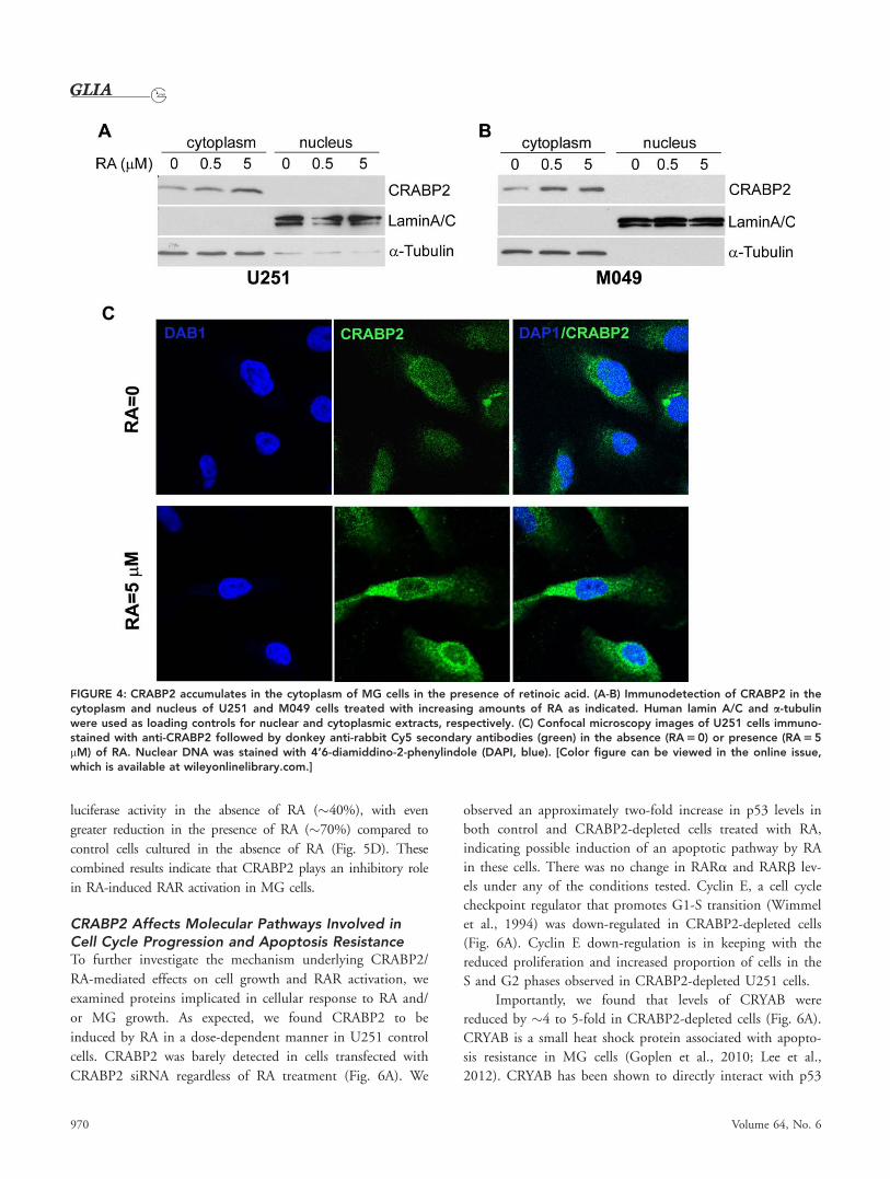
CRABP2 Accumulates in the Cytoplasmof MG Cells in Response to RA TreatmentAs discussed earlier, the subcellular distribution of CRABP2 in
response to RA is believed to be a key factor in cellular response
to RA. To this end, we treated both U251 and M049 MG cell
lines with increasing amounts of all-trans RA and then prepared
966 Volume 64, No. 6
FIGURE 1: Association of CRABP2 levels and subcellular distribution with clinical outcomes in GBM. (A-C) CRABP2 immunohistochemicalscores were obtained by immunostaining a tissue microarray prepared from 103 GBM patients. (A) Selected TMA sections showing cyto-plasmic, nuclear or negative CRABP2 immunostaining. A magnified region from each image is shown in the box. (B) Relative frequencyof tumors showing negative or positive CRABP2 immunostaining in the cytoplasm or nucleus. (C) Recurrence-free survival (RFS) probabil-ity based on cytoplasmic (left) and nuclear (right) immunostaining scores from the 103-patient GBM cohort. The patient population wasdivided into high and low immunoreactivity for cytoplasmic CRABP2, or positive (Pos) and negative (Neg) immunoreactivity for nuclearCRABP2, as defined in Materials and Methods. (D) Overall (OS; left) and disease-free (DFS; right) survival curves were generated withlow or high CRABP2 RNA levels in GBM tumor tissues obtained from a TCGA dataset (Brennan et al., 2013) deposited in cBioportal net-work (Cerami et al., 2012; Gao et al., 2013). The hazard ratio (HR) and statistical significance level (p) are indicated. “n” indicates samplesize. [Color figure can be viewed in the online issue, which is available at wileyonlinelibrary.com.]
tional activity compared to control cells (Fig. 5B).
To ensure that the observed effect of CRABP2 on RA sig-
naling through RAR was not cell line-specific, we repeated the
experiment in a second MG cell line that expresses CRABP2,
M049. Of note, M049 expresses the highest CRABP2 levels of
all 10 MG lines tested. Transfections were carried out as detailed
for U251. In contrast to U251, we observed a significant induc-
tion in RAR activation (�5-fold increase) upon CRABP2 deple-
tion in the absence of RA (Fig. 5C). The level of RAR induction
in control cells in the presence of RA was similar to that of U251
(4- to 5-fold increase). However, a much higher increase in lucif-
erase activity, 45- to 46-fold, was observed in CRABP2-depleted
M049 cells in the presence of RA (Fig. 5C). These results indi-
cate that CRABP2 plays a negative role in RAR activation in the
presence of RA.
Next, we used a gain-of-function approach to address the
inhibitory effect of CRABP2 on RAR activity. We co-transfected
the CRABP2-negative T98 MG cell line with the RARE-driven
luciferase reporter gene along with a CRABP2 expression con-
struct (pcDNA3 carrying full-length CRABP2 cDNA). RA
treatment (0.5 2 5 mM) resulted in a 2.5- to 2.7-fold increase in
luciferase activity in T98 cells transfected with empty vector.
Ectopic expression of CRABP2 resulted in significantly reduced
FIGURE 2: CRABP2 affects the growth rate of U251 MG cells.(A) Western blot analysis of whole cell lysates prepared from tenMG cell lines using anti- CRABP2 and anti-b-actin (loading con-trol) antibodies. (B) RT-PCR analysis showing >80% reduction inCRABP2 RNA levels in U251 cells transfected with CRABP2siRNA. (C) Western blot analysis showing >80% depletion ofCRABP2 protein in U251 cells transfected with CRABP2 siRNA.(D) Growth rate curves of U251 cells transfected with control orCRABP2 siRNA. Cells were seeded in 12-well plates at 10,000cells per well and cultured in DMEM supplemented with 10%FBS until harvested and counted at the indicated time points.The results of one experiment is shown in (D), with similar dataobtained in one additional experiment. (E) The MTS assay wasused to confirm the effect of CRABP2 depletion on cell prolifera-tion. Both CRABP2 siRNA1 and 2 were included in this analysis.“*” denotes p < 0.05; “**” p < 0.01.
968 Volume 64, No. 6
FIGURE 3: Effect of CRABP2 depletion on the cell cycle. U251 cells were transfected with scrambled (control) siRNAs or CRABP2 siRNAs.(A) Western blot analysis shows a significant decrease in CRABP2 levels (>90%) in CRABP2-depleted (labeled 1-4) compared to control(Ctrl) cells. (B-F) Representative images of the cell cycle distribution of cells transfected with scrambled (control) or CRABP2 siRNAs.(G) Histograms showing the average percentage of cells in the G1, S and G2 phases of the cell cycle in control versus CRABP2-depletedcells. The values shown in individual columns represent the arithmetic mean of cell counts from triplicate wells transfected with thesame siRNA. The two-sided student t-test was used to analyze the significance of differences in cell count percentages in the G1, S andG2 phases of the cell cycle between control and CRABP2-depleted cells [Ctrl, black columns; CRABP2-depleted (1-4), gray columns].Standard deviation is indicated by the error bars. Three independent experiments were carried out with CRABP2 siRNA1 and 2. Thedata were confirmed using CRABP2 siRNA3 and 4. ** denotes p < 0.01.
Liu et al.: CRABP2 in Glioblastoma
June 2016 969
luciferase activity in the absence of RA (�40%), with even
greater reduction in the presence of RA (�70%) compared to
control cells cultured in the absence of RA (Fig. 5D). These
combined results indicate that CRABP2 plays an inhibitory role
in RA-induced RAR activation in MG cells.
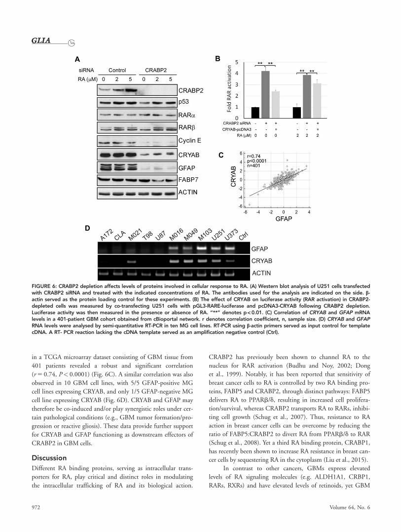
CRABP2 Affects Molecular Pathways Involved inCell Cycle Progression and Apoptosis ResistanceTo further investigate the mechanism underlying CRABP2/
RA-mediated effects on cell growth and RAR activation, we
examined proteins implicated in cellular response to RA and/
or MG growth. As expected, we found CRABP2 to be
induced by RA in a dose-dependent manner in U251 control
cells. CRABP2 was barely detected in cells transfected with
CRABP2 siRNA regardless of RA treatment (Fig. 6A). We
observed an approximately two-fold increase in p53 levels in
both control and CRABP2-depleted cells treated with RA,
indicating possible induction of an apoptotic pathway by RA
in these cells. There was no change in RARa and RARb lev-
els under any of the conditions tested. Cyclin E, a cell cycle
checkpoint regulator that promotes G1-S transition (Wimmel
et al., 1994) was down-regulated in CRABP2-depleted cells
(Fig. 6A). Cyclin E down-regulation is in keeping with the
reduced proliferation and increased proportion of cells in the
S and G2 phases observed in CRABP2-depleted U251 cells.
Importantly, we found that levels of CRYAB were
reduced by �4 to 5-fold in CRABP2-depleted cells (Fig. 6A).
CRYAB is a small heat shock protein associated with apopto-
sis resistance in MG cells (Goplen et al., 2010; Lee et al.,
2012). CRYAB has been shown to directly interact with p53
FIGURE 4: CRABP2 accumulates in the cytoplasm of MG cells in the presence of retinoic acid. (A-B) Immunodetection of CRABP2 in thecytoplasm and nucleus of U251 and M049 cells treated with increasing amounts of RA as indicated. Human lamin A/C and a-tubulinwere used as loading controls for nuclear and cytoplasmic extracts, respectively. (C) Confocal microscopy images of U251 cells immuno-stained with anti-CRABP2 followed by donkey anti-rabbit Cy5 secondary antibodies (green) in the absence (RA 5 0) or presence (RA 5 5mM) of RA. Nuclear DNA was stained with 4’6-diamiddino-2-phenylindole (DAPI, blue). [Color figure can be viewed in the online issue,which is available at wileyonlinelibrary.com.]
and caspase 3 to repress their apoptotic function (Liu et al.,
2007; Shin et al., 2009). Similar to CRYAB, GFAP levels
were reduced by �6 to 7-fold in CRABP2-depleted cells
compared to control cells. GFAP is an intermediate filament
protein that is normally expressed in differentiated astrocytes
as well as in astrocytoma tumors (Brehar et al., 2015; Choi
et al., 2009). GFAP physically associates with CRYAB in
astrocytomas cells (Wisniewski and Goldman 1998). The
expression of the neural stem cell/radial glial cells marker
FABP7 (Feng et al., 1994; Gomez-Lopez et al., 2011; Li
et al., 2008), which is also expressed in neurospheres derived
from GBM patients and which is associated with MG cell
migration (De Rosa et al., 2012; Liang et al., 2005; Mita
et al., 2007), was not significantly affected by RA in control
cells, with a slight decrease in FABP7 levels observed upon
RA treatment in CRABP2-depleted cells.
The possibility of crosstalk between CRABP2 and CRYAB
in mediating RA signaling to RARs was examined by co-
transfecting a CRYAB expression construct, along with the
RARE-driven luciferase reporter, in CRABP2-depleted U251
cells. As previously observed, there was a significant increase in
luciferase activity in CRABP2-depleted cells in either the absence
or presence of RA. However, this increase in luciferase activity
was mitigated by restoration of CRYAB expression, especially in
the absence of RA (Fig. 6B). As shown in Fig. 6A, there was an
increase in CRYAB upon RA treatment in CRABP2-depleted
cells. This increase in endogenous CRYAB levels may explain the
relatively small effect of CRYAB restoration on RAR activation
in the presence of RA in CRABP2-depleted cells. These results
suggest that the roles of CRABP2 (promoting cell survival and
attenuating RA signaling) in GBM cells are at least partially
mediated through modulation of CRYAB.
Both CRYAB and GFAP are induced in reactive astrocytes
under various pathological conditions (Hol and Pekny 2015;
Renkawek et al., 1994), with both proteins co-localizing to
intermediate filaments in astrocytoma cells (Goplen et al.,
2010). As the levels of CRYAB and GFAP are similarly reduced
upon CRABP2 depletion in U251 cells, we investigated the
possibility of a similar correlation in GBM tumor tissues and
other MG cell lines. Analysis of CRYAB and GFAP RNA levels
FIGURE 5: CRABP2 inhibits RA-induced RAR activation in MG cells. (A-B) Dose-dependent activation of RAR by RA in U251 cells afterCRABP2 depletion using two different CRABP2 siRNAs that target distinct regions of the CRABP2 RNA. Cells were transfected with aluciferase reporter vector driven by retinoic acid response element (RARE) and cultured in the presence of different amounts of RA asindicated. Luciferase activity (an indication of RAR activation) was corrected for protein concentration in each lysate and is shown as foldrelative to control cells transfected with scrambled siRNAs cultured in the absence of RA. (C) CRABP2 was depleted by siRNA transfec-tion in M049 cells (left corner insert). CRABP2-depleted cells showed greatly enhanced RA-induced luciferase activity. (D) T98 cells wereco-transfected with a RARE-driven luciferase construct (pGL3-RARE-Luciferase) and a CRABP2 expression construct (pcDNA3-CRABP2)and treated with increasing amounts of RA as indicated. RA-induced increase in luciferase activity was suppressed by CRABP2 overex-pression. Western blot analysis of CRABP2 in T98 cells is shown in the right corner. “*” denotes p < 0.05, “**” p < 0.01.
Liu et al.: CRABP2 in Glioblastoma
June 2016 971
in a TCGA microarray dataset consisting of GBM tissue from
401 patients revealed a robust and significant correlation
(r 5 0.74, P< 0.0001) (Fig. 6C). A similar correlation was also
observed in 10 GBM cell lines, with 5/5 GFAP-positive MG
cell lines expressing CRYAB, and only 1/5 GFAP-negative MG
cell line expressing CRYAB (Fig. 6D). CRYAB and GFAP may
therefore be co-induced and/or play synergistic roles under cer-
gression or reactive gliosis). These data provide further support
for CRYAB and GFAP functioning as downstream effectors of
CRABP2 in GBM cells.
Discussion
Different RA binding proteins, serving as intracellular trans-
porters for RA, play critical and distinct roles in modulating
the intracellular trafficking of RA and its biological action.
CRABP2 has previously been shown to channel RA to the
nucleus for RAR activation (Budhu and Noy, 2002; Dong
et al., 1999). Notably, it has been reported that sensitivity of
breast cancer cells to RA is controlled by two RA binding pro-
teins, FABP5 and CRABP2, through distinct pathways: FABP5
delivers RA to PPARb/d, resulting in increased cell prolifera-
tion/survival, whereas CRABP2 transports RA to RARs, inhibi-
ting cell growth (Schug et al., 2007). Thus, resistance to RA
action in breast cancer cells can be overcome by reducing the
ratio of FABP5:CRABP2 to divert RA from PPARb/d to RAR
(Schug et al., 2008). Yet a third RA binding protein, CRABP1,
has recently been shown to increase RA resistance in breast can-
cer cells by sequestering RA in the cytoplasm (Liu et al., 2015).
In contrast to other cancers, GBMs express elevated
levels of RA signaling molecules (e.g. ALDH1A1, CRBP1,
RARs, RXRs) and have elevated levels of retinoids, yet GBM
FIGURE 6: CRABP2 depletion affects levels of proteins involved in cellular response to RA. (A) Western blot analysis of U251 cells transfectedwith CRABP2 siRNA and treated with the indicated concentrations of RA. The antibodies used for the analysis are indicated on the side. b-actin served as the protein loading control for these experiments. (B) The effect of CRYAB on luciferase activity (RAR activation) in CRABP2-depleted cells was measured by co-transfecting U251 cells with pGL3-RARE-luciferase and pcDNA3-CRYAB following CRABP2 depletion.Luciferase activity was then measured in the presence or absence of RA. “**” denotes p< 0.01. (C) Correlation of CRYAB and GFAP mRNAlevels in a 401-patient GBM cohort obtained from cBioportal network. r denotes correlation coefficient, n, sample size. (D) CRYAB and GFAPRNA levels were analysed by semi-quantitative RT-PCR in ten MG cell lines. RT-PCR using b-actin primers served as input control for templatecDNA. A RT- PCR reaction lacking the cDNA template served as an amplification negative control (Ctrl).
972 Volume 64, No. 6
tumors are highly resistant to the growth-controlling effects
of RA (Campos et al., 2011, 2015). The ratio of FABP5 to
CRABP2 transcripts is even lower in GBM cells than in nor-
mal astrocytes, suggesting that RA resistance in GBM cells is
not caused by PPARb activation (Campos et al., 2015).
Accordingly, the relative levels of FABP5:CRABP2 are neither
related to tumor grade nor implicated for RA sensitivity in
GBM (Xia et al., 2015). Our results indicate that CRABP2
plays an unconventional role in GBM tumors. Specifically, we
show that CRABP2 accumulates in the cytoplasm of GBM
tumor cells, thereby promoting cell survival, attenuating the
transcriptional activity of RAR and inhibiting RA-induced
apoptosis by upregulating an anti-apoptotic pathway. Thus,
our findings help explain why overall CRABP2 (or relative
CRABP2 and FABP5 levels), fail to predict RA response in
certain cancers, including GBM (Campos et al., 2011, 2015;
Chen et al., 2012; Xia et al., 2015).
Campos et al. (2011) showed absence of CRABP2 in
the nucleus of �70% of GBM tumors compared to �50%
of grades II and III astrocytomas, suggesting a trend towards
reduced nuclear CRABP2 levels with increasing malignancy.
These authors also showed that the levels of proteins favoring
cellular RA availability (CRBP1, ALDH1A1) were elevated in
astrocytoma tumor tissue compared to non-neoplastic brain
tissue, with increasing levels associated with increasing tumor
malignancy and shorter patient survival, suggesting impaired
RA signaling in astrocytomas (Campos et al., 2011). In our
study, we found an inverse correlation between cytoplasmic,
but not nuclear, CRABP2 levels, and recurrence-free survival,
with recurrence considered an end point for GBM patients.
Our findings that cytoplasmic CRABP2 predicts decreased
recurrence-free survival in GBM patients and that cytoplasmic
CRABP2 is a negative mediator of RA signalling in GBM
cells suggest a causative role for cytoplasmic CRABP2 in
patient recurrence and tumor progression, perhaps mediated
through an attenuated physiological response to RA.
It is noteworthy that the cytoplasmic/nuclear distribu-
tion of CRABP2 is considerably less skewed towards the cyto-
plasm in breast cancer compared to GBM tumors (Liu et al.,
2015). For example, in ER-positive breast cancers which are
typically sensitive to RA, 51% and 91% of cancers showed
high levels (scores of 2 and 3) of CRABP2 in the cytoplasm
and nucleus, respectively. In ER-negative breast cancers, typi-
cally resistant to RA, 39% and 61% of cancers showed high
levels in the cytoplasm versus nucleus, respectively (Liu et al.,
2015). In comparison, we observed high levels of cytoplasmic
and nuclear CRABP2 in 27.2% and 6.8% of the GBM
tumors tested, respectively.
Little is known about the biological activity of CRABP2
in the cytoplasm. An obvious role is to sequester RA in the
cytoplasm, thus rendering it unavailable for activation of RARs
in the nucleus. Cytoplasmic CRABP2 may also target RA to
certain metabolizing enzymes such as CYP26, facilitating RA
catabolism to yield polar metabolites (Njar et al., 2006). RA
catabolism is believed to be one of the key players in cellular
resistance to RA (Njar et al., 2006; Petty et al., 2005; Sessler
and Noy, 2005). While we show that CRABP2 accumulates in
the cytoplasm in a RA dose-dependent manner in MG cell
lines, it remains to be determined whether CRABP2 simply
sequesters RA or transports RA to specific sites in the cytoplasm
for catabolism. As a third possibility, RA-bound cytoplasmic
CRABP2 may activate the extracellular signal regulated kinase
1/2 (ERK1/2), as previously demonstrated for CRABP1,
thereby affecting cell cycle progression, cell proliferation and
stemness (Persaud et al., 2013). Yet another possibility is that
cytoplasmic CRABP2 may directly bind to the RNA-binding
protein HuR, thereby enhancing its interaction with target
RNAs, leading to their increased stability and expression (Vree-
land et al., 2014a). Our discovery that CRABP2 is primarily
found in the cytoplasm of GBM cells warrants further studies
to examine its exact mechanism(s) of action in these tumors.
CRYAB (aB-crystallin) belongs to an evolutionarily con-
served family of small heat shock proteins that function as cyto-
protective chaperones (Horwitz, 1992). Induction of CRYAB
under various stress conditions confers protection against apo-
ptotic stimuli in different cancer cells (Arrigo et al., 2007).
CRYAB is upregulated in MG cells (Goplen et al., 2010; Odre-
man et al., 2005), and its silencing sensitizes these cells to dif-
ferent apoptotic inducers (Goplen et al., 2010; Lee et al.,
2012). In this study, we demonstrate that CRABP2 knockdown
sensitizes MG cells to RA-induced RAR activation, with
concomitant downregulation of CRYAB. We speculate that
CRABP2 may contribute to RA resistance in MG cells through
upregulation of CRYAB, which then acts as an inhibitor of
RA-induced apoptosis.
GFAP, normally expressed in astrocytes, is up-regulated
in reactive astrocytes and serves as a marker for astrocytoma
tumors (Brehar et al., 2015; Choi et al., 2009). GFAP levels
were markedly reduced upon CRABP2 depletion in MG
cells. CRYAB as well as other small heat shock proteins have
been shown to interact with GFAP in astrocytes and astrocy-
toma cells (Goldbaum et al., 2009; Nicholl and Quinlan,
1994; Perng et al., 1999; Wisniewski and Goldman, 1998).
This interaction protects the cells against cellular stress. Inter-
estingly, there is a strong correlation between GFAP and
CRYAB RNA levels in GBM tumor tissues as well as MG cell
lines, suggesting that these two genes may be co-regulated.
Although our study did not address whether GFAP and
CRYAB expression is directly regulated through a CRABP2-
dependent mechanism, it does show that reduction of GFAP
and CRYAB levels is independent of RA. It is possible that
by binding to the RNA-binding protein HuR, CRABP2
Liu et al.: CRABP2 in Glioblastoma
June 2016 973
enhances HuR’s interaction with the CRYAB and GFAP
RNAs, leading to their increased stability and translation
(Vreeland et al., 2014a).
In summary, we report the intriguing finding that
CRABP2 is predominantly localized in the cytoplasm of
GBM tumors, and provide evidence that cytoplasmic
CRABP2 is a significant prognostic factor associated with
shorter survival in GBM patients. We demonstrate that cyto-
plasmic CRABP2 promotes MG cell survival and attenuates
RA-mediated nuclear RAR activity. Our data suggest that
CRABP2 triggers an anti-apoptotic or cyto-protective
response in MG cells by upregulating CRYAB and GFAP to
resist RA signaling and action. This study underlines the
importance of determining the subcellular localization of
proteins involved in the regulation of RA bioavailability to
predict response to RA. We propose that CRABP2 accumula-
tion in the cytoplasm represents a critical factor causing RA
resistance in GBM and that reducing CRABP2 levels may
enhance the efficacy of RA therapy.
Acknowledgment
Grant sponsor: Canadian Institutes of Health Research; Grant
number: 130314.
The authors thank Elizabeth Monckton and Lei Li for
technical assistance. They also thank Xuejun Sun, Jingzhou
Huang, Gerry Barron, and the Cross Cancer Institute Cell
Imaging Facility for their assistance with cell cycle analysis
and imaging of GBM tumor tissue and cells.
ReferencesAdam SA, Schnell O, Poschl J, Eigenbrod S, Kretzschmar HA, Tonn JC,Schuller U. 2012. ALDH1A1 is a marker of astrocytic differentiation duringbrain development and correlates with better survival in glioblastomapatients. Brain Pathology 22:7882797.
Arrigo AP, Simon S, Gibert B, Kretz-Remy C, Nivon M, Czekalla A, Guillet D,Moulin M, Diaz-Latoud C, Vicart P. 2007. Hsp27 (HspB1) and alphaB-crystallin(HspB5) as therapeutic targets. FEBS Lett 581:366523674.
Boorjian SA, Milowsky MI, Kaplan J, Albert M, Cobham MV, Coll DM,Mongan NP, Shelton G, Petrylak D, Gudas LJ, Nanus DM. 2007. Phase 1/2clinical trial of interferon alpha2b and weekly liposome-encapsulated all-transretinoic acid in patients with advanced renal cell carcinoma. J Immunother30:6552662.
Brehar FM, Arsene D, Brinduse LA, Gorgan MR. 2015. Immunohistochemicalanalysis of GFAP-delta and nestin in cerebral astrocytomas. Brain TumorPathol 32:90298.
Brennan CW, Verhaak RG, McKenna A, Campos B, Noushmehr H, SalamaSR, Zheng S, Chakravarty D, Sanborn JZ, Berman SH, et al. 2013. Thesomatic genomic landscape of glioblastoma. Cell 155:4622477.
Brun M, Coles JE, Monckton EA, Glubrecht DD, Bisgrove D, Godbout R.2009. Nuclear factor I regulates brain fatty acid-binding protein and glialfibrillary acidic protein gene expression in malignant glioma cell lines. J MolBiol 391:282–300.
Budhu AS, Noy N. 2002. Direct channeling of retinoic acid between cellularretinoic acid-binding protein II and retinoic acid receptor sensitizes mammary
Campos B, Centner FS, Bermejo JL, Ali R, Dorsch K, Wan F, Felsberg J,Ahmadi R, Grabe N, Reifenberger G, et al. 2011. Aberrant expression of reti-noic acid signaling molecules influences patient survival in astrocytic gliomas.Am J Pathol 178:195321964.
Campos B, Wan F, Farhadi M, Ernst A, Zeppernick F, Tagscherer KE, AhmadiR, Lohr J, Dictus C, Gdynia G, et al. 2010. Differentiation therapy exerts anti-tumor effects on stem-like glioma cells. Clinical Cancer Res 16:271522728.
Campos B, Weisang S, Osswald F, Ali R, Sedlmeier G, Bageritz J, Mallm JP,Hartmann C, von Deimling A, Popanda O, et al. 2015. Retinoid resistanceand multifaceted impairment of retinoic acid synthesis in glioblastoma. Glia63:185021859.
Cerami E, Gao J, Dogrusoz U, Gross BE, Sumer SO, Aksoy BA, Jacobsen A,Byrne CJ, Heuer ML, Larsson E, et al. 2012. The cBio cancer genomics portal:An open platform for exploring multidimensional cancer genomics data. Can-cer Dis 2:4012404.
Chambon P. 1996. A decade of molecular biology of retinoic acid receptors.FASEB J 10:9402954.
Chen NN, Li Y, Wu ML, Liu ZL, Fu YS, Kong QY, Chen XY, Li H, Liu J. 2012.CRABP-II- and FABP5-independent all-trans retinoic acid resistance in COLO16 human cutaneous squamous cancer cells. Exp Dermatol 21:13218.
Choi KC, Kwak SE, Kim JE, Sheen SH, Kang TC. 2009. Enhanced glial fibril-lary acidic protein-delta expression in human astrocytic tumor. Neurosci Lett463:1822187.
De Rosa A, Pellegatta S, Rossi M, Tunici P, Magnoni L, Speranza MC, MalusaF, Miragliotta V, Mori E, Finocchiaro G, et al. 2012. A radial glia gene marker,fatty acid binding protein 7 (FABP7), is involved in proliferation and invasionof glioblastoma cells. PloS One 7:e52113.
Dong D, Ruuska SE, Levinthal DJ, Noy N. 1999. Distinct roles for cellular reti-noic acid-binding proteins I and II in regulating signaling by retinoic acid.J Biol Chem 274:23695223698.
Donovan M, Olofsson B, Gustafson AL, Dencker L, Eriksson U. 1995. The cel-lular retinoic acid binding proteins. J Steroid Biochem Mol Biol 53:4592465.
Duester G. 2008. Retinoic acid synthesis and signaling during early organo-genesis. Cell 134:9212931.
Esteller M, Guo M, Moreno V, Peinado MA, Capella G, Galm O, Baylin SB,Herman JG. 2002. Hypermethylation-associated inactivation of the cellularretinol-binding-protein 1 gene in human cancer. Cancer Res 62:590225905.
Feng L, Hatten ME, Heintz N. 1994. Brain lipid-binding protein (BLBP): A novelsignaling system in the developing mammalian CNS. Neuron 12:895–908.
Fiorella PD, Napoli JL. 1994. Microsomal retinoic acid metabolism. Effects ofcellular retinoic acid-binding protein (type I) and C18-hydroxylation as an ini-tial step. J Biol Chem 269:10538210544.
Gao J, Aksoy BA, Dogrusoz U, Dresdner G, Gross B, Sumer SO, Sun Y,Jacobsen A, Sinha R, Larsson E, et al. 2013. Integrative analysis of complexcancer genomics and clinical profiles using the cBioPortal. Sci Signal 6:pl1.
Garattini E, Gianni M, Terao M. 2007. Retinoids as differentiating agents inoncology: A network of interactions with intracellular pathways as the basisfor rational therapeutic combinations. Curr Pharm Des 13:137521400.
Godbout R, Bisgrove DA, Shkolny D, Day RS III. 1998. Correlation of B-FABPand GFAP expression in malignant glioma. Oncogene 16:195521962.
Godbout R, Miyakoshi J, Dobler KD, Andison R, Matsuo K, Allalunis-TurnerMJ, Takebe H, Day RS, 3rd 1992. Lack of expression of tumor-suppressorgenes in human malignant glioma cell lines. Oncogene 7:187921884.
Goldbaum O, Riedel M, Stahnke T, Richter-Landsberg C. 2009. The smallheat shock protein HSP25 protects astrocytes against stress induced by pro-teasomal inhibition. Glia 57:156621577.
Gomez-Lopez S, Wiskow O, Favaro R, Nicolis SK, Price DJ, Pollard SM, SmithA. 2011. Sox2 and Pax6 maintain the proliferative and developmental poten-tial of gliogenic neural stem cells In vitro. Glia 59:158821599.
974 Volume 64, No. 6
Goplen D, Bougnaud S, Rajcevic U, Boe SO, Skaftnesmo KO, Voges J, EngerPO, Wang J, Tysnes BB, Laerum OD, et al. 2010. alphaB-crystallin is elevatedin highly infiltrative apoptosis-resistant glioblastoma cells. Am J Pathol 177:161821628.
Hol EM, Pekny M. 2015. Glial fibrillary acidic protein (GFAP) and the astro-cyte intermediate filament system in diseases of the central nervous system.Curr Opin Cell Biol 32:1212130.
Horwitz J. 1992. Alpha-crystallin can function as a molecular chaperone. ProcNatl Acad Sci USA 89:10449210453.
Huang ME, Ye YC, Chen SR, Chai JR, Lu JX, Zhoa L, Gu LJ, Wang ZY. 1988.Use of all-trans retinoic acid in the treatment of acute promyelocytic leuke-mia. Blood 72:5672572.
Johnson DR, O’Neill BP. 2012. Glioblastoma survival in the United Statesbefore and during the temozolomide era. J Neurooncol 107:3592364.
Lee JS, Kim HY, Jeong NY, Lee SY, Yoon YG, Choi YH, Yan C, Chu IS, KohH, Park HT, et al. 2012. Expression of alphaB-crystallin overrides the anti-apoptotic activity of XIAP. Neuro-Oncology 14:133221345.
Li H, Chang YW, Mohan K, Su HW, Ricupero CL, Baridi A, Hart RP, GrumetM. 2008. Activated Notch1 maintains the phenotype of radial glial cells andpromotes their adhesion to laminin by upregulating nidogen. Glia 56:6462658.
Liang Y, Diehn M, Watson N, Bollen AW, Aldape KD, Nicholas MK, LambornKR, Berger MS, Botstein D, Brown PO, et al. 2005. Gene expression profilingreveals molecularly and clinically distinct subtypes of glioblastoma multi-forme. Proc Natl Acad Sci USA 102:581425819.
Liu RZ, Garcia E, Glubrecht DD, Poon HY, Mackey JR, Godbout R. 2015.CRABP1 is associated with a poor prognosis in breast cancer: Adding to thecomplexity of breast cancer cell response to retinoic acid. Mol Cancer 14:129.
Liu RZ, Graham K, Glubrecht DD, Germain DR, Mackey JR, Godbout R. 2011.Association of FABP5 expression with poor survival in triple-negative breastcancer: Implication for retinoic acid therapy. Am J Pathol 178:997–1008.
Liu S, Li J, Tao Y, Xiao X. 2007. Small heat shock protein alphaB-crystallinbinds to p53 to sequester its translocation to mitochondria during hydrogenperoxide-induced apoptosis. Biochem Biophys Res Commun 354:1092114.
Lotan R. 1996. Retinoids in cancer chemoprevention. FASEB J 10:103121039.
Mangelsdorf DJ. 1994. Vitamin A receptors. Nutr Rev 52:S32-S44.
Means AL, Gudas LJ. 1995. The roles of retinoids in vertebrate development.Annu Rev Biochem 64:2012233.
Mita R, Coles JE, Glubrecht DD, Sung R, Sun X, Godbout R. 2007. B-FABP-expressing radial glial cells: the malignant glioma cell of origin? Neoplasia 9:7342744.
Mongan NP, Gudas LJ. 2007. Diverse actions of retinoid receptors in cancerprevention and treatment. Differentiation 75:8532870.
Napoli JL. 1996. Biochemical pathways of retinoid transport, metabolism,and signal transduction. Clin Immunol Immunopathol 80:S52-S62.
Njar VC, Gediya L, Purushottamachar P, Chopra P, Vasaitis TS, Khandelwal A,Mehta J, Huynh C, Belosay A, Patel J. 2006. Retinoic acid metabolism block-ing agents (RAMBAs) for treatment of cancer and dermatological diseases.Bioorg Med Chem 14:432324340.
Noy N. 2010. Between death and survival: Retinoic acid in regulation of apo-ptosis. Annu Rev Nutr 30:2012217.
Odreman F, Vindigni M, Gonzales ML, Niccolini B, Candiano G, Zanotti B,Skrap M, Pizzolitto S, Stanta G, Vindigni A. 2005. Proteomic studies on low-and high-grade human brain astrocytomas. J Proteome Res 4:698–708.
Ostrom QT, Gittleman H, Fulop J, Liu M, Blanda R, Kromer C, Wolinsky Y,Kruchko C, Barnholtz-Sloan JS. 2015. CBTRUS statistical report: Primary brainand central nervous system tumors diagnosed in the United States in 2008-2012. Neuro-Oncology 17:iv1–iv62.
Perng MD, Cairns L, van den IP, Prescott A, Hutcheson AM, Quinlan RA.1999. Intermediate filament interactions can be altered by HSP27 andalphaB-crystallin. J Cell Sci 112:209922112.
Persaud SD, Lin YW, Wu CY, Kagechika H, Wei LN. 2013. Cellular retinoicacid binding protein I mediates rapid non-canonical activation of ERK1/2 byall-trans retinoic acid. Cell Signal 25:19–25.
Petrie K, Zelent A, Waxman S. 2009. Differentiation therapy of acute myeloidleukemia: Past, present and future. Curr Opin Hematol 16:84–91.
Petty WJ, Li N, Biddle A, Bounds R, Nitkin C, Ma Y, Dragnev KH,Freemantle SJ, Dmitrovsky E. 2005. A novel retinoic acid receptor beta iso-form and retinoid resistance in lung carcinogenesis. J Natl Cancer Inst 97:164521651.
Recchia F, Lalli A, Lombardo M, De Filippis S, Saggio G, Fabbri F, RosselliM, Capomolla E, Rea S. 2001. Ifosfamide, cisplatin, and 13-Cis retinoic acidfor patients with advanced or recurrent squamous cell carcinoma of the headand neck: A phase I-II study. Cancer 92:8142821.
Renkawek K, Voorter CE, Bosman GJ, van Workum FP, de Jong WW. 1994.Expression of alpha B-crystallin in Alzheimer’s disease. Acta Neuropathol 87:1552160.
Schug TT, Berry DC, Shaw NS, Travis SN, Noy N. 2007. Opposing effects ofretinoic acid on cell growth result from alternate activation of two differentnuclear receptors. Cell 129:7232733.
Schug TT, Berry DC, Toshkov IA, Cheng L, Nikitin AY, Noy N. 2008. Over-coming retinoic acid-resistance of mammary carcinomas by diverting reti-noic acid from PPARbeta/delta to RAR. Proc Natl Acad Sci USA 105:754627551.
Sessler RJ, Noy N. 2005. A ligand-activated nuclear localization signal in cel-lular retinoic acid binding protein-II. Mol Cell 18:3432353.
Shin DM, Glisson BS, Khuri FR, Clifford JL, Clayman G, Benner SE, ForastiereAA, Ginsberg L, Liu D, Lee JJ, et al. 2002. Phase II and biologic study ofinterferon alfa, retinoic acid, and cisplatin in advanced squamous skin cancer.J Clin Oncol 20:3642370.
Shin JH, Kim SW, Lim CM, Jeong JY, Piao CS, Lee JK. 2009. alphaB-crystallinsuppresses oxidative stress-induced astrocyte apoptosis by inhibitingcaspase-3 activation. Neurosci Res 64:3552361.
Singletary SE, Atkinson EN, Hoque A, Sneige N, Sahin AA, Fritsche HA, Jr.,Lotan R, Lu T, Hittelman WN, Bevers TB, et al. 2002. Phase II clinical trial ofN-(4-Hydroxyphenyl)retinamide and tamoxifen administration before defini-tive surgery for breast neoplasia. Clin Cancer Res 8:283522842.
Sottoriva A, Spiteri I, Piccirillo SG, Touloumis A, Collins VP, Marioni JC,Curtis C, Watts C, Tavare S. 2013. Intratumor heterogeneity in human glio-blastoma reflects cancer evolutionary dynamics. Proc Natl Acad Sci USA 110:400924014.
Tan NS, Shaw NS, Vinckenbosch N, Liu P, Yasmin R, Desvergne B, Wahli W,Noy N. 2002. Selective cooperation between fatty acid binding proteins andperoxisome proliferator-activated receptors in regulating transcription. MolCell Biol 22:511425127.
Tanaka T, De Luca LM. 2009. Therapeutic potential of “rexinoids” in cancerprevention and treatment. Cancer Res 69:494524947.
Verma AK, Conrad EA, Boutwell RK. 1982. Differential effects of retinoic acidand 7,8-benzoflavone on the induction of mouse skin tumors by the completecarcinogenesis process and by the initiation-promotion regimen. Cancer Res42:351923525.
Vreeland AC, Levi L, Zhang W, Berry DC, Noy N. 2014a. Cellular retinoicacid-binding protein 2 inhibits tumor growth by two distinct mechanisms.J Biol Chem 289:34065234073.
Liu et al.: CRABP2 in Glioblastoma
June 2016 975
Vreeland AC, Yu S, Levi L, de Barros Rossetto D, Noy N. 2014b. Transcriptstabilization by the RNA-binding protein HuR is regulated by cellular retinoicacid-binding protein 2. Mol Cell. Biol 34:213522146.
Warrell RP, Jr., Frankel SR, Miller WH, Jr., Scheinberg DA, Itri LM, HittelmanWN, Vyas R, Andreeff M, Tafuri A, Jakubowski A, et al. 1991. Differentiationtherapy of acute promyelocytic leukemia with tretinoin (all-trans-retinoic acid).N Engl J Med 324:138521393.
Williams SJ, Cvetkovic D, Hamilton TC. 2009. Vitamin A metabolism isimpaired in human ovarian cancer. Gynecol Oncol 112:6372645.
Wimmel A, Lucibello FC, Sewing A, Adolph S, Muller R. 1994. Inducibleacceleration of G1 progression through tetracycline-regulated expression ofhuman cyclin E. Oncogene 9:9952997.
Wisniewski T, Goldman JE. 1998. Alpha B-crystallin is associated with inter-mediate filaments in astrocytoma cells. Neurochem Res 23:3852392.
Xia SL, Wu ML, Li H, Wang JH, Chen NN, Chen XY, Kong QY, Sun Z,Liu J. 2015. CRABP-II- and FABP5-independent responsiveness ofhuman glioblastoma cells to all-trans retinoic acid. Oncotarget 6:588925902.