CASE REPORT Association of Chiari malformation and vitamin B12 deficit in a family Melanie Welsch & Sebastian Antes & Michael Kiefer & Sascha Meyer & Regina Eymann Received: 4 January 2013 / Accepted: 15 February 2013 # Springer-Verlag Berlin Heidelberg 2013 Abstract Purpose A clear etiology of Chiari malformation is still lack- ing. Some associations between this disorder and genetical variations have been reported. Documented cases of familial Chiari malformation in three consecutive generations are rare. Furthermore, an association of Chiari disorder and vitamin B12 deficit has rarely been described in literature. Methods In this study, three generations of a family suffering from Chiari 1 or Chiari 0 malformation have been examined with MRI, clinically and laboratory (hemograms). Results Chiari malformation could be confirmed in all presented patients: While the F2 generation (children: 1× ♀,1× ♂) and the female F1 generation (mother and sister of mother) suffered from Chiari type 1, the male F0 gener- ation showed Chiari 0 malformation. F0 and F1 generation further presented with syringomyelia (F0: C4-D1; F1: C4- D2). All patients except the grandfather (F0) underwent surgical posterior fossa decompression to relive successfully cerebellar and hydrocephalus associated progressing clinical symptoms. The hemograms of generation 1 and 2 revealed familial vitamin B12 deficit. Conclusions A hereditary component is discussed in Chiari malformation, yet proved etiology is still lacking. As folic acid plays an important role in the development of the neural tube, vitamin B12 deficit might have some impact on the development of Chiari malformations. Keywords Chiari malformation . Vitamin B12 deficiency . Familial Chiari . Genetic disorders Introduction The Chiari disorder describes four types of hindbrain malformations. The so-called types 1 and 2 form the major- ity, the types 3 and 4 occurring less frequently. Chiari type 1 is characterized by cerebellar tonsillar her- niation of more than 5 mm below the foramen magnum, which is considered to be clearly pathologic [1]. Following other authors, a tonsillar position of 2 to 3 mm is declared as the lowest normal position [3]. Unlike Chiari 2 malformation, the medulla is not caudal- ly displaced and the brainstem is not involved in type 1. The foramen magnum is often of regular size, further stigmata as cysts for example are usually lacking. The Chiari 1 disorder leads to an impaired cerebrospinal fluid (CSF) circulation through the foramen magnum. There is an association with syringomyelia in 30 to 70 % and with hydrocephalus in 7 to 9 % [17]. A clear etiology of Chiari 1 malformation is lacking. An association of this disorder has been described with a small posterior fossa, e.g., in cases of an underdeveloped occipital bone. Also a co-occurrence of Chiari 1 and Ehlers–Danlos syndrome or craniosynostosis has been reported. Abnormali- ties of the upper cervical spine as a hypermobility of the craniovertebral junction may be responsible for Chiari 1 mal- formation [25]. As the case may be, space occupying process- es as hydrocephalus or chronic subdural hematomas may promote the development of Chiari-like conditions as well. The clinical symptoms Chiari 1 patients suffer from are due to brain stem compression, hydrocephalus, or syringomyelia M. Welsch (*) Department of Neurosurgery, Mannheim University Hospital, Theodor-Kutzer-Ufer 1-3, 68167 Mannheim, Germany e-mail: [email protected]S. Antes : M. Kiefer : R. Eymann Department of Neurosurgery, Medical School, Saarland University, Kirrberger Straße, Building 90.5, 66421 Homburg-Saar, Germany S. Meyer Department of Pediatrics and Neonatology; Section Neuropediatrics, Saarland University, Homburg, Germany Childs Nerv Syst DOI 10.1007/s00381-013-2056-1
Transcript
CASE REPORT
Association of Chiari malformation and vitamin B12deficit in a family
Melanie Welsch & Sebastian Antes & Michael Kiefer &
Sascha Meyer & Regina Eymann
Received: 4 January 2013 /Accepted: 15 February 2013# Springer-Verlag Berlin Heidelberg 2013
AbstractPurpose A clear etiology of Chiari malformation is still lack-ing. Some associations between this disorder and geneticalvariations have been reported. Documented cases of familialChiari malformation in three consecutive generations are rare.Furthermore, an association of Chiari disorder and vitaminB12 deficit has rarely been described in literature.Methods In this study, three generations of a family sufferingfrom Chiari 1 or Chiari 0 malformation have been examinedwith MRI, clinically and laboratory (hemograms).Results Chiari malformation could be confirmed in allpresented patients: While the F2 generation (children:1×♀, 1×♂) and the female F1 generation (mother and sisterof mother) suffered from Chiari type 1, the male F0 gener-ation showed Chiari 0 malformation. F0 and F1 generationfurther presented with syringomyelia (F0: C4-D1; F1: C4-D2). All patients except the grandfather (F0) underwentsurgical posterior fossa decompression to relive successfullycerebellar and hydrocephalus associated progressing clinicalsymptoms. The hemograms of generation 1 and 2 revealedfamilial vitamin B12 deficit.Conclusions A hereditary component is discussed in Chiarimalformation, yet proved etiology is still lacking. As folicacid plays an important role in the development of the neural
tube, vitamin B12 deficit might have some impact on thedevelopment of Chiari malformations.
The Chiari disorder describes four types of hindbrainmalformations. The so-called types 1 and 2 form the major-ity, the types 3 and 4 occurring less frequently.
Chiari type 1 is characterized by cerebellar tonsillar her-niation of more than 5 mm below the foramen magnum,which is considered to be clearly pathologic [1]. Followingother authors, a tonsillar position of 2 to 3 mm is declared asthe lowest normal position [3].
Unlike Chiari 2 malformation, the medulla is not caudal-ly displaced and the brainstem is not involved in type 1. Theforamen magnum is often of regular size, further stigmata ascysts for example are usually lacking.
The Chiari 1 disorder leads to an impaired cerebrospinalfluid (CSF) circulation through the foramen magnum. Thereis an association with syringomyelia in 30 to 70 % and withhydrocephalus in 7 to 9 % [17].
A clear etiology of Chiari 1 malformation is lacking. Anassociation of this disorder has been described with a smallposterior fossa, e.g., in cases of an underdeveloped occipitalbone. Also a co-occurrence of Chiari 1 and Ehlers–Danlossyndrome or craniosynostosis has been reported. Abnormali-ties of the upper cervical spine as a hypermobility of thecraniovertebral junction may be responsible for Chiari 1 mal-formation [25]. As the case may be, space occupying process-es as hydrocephalus or chronic subdural hematomas maypromote the development of Chiari-like conditions as well.
The clinical symptoms Chiari 1 patients suffer from are dueto brain stem compression, hydrocephalus, or syringomyelia
M. Welsch (*)Department of Neurosurgery, Mannheim University Hospital,Theodor-Kutzer-Ufer 1-3,68167 Mannheim, Germanye-mail: [email protected]
S. Antes :M. Kiefer : R. EymannDepartment of Neurosurgery, Medical School,Saarland University, Kirrberger Straße, Building 90.5,66421 Homburg-Saar, Germany
S. MeyerDepartment of Pediatrics and Neonatology; SectionNeuropediatrics, Saarland University, Homburg, Germany
Childs Nerv SystDOI 10.1007/s00381-013-2056-1
although up to 15 to 30 % of patients with Chiari 1 can beasymptomatic [5]. The most common symptom is pain, espe-cially in the suboccipital region. Also cerebellar signs, ataxia,and lower cranial nerve palsies may be seen. In case ofcentral cord syndrome, dissociated sensory loss or seg-mental weakness occur.
The diagnostic test of choice to detect Chiari 1 malfor-mation is MRI. The surgical treatment consists of posteriorfossa decompression (suboccipital craniectomy, enlarge-ment of foramen magnum), possibly in combination withdural patch grafting and cervical laminectomy of C1.
Chiari 2 malformation is marked by a caudal dislocation ofthe pons, the fourth ventricle, the medulla, or thecervicomedullary junction. The cerebral tonsils are at or belowthe foramen magnum. The etiology of Chiari 2 appears to bedue to a primary dysgenesis of the brainstem [29].
Clinically, resulting from brain stem dysfunction or lowercranial nerve dysfunction, neurogenic dysphagia, apneicspells, aspiration, nystagmus, or arm weakness up toquadriparesis can occur [19].
In MRI, Chiari 2 can be associated with hydrocephalus,syringomyelia, trapped fourth ventricle, agenesis, or dys-genesis of corpus callosum and cerebellomedullarycompression. The treatment consists of posterior fossa de-compression and CSF shunting in case of hydrocephalus.
Chiari type 3 is very rare and at once the most severe Chiarimalformation. The cerebellum is herniated through the foramenmagnum into the cervical spinal canal, often in combinationwith a high cervical or suboccipital encephalomeningocele.Chiari 3 is usually incompatible with life.
Chiari 4, on the other hand, is characterized by cerebellarhypoplasia without cerebellar herniation.
Avariety of Chiari disorder is Chiari 0 malformation beingconsidered as syringomyelia without hindbrain herniation,symptoms improving by posterior fossa decompression [38].
As described, a clear etiology of Chiari malformation isstill lacking, yet various reasons are discussed, especiallygenetic factors.
Concerning this, deletions of the short arm of chro-mosome 5 [4], rearrangement of the chromosomal re-gion 16p11.2 [33] or alterations on chromosome 9 and15 are taken into consideration [7]. Furthermore, a co-occurrence of Chiari 1 malformation and several genet-ic syndromes is described in literature: Chiari malfor-mation related to macrocephaly-cutis marmoratateleangiectatica congenital [22], hereditary disorders ofconnective tissue [25], Fabry disease [14], Crouzonsyndrome [13], renal coloboma syndrome [34], dyspla-sia tarda [16], Williams syndrome [30], or trisomy 18[10] has been reported. However, and to the best ofour knowledge, an association of Chiari malformationwith vitamin B12 deficiency in a family over threegenerations has not been published.
Here, we report on the association of Chiari malformationin three generations of one family, whereof two generationsare suffering from concomitant vitamin B12 deficiency.
Patients and methods/case material
In the aforementioned family, three generations (F0 to F2)are suffering from Chiari malformation (type 0 and 1).These patients are regularly seen in our neurosurgical andneuropediatric outpatient clinics. In case of an uneventfulclinical course without complications, patients are examinedin our outpatient clinics 6 weeks after surgery, and subse-quently after 3, 6, and 12-month intervals. Upon recognitionof a familial cluster of Chiari disorder in this family (firstpatients: the two members of F2 generation), we asked allaffected family members to be seen in our outpatient clinicsfor clinical assessment. All patients gave their consent todata collection. Informed consent in case of underage (youn-ger than 18 years of age) was obtained from the parents.
Results
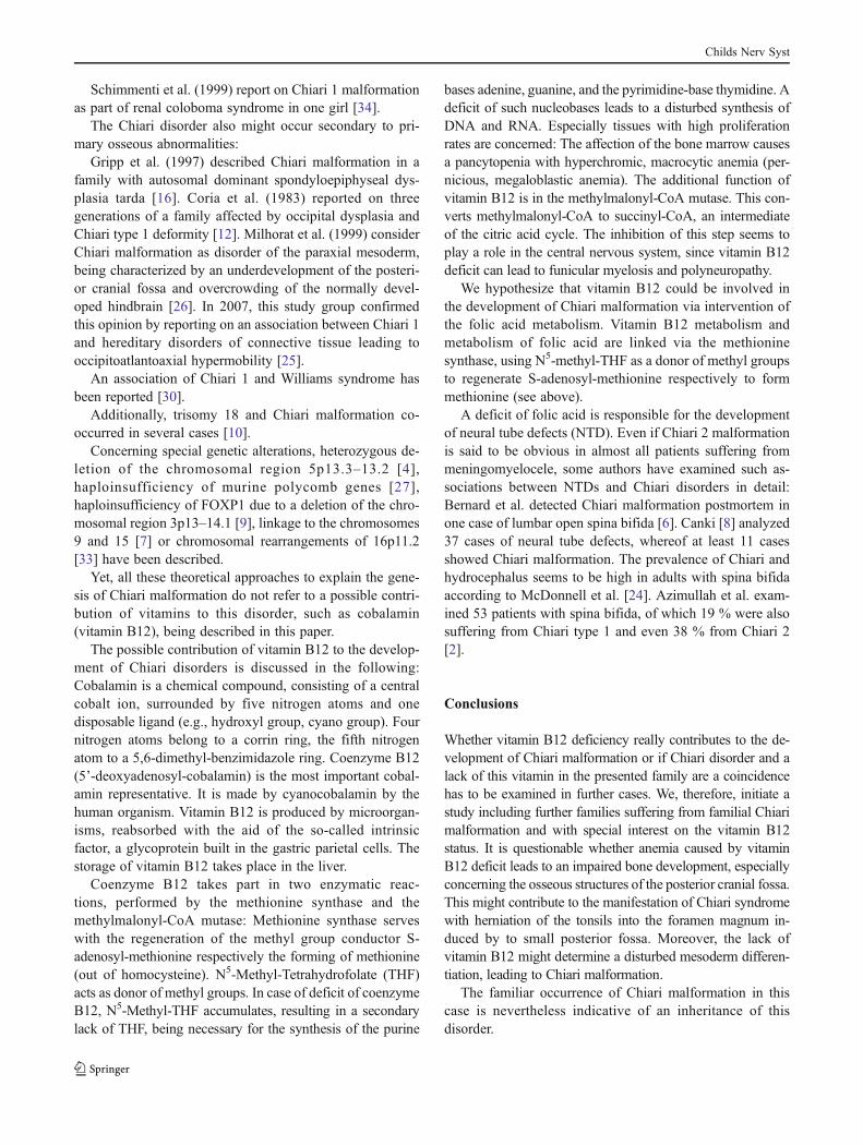
For a better understanding, a family tree of the familymembers affected by Chiari malformation is enclosed(see Fig. 1).
MRI revealed Chiari 0 malformation in the grandfa-ther (F0 generation). Moreover, MRI of the cervicalspine detected syringomyelia from C4 to D1. One ofhis two daughters (F1 generation), presented with Chiari1 malformation in combination with syringomyelia (C4-D2) too. Her sister (F1) suffers from Chiari 1 withoutsyringomyelia.
One girl, aged 14 of the F2 generation, being the daugh-ter of the latter mentioned sister, was diagnosed with Chiari1 malformation as well, in this case in conjunction withmacrocephalus. Moreover, she suffered from initially hid-den benign intracranial hypertension, which was detected2 years after surgical intervention: Due to reoccurring clin-ical complaints as described below (vomiting, wake-upheadaches), we had performed telemetric measurement ofintracranial pressure (ICP) for 3 days, revealing pathologicalB-waves fraction of more than 70 % combined with highICP peaks during the night and accordingly, hidden benignintracranial hypertension (BIH).
In her brother, aged 10 (F2), Chiari 1 malformationcombined with hydrocephalus was detected.
Syringomyelia could not be detected in F2 generation.In F0 generation, the grandfather, who had presented in
our neurosurgical department due to the medical records ofhis family, could state occasional dysesthesia in the upperand lower limb, yet less headache sensations.
Childs Nerv Syst
Clinically, both sisters of F1 generation suffered fromheadaches most prominent in the occipital region as typicalfor Chiari patients. Furthermore, both patients complainedabout occasional dysesthesia in the upper limbs.
The girl of F2 generation initially presented with recur-ring headaches and disturbance of fine motor skills andcoordination. After surgical decompression of posterior cra-nial fossa, the clinical symptoms, especially the headaches,improved for 2 years. After that period of time, the patientsuffered from the abovementioned vomiting early in themorning in combination with wake-up headaches.
Her brother (F2) was born with hydrocephalus andmacrocephaly.
Concerning therapy, all the concerned family membersexcept the grandfather obtained posterior fossa decompres-sion (enlargement of foramen magnum combined with duralpatch grafting and cervical laminectomy of C1) leading topain release. This surgical intervention relived successfullyprogressing cerebellar clinical symptoms (improvement offine motor skills and coordination in female F2 forexample) and symptoms resulting from syringomyeliain F1 generation (dysesthesia).
The grandfather (F0) did not undergo surgery on his owndemand.
The hydrocephalus of the boy (F2 generation) was treatedby CSF shunting (ventriculoperitoneal shunt).
His sister was finally shunted after detection of the hid-den BIH, resulting in considerable improvement of theheadaches and vanishing of nausea and vomiting.
Since the two children’s mother (F1) suffered frompernicious anemia as a consequence of vitamin B12deficit, a hematological screening of the whole familywas performed. The hemograms revealed familial vita-min B12 deficit additionally in her daughter (F2). Asubcutaneous substitution with cobalamin was the con-sequence of the anemia.
Discussion
The reason for the development of Chiari 1 malformationremains unknown. Yet many factors, especially of geneticcharacter, are discussed. To consider genetics to play a rolein the genesis of Chiari syndrome results particularly from thefact that this deformity occurs occasionally, familiarly cumu-lative, and albeit rare [11, 15, 18, 21, 23, 35, 36, 37, 39].
Apart from human medicine, the inheritance of Chiari mal-formation even in combination with syringomyelia is known indog breeding (Cavalier King Charles Spaniel) [31, 32].
On the other hand, several genetic syndromes have beendescribed in literature, co-occurring with Chiari malformation:
Concerning a publication of Germain et al. (2006) [14],Chiari type 1 malformation has been described in fourunrelated patients affected with Fabry disease, an X-linkedinborn error of metabolism resulting from deficient activityof alpha-galactosidase A.
Furthermore, Chiari type 1 could be proved in patientswith FG syndrome (FGS), also an X-linked disorder, com-prising congenital hypotonia, macrocephaly, agenesis ofcorpus callosum, psychomotor delay, and disturbances ofgastrointestinal function. This study group postulated thatChiari malformation is more common in individuals withFGS than in the general population. The FG syndrome isalso called Opitz–Kaveggia syndrome and is namedaccording to the first letters of two sisters, having sons withthis syndrome [28].
Holder-Espinasse and Winter (2003) described thefourth case of Chiari malformation in patients withNoonan syndrome [20].
Due to premature synostosis of the lambdoid suture inpatients with Crouzon syndrome, more than 70 % of thesepatients suffer from Chiari type 1 malformation. In familialCrouzon syndrome with Chiari 1 and syringomyelia, a novelFGFR2 mutation could be detected [13].
Chiari 0 Syringomyelia
Chiari I Chiari I
Syringomyelia
Chiari I Macrocephalus
BIH
Chiari I Hydrocephalus
Fig. 1 Illustrates the familytree of the Chiari patients. Thegrandfather has two daughters,whereof one has two children(one daughter and one son).Detailed information of Chiarimanifestations are elucidated inthe text
Childs Nerv Syst
Schimmenti et al. (1999) report on Chiari 1 malformationas part of renal coloboma syndrome in one girl [34].
The Chiari disorder also might occur secondary to pri-mary osseous abnormalities:
Gripp et al. (1997) described Chiari malformation in afamily with autosomal dominant spondyloepiphyseal dys-plasia tarda [16]. Coria et al. (1983) reported on threegenerations of a family affected by occipital dysplasia andChiari type 1 deformity [12]. Milhorat et al. (1999) considerChiari malformation as disorder of the paraxial mesoderm,being characterized by an underdevelopment of the posteri-or cranial fossa and overcrowding of the normally devel-oped hindbrain [26]. In 2007, this study group confirmedthis opinion by reporting on an association between Chiari 1and hereditary disorders of connective tissue leading tooccipitoatlantoaxial hypermobility [25].
An association of Chiari 1 and Williams syndrome hasbeen reported [30].
Additionally, trisomy 18 and Chiari malformation co-occurred in several cases [10].
Concerning special genetic alterations, heterozygous de-letion of the chromosomal region 5p13.3–13.2 [4],haploinsufficiency of murine polycomb genes [27],haploinsufficiency of FOXP1 due to a deletion of the chro-mosomal region 3p13–14.1 [9], linkage to the chromosomes9 and 15 [7] or chromosomal rearrangements of 16p11.2[33] have been described.
Yet, all these theoretical approaches to explain the gene-sis of Chiari malformation do not refer to a possible contri-bution of vitamins to this disorder, such as cobalamin(vitamin B12), being described in this paper.
The possible contribution of vitamin B12 to the develop-ment of Chiari disorders is discussed in the following:Cobalamin is a chemical compound, consisting of a centralcobalt ion, surrounded by five nitrogen atoms and onedisposable ligand (e.g., hydroxyl group, cyano group). Fournitrogen atoms belong to a corrin ring, the fifth nitrogenatom to a 5,6-dimethyl-benzimidazole ring. Coenzyme B12(5’-deoxyadenosyl-cobalamin) is the most important cobal-amin representative. It is made by cyanocobalamin by thehuman organism. Vitamin B12 is produced by microorgan-isms, reabsorbed with the aid of the so-called intrinsicfactor, a glycoprotein built in the gastric parietal cells. Thestorage of vitamin B12 takes place in the liver.
Coenzyme B12 takes part in two enzymatic reac-tions, performed by the methionine synthase and themethylmalonyl-CoA mutase: Methionine synthase serveswith the regeneration of the methyl group conductor S-adenosyl-methionine respectively the forming of methionine(out of homocysteine). N5-Methyl-Tetrahydrofolate (THF)acts as donor of methyl groups. In case of deficit of coenzymeB12, N5-Methyl-THF accumulates, resulting in a secondarylack of THF, being necessary for the synthesis of the purine
bases adenine, guanine, and the pyrimidine-base thymidine. Adeficit of such nucleobases leads to a disturbed synthesis ofDNA and RNA. Especially tissues with high proliferationrates are concerned: The affection of the bone marrow causesa pancytopenia with hyperchromic, macrocytic anemia (per-nicious, megaloblastic anemia). The additional function ofvitamin B12 is in the methylmalonyl-CoA mutase. This con-verts methylmalonyl-CoA to succinyl-CoA, an intermediateof the citric acid cycle. The inhibition of this step seems toplay a role in the central nervous system, since vitamin B12deficit can lead to funicular myelosis and polyneuropathy.
We hypothesize that vitamin B12 could be involved inthe development of Chiari malformation via intervention ofthe folic acid metabolism. Vitamin B12 metabolism andmetabolism of folic acid are linked via the methioninesynthase, using N5-methyl-THF as a donor of methyl groupsto regenerate S-adenosyl-methionine respectively to formmethionine (see above).
A deficit of folic acid is responsible for the developmentof neural tube defects (NTD). Even if Chiari 2 malformationis said to be obvious in almost all patients suffering frommeningomyelocele, some authors have examined such as-sociations between NTDs and Chiari disorders in detail:Bernard et al. detected Chiari malformation postmortem inone case of lumbar open spina bifida [6]. Canki [8] analyzed37 cases of neural tube defects, whereof at least 11 casesshowed Chiari malformation. The prevalence of Chiari andhydrocephalus seems to be high in adults with spina bifidaaccording to McDonnell et al. [24]. Azimullah et al. exam-ined 53 patients with spina bifida, of which 19 % were alsosuffering from Chiari type 1 and even 38 % from Chiari 2[2].
Conclusions
Whether vitamin B12 deficiency really contributes to the de-velopment of Chiari malformation or if Chiari disorder and alack of this vitamin in the presented family are a coincidencehas to be examined in further cases. We, therefore, initiate astudy including further families suffering from familial Chiarimalformation and with special interest on the vitamin B12status. It is questionable whether anemia caused by vitaminB12 deficit leads to an impaired bone development, especiallyconcerning the osseous structures of the posterior cranial fossa.This might contribute to the manifestation of Chiari syndromewith herniation of the tonsils into the foramen magnum in-duced by to small posterior fossa. Moreover, the lack ofvitamin B12 might determine a disturbed mesoderm differen-tiation, leading to Chiari malformation.
The familiar occurrence of Chiari malformation in thiscase is nevertheless indicative of an inheritance of thisdisorder.
Childs Nerv Syst
Acknowledgments Special thanks to all the colleagues who contrib-uted to this paper and thanks for the positive suggestions. The corre-sponding author and the co-authors have received financial support forothers than this study by the German Federal Ministry of Educationand Research (BMBF). Dr. M. Welsch (née Schmitt) and Dr. S. Antesfurther on received financial support for purpose of education byCodman (Johnson and Johnson Company)/ Raynham, MA andAesculap (Miethke)/ Tuttlingen, Germany, not concerning this study.Dr. R. Eymann and Prof. M. Kiefer received some financial supportduring the past for other than this research work by the Raumedic AG/Helmbrechts, Germany.
Conflict of interest The authors report no conflict of interestconcerning the materials or methods used in this study or the findingsspecified in this paper.
References
1. Aboulezz AO, Sartor K, Geyer CA, Gado MH (1985) Position ofcerebellar tonsils in the normal population and in patients withChiari malformation: a quantitative approach with MR imaging. JComput Assist Tomogr 9:1033–1036
2. Azimullah PC, Smit LM, Rietveld-Knol E, Valk J (1991)Malformations of the spinal cord in 53 patients with spina bifidastudied by magnetic resonance imaging. Childs Nerv Syst 7:63–66
3. Barkovich AJ, Wippold FJ, Sherman JL, Citrin CM (1986)Significance of cerebellar tonsillar position on MR. AJNR Am JNeuoradiol 7:795–799
4. Bayrakli F, Bilguvar K, Ceyhan D, Ercan-Sencicek AG, CankayaT, Gunel M et al (2010) Heterozygous 5p13.3–13.2 deletion in apatient with type I Chiari malformation and bilateral Duane retrac-tion syndrome. Clin Genet 77:499–502
6. Bernard JP, Suarez B, Rambaud C, Muller F, Ville Y (1997)Prenatal diagnosis of neural tube defect before 12 weeks gestation:direct and indirect ultrasonographic semeiology. Ultrasound ObstetGynecol 10:406–409
7. Boyles AL, Enterline DS, Hammock PH, Siegel DG, Slifer SH,Speer MC et al (2006) Phenotypic definition of Chiari type Imalformation coupled with high-density SNP genome screenshows significant evidence for linkage to regions on chromosomes9 and 15. Am J Med Genet A 140:2776–2785
8. Canki N (1984) Development of neural tube defects and geneticcounselling. Jugosl Ginekol Obstet 24:45–48
9. Carr CW, Moreno-De-Luca D, Parker C, Zimmerman HH,Ledbetter N, Abdul-Rahman OA et al (2010) Chiari I malforma-tion, delayed gross motor skills, severe speech delay, and epilep-tiform discharges in a child with FOXP1 haploinsufficiency. Eur JHum Genet 18:1216–1220
10. Case ME, Sarnat HB, Monteleone P (1977) Type II Arnold–Chiarimalformation with normal spine in trisomy 18. Acta Neuropathol37:259–262
11. Cavender RK, Schmidt JH 3rd (1995) Tonsillar ectopia and Chiarimalformations: monozygotic triplets. Case report. J Neurosurg82:497–500
12. Coria F, Quintana F, Rebollo M, Combarros O, Berciano J (1983)Occipital dysplasia and Chiari type I deformity in a family. Clinicaland radiological study of three generations. J Neurol Sci 62:147–158
13. Fujisawa H, Hasegawa M, Kida S, Yamashita J (2002) A novelfibroblast growth factor receptor 2 mutation in Crouzon syndromeassociated with Chiari type I malformation and syringomyelia. JNeurosurg 97:396–400
14. Germain DP, Benistan K, Halimi P (2006) Chiari type I malfor-mation in four unrelated patients affected with Fabry disease. Eur JMed Genet 49:419–425
16. Gripp KW, Scott CI Jr, Nicholson L, Magram G, Grissom LE(1997) Chiari malformation and tonsillar ectopia in twin brothersand father with autosomal dominant spondylo-epiphyseal dyspla-sia tarda. Skeletal Radiol 26:131–133
17. Guinto G, Zamorano C, Dominguez F et al (2004) Chiari I mal-formation Part I. Contemp Neurosurg 26:1–7
18. Herman MD, Cheek WR, Storrs BB (1990–1991) Two siblingswith the Chiari I malformation. Pediatr Neurosurg 16:183–184
19. Hoffman HJ, Hendrick EB, Humphreys RP (1975) Manifestationsand management of Arnold–Chiari malformation in patients withmeningomyelocele. Childs Brain 1:255–259
20. Holder-Espinasse M, Winter RM (2003) Type 1 Arnold–Chiarimalformation and Noonan syndrome. A new diagnostic feature?Clin Dysmorphol 12:275
21. Iwasaki Y, Hida K, Onishi K, Nanba R (2000) Chiari malformationand syringomyelia in monozygotic twins: birth injury as a possiblecause of syringomyelia–case report. Neurol Med Chir 40:176–178
22. Martínez-Lage JF, Guillén-Navarro E, Almagro MJ, Felipe-MurciaM, López-Guerrero A, Galarza M (2010) Hydrocephalus and Chiaritype 1 malformation in macrocephaly-cutis marmorata telangiectaticacongenita: a case-based update. Childs Nerv Syst 26:13–18
23. Mavinkurve GG, Sciubba D, Amundson E, Jallo GI (2005)Familial Chiari type I malformation with syringomyelia in twosiblings: case report and review of the literature. Childs Nerv Syst21:955–999
24. McDonnell GV, McCann JP, Craig JJ, Crone M (2000) Prevalenceof the Chiari/hydrosyringomyelia complex in adults with spinabifida: preliminary results. Eur J Pediatr Surg 10(Suppl 1):18–19
25. Milhorat TH, Bolognese PA, Nishikawa M, McDonnell NB,Francomano CA (2007) Syndrome of occipitoatlantoaxialhypermobility, cranial settling, and Chiari malformation type I inpatients with hereditary disorders of connective tissue. J NeurosurgSpine 7:601–609
26. Milhorat TH, Chou MW, Trinidad EM, Kula RW, Mandell M,Speer MC et al (1999) Chiari I malformation redefined: clinicaland radiographic findings for 364 symptomatic patients.Neurosurgery 44:1005–1017
27. Miró X, Zhou X, Boretius S, Michaelis T, Kubisch C, Gruss P et al(2009) Haploinsufficiency of the murine polycomb gene Suz12results in diverse malformations of the brain and neural tube. DisModel Mech 2:412–418
28. Opitz JM, Kaveggia EG (1974) Studies of malformation of man 33:the FG syndrome. An X-linked recessive syndrome of multiple con-genital anomalies and mental retardation. Z Kinderheilkg 117:1–18
29. Peach B (1965) The Arnold–Chiari malformation; morphogenesis.Arch Neurol 12:527–535
30. Pober BR, Filiano JJ (1995) Association of Chiari I malformationand Williams syndrome. Pediatr Neurol 12:84–88
31. Rusbridge C, Knowler SP (2003) Hereditary aspects of occipitalbone hypoplasia and syringomyelia (Chiari type I malformation) incavalier King Charles spaniels. Vet Rec 153:107–112
32. Rusbridge C, Knowler SP (2004) Inheritance of occipital bonehypoplasia (Chiari type I malformation) in Cavalier King CharlesSpaniels. J Vet Intern Med 18:673–678
33. Schaaf CP, Goin-Kochel RP, Nowell KP, Hunter JV, Aleck KA,Shinawi M et al (2011) Expanding the clinical spectrum of the16p11.2 chromosomal rearrangements: three patients with syrin-gomyelia. Eur J Hum Genet 19:152–156
34. Schimmenti LA, Shim HH, Wirtschafter JD, Panzarino VA,Kashtan CE, Dobyns WB et al (1999) Homonucleotide expansionand contraction mutations of PAX2 and inclusion of Chiari 1
Childs Nerv Syst
malformation as part of renal-coloboma syndrome. Hum Mutat14:369–376
35. Speer MC, George TM, Enterline DS, Franklin A, WolpertCM, Milhorat TH (2000) A genetic hypothesis for Chiari Imalformation with or without syringomyelia. Neurosurg Focus8(3):E12
36. Stovner LJ, Cappelen J, Nilsen G, Sjaastad O (1992) The Chiaritype I malformation in two monozygotic twins and first-degreerelatives. Ann Neurol 31:220–222
37. Szewka AJ, Walsh LE, Boaz JC, Carvalho KS, Golomb MR(2006) Chiari in the family: inheritance of the Chiari I malforma-tion. Pediatr Neurol 34:481–485
38. Tubbs RS, Wellons JC 3rd, Blount JP, Oakes WJ (2004)Syringomyelia in twin brothers discordant for Chiari I malforma-tion: case report. J Child Neurol 19:459–462
39. Weisfeld-Adams JD, Carter MR, Likeman MJ, Rankin J (2007)Three sisters with Chiari I malformation with and without associ-ated syringomyelia. Pediatr Neurosurg 43:533–538