Page 1
1
ASSOCIATION OF MICROPARTICLES AND NEUTROPHIL ACTIVATION WITH 1
DECOMPRESSION SICKNESS 2
3
Stephen R. Thom1, Michael Bennett2, Neil D. Banham3, Walter Chin4, Denise F. Blake5, 4
Anders Rosen6, Neal W. Pollock7, Dennis Madden8, Otto Barak8, Alessandro Marroni9, 5
Costantino Balestra9, 10, Peter Germonpre11, Massimo Pieri9, Danilo Cialoni9, Phi-Nga 6
Jeannie Le12, Christopher Logue12, David Lambert12, Kevin R. Hardy12, Douglas 7
Sward1, Ming Yang 1, Veena B. Bhopale1, Zeljko Dujic8 8
9
1Department of Emergency Medicine, University of Maryland School of Medicine 10 Baltimore MD, 2Dept. of Diving and Hyperbaric Medicine, Prince of Wales Hospital, New 11 South Wales, Australia, 3Dept. of Hyperbaric Medicine, Fiona Stanley Hospital, Perth, 12 Western Australia, 4Dept. of Hyperbaric Medicine, UCLA Medical Center, Los Angeles, CA, 13 5Dept.of Emergency Medicine, The Townsville Hospital and College of Marine and 14 Environmental Sciences, James Cook University, Townsville, Queensland, Australia, 15 6Sahlgrenska University Hospital/Omrade2, Gothenburg, Sweden, 7Divers Alert Network, 16 Durham, NC, 8Department of Integrative Physiology, University of Split School of Medicine, 17 Soltanska, Split, Croatia, 9Divers Alert Network Europe Foundation and Diving Safety 18 Laboratory, 10Environmental, Occupational, Ageing and Integrative Physiology Laboratory, 19 Haute Ecole Paul-Henri Spaak, Brussels, Belgium, 11Centre for Hyperbaric Oxygen Therapy, 20 Military Hospital Brussels, Bruynstraat 200, Brussels 1120, Belgium, 12Dept. of Emergency 21 Medicine, Pereleman School of Medicine, University of Pennsylvania, Philadelphia, PA. 22
23 Running Title: Microparticles and decompression 24
25 Key Words: decompression sickness, myeloperoxidase, CD41, CD235, CD14, 26
tissue factor, von Willebrand factor, platelet-endothelial cell adhesion 27 molecule 28
29 Address correspondence to: Stephen R. Thom, M.D., Ph.D., Department of Emergency 30 Medicine, University of Maryland School of Medicine, 655 W. Baltimore St., Bressler 31 Control Building Room 4-013, Baltimore, MD 21201, Telephone: 410-706-8294, Fax: 410-32 328-8028. E-mail: [email protected] 33
34
Articles in PresS. J Appl Physiol (July 2, 2015). doi:10.1152/japplphysiol.00380.2015
Copyright © 2015 by the American Physiological Society.
by 10.220.32.247 on April 3, 2017
http://jap.physiology.org/D
ownloaded from
Page 2
2
ABSTRACT 35 36
Decompression sickness (DCS) is a systemic disorder assumed due to gas bubbles, but 37
additional factors are likely to play a role. Circulating microparticles, vesicular structures 38
with diameters of 0.1 to 1.0 µm, have been implicated but data in human divers has been 39
lacking. We hypothesized that the number of blood-borne annexin V-positive microparticles 40
(MPs) and neutrophil activation assessed as surface myeloperoxidase (MPO) staining would 41
differ between SCUBA divers suffering from DCS versus asymptomatic divers. Blood was 42
analyzed from 280 divers who had been exposed to maximum depths from 7 to 105 meters; 43
185 were control/asymptomatic divers and 90 were diagnosed with DCS. Elevations of MPs 44
and neutrophil activation occurred in all divers but normalized within 24 hours in those who 45
were asymptomatic. MPs bearing the following proteins: CD66b, CD41, CD31, CD142, 46
CD235 and von Willebrand factor were between 2.4 and 11.7-fold higher in blood from 47
divers with DCS versus asymptomatic divers matched for time of sample acquisition, 48
maximum diving depth and breathing gas. Multiple logistic regression analysis documented 49
significant associations (p<0.001) between DCS and MPs and for neutrophil MPO staining. 50
Effect estimates were not altered by gender, body mass index, use of non-steroidal anti-51
inflammatory agents or emergency oxygen treatment, and modestly influenced by divers’ 52
age, choice of breathing gas during diving, maximum diving depth, and whether repetitive 53
diving had been performed. There were no significant associations between DCS and number 54
of MPs without surface proteins listed above. We conclude that MPs production and 55
neutrophil activation exhibit strong associations with DCS. 56
57
by 10.220.32.247 on April 3, 2017
http://jap.physiology.org/D
ownloaded from
Page 3
3
INTRODUCTION 58
Decompression sickness (DCS) is a risk associated with compressed gas diving, tunneling, 59
high altitude aviation, and space exploration. Gas bubbles, long thought to be the inciting 60
factor for DCS, are common and often asymptomatic; hence, additional pathophysiological 61
factors have been sought to explain the development of the syndrome (7, 15, 16). There is 62
now considerable evidence that microparticles (MPs), cell-derived membrane vesicles with 63
diameters of 0.1 to 1.0 µm, are elevated in association with simulated as well as bona fide 64
underwater diving(17-19, 23, 27, 28, 32). Maneuvers which decrease the incidence of DCS 65
also diminish MPs production (17, 18). Murine studies suggest that MPs play a role in high 66
pressure gas pathophysiology and possibly with gas bubble nucleation (29, 30, 35, 36). In the 67
mouse model, MPs have been shown to initiate a systemic inflammatory process that is 68
related to neutrophil activation following decompression (29, 30, 35, 36). Injuries identified 69
in decompressed animals can be recapitulated by injecting decompression-induced MPs into 70
naïve mice (30, 35, 36). 71
72
There are as yet no data associating MPs with DCS in humans. The goal of this study was to 73
examine MPs and neutrophil activation in blood obtained from SCUBA (Self-contained 74
underwater breathing apparatus) divers. MPs were characterized, as is standard, by surface 75
expression of antigenic markers from parent cells and based on annexin V binding because as 76
MPs are formed, negatively charged phosphatidylserine residues become exposed. We 77
hypothesized that differences would be identified between healthy, asymptomatic divers and 78
those suffering from DCS. Blood samples were obtained from divers by a consortium of 79
investigators around the globe who supervise diving and/or treat divers with DCS. As the 80
by 10.220.32.247 on April 3, 2017
http://jap.physiology.org/D
ownloaded from
Page 4
4
study progressed it became obvious that those with DCS exhibited marked differences from 81
asymptomatic divers. This prompted an examination of blood-borne changes following a 82
variety of dives to different depths, using different breathing gases, and after multiple dive 83
sessions. 84
85
EXPERIMENTAL PROCEDURES 86
Subjects: All procedures were completed in accordance with the Declaration of Helsinki and 87
approved by Ethical Committees of all organizations involved with this investigation. 88
Participants provided informed, written consent. Divers with DCS symptoms were 89
approached by clinical teams when they presented to hospitals for evaluation and treatment. 90
A comparison group of divers who were not suffering DCS was developed by soliciting 91
cooperation from sport SCUBA divers. These were experienced, certified divers monitored 92
by one or more of the co-authors. The dive profiles, frequency of diving, and choice of 93
breathing gases were selected by the divers and were independent of the study protocol. 94
Activities were planned for other purposes, often as recreation, and the research component 95
was solely a willingness to undergo phlebotomy prior to and at a range of times after diving. 96
Under supervision, these divers swam continuously while at depth at a pace assumed similar 97
to that which a normal diver would follow; activity that for most represents a sustained 98
moderate work-rate. Diving profiles were chosen to be within accepted standards so there 99
would be no decompression requirement. Total dive times ranged from 17 to 178 minutes. 100
101
Divers evaluated for DCS: The signs and symptoms reported covered the gamut that is 102
typically seen and well described in other publications (22). Pain as one of the primary 103
by 10.220.32.247 on April 3, 2017
http://jap.physiology.org/D
ownloaded from
Page 5
5
complaints was reported by 69 (72.6%), sensory abnormalities by 52 (54.7%), weakness by 104
45 (47.4%), and central nervous symptoms by 15 (15.8%). The median interval of time from 105
termination of the last dive to onset of DCS signs and/or symptoms was 0.55 (25th and 75th 106
percentile: 0.08, 4.25) hours. There were no obvious violations of decompression algorithms 107
based on divers’ reports and – where possible – confirmed by interrogation of dive computers 108
in 72 (80%) cases. Violations of dive tables and/or alerts from dive computers were identified 109
in 18 (20%) cases, typically due to diver decisions or equipment failure that resulted in an 110
uncontrolled ascent from depth. Some divers had first aid/emergency interventions prior to 111
arrival at hospital: 42 divers received supplemental O2, and 6 received nonsteroidal anti-112
inflammatory agents (NSAID). 113
114
There is no definitive test by which to establish a diagnosis of DCS. We defined DCS as 115
having occurred when a diver presented with complaints consistent with DCS such that a 116
clinical decision was made to initiate therapeutic recompression and where recompression 117
was associated with an improvement in signs or symptoms. Participation in the study 118
involved obtaining blood at the time of the initial evaluation and after recompression 119
treatment. Divers were also asked to provide a sample when returning to clinic for a late 120
follow-up medical evaluation prior to any return to diving. 121
122
Control divers: Divers participating in this project used their own equipment. Venous blood 123
was collected from an antecubital arm vein by a trained phlebotomist prior to and at one or 124
more times between 15 minutes and 144 hours after diving. Phlebotomy was often carried out 125
at a remote beach site but, where feasible, it was done at shore-based laboratory facilities. 126
by 10.220.32.247 on April 3, 2017
http://jap.physiology.org/D
ownloaded from
Page 6
6
When results were compared among the field sites or according to the location where 127
phlebotomy was done (remote beach site versus laboratory) and matched for time when 128
samples were obtained post-diving, there were no statistically significant differences. 129
130
Materials and standard laboratory procedures: Blood (~ 5 ml) was drawn into Cyto-Chex 131
BCT test tubes that contain a proprietary preservative (Streck, Inc., Omaha, NE). Samples 132
were sent by express mail to the first author’s laboratory where all analyses were performed 133
following published techniques within 48 hours after arrival; from 4 to 9 days after collection 134
(27). Prior work has shown that MPs and neutrophil characteristics remain unchanged when 135
samples are processed within 3 weeks from time of acquisition (27). All supplies, reagents 136
and manufacturer sources have been described in previous publications (17, 18, 27, 28). 137
138
Flow Cytometry: Early studies were performed with a 10-color FACSCanto (Becton 139
Dickinson, San Jose, CA), the majority were performed with an 8-color, triple laser 140
MACSQuant (Miltenyi Biotec Corp., Auburn, CA) using manufacturers’ acquisition 141
software. MPs were stained with annexin V and analyzed exactly as previously described (27, 142
28). Surface markers were evaluated with use of the ‘‘Fluorescence-Minus-One Control Test” 143
(31). This analysis provides a way to define the boundary between positive and negative 144
particles in an unbiased manner by defining the maximum fluorescence expected for a given 145
subset after outlining the area in a two-dimensional scatter diagram when a fluorophore-146
tagged antibody is omitted from the stain set. This analysis allows a simple decision as to 147
where to place the upper boundary for non-staining particles in a fluorescence channel. We 148
define MPs as annexin V-positive particles with diameters up to 1 µm. Neutrophils in whole 149
by 10.220.32.247 on April 3, 2017
http://jap.physiology.org/D
ownloaded from
Page 7
7
blood were identified by CD66b staining and surface expression of myeloperoxidase (MPO) 150
assayed as previously described (27, 28). MPO% indicates the fraction of all CD66b-positive 151
cells exhibiting positive staining for MPO, MPO-median the geometric median fluorescence 152
value. 153
154
Statistical analysis: Results are summarized as median, 25th and 75th percentiles. Log 155
transformations were used for logistic regression analysis and to carry out two-way ANOVA. 156
Correlations were evaluated by the Spearman rank order test. We used Sigmastat software 157
(Systat, Point Richmond, CA) for the statistical analysis. Statistical significance level was set 158
as p<0.05. 159
160
RESULTS 161
Blood samples were obtained from 280 divers. Table 1 displays characteristics of the study 162
population. Among 95 divers who presented to hospitals with signs and/or symptoms thought 163
to be due to DCS, complaints improved with recompression therapy in 90. They had 164
performed a median of two (1.5, 3.0) dives prior to developing DCS; only 18% developed 165
DCS after a single dive. Most repetitive dives were performed on the same day, a minority 166
over two or more consecutive days. There was no statistically significant correlation between 167
DCS and the maximum depth of the most recent dive before presentation. 168
169
There were 185 divers in the control group. Age, gender distribution, body mass index and 170
median of the maximum depth of diving were not statistically significantly different from the 171
DCS group (Table 1). The majority of divers in both groups used compressed air as breathing 172
by 10.220.32.247 on April 3, 2017
http://jap.physiology.org/D
ownloaded from
Page 8
8
gas, the rest used either ‘enriched air nitrox’ (EAN) comprised of 32% oxygen (O2)/68% 173
nitrogen (N2), or Tri-mix gas which, depending on the dive depth and an individual diver’s 174
preference, contained variable amounts of O2 (7-22%), helium (5-35%) and N2 (40-60%). 175
Tri-mix divers in both groups used rebreather apparatus with fixed, user-selected O2 partial 176
pressures that typically involved 1.0 to 1.3 ATA O2 at depth and up to 1.4 to 1.64 ATA O2 177
during the final stages of decompression. A higher percentage of divers used Tri-mix in the 178
control group than in the DCS group. 179
180
Analysis of blood from control divers demonstrated that neutrophil activation assessed as 181
MPO on the cell surface and elevations of MPs did occur from 15 minutes to four hours after 182
diving (Figure 1), but changes resolved within 24 hours. Obtaining blood at two hours post-183
diving was a frequent part of the research protocol. When results from control divers were 184
separated by decades of maximum diving depth (e.g.10-19.9 meters, 20-29.9 meters, etc.), all 185
two hour-post diving values for neutrophil activation and MPs elevations were statistically 186
significantly different from pre-dive values but none were significantly different from each 187
other (data not shown). 188
189
Some individuals in the control group dove only once and blood was obtained at one or more 190
times from 0.25 to 96 hours later. Others performed one or two dives per day for up to 6 days. 191
Figure 2 displays the pattern of MPs and neutrophil MPO staining when control divers 192
conducted repetitive dives. Pre-dive values on all days were not statistically significantly 193
different from each other. Statistically significant elevations were found in all two hour-post 194
dive samples, but only post-dive neutrophil MPO on days three and six were greater than 195
by 10.220.32.247 on April 3, 2017
http://jap.physiology.org/D
ownloaded from
Page 9
9
post-dive values on days one and two (Figure 2). The pattern of elevations with each day of 196
diving and normalization of values prior to diving the next day was also observed within MPs 197
sub-groups (Table 2). Most of the repetitive divers performed just one dive per day, but data 198
were not significantly different among 22 divers who performed two dives in one day and 199
blood obtained 24 hours after the second dive (data not shown). 200
201
Table 3 displays data from control divers and those with DCS. The first column contains pre-202
dive data from the control group. The second column shows post-dive results from control 203
divers, but this data set differs slightly from the pre-dive set. As was outlined in Table 1, the 204
control subjects used Tri-mix gas ~ 15% of the time (27 divers). Because it is unknown how 205
this usage may influence neutrophil activation and MPs, data from only the first four Tri-mix 206
divers enrolled in the study were used so that the proportions matched those for divers with 207
DCS (a post-hoc analysis demonstrated that data for these four were not statistically 208
significantly different from the 23 other control Tri-mix divers). Hence, data represent results 209
from 162 divers; 130 (80.2%) used compressed air, 28 (17.3%) used EAN and four (2.5%) 210
used Tri-mix. 211
212
The post-dive data in the second column of Table 3 were generated using only one blood 213
sample from each control diver. The sample chosen was always the last obtained from those 214
who did a single dive (n= 65). For repetitive divers (n = 97) the sample chosen for analysis 215
was obtained just before diving on the last day. This approach was taken to better match the 216
data set for DCS divers, where blood samples were obtained at a median time of 24 (11.8, 217
55.0; range 0.5 – 144) hours after diving. By compiling the control diver data set in the 218
by 10.220.32.247 on April 3, 2017
http://jap.physiology.org/D
ownloaded from
Page 10
10
manner described, neutrophil and MPs changes could be assessed at a median time of 24 (7.4, 219
96) hours post-diving (range 0.5 – 144) hours. 220
221
Data from DCS diver samples obtained on presentation to hospital are identified as “acute” in 222
the third column of Table 3. Values for neutrophil MPO staining and MPs expressing each of 223
the protein sub-types were statistically significantly different from the post-dive control 224
values. Among the 90 divers with DCS, 35 returned to clinics for follow-up evaluations and 225
provided blood samples at a median time of 28 (13.5, 35) days after treatment (last column in 226
Table 3). Divers were all asymptomatic at this time. All values in this group were statistically 227
significantly different from pre-dive values from the control group (column 1), but few were 228
significantly different from post-dive control group values (column 2). Leukocyte and platelet 229
counts were not significantly different among samples from control subjects, the acute 230
samples obtained from DCS divers and the late follow-up samples (data not shown). 231
232
We also compared DCS diver data sets between those who performed a single dive and those 233
who had conducted repetitive dives prior to onset of DCS. The only values exhibiting 234
statistically significant differences were time when blood samples were obtained post-235
symptom onset and the number of CD142-positive MPs. All other blood analyses listed in 236
Table 3 were not significantly different. The median time of blood sample acquisition for 237
those performing a single dive was 12 (4, 48) hours versus 26 (12, 72) hours (p=0.002) for 238
those having performed repetitive dives. The CD142-positive MPs count for those performing 239
a single dive was 18 (11, 50) MPs/µl versus 67 (29, 186) (p=0.012) MPs/µl for those having 240
by 10.220.32.247 on April 3, 2017
http://jap.physiology.org/D
ownloaded from
Page 11
11
performed repetitive dives. Interestingly, a trend for elevations in CD142 MPs with repetitive 241
dives in the control group also appeared in Table 2. 242
243
It was feasible that ingestion of NSAID medication or emergency use of supplemental O2 244
prior to DCS diver presentation might influence various blood test results. However, using 245
either NSAID or O2 use as the binary, dependent variable for multiple logistic regression 246
analysis, we found no significant impact for these interventions on total MPs, any MPs sub-247
type or neutrophil activation. 248
249
Logistic regression of log transformed data found a positive association between both the 250
magnitude of neutrophil activation and numbers of MPs in all sub-groups and DCS with odds 251
ratios (ORs) from 1.4 to 6.9 (Table 4). These ORs increased when adjusted for the time when 252
the blood samples were obtained after diving. Factoring in diving characteristics such as 253
maximum depth, breathing gas, and whether repetitive diving was performed along with time 254
of sample acquisition had little impact on ORs (data not shown). The OR for MPO-median 255
adjusted for a diver’s age increased the value to 8.6 (95 % confidence limits: 2.4, 31.5, 256
p=0.001) and by including time of sample acquisition and age the OR was 9.0 (2.4, 33.3, 257
p=0.001). A diver’s age did not modify the effect estimate of any other variable. Gender and 258
body mass index did not modify the effect estimate of any variable. Adjustment of ORs by 259
including factors related to dive characteristics and also diver age increased ORs for some 260
variables modestly while diminishing the OR for MPO-median (last row in Table 4). Adding 261
gender, body mass index or including multiple MPs sub-types in this multiple variable 262
by 10.220.32.247 on April 3, 2017
http://jap.physiology.org/D
ownloaded from
Page 12
12
analysis did not alter the ORs. There was no significant association between DCS and total 263
number of annexin V-positive MPs. 264
265
Consistent with the regression analysis, DCS exhibited a significant correlation with all 266
variables (Table 5). There were weak, statistically significant correlations between MPO on 267
neutrophils and most MPs subtypes, and strong correlations among the number of each of the 268
MPs subtypes. No correlation was found between DCS and maximum diving depth, 269
performing repetitive versus a single dive, or gender. Age was negatively correlated with 270
DCS (-0.25, p<0.0001). 271
272
DISCUSSION: 273
Our results provide a number of insights regarding human responses to SCUBA diving. 274
Elevations of MPs and neutrophil MPO surface staining occur predictably, but there are no 275
statistically significant differences in the responses based on depth of diving. It does appear, 276
however, that repetitive diving augments neutrophil activation as shown in Figure 2. We 277
interpret elevations of MPs as a response to high gas pressure exposures. The mechanism has 278
been discerned for neutrophils as an oxidative stress response due to an interaction between 279
O2 and ballast, or what are viewed as inert gases such as nitrogen (26); studies with other 280
vascular cells are underway. Similar MPs and neutrophil activation responses occur in the 281
murine model, but a dose-response between gas pressure versus MPs numbers and neutrophil 282
activation can be discerned with inbred laboratory animals (29). 283
284
by 10.220.32.247 on April 3, 2017
http://jap.physiology.org/D
ownloaded from
Page 13
13
With regard to divers with DCS, neutrophil and MPs responses are markedly greater than 285
among asymptomatic divers. There are statistically significant associations between these 286
variables and DCS. Diving depth, breathing gas and participation in repetitive diving had no 287
meaningful impact on the associations between DCS and neutrophil activation or MPs sub-288
types. Post-decompression MPs elevations and neutrophil activation are clearly linked to 289
injuries to the vasculature and brain in the murine model but obviously, results from this 290
project do not identify the pathophysiological relationship between these blood-borne 291
changes and clinical findings in humans (30, 35, 36). 292
293
Post-dive values for control divers in the second column of Table 3, obtained at a median 294
time of 24 hours post-diving, are significantly different from the pre-dive values. This may 295
appear to contradict findings in Figure 1 where resolution of dive-induced changes occur 296
within 24 hours. It is important to note, however, that 65 (40.1%) of the samples in the Table 297
3 analysis were obtained from divers 0.5 to 2 hours after diving. It was our belief that 298
including these early post-dive data provided a more balanced comparison for findings to the 299
DCS diver group, as many of the injured presented to hospital within a few hours of diving. 300
301
There appears to be persistent neutrophil activation and elevations in some MPs sub-types 302
long after divers suffered DCS (last column in Table 3). These divers had been instructed not 303
to participate in SCUBA diving until presenting for follow-up evaluations. Assuming that 304
most were compliant with instructions, the findings could suggest that on-going or long-term 305
changes occur after DCS. It is important to note, however, that the magnitude of neutrophil 306
activation related to diving is much less than that in response to chemicals thought similar to 307
by 10.220.32.247 on April 3, 2017
http://jap.physiology.org/D
ownloaded from
Page 14
14
some pathological stimuli (27). These results are not evidence for a fulminant systemic 308
inflammatory response syndrome. Indeed, the late follow-up divers did not express physical 309
complaints. 310
311
Persistent elevations of some MPs sub-types weeks after DCS could be related to rates of 312
MPs clearance. Surface phosphatidylserine on MPs constitutes a recognition signal that 313
enables phagocytosis (1). In the mouse model there are marked differences in clearance rates 314
among MPs, but data in humans are lacking (36). The results may be interpreted as a feed-315
back loop whereby persistent elevations of MPs are causing neutrophil activation, a 316
phenomenon shown to occur in the murine model (30, 35, 36). An alternative possibility, 317
however, is that the elevations found late after treatment actually represent these individuals’ 318
baseline or pre-dive characteristics such that attributes of MPs or sensitivity for neutrophil 319
activation place them at greater risk for DCS. 320
321
The relationship between MPs elevations and neutrophil activation is complex and each can 322
lead to the other, as well as the development of vascular injuries (30, 35, 36). Human divers 323
exhibit vascular dysfunction assessed as a decrease in conduit artery endothelial function. 324
Measured as flow-mediated dilatation, it occurs after a single dive and can persist for several 325
days (2, 21). Correlations between neutrophil activation and MPs elevations and among MPs 326
sub-types (Table 5) are consistent with murine studies (29, 30, 35, 36). Additionally, 327
neutrophil activation results in some MPO adhering to the cell surface and MPO on the 328
neutrophil surface can cause auto-activation (14). 329
330
by 10.220.32.247 on April 3, 2017
http://jap.physiology.org/D
ownloaded from
Page 15
15
The dynamics between MPs and neutrophil activation may also be responsible for the trends 331
with elevations of MPs sub-types in repetitive divers (Table 2), and elevations of CD142-332
positive MPs in repetitive DCS divers versus those diving only once. Intravascular expression 333
of CD142 (tissue factor), is a primary mechanism of inflammation-induced coagulation 334
activation and it is the most important initiator of thrombin formation (4). In this regard, it is 335
of interest that reductions of plasma fibrinogen occur with repetitive diving and, on rare 336
occasions, coagulopathy occurs with DCS (6). 337
338
Of course these results do not resolve the role for bubbles in DCS. The relationship between 339
intravascular bubbles, MPs, and neutrophil activation is influenced by variables such as diver 340
exertion as well as breathing gases, and possibly diet or dietary supplements (28, 33, 34). 341
There is evidence supporting the presence of a gas phase in some MPs (36). These could 342
serve as bubble nucleation sites and, given that MPs enlargement occurs post-decompression, 343
MPs may be a source of decompression-induced vascular bubbles which have reported 344
diameters of 24 to 160 μm (9, 10, 12). 345
346
Finally, the data provide some insight into perceived risks of DCS. There is on-going debate 347
whether women have greater risk, possibly linked to menstrual changes (24). We found no 348
statistically significant association between gender and DCS, nor did gender influence the 349
effect estimates of the various blood-borne measurements (Table 4). In some but not all 350
studies, obesity appears to be one of the factors which increases the risk of DCS (5, 11), 351
however, we did not find body mass index to be associated with DCS. Surprisingly, age was 352
negatively correlated with DCS. There has not been much effort focused on investigating the 353
by 10.220.32.247 on April 3, 2017
http://jap.physiology.org/D
ownloaded from
Page 16
16
association between age and DCS. One study reported that age was a contributing factor to 354
intravascular bubble formation, and another found an increased incidence of altitude-induced 355
DCS in those over 42 years of age (3, 25). Another study found no age influence on DCS, 356
however, there were no divers over age 50 in the series (13). We found age to significantly 357
influence the effect estimate of MPO-median value on DCS. This is an interesting issue, as 358
age is generally viewed as having a negative influence on neutrophil functions such as 359
priming and degranulation (8, 20). 360
361
In conclusion, whereas neutrophil activation and MPs elevations are a common response to 362
diving, individuals who develop DCS exhibit more exuberant responses than do the 363
control/asymptomatic divers that were studied. Increased levels of MPs and activated 364
neutrophils are associated with the development of DCS symptoms when compared to divers 365
who have not experienced DCS symptoms while conducting dives with similar profiles. Time 366
of blood sample acquisition post-diving greatly impacts measurements. At least among those 367
divers who present to hospital at later times, the blood-borne changes described here might be 368
useful as biomarkers to aid in diagnosing DCS (Figure 3). Further work will be needed, 369
however, because values from the DCS and control groups exhibit some overlap. Some 370
interventions that inhibit MPs elevations and tissue injuries in mice also diminish MPs 371
elevations and neutrophil activation in human divers (33, 34). This offers an opportunity to 372
examine whether similar interventions could improve the safety of provocative diving. It 373
remains unclear, however, whether there are pre-existing differences within the population 374
that contribute to development of DCS. 375
376
by 10.220.32.247 on April 3, 2017
http://jap.physiology.org/D
ownloaded from
Page 17
17
ACKNOWLEDGMENTS 377
Funding sponsors played no role in the study design. This work was supported by grants from 378
the U.S. Office of Naval Research, Divers Alert Network, Unity through Knowledge Fund 379
(Grant no. 33/08) and Croatian Ministry of Science, Education and Sports (Grant no. 216-380
2160133-0130). We thank Jelena Pavic, Jenna M. Wiley and Stefanie D. Martina for field 381
testing assistance and Marina Bogush and Tatyana N. Milovanova at the University of 382
Pennsylvania for technical assistance in the laboratory. 383
384
385
386
REFERENCES 387
1. Bevers EM, and Williamson PL. Phospholipid scramblase: an update. . FEBS Lett 388
584: 2724-2730, 2010. 389
2. Brubakk AO, Duplancic D, Valic Z, Palada I, Obad A, Bakovic D, Wisloff U, and 390
Dujic Z. A single air dive reduces arterial endothelial function. J Physiol 566.3: 901-906, 391
2005. 392
3. Carturan D, Boussuges A, Vanuxem P, Bar-hen A, Burnet H, and Gardette B. 393
Ascent rate, age, maximal oxygen uptake, adiposity, and circulating venous bubbles in 394
diving,. J Appl Physiol 93: 1349-1356, 2002. 395
4. Chu AJ. Tissue factor mediates inflammation. Arch Biochem Biophys 440: 123-132, 396
2005. 397
by 10.220.32.247 on April 3, 2017
http://jap.physiology.org/D
ownloaded from
Page 18
18
5. Dembert ML, Jekel JF, and Mooney LW. Health risk factors for the development 398
of decompression sickness among U. S. Navy divers. UnderseaBiomedRes 11: 395-406, 399
1984. 400
6. Eckenhoff RG, and Hughes JS. Hematologic and hemostatic changes with repetative 401
air diving. Aviat Space Environ Med 55: 592-597, 1984. 402
7. Eftedal OS, Lydersen S, and Brubakk AO. The relationship between venous gas 403
bubbles and adverse effects of decompression after air dives. Undersea and Hyperbaric Med 404
34: 99-105, 2007. 405
8. Fulop T, Larbi A, Douziech N, Fortin C, Guerard KP, Lesur O, Khalil A, and 406
Dupuis G. Signal transduction and functional changes in neutrophils with aging. Aging Cell 407
3: 217-226, 2004. 408
9. Gersh I, Hawkinson GE, and Rathburn EN. Tissue and vascular bubbles after 409
decompression from high pressure atmospheres - correlation of specific gravity with 410
morphological changes. J Cell Comp Phy 24: 35-70, 1944. 411
10. Grulke DC, Marsh PL, and Hills BA. Experimental air embolism: Measurement of 412
microbubbles using the coulter counter. Br J Exp Path 54: 684-691, 1973. 413
11. Hagberg M, and Ornhagen H. Incidence and risk factors for symptoms of 414
decompression sickness among male and female dive masters and instructors - a retrospective 415
cohort study. Undersea and Hyperbaric Med 30: 93-102, 2003. 416
12. Hills BA, and Butler BD. Size distribution of intravascular air emboli produced by 417
decompression. UnderseaBiomedRes 8: 163-170, 1981. 418
13. Hoiberg A. Consequences of U.S. Navy diving mishaps: decompression sickness. 419
Undersea Biomed Res 13: 383-394, 1986. 420
by 10.220.32.247 on April 3, 2017
http://jap.physiology.org/D
ownloaded from
Page 19
19
14. Lau D, Mollnau H, Eiserich JP, Freeman BA, Daiber A, Gehling UM, Brummer 421
J, Rudolph V, Munzel T, Heitzer T, Meinertz T, and Baldus S. Myeloperoxidase mediates 422
neutrophil activation by association with CD11b/CD18 integrins. Proc Natl Acad Sci U S A 423
102: 431-436, 2005. 424
15. Ljubkovic M, Dujic Z, Mollerlokken A, Bakovic S, Obad A, Breskovic T, and 425
Brubakk AO. Venous and arterial bubbles at rest after no-decompression air dives. Med Sci 426
Sports Exerc 43: 990-995, 2011. 427
16. Ljubkovic M, Marinovic J, Obad A, Breskovic T, Gaustad SE, and Dujic Z. High 428
incidence of venous and arterial gas emboli at rest after trimix diving without protocol 429
violations. J Appl Physiol 109: 1670-1674, 2010. 430
17. Madden D, Thom SR, Milovanova TN, Yang M, Bhopale VM, Ljubkovic M, and 431
Dujic Z. Exercise before SCUBA diving ameliorates decompression-induced neutrophil 432
activation. Med Sci Sports Exerc 46: 1928-1935, 2014. 433
18. Madden D, Thom SR, Yang M, Bhopale VM, Milovanova TN, Ljubkovic M, and 434
Dujic Z. High intensity cycling before SCUBA diving reduces post-decompression 435
microparticle production and neutrophil activation. Eur J Appl Physiol 114: 1955-1961, 2014. 436
19. Madden LA, Chrismas BC, Mellor D, Vince RV, Midgley AW, McNaughton LR, 437
Atkins SL, and Laden G. Endothelial function and stress response after simulated dives to 438
18 msw breathing air or oxygen. Aviat Space Environ Med 81: 41-51, 2010. 439
20. McLaughlin B, O'Malley K, and Cotter TG. Age-related differences in granulocyte 440
chemotaxis and degranulation. Clin Sci 70: 59-62, 1986. 441
by 10.220.32.247 on April 3, 2017
http://jap.physiology.org/D
ownloaded from
Page 20
20
21. Obad A, Palada I, Valic Z, Ivancev V, Bakovic D, Wisloff U, Brubakk AO, and 442
Dujic Z. The effects of acute oral antioxidants on diving-induced alterations in human 443
cardiovascular function. J Physiol 578: 859-870, 2007. 444
22. Ozyigit T, Egi SM, Denoble P, Balestra C, Aydin S, Vann RD, and Marroni A. 445
Decompression illness medically reported by hyperbaric treatment facilities: Cluster analysis 446
of 1929 cases. Aviat Space Environ Med 81: 3-7, 2010. 447
23. Pontier JM, Gempp E, and Ignatescu M. Blood platelet-derived microparticles 448
release and bubble formation after an open-sea dive. Appl Physiol Nutr Metab 37: 1-5, 2012. 449
24. St. Leger Dowse M, Gunby A, Phil D, Moncad R, Fife C, Morsman J, and Bryson 450
P. Problems associated with scuba diving are not evenly distributed across a menstrual cycle. 451
J Obstetrics and Gynecology 26: 216-221, 2006. 452
25. Sulaiman ZM, Pilmanis AA, and O'Connor RB. Relationship between age and 453
susceptibility to altitude decompression sickness. Aviat Space Environ Med 68: 695-698, 454
1997. 455
26. Thom SR, Bhopale VM, and Yang M. Neutrophils generate microparticles during 456
exposure to inert gases due to cytoskeletal oxidative stress. J Biol Chem 289: 18831-18845, 457
2014. 458
27. Thom SR, Milovanova TN, Bogush M, Bhopale VM, Yang M, Bushmann K, 459
Pollock NW, Ljubkovic M, Denoble P, and Dujic Z. Microparticle production, neutrophil 460
activation and intravascular bubbles following open-water SCUBA diving. J Appl Physiol 461
112: 1268-1278, 2012. 462
28. Thom SR, Milovanova TN, Bogush M, Yang M, Bhopale VM, Pollock NW, 463
Ljubkovic M, Denoble P, Madden D, Lozo M, and Dujic Z. Bubbles, microparticles and 464
by 10.220.32.247 on April 3, 2017
http://jap.physiology.org/D
ownloaded from
Page 21
21
neutrophil activation: Changes with exercise level and breathing gas during open-water 465
SCUBA diving. J Appl Physiol 114: 1396-1405, 2013. 466
29. Thom SR, Yang M, Bhopale VM, Huang S, and Milovanova TN. Microparticles 467
initiate decompression-induced neutrophil activation and subsequent vascular injuries. J Appl 468
Physiol 110: 340-351, 2011. 469
30. Thom SR, Yang M, Bhopale VM, Milovanova TN, Bogush M, and Buerk DG. 470
Intra-microparticle nitrogen dioxide is a bubble nucleation site leading to decompression-471
induced neutrophil activation and vascular injury. J Appl Physiol 114: 550-558, 2013. 472
31. Tung JW, Parks DR, Moore WA, Herzenberg LA, and Herzenberg LA. New 473
approaches to fluorescence compensation and visualization of FACS data. Clin Immunol 110: 474
277-283, 2004. 475
32. Vince RV, McNaughton LR, Taylor L, Midgley AW, Laden G, and Madden LA. 476
Release of VCAM-1 associated endothelial microparticles following simulated SCUBA 477
dives. Eur J Appl Physiol 105: 507-513, 2009. 478
33. Yang M, Barak OF, Dujic Z, Madden D, Bhopale VM, Bhullar J, and Thom SR. 479
Ascorbic acid diminishes microparticle elevations and neutrophil activaiton folloowing 480
SCUBA diving. Am J Physiol in press: 2015. 481
34. Yang M, Bhopale VM, and Thom SR. Ascorbic acid abrogates microparticle 482
generation and vascular injuries associated with high pressure exposure. J Appl Physiol In 483
press.: 2015. 484
35. Yang M, Kosterin P, Salzberg BM, Milovanova TN, Bhopale VM, and Thom SR. 485
Microparticles generated by decompression stress cause central nervous system injury 486
by 10.220.32.247 on April 3, 2017
http://jap.physiology.org/D
ownloaded from
Page 22
22
manifested as neurohypophiseal terminal action potential broadening. J Appl Physiol 115: 487
1481-1486, 2013. 488
36. Yang M, Milovanova TN, Bogush M, Uzan G, Bhopale VM, and Thom SR. 489
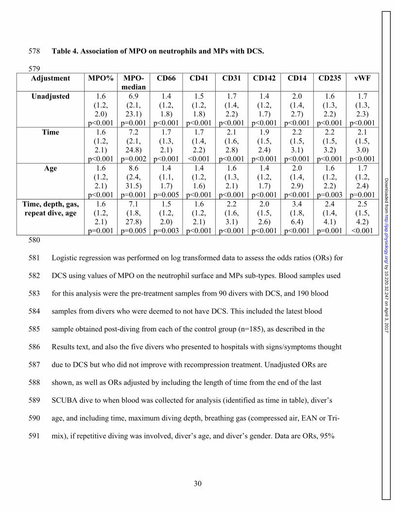
Microparticle enlargement and altered surface proteins after air decompression are associated 490
with inflammatory vascular injuries. J Appl Physiol 112: 204-211, 2012. 491
492
493
by 10.220.32.247 on April 3, 2017
http://jap.physiology.org/D
ownloaded from
Page 23
23
FIGURE LEGENDS 494
495
Figure 1. Blood-borne annexin V-positive MPs and neutrophil myeloperoxidase (MPO) 496
staining in blood from control divers before and after a single dive. Blood was obtained 497
prior to a dive and from 15 minutes to 24 hours post-dive. As discussed in Methods, 498
participants performed dives based on their own choices and involvement in this project 499
entailed their willingness to undergo phlebotomy at intervals. Therefore, the number of 500
samples (shown in brackets below the figure) differed at each time point. The number of 501
MPs/µl of plasma is shown in the top figure. MPO% indicates the fraction of CD66b-positive 502
cells exhibiting myeloperoxidase fluorescence above the fluorescence-minus-one threshold, 503
MPO-median indicates the geometric median fluorescence value for MPO on CD66b-positive 504
cells. The figure indicates median value as the horizontal line within grey boxes, the boxes 505
display 25th and 75th percentile, error bars show 10th and 90th percentile, with outliers shown 506
as single dots. *p<0.05 versus pre-dive sample. 507
508
509
Figure 2. MPs and neutrophil MPO staining in blood from control divers performing 510
repetitive dives. Data show pre-dive values for the same parameters as described in Figure 1 511
within one hour before diving and at two hours post-diving. Dives were conducted at 512
approximately 24 hour intervals, thus the pre-dive values for days two, three and six are 513
values in blood about 22 hours after diving on days one, two and five. All post-dive values 514
are significantly different (p<0.001) from pre-dive values on the same day. The (*) indicates 515
by 10.220.32.247 on April 3, 2017
http://jap.physiology.org/D
ownloaded from
Page 24
24
p<0.001 versus day one and day two post-dive values based on two-way repeated measures 516
ANOVA of log-transformed data. 517
518
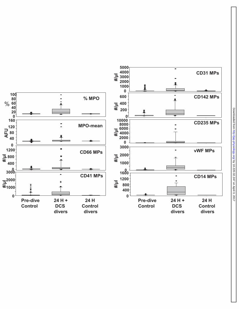
Figure 3. Differences between DCS and control/asymptomatic divers’ blood-borne 519
annexin V-positive MPs and neutrophil myeloperoxidase (MPO) staining in blood at 24 520
or more hours post-diving. The first bar in each graph are pre-dive data for control divers 521
(n=185), the second bar are data from DCS divers obtained 24 or more hours after the dive 522
that incited DCS (n=59) and the third bar are data for control divers obtained 24 hours after 523
their first dive (n=27). The figure illustrates the persistent elevations of blood-borne changes 524
in DCS divers versus control/asymptomatic divers. Panel labels are the same as described for 525
Table 3. The figure indicates median value as the horizontal line within grey boxes, the boxes 526
display 25th and 75th percentile, error bars show 10th and 90th percentile, with outliers shown 527
as single dots. For all measurements the data for DCS divers are statistically significantly 528
different from the first and last control diver panels (p<0.05 ANOVA) and there are no 529
significant differences between pre- and post-control diver values. 530
531
532
by 10.220.32.247 on April 3, 2017
http://jap.physiology.org/D
ownloaded from
Page 25
25
Table 1. Characteristics of study population. 533
DCS Group
(90)
Control Subjects
(185)
Age (years) 34 (27, 42)
Range: 16 - 73
40 (37, 44)
Range: 21 - 72
Dive depth (meters) 22 (16, 34) 18 (18, 33)
# Female 23 (34%) 30 (20 %)
Compressed air 69 (76.7%) 130 (70.3%)
Body mass index 25.8 (22.7, 28.3) 26.9 (24.1, 28.7)
EAN 19 (21.1%) 28 (15.1%)
Tri-mix 2 (2.2%) 27 (14.6%)*
534
Age, diving depth, gender distribution and body mass index between the divers with DCS and 535
control subjects were not statistically significantly different. The last three rows indicate the 536
breathing gas used by the divers. *p<0.001. 537
538
539
by 10.220.32.247 on April 3, 2017
http://jap.physiology.org/D
ownloaded from
Page 26
26
Table 2: MPs sub-types in blood from repetitive diver control subjects. 540
541 Day 1
Pre- (20)
Day 1 Post-
Day 2 Pre- (11)
Day 2 Post-
Day 3 Pre- (11)
Day 3 Post
Day 6 Pre- (8)
Day 6 Post-
CD66 3.6 (2.2, 8.3)
93 (35, 230)
8.1 (4.2, 14.1)
228 (39, 532)
5.6 (3.7, 11.3)
200 (115, 425)
5.1 (4.0, 12.3)
128 (55, 206)
CD41 6.6 (3.6, 9.6)
77.2 (31.8, 229)
7.0 (4.3, 11.5)
58.3 (39.5, 333)
6.2 (3.7, 10.5)
193.7 (108, 424)
8.0 (4.1, 15.7)
195.9 (114, 390)
CD31 6.3 (2.8, 10.0)
103 (37, 233)
9.0 (3.5, 18.5)
233 (40, 365)
7.7 (5.5, 12.1)
220 (176, 290)
6.5 (4.7, 12.0)
226 (138, 357)
CD142 0.3 (0.06, 2.9)
19 (9, 39)
5.4 (2.1, 5.5)
22 (10, 40)
0.7 (0.5, 0.9)
86 (43, 127)
0.9 (0.5, 1.3)
50 (41, 189)
CD14 4.9 (3.9, 6.8)
11 (6, 27)
5.9 (3.3, 6.3)
17 (7, 72)
7.7 (6.2, 9.2)
122 (56, 157)
7.5 (6.3, 11.6)
79 (59, 299)
CD235 5.4 (3.7, 6.3)
15 (9, 72)
5.0 (4.1, 5.8)
12 (8, 34)
8.7 (5.7, 9.1)
98 (51, 315)
8.4 (6.4, 10.3)
134 (81, 307)
vWF 5.5 (3.6, 6.3)
21 (11, 73)
5.6 (4.8, 6.4)
10 (7.3, 217)
5.2 (3.2, 6.6)
80 (44, 118)
6.0 (4.7, 10.9)
52 (38, 184)
542 Data are median, 25th and 75th percentiles for repetitive control subject divers, (n) in the pre-543
dive columns indicates the number of individual diver samples in each pre/post-dive set. All 544
post-dive values are statistically significantly greater than pre-dive values (p<0.001). 545
Although a trend appears suggesting that post-dive values for days three and six are greater 546
than the post-dive values for day one and/or day two, the differences are not statistically 547
significant based on two-way ANOVA on log-transformed data. All rows indicate the 548
number/µl plasma for MPs manifesting the following surface markers: CD66b (neutrophil 549
specific), CD41 (platelet specific), CD31 (platelet-endothelial cell adhesion molecule), 550
by 10.220.32.247 on April 3, 2017
http://jap.physiology.org/D
ownloaded from
Page 27
27
CD142 (tissue factor), CD14 (leukocyte common antigen), CD235 (erythrocyte specific), 551
vWF (von Willebrand factor). 552
553
554
555
556
557
558
by 10.220.32.247 on April 3, 2017
http://jap.physiology.org/D
ownloaded from
Page 28
28
Table 3. MPs and neutrophil activation data on blood samples. 559 Pre-dive
Control subjects (185)
Post-dive Control
Subjects (n=162)
DCS Divers-Acute (n=90)
DCS Divers-follow-up
(n=35) Total MPs/µl 1448
(946, 2165) 2391
(1258, 5123)* 2716
(945, 6920)* 2047
(779, 3682) MPO % 2.6 (1.9, 3.5) 5.8 (2.7, 12.2) 11.5 (4.3, 24.4)* 10.3 (4.6, 18.3)*
MPO-median 12.2 (11.4, 14.0)
15.3 (13.5, 21.6)* 16.4 (13.8, 19.9)*
14.9 (12.8, 22.3)
MPs-CD66b/µl 4.8 (1.8, 38.2) 31.1 (15.9, 44.3)* 84.0 (22.8, 148.9)*†
55.7 (18.8, 128.9)*
MPs-CD41/µl 9.6 (4.5, 33.7) 45.0 (18.4, 87.5)* 110.4 (52.2, 400.9)*†
88.2 (19.5, 337.4)*
MPs-CD31/µl 13.1 (5.3, 64.2)
37.9 (21.0, 242.8)*
186.9 (70.3, 606.3)*†
137.6 (53.5, 298.3)* †
MPs-CD142/µl 1.4 (0.3, 16.9) 16.4 (2.1, 132.3)* 66.7 (24.5, 194.6)*†Δ
19.4 (8.1, 46.9)*
MPs-CD235/µl 6.4 (3.9, 15.8) 32.5 (9.0, 126.7)* 385.9 (56.6, 692.6)*†Δ
73.4 (34.1, 248.6)* †
MPs-vWF/µl 6.5 (4.0, 18.0) 38.2 (6.6, 148.3)* 248.9 (36.0, 558.0)*†Δ
58.1 (4.1, 272.0)*
MPs-CD14/µl 7.7 (4.1, 18.0) 25.5 (6.2, 81.8)* 271.2 (101.4, 765.0)*†
206.6 (26.5, 309.1)* †
560 561
Data are pre-dive values for control subjects (n=185, first column), post-dive control subjects 562
(n=162, second column) included only four divers who used Tri-mix gas, as described in the 563
text, and the samples analyzed were obtained at the longest time after diving to match the 564
time when samples were obtained in the acute DCS group (column 3). Column 4 displays 565
data from divers with DCS who returned for follow-up evaluations after treatment. Data are 566
median (25th and 75th percentiles), (*) indicates p<0.001 versus pre-dive control subject 567
values; †indicates p<0.05 vs post-dive control subject values, Δ=p<0.05 versus late follow-up 568
DCS values. Rows are labeled as follows: MPO% indicates the fraction of CD66b-positive 569
cells exhibiting myeloperoxidase fluorescence above the fluorescence-minus-one threshold 570
(see Methods), MPO-median indicates the geometric median fluorescence value for MPO on 571
by 10.220.32.247 on April 3, 2017
http://jap.physiology.org/D
ownloaded from
Page 29
29
CD66b-positive cells. All other rows indicate the number/µl plasma for MPs manifesting the 572
following surface markers: CD66b (neutrophil specific), CD41 (platelet specific), CD31 573
(platelet-endothelial cell adhesion molecule), CD142 (tissue factor), CD14 (leukocyte 574
common antigen), CD235 (erythrocyte specific), vWF (von Willebrand factor). 575
576
577
by 10.220.32.247 on April 3, 2017
http://jap.physiology.org/D
ownloaded from
Page 30
30
Table 4. Association of MPO on neutrophils and MPs with DCS. 578
579 Adjustment MPO% MPO-
median CD66 CD41 CD31 CD142 CD14 CD235 vWF
Unadjusted 1.6 (1.2, 2.0)
p<0.001
6.9 (2.1, 23.1)
p=0.001
1.4 (1.2, 1.8)
p<0.001
1.5 (1.2, 1.8)
p<0.001
1.7 (1.4, 2.2)
p<0.001
1.4 (1.2, 1.7)
p<0.001
2.0 (1.4, 2.7)
p<0.001
1.6 (1.3, 2.2)
p<0.001
1.7 (1.3, 2.3)
p<0.001Time 1.6
(1.2, 2.1)
p<0.001
7.2 (2.1, 24.8)
p=0.002
1.7 (1.3, 2.1)
p<0.001
1.7 (1.4, 2.2)
<0.001
2.1 (1.6, 2.8)
p<0.001
1.9 (1.5, 2.4)
p<0.001
2.2 (1.5, 3.1)
p<0.001
2.2 (1.5, 3.2)
p<0.001
2.1 (1.5, 3.0)
p<0.001Age 1.6
(1.2, 2.1)
p<0.001
8.6 (2.4, 31.5)
p=0.001
1.4 (1.1, 1.7)
p=0.005
1.4 (1.2, 1.6)
p<0.001
1.6 (1.3, 2.1)
p<0.001
1.4 (1.2, 1.7)
p<0.001
2.0 (1.4, 2.9)
p<0.001
1.6 (1.2, 2.2)
p=0.003
1.7 (1.2, 2.4)
p=0.001Time, depth, gas, repeat dive, age
1.6 (1.2, 2.1)
p=0.001
7.1 (1.8, 27.8)
p=0.005
1.5 (1.2, 2.0)
p=0.003
1.6 (1.2, 2.1)
p<0.001
2.2 (1.6, 3.1)
p<0.001
2.0 (1.5, 2.6)
p<0.001
3.4 (1.8, 6.4)
p<0.001
2.4 (1.4, 4.1)
p=0.001
2.5 (1.5, 4.2)
<0.001 580
Logistic regression was performed on log transformed data to assess the odds ratios (ORs) for 581
DCS using values of MPO on the neutrophil surface and MPs sub-types. Blood samples used 582
for this analysis were the pre-treatment samples from 90 divers with DCS, and 190 blood 583
samples from divers who were deemed to not have DCS. This included the latest blood 584
sample obtained post-diving from each of the control group (n=185), as described in the 585
Results text, and also the five divers who presented to hospitals with signs/symptoms thought 586
due to DCS but who did not improve with recompression treatment. Unadjusted ORs are 587
shown, as well as ORs adjusted by including the length of time from the end of the last 588
SCUBA dive to when blood was collected for analysis (identified as time in table), diver’s 589
age, and including time, maximum diving depth, breathing gas (compressed air, EAN or Tri-590
mix), if repetitive diving was involved, diver’s age, and diver’s gender. Data are ORs, 95% 591
by 10.220.32.247 on April 3, 2017
http://jap.physiology.org/D
ownloaded from
Page 31
31
confidence limits and p values. Columns are labeled as was described for rows in the caption 592
of Table 3. 593
594
by 10.220.32.247 on April 3, 2017
http://jap.physiology.org/D
ownloaded from
Page 32
32
Table 5. Spearman Correlation Analysis. 595
596 MPO% MPO-
median CD66b CD41 CD31 CD142 CD14 CD235 vWF
DCS 0.38 <0.0001
0.19 <0.01
0.28 <0.0001
0.29 <0.0001
0.41 <0.0001
0.33 <0.0001
0.46 <0.0001
0.43 <0.0001
0.42 <0.0001
MPO% 0.41 <0.0001
0.26 <0.0001
NS 0.30 <0.0001
0.25 <0.01
0.54 <0.0001
0.32 <0.0001
0.53 <0.0001
MPO-median
0.23 <0.01
0.14 <0.0001
0.21 0.01
0.26 <0.01
0.44 <0.0001
0.35 <0.0001
0.59 <0.0001
CD66b 0.70 <0.0001
0.69 <0.0001
0.78 <0.0001
0.76 <0.0001
0.61 <0.0001
0.74 <0.0001
CD41 0.80 <0.0001
0.73 <0.0001
0.78 <0.0001
0.71 <0.0001
0.80 <0.0001
CD31 0.72 <0.0001
0.70 <0.0001
0.88 <0.0001
0.87 <0.0001
CD142 0.73 <0.0001
0.66 <0.0001
0.78 <0.0001
CD14 0.74 <0.0001
0.82 <0.0001
CD235 0.79 <0.0001
597
Correlation coefficients and p values are shown for analyses conducted using the same data 598
set as described for Table 4. Values are more highly correlated with coefficients approaching 599
1.0. 600
by 10.220.32.247 on April 3, 2017
http://jap.physiology.org/D
ownloaded from
Page 33
0
5000
10000
15000
20000
0
20
40
60
80
0
10
20
30
40
50
Pre- 15 min 2 hrs 4 hrs 24 hrs(185) (54) (137) (19) (56)
** *
* *
*
* **
Total MPs
MPO %
MPO median
# M
Ps/μ
l%
of N
eutr
ophi
lsFl
uore
scen
ce u
nits
by 10.220.32.247 on April 3, 2017
http://jap.physiology.org/D
ownloaded from
Page 34
02000400060008000
100001200014000
01020304050
Total MPs
MPO%
01020304050 MPO-median
Pre- Post- Pre- Post- Pre- Post- Pre- Post-Day 1 (46) Day 2 (46) Day 3 (21) Day 6 (17)
**
**
# M
Ps/μ
l%
of n
eutr
ophi
lsFl
uore
scen
ceun
its by 10.220.32.247 on April 3, 2017
http://jap.physiology.org/D
ownloaded from
Page 35
020406080
100% MPO
MPO-mean
04080
120160
%
AFU
#/μl
CD66 MPs
0
1000
2000
3000CD41 MPs
0400800
1200
#/μl
010002000300040005000
CD31 MPs
#/μl
CD142 MPs
#/μl
CD235 MPs
0
1000
2000
3000vWF MPs
0400800
12001600
CD14 MPs
0200400600
02000400060008000
10000#/
μl#/
μl#/
μl
Pre-diveControl
24 H +DCS divers
24 H Control divers
Pre-diveControl
24 H +DCS divers
24 H Control divers
by 10.220.32.247 on April 3, 2017
http://jap.physiology.org/D
ownloaded from