www.Larry-Swearingen.com Joye M. Carter, M.D., FCAP Chief Medical Examiner (713) 796-9292 (713) 796-6815 FAX: (713) 796-6842 OFFICE OF THE MEDICAL EXAMINER OF HARRIS COUNTY JOSEPH A. JACHIMCZYK FORENSIC CENTER 1885 OLD SPANISH TRAIL HOUSTON. TEXAS 77054-2098 AUTOPSY REPORT OC 99 - 2 January 3, 1999 ON THE BODY OF Melissa Trotter 7201 F.M. 3081 Willis, Texas CAUSE OF DEATH: Asphyxia due to ligature strangulation. MANNER OF DEATH: Homicide. Reviewed and signed by: ye M. Carter, M.D., FeAP hief Medical Examiner www.Larry-Swearingen.com
Transcript
www.Larry−Swearingen.com
Joye M. Carter, M.D., FCAPChief Medical Examiner
(713) 796-9292
(713) 796-6815
FAX: (713) 796-6842
OFFICE OF THE MEDICAL EXAMINER OF HARRIS COUNTY
JOSEPH A. JACHIMCZYK FORENSIC CENTER1885 OLD SPANISH TRAIL
HOUSTON. TEXAS 77054-2098
AUTOPSY REPORT
OC 99 - 2
January 3, 1999
ON THE BODY OF
Melissa Trotter7201 F.M. 3081Willis, Texas
CAUSE OF DEATH: Asphyxia due to ligaturestrangulation.
MANNER OF DEATH: Homicide.
Reviewed and signed by:
ye M. Carter, M.D., FeAPhief Medical Examiner
www.Larry−Swearingen.com
www.Larry−Swearingen.com
OC 99 - 2
-2-
POSTHORTEH ,EXAMINATION ON TIm BODY OF
Melissa Trotter7201 F.M. 3081Willis, Texas
HISTORY: The body of an unidentified Caucasian female,subsequently identified as that of 19 year old Melissa Trotter r wasfound January 2, 1999, in the National Forest of Montgomery County,Texas. The body was received at the Harris County MedicalExaminers Office for postmortem examination and identification.The sealed body bag was Dpened at 12:35 p.m. on January 3, 1999, byMr. Reynolds, Harris County Medical Examiner's Forensic Scientist,in the presence of. Dr. Joy£? Ii. Ca;r:ter r Chief Medical Examiner.Prior to autopsy examination, trace evidence was collected and themissing persons repDrt filed on behalf of Melissa Trotter wasreviewed r dental records of Melissa Trotter were reviewed, andmedical records of Melissa Trotter were reviewed. Total bodyX-rays were obtained and reviewed. A sexual assault kit wasperfDrmed.
IN ATTENDANCE AT THE AUTOPSY: 11r. David Tanner and Detective BrianM. DuBDse, MDntgomery County Sheriff's Office; Hr. Mike McDDugal,District Attorney's Officer MDntgDmery County; Dr. DeLattre and Dr.StimsDn; AutDpSy ASSistant, Mr. Michael JDnes; and ForensicPhDtographer, Ms. Marlene Suarez.
AUTOPSY: The autopsy was performed in the Joseph A. JachimezykForensic Center of Harris County by Chief Medical Examiner JoyeM. Carter,M.D.·Feat the request and upon .the written authorizationof The HonDrable John R. Kleimann, Justice of. the Peace r Pree,inetI, MDntgomery County, Texas, beginning at 2:15 p.m., Dn January3, 1999.
CLOTHING: The body was recei'\fed clad in blue denim jeans, whitesOEks, one black and white gym spoe on the left fDot. The sole ofthe gym shoe r with treads, was clean with minimal debris no.ted.There was no caked mud Dr grass observed Dn the bottom Df theshoe. On the top of the body was a green, long-sleeved r
oversweater which zipped in front with burgundy and white stripesat the elbows. Beneath this sweater was a IDng-sleeved blacksweater and a bra which had been pushed up over the breasts.Beneath the blue denim jeans were red underpants. No personaleffects were received with the body. Prior to examination,fingernail scrapings were performed and fingerprints were taken.During fingerprinting r the skin and nails of the right hand removedin a glove formation secondary to decompositiDn. The outer greensweater and black sweater underneath were cut open with evidencetape along the cut edges marked for further examination. The bodywas then undressed fDr further examinatiDn. Removed from the front
www.Larry−Swearingen.com
www.Larry−Swearingen.com
Melissa Trotter OC 99 - 2セSM
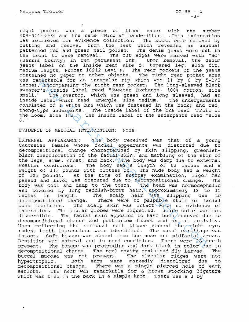
right pocket was a piece of lined paper with the number409-524-3008 and the name "Nicole" handwritten. This informationwas retrieved for evidence collection. The socks were removed bycutting and removal from the feet which revealed an unusualpatterned red and green nail polish. The denim jeans were cut inthe front to ease removal. The cut edges were marked with "HC"(Harris County) in red permanent ink. Upon removal, the denimjeans label on the inside read size 5, tapered leg, slim fit,medium length, Number 10912 Levi's. The rear pockets of the jeanscontained no paper or other objects. The right rear pocket areawas remarkable for an irregular rip which was 11 by 6 by 5-1/2inches, encompassing the right rear pocket. The long-sleeved blacksweater's inside label read "Sweater Exchange, 100% cotton, sizesmall. " The overtop, which was green and long sleeved, had aninside label which read "Energie, size medium." The undergarmentsconsisted of a white bra which was fastened in the back; and red,thong-type underpants. The inside label of the bra read "Fruit ofthe Loom, size 36B." The inside label of the underpants read "size6 " II
EVIDENCE OF MEDICAL INTERVENTION: None.
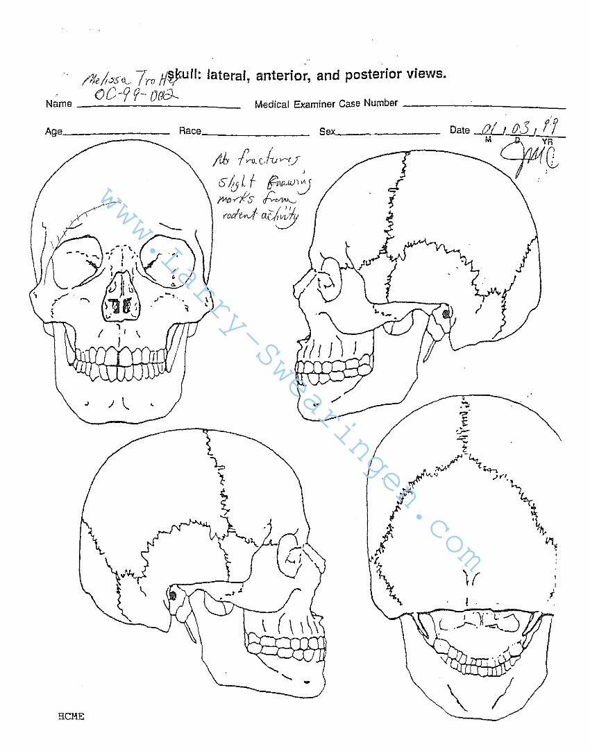
EXTERNAL APPEARANCE: The body received was that of a youngCaucasian female whose facial appearance was distorted due todecompositional change characterized by skin slipping, greenishblack discoloration of the facial skin, and marbling of the skin ofthe legs, arms, chest, and back. The body was damp due to externalweather conditions. The body had a length of 63 inches and aweight of 113 pounds with clothes on. The nude body had a weightof 105 pounds. At the time of autopsy examination, rigor hadpassed and livor was obscured due to decompositional change. Thebody was cool and damp to the touch. The head was normocephalicand covered by long reddish-brown hair, approximately 12 to 15inches in length. The scalp hair was slipping due todecomposi tional change. There were no palpable skull or facialbone fractures. The scalp skin was intact with no evidence oflaceration. The ocular globes were liquefied. Iride color was notdiscernible. The facial skin appeared to have been removed due todecomposi tional change and postmortem insect and animal activi ty.Upon reflecting the residual soft tissue around the right eye,rodent teeth impress ions were identi fied. The nasal cartilage wasintact. Soft tissue was absent from the nose and midfacial areas.Denti tion was natural and in good condition. There were 28 teethpresent. The tongue was protruding and dark black in color due todecompositional change. The oral cavity contained fly larvae. Thebuccal mucosa was not present. The alveolar ridges were nothypertrophic. Both ears were markedly discolored due todecomposi tional change. There was a single pierced hole of eachearlobe. The neck was remarkable for a brown stocking ligaturewhich was tied in the back in a simple knot. There was a 3 by
www.Larry−Swearingen.com
www.Larry−Swearingen.com
Melissa Trotter OC 99 - 2-4-
2-3/4 inch defect on the anterior neck with liquefaction of tissue,maggot activity, and blood present. The anterior chest withbreasts and the abdominal cont·our were remarkable only fordecornpositional change. There were no palpable masses or Bcarsappreciated. There were no tattoos present. The externalァ ・ セ ゥ エ 。 ャ ゥ 。 were those of an adult female. The body hair was femalein distribution. The upper extremities were synunetrical and wellformed and were remarkable for decornpositional change. There wereno tattoos, venous track marks, or unusual scars of the upperextremi ties. The fingernails were remarkable for a green-and-redstriped fingernail polish. There was marked skin slippage' of theskin of the right hand with only the fingernail remaining on the2nd digit. The skin of the left hand was intact but was too moistfor fingerprinting. The fower extremities were !3yrnmetrical, wellfo=ed, and remarkable for decompositional change. The dorsum ofthe left foot was remarkable for a .3 inch curvilinear,hyperpigmented scar which was well healed. The toenails of bothfeet were remarkable for the same patterned red and green nailpolish. The posterior surface was remarkable for plant vegetationadherent to the body. There was no discreet pattern of livormortis. The anal and vaginal orifices were unremarkable with theexception of decompositional changes. The skin of the· bodydiffusely showed splotchy areas of red, green, and graydiscoloration secondary to postmortem mold growth. There wasgeneralized skin slippage with discoloration and marbling over theentire body surface.
INTERNAL EXAMINATION: Section: The thoracoabdominal region wasexplored using an H-shaped incision. The sternum and rib cage キ セ イ ・
intact. The midline·" fatty subcutaneous tissue at the level of 'theumbilicus was 1/2 inch in depth. The right hemithoracic cavity _ .contained approximately 150 cUbic centimeters of sanguineous fluid.The left hemithoracic cavity contained approximately 100 cubiccentimeters of sanguineous fluid. The pericardial sac was intactand contained approximately 10 cubic centimeters of sanguineousflBid. The peritoneal cavity contained a minimal amount ofsanguineous fluid. All organs maintained their usual anatomic.relationships. The appendix was observed, as was the gallbladder.There appeared to. be no previous. intervening surgical procedu.I'ethrough the skin of the abdominal or chest wall. The breast tissuewas firm and intact. There were no appreciable masses upon !3erialsectioning of the fibrofatty tissue of the breasts.
HEART: The heart was intact with a residual weight of 175 grams.The external epicardial surface was dull tan-brown. The heart hada normal configuration and a flabby consistency. There was minimalsubepicardial fatty tissue deposition noted on both anterior andposterior surfaces. The coronary arteries arose normally in theaortic root. The vessels were widely patent and contained noevidence of narrowing due to atheromatous plaque buildup. The
www.Larry−Swearingen.com
www.Larry−Swearingen.com
-Melissa Trotter OC 99 - 2-5-
aorta was intact with a smooth intimal surface and had minimalfatty streaking along its entire length. The myocardium wasred-brown in appearance, dull, and homogeneous with noteddecompositional change. There was extreme laxity of theconsistency due to decomposi tional change. The heart valves weredark in color and thin with no evidence of malformation. theendocardial surface was dull and tan-gray. The aorta was intactand remarkable for decompositional change.
LUNGS: The right lung weighed a residual 325 grams while the leftweighed 450 grams. The pleural aspects were red-brown, somewhatmottled, and glistening in appearance. Upon sectioning, thepulmonary arterial tree bilaterally was patent with no evidence ofthromboemboli. There was no evidence of vascular malformation.The bronchial tree was dissected bilaterally. There was noobstruction by mucus plug, foreign body, or insect activity. Thepulmonary parenchyma bilaterally was markedly congested. Therewere no palpable areas of consolidation. There was no apparentanthracotic pigment deposition due to the decompositional state andthe dark burgundy-purple discoloration of the parenchyma.
LIVER: The liver had a residual weight of 775 grams. The capsularsurface was intact and dull gray-brown. The anterior margin wassharp and the parenchyma had a soft consistency. Upon sectioning,the parenchyma was red-brown and dull in appearance. It appearedto be homogeneous with no evidence of preexisting tumor mass orinfectious disease process. There was no nodularity upon serialsectioning of the hepatic parenchyma. The gallbladder bed wasintact and contained approximately 5 cubic centimeters of thinbrown bile. The bile passages were patent. There was no evidenceof cholelithiasis. The gallbladder mucosa was yellow-green andvelvety in appearance.
Pancreas: The pancreas was intact with a residual weight of 100grams. The external surface was dark red-brown and dull due todecomposition and autolysis. There was loss of the normal tissuearchi tecture with very limited lobulation noted. Upon sectioning,the duct was patent. The cut surface was yellow-red and autolyzed.
Adrenals:autolyzed.
The left and right adrenal glands were markedly
SPLEEN: The spleen was intact with a weight of 100 grams. Thecapsular surface was red-brown, dUll, and smooth. There were nodefects noted to the hilar region. The entire spleen was retainedfor toxicological analysis. There were no areas of lymphadenopathyobserved.
www.Larry−Swearingen.com
www.Larry−Swearingen.com
Melissa Trotter OC 99 - 2
-6-
GENITOURINARY TRACT: The right and left kidneys each weighed 50grams. The renal capsules stripped with ease to reveal dullred-brown, smooth cortical surfaces. Both kidneys had a flabbyconsistency. Upon sectioning, normal tissue architectural changewas obscured due to decompositional change. The medullary regionswere demarcated and appeared darker in coloration. The collectingsystems were unremarkable. The proximal ureters were not dilatedor obstructed. The urinary bladder was void of urine. The vaginaltract was lined by a thick tan-brown material. The approximateamount was 5 to 8 cubic centimeters. Upon rinsing, the vaginalwall displayed focal areas of pink-red discoloration. The internalgenitalia were remarkable for mild decomposition change. Thecervical os was examined and was circular with focal areasconsistent with preexisting surgical manipulation of theendocervical surface. The uterus was nongravid and measured 2-3/4by 2 inches in area. Both right and left ovaries were observedwith their respective fallopian tubes. The left ovary contained ahemorrhagic cyst. The right ovary was unremarkable uponsectioning. Upon sectioning the uterus, the endometrial contentswere a small amount of tan mucoid material.
GASTROINTESTINAL TRACT: The esophagus was intact and remarkablefor decompositional change. The esophageal mucosa was purple-tan.The esophageal gastric junction was free of laceration. Thegastric contents were approximately 200 cubic centimeters ofpoorly-masticated tan liquid in which large pieces of white meat,consistent with chicken, and pieces of potato, consistent withfrench fries, were observed. There were small portions of whatappeared to be yellow and green vegetables, as well. There was notablet or pill residue observed in the gastric contents. Thepyloric region was patent. The first part of the duodenumcontained a moderate amount of pink-tan chyme material. There wasno evidence of ulceration of the proximal duodenum. The gastricmucosa, upon rinsing, displayed no evidence of ulceration. Therugal folds were normal in appearance. Decompos i tional changeswere noted to the mucosal surface of the stomach. The remainder ofthe small and large intestine was intact and displayed mild tomoderate decompositional change characterized by bubbling andtransparency of the serosal surfaces. There were no foreign bodieswi thin the rectal vault. The appendix was in its normal anatomiclocation and was unremarkable upon sectioning.
NECK: The neck region was explored initially. The skin wasreflected laterally to reveal markedly decomposed anterior neckstructures. There appeared to be a clean separation between theportion of larynx on the posterior aspect of the neck organs. Thetongue was intact and was remarkable for a 1/2 inch intramuscularcontusion on the right anterior aspect of the tip. The hyoid bonewas palpated. It was flexible and was not fractured. The proximalportion of the thyroid cartilage was palpated and was intact. The
www.Larry−Swearingen.com
www.Larry−Swearingen.com
Melis,sa Trotter OC 99 - 2-7-
anterior edge of the thyroid cartilage appeared to be gnawed andirregular. The posterior segment of the larynx remnant was 3/4inch in length by 7/8 inch. The inferior portion was smooth anddark brown. The remnants of the thyroid cartilage horns wereirregular and appeared to be gnawed. The lower segment of thelarynx measured 3/4 by 3/4 inch. The surface of the superiormargin was remarkable for fine ridges. Opening the inferiorsegment of larynx revealed the left vocal fold to be intact. Theright vocal fold was not identified. Upon reexamination, theinferior remnant of the larynx was irregular in the midline andthere were tiny notched ridges from postmortem gnawing. Thetrachea was intact and the rings were not collapsed. There was nounusual material within the trachea. There was no foreign body ormucus plug. There were scattered maggots within the neck organsupon dissection. The aortic arch was dissected in proximity to theneck organs. The great vessels were given off normally. The greatvessels in the proximate transection of the trachea had apparentteeth and gnaw marks. The thyroid gland was absent. Themidportion of the larynx was absent. The cricoid cartilage wasabsent. The underlying cervical spine was intact with no palpableridges from sharp force injury.
HEAD: The scalp was reflected forward via a bimastoid incisionrevealing no evidence of galeal or subgaleal hematoma. Thecalvarium was intact and free of fracture line. Upon removal ofthe calvarium, the dura mater was intact with no evidence ofepidural hematoma formation. Upon removing the dura mater, therewas no evidence of subdural hematoma formation. The brain had aresidual weight of 1100 grams. There was no evidence ofsubarachnoid hematoma formation. Upon removal of the brain fromthe cranial cavity, the tissue was semiliquid and pink-green incolor. There was complete loss of normal tissue architecture uponremoving the brain tissue. There was no evidence of preexistinglesion or mass defect in the parenchyma. There was no evidence ofventricular abnormality. The vessels forming the circle of Williswere thin walled. The pituitary gland was markedly autolyzed. Thebasilar skull was free of fracture line. After examining the head,the jaws were incised and dental examination was obtained.
Total body X-rays revealed two metal screws in the metatarsal areaof the left foot. There were no fractures to the skeletal systemobserved. Examination of the lower alveolar ridge revealed aretention appliance for orthodontic procedure.
EVIDENCE OF INJURY: Ligature Around the Neck: The ligatureconsisted of a piece of brown hosiery. The toe was intact on onelimb of the ligature and it appeared to be a sandal-foot-type toe.The second limb appeared to be cut irregularly. The ligature wastied around the posterior surface of the neck. There was a singlesimple knot. The ligature was removed from around the neck by
www.Larry−Swearingen.com
www.Larry−Swearingen.com
Melissa Trotter OC 99 - 2-8-
cutting. The cut edges were marked with red identifying tape. Thediameter of the ligature was 3-1/4 inches. The ligature appearedto be a cut leg of pantyhose. There was a 1/4 to 1/2 inch remnantof the panty portion on the cut end. The cut ends were slightlyrolled. There was a run going through the entire length of thehosiery. The ligature imprint was poorly discernible due to thedecompositional change of the neck. There was a dark purple, 2-1/2inch, raised mark on the posterior surface of the neck. Uponinspection of the anterior neck wound, there appeared to be atransection of the larynx. The edges of the severed larynx wereirregular due to postmortem animal activity. The surroundingtissue appeared to be hemorrhagic and dark secondary to postmorteminsect and animal degradation. This was not discernible completelydue to decompositional change.
TOXICOLOGY: Samples ofgastric contents, andtoxicological analysis.kidney, brain tissue, and
heart fluid, chest fluid, bile, liver,the entire spleen were retained for
Also retained were samples of lung,heart.
HISTOLOGY: Representative sections of all major organs wereretained in formalin.
PATHOLOGICAL FINDINGS
1. Ligature around neck.2. Decompositional change with maggot activity.
www.Larry−Swearingen.com
www.Larry−Swearingen.com
OFFICE OF THE MEDICAL EXAMINER OF HARRIS COUNTY
JOSEPH A. JACHIMCZYK FORENSIC CENTER
1885 OLD SPANISH TRAIL
HOUSTON, TEXAS 77054-2098
REPORT OF ANALYSIS
January 13, 1999
TO: Joye M. Carter, M.D.
Chief Medical Examiner
CASE#: OC99-002
Evidence submitted on 01/04/99.
RESULTS:
Blood:
Bile:
Liver:
Ethanol- 0.06 gleIL
Methanol, Acetone, Isopropanol- Not Detected.
Norcarboxytetrahydrocannabinol- Not Detected.
Delta-9-tetrahydrocannabinol- Not Detected.
Standard Basic Drug Screen- Not Detected.
Cocaine Metabolite, AmphetaminelMethamphetamine,
Phencyclidine, Opiate- Not Detected.
Ethanol- 0.03 g/dL
Methanol, Acetone, Isopropanol- Not Detected.
Ethanol- 0.02 gigMethanol, Acetone, Isopropanol- Not Detected.
..--t1'l"H''''''4Itherwise requested, specimens will be discarded one year after date of receipt.
Patricia 1. Small, B.S., MT, ASCP
Assistant Toxicologist
AshrafMozayani, Ph. ., DABFT
Chief Toxicologist
Medical Examiner's Initial _
www.Larry−Swearingen.com
www.Larry−Swearingen.com
Name
PERINEUM, FEMALE
IiAセセsヲBM Irv fft,r {;t,PV--00:9___セNNNZNNZNNZNセZMMMNNZNNZNNNZNM M.E. Case No. .
www.Larry−Swearingen.com
www.Larry−Swearingen.com
ML OC- 9f- ooo? 19 f'f.:.....L.__
E XTE RNA L V lEW S
OF THE HEAD
Head, Left Lateral (I)
" " .." /
Head, Anterior (3)
Head, Posterior (4)
Head, Right Lateral (2)
Head, Superior (5)
www.Larry−Swearingen.com
www.Larry−Swearingen.com
[QォOエセUHl 7'TO セオOャZ lateral, anterior, and posterior views.
oC-y f- OCICJ--Name セ⦅⦅⦅⦅ Medical Examiner Case Number ---'- _
iI .
I(
\
Sex______ Date of I 0'3 I If
B セ エ
.;" ? ..• \
" '1
I . I \
I
" ) \...
HCME
Age_----::::::==:::::_ Race _
www.Larry−Swearingen.com
www.Larry−Swearingen.com
· CASE NO. of.-: 79 OOld--
AGE RACE---- S EX !IE I GilT._- ----__\\E I GlIT _


![.7'hemeroteca.ciasc.sc.gov.br/jornais/O Cacique/CAC1870013.pdf'. M [N GN ⦅G M M GNセ セ NZNN@ Z セM. ,.7 ' .. N M]M G M Nセ MM G NM Z@ N セ@ '. LMM[MセMᆳ Mᆳ M l '](https://static.documents.pub/doc/80x56/6032344063984663a15e4415/7-caciquecac1870013pdf-m-n-gn-ig-m-m-gni-i-nznni-z-im.jpg)


![. (C{)i[f'J']HlH D .,.. セ@ •• セZZ@ N セQ@hemeroteca.ciasc.sc.gov.br › jornais › avozdopovo › AVO1885027.pdfJ'clII-,;e nl pcrmut:l. pn l'i:lm05 nn,,"'; slncc- Illlde](https://static.documents.pub/doc/80x56/60d94129b0d2714d30726c44/-cifjhlh-d-ii-aa-izzi-n-iqi-a-jornais-a-avozdopovo.jpg)


![Hypolito セゥエ・オ セ セB@ Themeroteca.ciasc.sc.gov.br/jornais/oolhosemanario/OL... · 2016-11-23 · ⦅MセBB]G Mi M ェNNMMGMMG cTャャQMGャ BZZ ーゥZゥュ・ゥゥNッ](https://static.documents.pub/doc/80x56/5f8895082ed72267030d9519/hypolito-iiiii-i-ibi-2016-11-23-imibbg-mi-m-innmmgmmg-ctiiqmgi.jpg)
