Page 1
HAL Id: inserm-00914357https://www.hal.inserm.fr/inserm-00914357
Submitted on 5 Dec 2013
HAL is a multi-disciplinary open accessarchive for the deposit and dissemination of sci-entific research documents, whether they are pub-lished or not. The documents may come fromteaching and research institutions in France orabroad, or from public or private research centers.
L’archive ouverte pluridisciplinaire HAL, estdestinée au dépôt et à la diffusion de documentsscientifiques de niveau recherche, publiés ou non,émanant des établissements d’enseignement et derecherche français ou étrangers, des laboratoirespublics ou privés.
Attention orienting dysfunction with preservedautomatic auditory change detection in migraine.
Dominique Morlet, Geneviève Demarquay, Frédérique Brudon, CatherineFischer, Anne Caclin
To cite this version:Dominique Morlet, Geneviève Demarquay, Frédérique Brudon, Catherine Fischer, Anne Caclin. At-tention orienting dysfunction with preserved automatic auditory change detection in migraine.. Clin-ical Neurophysiology, Elsevier, 2014, pp.500-11. �10.1016/j.clinph.2013.05.032�. �inserm-00914357�
Page 2
1
Attention orienting dysfunction with preserved automatic
auditory change detection in migraine
Dominique Morletabc
, Geneviève Demarquayd, Frédérique Brudon
e, Catherine Fischer
abcf,
Anne Caclinabc
a INSERM U1028, Lyon Neuroscience Research Center, Brain Dynamics and Cognition Team, Lyon, F-69000,
France
b CNRS UMR5292, Lyon Neuroscience Research Center, Brain Dynamics and Cognition Team, Lyon, F-69000,
France
c Université Lyon 1, Lyon, F-69000, France
d Hospices Civils de Lyon, Croix-Rousse Hospital, Neurology Department, Lyon, F-69000, France
e Tonkin Clinic, Villeurbanne, F-69100, France
f Hospices Civils de Lyon, Neurological Hospital, Functional Neurology and Epileptology Department, Lyon, F-
69000, France
Corresponding author: Dominique Morlet
Lyon Neuroscience Research Center, Dycog team
69675 Bron Cedex, France
tel: + 33 (0)4 72 13 89 03
fax: + 33 (0)4 72 13 89 01
e-mail: [email protected]
Acknowledgements: We wish to thank the control subjects and the patients who participated in this
experiment. We are also grateful to the technicians of the Department of
Functional Neurology and Epileptology for their help in recordings ERPs. The
authors have no conflict of interest.
Keywords : Migraine
Event-Related Potentials
Attention orienting
MMN
N2b
P3a
Page 3
2
Highlights:
The mismatch negativity (MMN) is normal in migraine patients.
The N1 orienting component and the N2b are increased in migraine patients.
Auditory processes up to attention triggering are preserved in migraine patients.
Attention orienting to sound income and sound deviance are exacerbated in migraine.
These observations suggest abnormal activation of attention-related frontal networks
Abstract
Objective
To investigate automatic event-related potentials (ERPs) to an auditory change in migraine patients.
Methods
Auditory ERPs were recorded in 22 female patients suffering from menstrually-related migraine and
in 20 age-matched control subjects, in three sessions: in the middle of the menstrual cycle, before and
during menses. In each session, 200 trains of tone-bursts each including two duration deviants were
presented in a passive listening condition.
Results
In all sessions, duration deviance elicited a mismatch negativity (MMN) showing no difference
between the two groups. However, migraine patients showed an increased N1 orienting component to
all incoming stimuli and a prolonged N2b to deviance. They also presented a different modulation of
P3a amplitude along the menstrual cycle, which tended to normalize during migraine attacks. None of
the studied ERP components showed a default of habituation.
Conclusions
This passive paradigm highlighted increased automatic attention orienting to auditory changes but
normal auditory sensory processing in migraineurs.
Significance
Our observations suggest normal auditory processing up to attention triggering but enhanced
activation of attention-related frontal networks in migraineurs.
Page 4
3
1. Introduction
Migraine is one of the most common headache disorders and affects 11% of the adult population
(Stovner et al., 2007). Attacks are characterized by recurrent throbbing headaches accompanied by
nausea, vomiting, photophobia, and/or phonophobia and are aggravated by movements (The
International Classification of Headache Disorders, 2004). Between attacks, migraine patients may
also show a hypersensitivity to visual, auditory, or olfactory stimuli (Main et al., 1997; 2000).
Moreover, sound/light-induced headaches are frequent in migraine patients (Vingen et al., 1999;
2004). The pathophysiological basis of such hypersensitivity to external stimuli is currently not
completely elucidated.
In line with these symptoms, electrophysiological studies have revealed changes in cortical excitability
between migraine attacks. Increased attention orienting to new incoming stimuli had been long ago
demonstrated in migraine patients. The initial (or early) Contingent Negative Variation (iCNV), which
reflects the orienting properties of a warning stimulus in an active paradigm, has been found to be
increased in migraine (Maertens de Noordhout et al., 1986), preferentially just before an attack (Kropp
and Gerber, 1998), but not systematically (Böcker et al., 1990; Mulder et al., 2001). A default in the
habituation of several event-related potentials (ERPs) to visual and auditory stimuli has also been
observed in migraine patients in numerous studies (for a review, see Coppola et al., 2009), although
not systematically (Oelkers et al., 1999; Sand and Vanagaite Vingen, 2000; Omland et al., 2013).
Using an auditory habituation paradigm made of small consecutive trains of tones (Woods and
Elmasian, 1986), we also found normal habituation pattern of the sensory N1 in migraineurs
(Demarquay et al., 2011). Interestingly, this passive paradigm allowed to investigate the automatic
response to the first stimuli of stimulation trains, and revealed that migraine patients exhibited a
drastically augmented N1 orienting component, a fronto-central negative component appearing after
the obligatory sensory N1 when the inter-stimulus interval is greater than 4 s (Näätänen and Picton,
1987; Alcaini et al., 1994). The response to standard stimuli inside the trains also exhibited enhanced
negative potentials in the descending slope of the N1 wave. By comparison with the augmented N1
Page 5
4
orienting component observed in response to the first stimuli of the trains, we called this additional
component “residual orienting component” and we concluded that migraine patients showed
exacerbated attention orienting not only to first stimuli after a silent gap but also to repeated similar
incoming stimuli.
The electrophysiological studies described above showed abnormal responses not only to attended
warning stimuli but also to passively endured stimuli. They emphasized attention-orienting
exacerbation and/or habituation deficit in migraine without clearly disentangling basic auditory
processing dysfunction and abnormal orienting processes. The use of passive oddball paradigms is of
particular interest here because it allows studying both automatic sensory responses to auditory
changes and automatic attention triggering. Indeed, in those paradigms, a rare and random change in
repetitive stimulation elicits a negative response (Mismatch Negativity or MMN) disclosed when the
response to the standard is subtracted from the response to the deviant. MMN is attributed to
discriminative processes using memory traces developed from the previous stimulation (review in
Näätänen et al., 2001; Näätänen et al., 2007; Näätänen et al., 2011). It is noteworthy that the memory
traces probed by the MMN are thought to reflect the outcome of perceptual processing (Näätänen and
Winkler, 1999; Näätänen et al., 2011) and further that MMN occurs automatically, even if subject’s
attention is not directed to the sounds (Näätänen et al., 1993). Nevertheless, if deviant stimuli are
salient enough, attention-orienting processes can be triggered, and an N2b-P3a complex (brain
orienting response) is further obtained following the MMN (Näätänen et al., 1982). In contrast,
presenting a second consecutive deviant drastically diminishes the MMN (and subsequent orienting
responses), which has been called “short-term habituation of the MMN” (Sams et al., 1984). This
phenomenon was explained by the simultaneous occurrence, for the repeated deviant, of mismatch
processes with the (declining) neuronal model of the standard and match processes with the neuronal
model of the deviant. Altogether, the study of ERPs triggered by a rare change in auditory stimulation
thus offers the advantage to assess, in a passive listening situation, the integrity of pre-attentive stages
of perceptual processing, as indexed by the MMN, as well as the automatic triggering of attention,
indexed by the N2b-P3a complex. For these reasons, passive auditory oddball paradigms have been
used in a very large number of ERP studies in various pathologies (review in Näätänen, 2003;
Page 6
5
Naatanen et al., 2012), but surprisingly very rarely in migraine.
Indeed, most of the oddball paradigm studies in migraine have required an active detection of deviant
tones and they mainly focused on the P300 in response to targets, which was found to be altered in
migraine patients (Drake et al., 1989; Mazzotta et al., 1995; Wang et al., 1995). The investigation of
brain mechanisms to a stimulus change during passive listening has been rarely reported in migraine
patients. Only one study investigated the MMN in adult migraineurs and reported increased N1 and
MMN latencies, suggesting a hypo-activity of automatic cortical processes (de Tommaso et al., 2004).
Two recent paediatric migraine studies also pointed to subtle MMN alterations in these patients
(Valeriani et al., 2009; Korostenskaja et al., 2011). One study (Wang and Schoenen, 1998) explored
the amplitude of an N2-P3a complex in response to deviant stimuli and reported for migraine patients
a potentiation of this response in successive blocks, contrasting with a habituation in controls.
The aim of the present study was thus to investigate the specific response to deviant stimuli in
migraine patients, from the pre-attentive processes (MMN) to the attention-orienting processes (N2b,
P3a), and to assess the habituation of the different components. Abnormalities of the MMN would
suggest dysfunction in basic auditory processing of migraine patients. In light of our previous results,
we expected a pathological brain orienting response to deviants.
Page 7
6
2. Methods
The present analysis is the second part of a larger auditory ERP study conducted in migraineurs using
small trains of repeated standard tones with occasional deviant stimuli. The first part of the analysis
focused on the responses to standards and their habituation patterns (Demarquay et al., 2011). Here we
explore the response to deviant stimuli.
2.1 Migraine patients and healthy subjects
Twenty-two female migraine patients suffering from menstrually-related migraine without aura
(A1.1.2 in ICHD 2004) were included in the study (mean age ± SD: 27 years ± 7; disease duration: 12
years ± 8; attack frequency: 2 ± 1 per month). Migraine patients and controls were recruited through
advertising in Lyon University and INSERM administration. A neurologist (CF, FB or GD)
subsequently examined eligible subjects.
Twenty age-matched female controls participated in the study (28 years ± 9). Exclusion criteria for all
subjects included chronic daily headache, known morphological brain abnormality, current substance
abuse, and migraine preventive medication. 14 migraine patients and 11 controls took oral
contraceptives (2 = 0.0649, p = 0.799).
All subjects gave their written informed consent (CCPPRB centre Léon-Bérard, Lyon, A 06-107,
04/20/06) and were remunerated for their contribution.
2.2 Recording procedure
The recording procedure has been described in detail previously (Demarquay et al., 2011).
Patients and healthy subjects were recorded in 3 different sessions, in the middle of the menstrual
cycle (session 0, S0, day 15 ± 4 of the on-going cycle), before menses (session 1, S1, day -1 ± 1 of the
cycle), and at the beginning of the menses (session 2, S2, day 1 ± 1 of the cycle). As a general rule, S2
immediately followed S1 (mean delay = 2 days ± 1), except for 1 healthy subject and for 3 patients,
Page 8
7
for whom S1 and S2 were in separate cycles. Session 0 randomly preceded or followed S1 and S2. S0
was before S1 in 12/22 patients and in 13/20 healthy subjects (2 = 0.923, p = 0.337).
Among the 22 migraineurs, ERPs were recorded during a migraine attack in 12 patients (session 1 for
5 patients; session 2 for 6 patients; session 0 for 1 patient). In all patients, the attack occurred before
the onset of the ERP recording and lasted until the end; medication was given at the end of recording
session.
During the recording sessions, the subject sat in a comfortable armchair in a quiet room. She was
instructed to watch a silent movie of her choice and not to pay attention to the auditory stimuli.
EEG was recorded continuously from 7 scalp electrodes placed at frontal (Fz, F3, F4), central (Cz),
and parietal (Pz) sites, and at the two mastoids (M1, M2). The reference electrode was placed on the
tip of the nose, the ground electrode on the forehead. One bipolar EOG derivation was recorded from
2 electrodes placed on the supra-orbital and infra-orbital ridges of the right eye. Using a Micromed
System98 EEG recording system, the signal was amplified (band-pass 0.3-100 Hz), digitized
(sampling frequency 1024 Hz) and stored for off-line analysis. One stimulation session lasted about 45
minutes without any interruption.
2.3 Auditory paradigm
The stimuli were delivered binaurally through insert earphones at an intensity of 65 dB HL.
In each recording session, 200 trains of auditory stimuli were presented with an average of 10 stimuli
per train (random number from 8 to 12). The inter-train interval ranged between 5.5 and 8.0 seconds
(mean value 6.75 s). Within trains, stimulus onset asynchrony was 610 ms.
The stimuli were spectrally rich tone bursts (fundamental frequency 800 Hz). The more frequent
stimuli (standards) had a duration of 75 ms, including 5-ms rise and fall times. Duration deviants
(duration 30 ms) were used, because duration changes (with deviants shorter than standards) are
known to provide robust and reproducible MMNs (Tervaniemi et al., 1999). Two deviant tones were
pseudo-randomly included in each train. The first deviant was randomly presented after 3 to 9
consecutive standards. In each recording session, one hundred trains (50% of the trains) obeyed the
rule routinely used in oddball paradigms, namely at least 2 standards were presented between the first
Page 9
8
and the second deviant. In the other half of the trains, the second deviant appeared immediately after
the first one (repeated deviant). One recording session thus included 300 “generic” deviants appearing
after at least 2 standards (200 generic deviants being first deviants and 100 generic deviants being
second deviants) and 100 “repeated deviants” appearing immediately after another deviant. Figure 1
illustrates the design and the timing of auditory stimulation.
Additionally, 20% of the trains ended with a salient stimulus (environmental sound). Evoked
responses for these novel stimuli are beyond the scope of the present report.
2.4 Event-related potentials
The software package for electrophysiological analysis (ELAN) developed at the Lyon Neuroscience
Research Center (Aguera et al., 2011) was used for ERP analysis. Responses to standards and to
deviants were considered for averaging for an epoch of 700 ms including a pre-stimulus period of 100
ms. Epochs showing peak-to-peak deflections larger than ± 100V were rejected. One patient showed
a large stimulation artifact at the mastoids 15 ms after stimulus onset in the 3 recording sessions. For
this patient, an independent component analysis (ICA) was used in order to isolate and remove this
unwanted component.
The ERPs were baseline-corrected by subtracting the mean value of the signal during the 100 ms prior
to the stimulus. A 30-Hz low-pass digital filter (bidirectional Butterworth, 6th order) and a 2-Hz high-
pass filter (bidirectional Butterworth, 2nd
order) were applied to the averaged epochs.
To fulfill the different aims of the study, we performed the following types of averages in each session
for each participant: 1) averages of “generic standards” (in black Figure 1), from which the first 3
standards in the trains and the standards that immediately followed a deviant were excluded; 2)
averages of “generic deviants” (in black Figure 1), from which the deviants that immediately followed
another deviant were excluded; 3) averages of “first” deviants, which included only the first deviant in
each train; 4) averages of “repeated deviants” (in grey Figure 1), which included only the second
deviants that immediately followed a first deviant. To assess long-term habituation averages 1 and 2
were performed separately in the first half (first 100 trains) and in the second half (last 100 trains) of
Page 10
9
each session. Averages 3 and 4 were used to investigate the so-called “short-term habituation” to
deviance.
2.5 Statistical analysis
In a first stage, we studied the response to generic deviants and to generic standards. In a second stage,
the response to generic standards was subtracted from the response to generic deviants in order to
classically assess the response specific to deviance. A mismatch negativity (MMN) was expected
around 100-150 ms after the point in time when the deviant stimulus turns out to be shorter than
standards (in our paradigm, 25 ms after stimulus onset). Based on previous studies using similar
paradigms (Jung et al., 2006; Ruby et al., 2008), we also expected a central negative N2b and a
positive P3a following the MMN. At both stages, analyses were performed along two streams. Firstly,
we assessed ERP amplitudes around the maxima of the expected components (N1 for the standard and
deviant ERPs; MMN, N2b, and P3a for the subtraction ERP). The time-windows and electrode sites of
interest for the assessment of the expected components were derived from the observation of the grand
averages over sessions and subjects. Secondly, in order to uncover possible abnormalities in the ERPs
of migraine patients away from the peaks of the main expected components, we also performed a
direct comparison of the ERPs of migraine patients and healthy subjects using Kruskal-Wallis tests at
each time sample (between 0 and 300 ms) and each electrode. For this analysis the data were pooled
over the three sessions (see also Demarquay et al., 2011). To correct for multiple comparisons, we
only considered as significant the effects lasting more than 15 ms (Guthrie and Buchwald, 1991). This
procedure allowed defining two other time windows of interest (one in the descending slope of N1 and
one in the descending slope of N2b), where we further assessed session and habituation effects. The
two streams of analysis provide objective measures of the different ERP components, as they do not
depend of visual selection of individual peaks.
In all cases, ANOVAs were applied to ERP mean amplitudes in the time-windows of interest to
investigate the effects of the between-subject factor Pathology (2 levels, 20 healthy subjects versus 22
migraine patients) and the within-subject factors Session (3 levels, S0 = middle of the menstrual cycle,
Page 11
10
S1 = before menses, and S2 = during menses) and Long-Term Habituation (LTH, 2 levels, first half
and second half of the recording sessions). When analyzing the N1, the within-subject factor Type of
stimulus (2 levels, generic standard versus generic deviant) was also considered. The so-called “short-
term habituation” to deviance was separately investigated in an ANOVA with Pathology and Session
factors, comparing the response to the first deviant and the response to the immediately following
deviant (within-subject factor Short-Term Habituation, STH, 2 levels, first deviant versus repeated
deviant).
A saturated ANOVA model was used to test for all possible factor interactions. When appropriate,
Greenhouse-Geisser (G-G) correction of the degrees of freedom was applied. For post-hoc
comparisons we used Tukey’s HSD test. Statistical analyses were performed with the Statistica
software (StatSoft Inc).
Page 12
11
3. Results
Across the three recording sessions, the mean number of accepted generic deviant trials was 266 ± 32
for the migraine patients and 268 ± 27 for the healthy subjects (i.e. 11% of rejected deviant trials on
average).
3.1 N1 sensory and orienting components to generic standards and deviants
In both populations, N1 peaked around 85 ms (Figure 2a) and displayed a central topography with the
typical polarity inversion at the mastoids of the sensory N1 component (Figure 2c). The sensory N1
component was assessed in the 75-95 ms time-window at electrode Cz. Sample-by-sample Kruskal-
Wallis tests comparing the responses to standard stimuli of patients and controls showed significant
differences in the 100-120 ms time-window at F3, Fz, F4, and Cz (see Figure 3). Similar differences
were obtained in this latency range for the deviant ERPs. This time-window corresponds to the
residual orienting component of the N1, hidden in the descending slope of the sensory N1 and
previously found enhanced in migraine patients in response to standard stimuli (Demarquay et al.,
2011). We assessed the amplitude of the N1 orienting component as the mean potential in the 100 -
120 ms time-window on a cluster including the 4 frontal-central electrodes.
The results of four-way ANOVAs on the measures of N1 sub-components, with Pathology as
between-subject factor and with Session, Type of stimulus, and Long-Term Habituation as within-
subject factors are displayed in Table 1. Both sub-components showed a significant long-term
habituation that did not interact with Pathology. The type of stimulus influenced the two components
with opposite effects: deviants triggered a larger N1 orienting component than standards, but a smaller
N1 sensory component. For the sensory N1, the effect of the type of stimulus can be attributed to the
acoustical difference between the two sounds: the less energetic shorter deviant gives rise to smaller
obligatory ERP components, as we already observed in a number of other studies using the same
MMN paradigm (Fischer et al., 1999; Ruby et al., 2008). In the N1 orienting component time-window,
Page 13
12
the effect of the type of stimulus can be attributed to an overlap with the onset of the MMN: the
response to deviants shows enhanced negative potentials at frontal sites starting from 200 ms, i.e. in
the descending slope of N1. As expected from the Kruskal-Wallis tests, a significant effect of
Pathology was found for the N1 orienting component. Migraine patients thus present an enhancement
of negative potentials at the latency of the orienting component in the late part of the N1, as already
observed for standard stimuli (Demarquay et al., 2011). The present study suggests that this
enhancement does not differ between deviant and standard stimuli (see Figure 3, c and d).
3.2 Main components of the deviance-specific response: MMN, N2b, P3a
A negative difference wave was observed in both populations after 100 ms in the specific response to
deviance (Figure 2b). Visual inspection of the difference wave and its topography (Figure 2, b and c)
made it possible to disentangle an MMN, maximal at frontal sites and characterized by a pronounced
polarity inversion at both mastoids around 135 ms, followed in both populations by a central
negativity without any accompanying polarity inversion (N2b) peaking around 165 ms and a central
positivity (P3a) peaking around 250 ms. We assessed MMN amplitude as the mean potential between
115 and 145 ms measured at the 3 frontal electrodes (F3, Fz, and F4). N2b and P3a amplitudes were
measured at Cz. N2b amplitude was measured as the mean potential between 145 and 200 ms. The
junction between N2b and P3a (i.e. the point where the response changes its polarity between the two
components) occurs around 200 ms and seems different in the two groups. For P3a we thus assessed
the mean of positive potentials between 200 and 300 ms, in order to avoid contaminating the estimate
with the latest part of the N2b. Figure 4 displays the measures of MMN (Fig. 4a), N2b (Fig. 4b), and
P3a (Fig. 4c) amplitudes for each group and in each recording session.
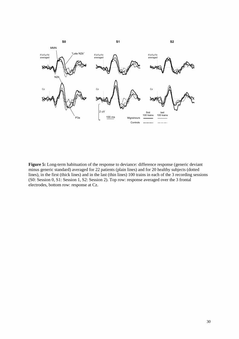
3.2.1 Long-term habituation of the components of the deviance-specific response
Figure 5 illustrates the effects of long-term habituation on the difference response in the three
recording sessions at frontal sites (average of F3, Fz, and F4) where MMN is measured and at Cz
where N2b and P3a are measured. The results of three-way ANOVAs performed on the measures of
Page 14
13
the three components, with Pathology as between-subject factor and Session and LTH as within-
subject factors are shown in Table 2. None of the three components showed a significant effect of
Pathology. Long-term habituation significantly affected N2b and P3a and showed no significant
interaction with Pathology. MMN did not habituate over the course of an entire session, in accordance
with previous unpublished data from our lab using a similar protocol.
For N2b, an effect of Session was observed, with larger N2b in the pre-menstrual session (post-hoc S1
versus S2 p = 0.04, S1 versus S0 p = 0.10, Fig. 4b). This N2b amplitude variation as a function of the
recording session combined with the LTH of this component resulted in an interaction between
Session and LTH. However, the effect of the menstrual cycle on N2b amplitude was not different
between patients and healthy subjects.
For P3a, an interaction between Pathology and Session was the result of a smaller P3a in the menstrual
session than in the session in the middle of the menstrual cycle for control subjects only (post-hoc S2
versus S0 p = 0.04 for healthy subjects, p = 0.41 for patients, Fig 4c). Thus, in the menstrual session,
i.e. in the session where migraine attacks preferentially occur, the migraine patients did not show the
decrease in P3a component observed in healthy subjects. In order to highlight a possible effect of a
migraine attack on P3a, we performed an additional analysis differentiating the 11 patients who
actually presented a migraine attack during one of the recording sessions (S1 or S2) and the 10
patients who did not, excluding the only subject who experienced headache during the session in the
middle of the cycle (S0). A 3-way ANOVA was applied to P3a amplitude, with the between subjects
factor “migraine-attack” (3 levels: control subjects, patients with and patients without migraine attack)
and LTH and Session as within-subject factors. In this analysis, the Session factor had only 2 levels,
the between menses session (S0) and a peri-menses session, possibly with a migraine attack. For
patients with a migraine attack, the peri-menses session was S1 for 5 patients and S2 for 6 patients.
For the patients who did not report a migraine attack and for the controls, the peri-menses session was
randomly chosen to be S1 (5 patients and 9 controls) or S2 (5 patients and 11 controls, see
(Demarquay et al., 2011), for a similar analysis). This additional ANOVA resulted in a significant
interaction between the intra-subject factor “Session” and the between-subject factor “migraine attack”
Page 15
14
(F(2,38) = 4.653, p = 0.016). Post-hoc tests were not significant. However, as displayed in Figure 4d,
patients with a migraine attack showed a tendency for a decrease in P3a amplitude in the peri-menses
session like the healthy subjects, while patients without a migraine attack showed a tendency for an
increase in P3a amplitude in the peri-menses session. Another 3-way ANOVA using LTH and Session
as within-subject factors, but restricted to the migraine patients (2 levels: patients with and without
migraine attack), confirmed this different modulation of P3a amplitude by migraine attacks, with a
marginally significant interaction between the intra-subject factor "Session" and the between-subject
factor "migraine attack" (F(1,19) = 4.300, p = 0.052).
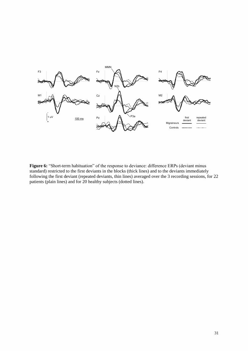
3.2.2 “Short-term habituation” of the components of the deviance-specific response
Figure 6 shows the difference responses restricted to the first deviants in the trains (first deviant minus
generic standard) and to the deviants that immediately followed the first deviants (repeated deviant
minus generic standard), grand averaged over the 3 sessions. It illustrates the effects of the so-called
“short-term habituation” on the components of the response to deviance. Table 3 displays the results
of ANOVAs performed on MMN, N2b, and P3a components with Pathology as between-subject
factor and with Session and STH as within-subject factors. There was a significant effect of STH on
the three components (MMN, N2b, P3a), the response globally collapsing to repeated deviance in both
populations. For N2b, there was an interaction between STH and Pathology due to a non-significant
tendency (p = 0.26) for this component to be larger in migraine patients than in controls in response to
the first deviant. For P3a an interaction between STH, Pathology and Session resulted from smaller
amplitude of the P3a to the first deviant in S2 than in S0 for the healthy subjects (post-hoc S2 versus
S0 p = 0.07 for healthy subjects), as also observed above for generic deviants.
3.3 Direct comparison of responses to deviance in migraine patients and in healthy subjects: “Late
N2b”
To investigate effects of pathology in the specific response to deviance outside the maxima of the
expected components, we considered the sample-by-sample difference between the difference
responses (generic deviants minus generic standards) of Patients and Controls averaged over the three
Page 16
15
sessions. This was done separately for the first half (first 100 trains) and the second half (last 100
trains) of the recordings because of the large LTH of the N2b and P3a components (see above). We
ran sample-by-sample Kruskal-Wallis tests at each electrode and each time sample between 0 and 300
ms post-stimulus to compare the two groups. A significant difference between the two groups was
observed at the 3 frontal electrodes in the 190 – 210 ms time-window in the first half of the sessions
(Figure 7). Indeed, the “Patients minus Controls” difference curve showed a maximum around 200 ms
at frontal sites, with the N2b thus appearing to last longer in migraine patients than in controls. To
further investigate the effects of Session on this “Late N2b” which was different between groups, we
performed a two-way ANOVA on the mean potentials measured between 190 and 210 ms for the first
100 trains of each session on a cluster including the 3 frontal electrodes with Pathology as between-
subject factor and Session as within-subject factor (see the measures in Figure 7d). This “late N2b”
was significantly enhanced in migraine patients (F(1,40) = 5.106, p = 0.029), which was expected
based on the Kruskal-Wallis tests reported above. There was also a global effect of Session (F(2,80) =
3.630, ε = 0.926, p = 0.035), with a collapse in the menstrual session (S2 versus S1 p = 0.04 and S2
versus S0 p = 0.06), but without any interaction with Pathology (F(2,80) = 1.470, p = 0.237). In other
words, migraine patients show enhanced negative frontal potentials in the late part of N2b, mostly
evident in the first half of the sessions, when N2b and P3a have not yet habituated.
Page 17
16
4. Discussion
Sensory processing has been extensively studied in migraine patients using ERPs (Schoenen et al.,
2003), and has mostly concerned habituation to repeated sensory stimulation in passive paradigms, as
well as brain responses to sensory changes in active paradigms. However, the specific response to
unexpected change in auditory stimulation during passive listening was scarcely reported. In this
study, we investigated the brain processes triggered by shorter tones randomly presented among
repeated tones in migraine patients in a situation of passive listening during three sessions along the
migraine cycle. The paradigm made it possible to study the MMN reflecting automatic mismatch
detection and subsequent N2b and P3a components reflecting attention orienting (Näätänen and
Gaillard, 1983). MMN was normal in migraine patients. However, along all recording sessions,
migraine disease modulated the responses at three stages of processing where attention-orienting
processes are thought to occur. First, around the descending slope of the N1, at the latency of a
possible residual orienting component (around 105 ms), fronto-central negative potentials were
enhanced in migraine patients, both for standard and for deviant tones. Second, in the differential
response to deviance (deviants minus standards), frontal potentials were negatively enhanced in
migraine patients during the descending slope from the N2b to the P3a (around 200 ms). Finally, P3a
amplitude was modulated differently along the menstrual cycle in the two groups, and tended to
normalize during migraine attacks.
4.1 Normal automatic mismatch detection in migraine
In the control group, randomly presented short-duration deviant tones elicited an MMN, followed by
N2b and P3a components. MMN is considered to be elicited pre-attentively (review in Näätänen and
Winkler, 1999), and to reflect automatic mismatch processes between the incoming deviant and a
memory trace of the previous repeated standard. As expected, immediately repeating the deviant tone
drastically diminished the amplitude of the MMN and of subsequent components (so-called “short-
Page 18
17
term habituation” of the MMN in Sams et al., 1984). In contrast, MMN amplitude did not evolve over
the course of the entire recording sessions (i.e., did not show any significant long-term habituation),
contrarily to subsequent attention-triggering mechanisms (N2b - P3a), in accordance with previous
observations (Lyytinen et al. (1992), and own unpublished observation over blocks of 3 hours of
stimulation).
Statistical analysis did not reveal any difference in MMNs between migraineurs and controls,
suggesting that up to the level of auditory processing where MMN operates, this processing does not
show any impairment in migraine. The current results extend our previous findings of preserved
sensory N1 component in migraine (Demarquay et al., 2011) by showing that the sensory memory
traces underlying MMN generation are also preserved in migraine. Our results contrast with a report
suggesting that MMN habituates in controls but not in migraineurs (de Tommaso et al., 2004). In this
earlier report, the stimulation paradigm was similar to our paradigm. However, MMN was measured
in the deviant ERP rather than in the usual deviant minus standard ERP. Considering this important
methodological discrepancy with our study and the fact that MMN latencies reported in de Tommaso
et al. (2004) were globally late in controls and in patients (around 200 ms), we can speculate that these
MMN measures rather concerned the N2b. Similarly, in their pediatric MMN study, Valeriani et al.
(2009) reported an habituation deficit of the MMN in migraine, but did not attempt to disentangle
MMN from N2b. In an attempt to dissociate the sensory mismatch processes from the subsequent
attention alerting processes, we assessed separately the mean amplitudes of the potentials at the
respective latencies of MMN and N2b. Other techniques such as trial-by-trial time-frequency analysis
of the EEG, beyond the scope of the present report, could be helpful to definitely exclude differences
in change detection processes between migraine patients and controls.
Neither the present study, nor our previous migraine study (Demarquay et al., 2011) of auditory ERPs
in a passive listening situation evidenced any abnormality in basic sensory processing, including
MMN generation. These observations are consistent with previous studies showing no significant
difference between migraineurs and controls with regard to sensory N1 and P2 component (Drake et
al., 1989; Sand and Vanagaite Vingen, 2000).
Page 19
18
4.2. Abnormal attention orienting in migraine
Contrasting with the fairly normal MMN observed in migraine patients, we observed at two different
processing stages enhanced frontal negative potentials in migraine patients, which suggest an
increased attention orienting (Halgren et al., 2011). Patients showed larger negative frontal potentials
firstly in the descending slope of the N1 for responses to both standard and deviant tones, and
secondly in the late part of the N2b in the response specific to deviance (deviants minus standards
difference wave).
In a previous report (Demarquay et al., 2011), we already reported such increased fronto-central
negative potentials for standard tones in migraine. This effect was peculiarly pronounced when tones
appeared at the beginning of the trains of stimuli (i.e. after a long silence), when the orienting
component (or component III, Näätänen and Picton, 1987) of the N1 is large (Alcaini et al., 1994). We
interpreted these enhanced fronto-central negative potentials in response to any standard tone as a
residual N1 orienting component. Here we show that a comparable effect is obtained with deviant
tones, suggesting that augmented orienting processes might actually be elicited by any incoming
auditory stimulus in migraine. Interestingly, the increase of the N1 orienting component observed in
migraine patients occurs earlier than the MMN (see Figure 2). As MMN appears fairly normal in
migraine, it can be hypothesized that these abnormal early orienting processes do not interfere with the
sensory memory and comparison processes underlying MMN generation.
Migraine patients also showed augmented frontal negative potentials in the deviance-specific response
located in the descending slope of N2b. This effect was mostly evident in the first half of all sessions.
N2b is a fronto-central subcomponent, which typically arises in active paradigms and has been
associated with attention-triggering (Snyder and Hillyard, 1976; Näätänen and Gaillard, 1983; Kiehl et
al., 2001) and cognitive control encompassing response inhibition to a non-target deviant, response
conflict, and error monitoring (Folstein and Van Petten, 2008; Schwartze et al., 2011). N2b was shown
to be associated with higher order processing than the MMN (Ritter et al., 1992). However N2b can
also occur in passive oddball paradigms, in particular when the difference between standards and
deviants is large enough (Näätänen et al. (1982); see also Ruby et al. (2008) for an example of N2b
Page 20
19
recorded in a passive oddball paradigm using duration deviants). Although participants in our study
were instructed to watch a silent movie and to ignore auditory stimuli, their attention might have been
involuntarily oriented to the deviant stimuli and the prolonged negative potentials at the end of N2b
observed in migraineurs could reflect an abnormal activity at the moment of this involuntary shift of
attention.
Moreover, P3a amplitude was modulated along the menstrual cycle in both groups. Controls showed a
smaller P3a during menses than in the session between menses. As far as we know, this was never
described in the literature. The overall migraine population showed an opposite pattern, with an
enhanced P3a in the menstrual session. This abnormality tended to normalize during migraine attacks.
In a previous study (Demarquay et al., 2011), we already observed P3a abnormalities depending on
the recording session. However, whereas the present result concerned the P3a individualized in the
response specific to deviance, the former result concerned the P3a in response to the first tones of the
trains, a component partially overlapping the sensory P2. Thus, the two results can hardly be
compared. Nevertheless, both results have in common a normalization of P3a amplitudes during
migraine attacks, in accordance with previous observations concerning several other components
(Kropp and Gerber, 1995; Kropp and Gerber, 1998; Evers et al., 1999; Judit et al., 2000).
As a whole, our results reveal ERP abnormalities arising in migraine patients at stages of processing
where attention-orienting mechanisms are susceptible to operate. They are in keeping with findings
using active paradigms (and thus involving attention), showing abnormal cognitive responses like the
P300 to targets (Drake et al., 1989; Wang et al., 1996; Evers et al., 1998) or the CNV to warning
stimuli (Maertens de Noordhout et al., 1986; Kropp and Gerber, 1993). As the main generators of N1
orienting component, N2b and P3a are thought to be located in frontal areas (see Alcaini et al. (1994)
for the N1 orienting component, Halgren et al. (2011) for the N2b and Polich and Criado (2006) for
P3a), the abnormal automatic attention orienting towards acoustic stimuli observed in migraineurs
may reflect abnormal attention-related frontal networks in migraine.
Page 21
20
4.3. Sensory processing and attention orienting in migraine
To sum up, the present ERP findings allow setting the working hypothesis of normal sensory stages of
auditory processing in migraine, but exacerbated mechanisms of automatic attention orienting to the
auditory stimulation, possibly relying on an abnormal involvement of frontal networks. As
emphasized above, our results are in line with previous studies that showed normal N1 and P2 sensory
responses but altered cognitive responses like the iCNV, and P300. The apparent discrepancy with
previous reports of a lack of habituation of auditory ERPs in migraine (Wang et al., 1996; Ambrosini
et al., 2003) could be related to the paradigms used in these studies. Indeed, lack of habituation was
observed in paradigms using intensity dependence of auditory potentials (IDAP), where sound
intensity varies randomly within blocks. No habituation deficit was observed when stimulus intensity
changes did not occur within blocks (Sand and Vanagaite Vingen, 2000), or in paradigms not
involving intensity variations such as ours (Demarquay et al., 2011). We can speculate that random
changes in the auditory stimulation might be more arousing than unvarying stimuli, and thus more
likely to automatically engage attentional networks. This is in keeping with the present findings of an
increased attention orienting to rare changes occurring in streams of otherwise repetitive tones.
Furthermore, a link between habituation deficits in migraine and abnormal orienting activity has been
already proposed by Siniatchkin et al. (Siniatchkin et al., 2000).
In the visual modality, the habituation of ERPs has been widely tested in migraine patients, mainly
using pattern-reversal stimuli, with contrasting results (Schoenen et al., 1995; Afra et al., 1998; Wang
et al., 1999). One study suggested that the visual habituation deficit in migraine depends on the
stimulus characteristics (Oelkers et al., 1999), but a recent one using individual identification of visual
evoked peaks in a blinded evaluation design found normal habituation in interictal migraineurs, with
similar check-size effects in patients and controls (Omland et al., 2013). Besides, recent studies
showed an increased automatic attentional response to sudden-onset visual events in migraineurs
(Mickleborough et al., 2011a). Interestingly, in line with our observation of an enhancement of the N1
orienting component in auditory ERPs, these authors showed an early attention effect in the visual N1
ERP component in migraine patients (Mickleborough et al., 2011b). Thus, in the visual modality also,
Page 22
21
further studies might possibly enlighten a link between observed habituation deficits and attention
orienting exacerbation.
4.4. Methodological issues
In the present study, in an attempt to follow a migraine cycle, we only recorded patients with
menstrually-related migraine. This strategy indeed allowed us to record 11 patients during a migraine
attack. However, as patients were not requested to fulfill migraine diaries, it might be possible that,
during some recordings, some patients were in a preictal state, a state where cortical excitability has
been shown to fluctuate. This would be more likely for sessions S1 and S2, i.e., the sessions around
menses. Indeed, before an attack, migraineurs show a normalization of the interictal lack of
habituation of CNV (Kropp and Gerber, 1995; Kropp and Gerber, 1998; Siniatchkin et al., 1999) and
of IDAP potentials (Judit et al., 2000). It is however noteworthy that, except for the P3a, the between-
group differences observed in the present study (enhanced N1 orienting component and enhanced late
N2b in migraine patients) did not depend on the recording sessions, suggesting that these effects are
not modulated along the migraine cycle. We cannot exclude that a precise sorting of the recording
sessions in "interictal" "pre-ictal" and "ictal" could allow to reveal further differences between
migraine patients and controls.
In conclusion, our study shows normal auditory sensory processing in migraine patients but increased
automatic attention orienting processes to auditory changes (and more generally to any incoming
auditory stimulus). The pathophysiologic basis of such heightened attentional response recorded in
migraineurs is still unknown, but could, at least partially, be linked to the close relationship observed
between environmental stimuli and migraine. Understanding the links between photo/phonophobia
and an increased automatic attention orienting in migraine is a challenge for future studies.
Page 23
22
References
Afra J, Cecchini AP, De Pasqua V, Albert A, Schoenen J. Visual evoked potentials during long periods of
pattern-reversal stimulation in migraine. Brain 1998; 121: 233-241.
Aguera PE, Jerbi K, Caclin A, Bertrand O. ELAN: a software package for analysis and visualization of MEG,
EEG, and LFP signals. Comput Intell Neurosci 2011; 2011: 158970.
Alcaini M, Giard MH, Thevenet M, Pernier J. Two separate frontal components in the N1 wave of the human
auditory cortex. Psychophysiology 1994; 31: 611-615.
Ambrosini A, Rossi P, De Pasqua V, Pierelli F, Schoenen J. Lack of habituation causes high intensity
dependence of auditory evoked cortical potentials in migraine. Brain 2003; 126: 2009-2015.
Böcker KB, Timsit-Berthier M, Schoenen J, Brunia CH. Contingent Negative Variation in migraine. Headache
1990; 30: 604-609.
Coppola G, Pierelli F, Schoenen J. Habituation and migraine. Neurobiol Learn Mem 2009; 92: 249-259.
de Tommaso M, Guido M, Libro G, Losito L, Difruscolo O, Sardaro M, et al. Interictal lack of habituation of
mismatch negativity in migraine. Cephalalgia 2004; 24: 663-668.
Demarquay G, Caclin A, Brudon F, Fischer C, Morlet D. Exacerbated attention orienting to auditory stimulation
in migraine patients. Clin Neurophysiol 2011; 122: 1755-1763.
Drake ME, Jr., Pakalnis A, Padamadan H. Long-latency auditory event related potentials in migraine. Headache
1989; 29: 239-241.
Evers S, Bauer B, Grotemeyer KH, Kurlemann G, Husstedt IW. Event-related potentials (P300) in primary
headache in childhood and adolescence. J Child Neurol 1998; 13: 322-326.
Evers S, Quibeldey F, Grotemeyer KH, Suhr B, Husstedt IW. Dynamic changes of cognitive habituation and
serotonin metabolism during the migraine interval. Cephalalgia 1999; 19: 485-491.
Fischer C, Morlet D, Bouchet P, Luaute J, Jourdan C, Salord F. Mismatch negativity and late auditory evoked
potentials in comatose patients. Clin Neurophysiol 1999; 110: 1601-1610.
Folstein JR, Van Petten C. Influence of cognitive control and mismatch on the N2 component of the ERP: a
review. Psychophysiology 2008; 45: 152-170.
Guthrie D, Buchwald JS. Significance testing of difference potentials. Psychophysiology 1991; 28: 240-244.
Halgren E, Sherfey J, Irimia A, Dale AM, Marinkovic K. Sequential temporo-fronto-temporal activation during
monitoring of the auditory environment for temporal patterns. Hum Brain Mapp 2011; 32: 1260-1276.
ICHD. The International Classification of Headache Disorders: 2nd edition. Cephalalgia 2004; 24 Suppl 1: 9-
160.
Judit A, Sandor PS, Schoenen J. Habituation of visual and intensity dependence of auditory evoked cortical
potentials tends to normalize just before and during the migraine attack. Cephalalgia 2000; 20: 714-719.
Jung J, Morlet D, Mercier B, Confavreux C, Fischer C. Mismatch negativity (MMN) in multiple sclerosis: An
event-related potentials study in 46 patients. Clin Neurophysiol 2006; 117: 85.
Kiehl KA, Laurens KR, Duty TL, Forster BB, Liddle PF. Neural sources involved in auditory target detection
and novelty processing: an event-related fMRI study. Psychophysiology 2001; 38: 133-142.
Page 24
23
Korostenskaja M, Pardos M, Kujala T, Rose DF, Brown D, Horn P, et al. Impaired auditory information
processing during acute migraine: a magnetoencephalography study. Int J Neurosci 2011; 121: 355-365.
Kropp P, Gerber W-D. Prediction of migraine attacks using a slow cortical potential, the contingent negative
variation. Neuroscience Letters 1998; 257: 73-76.
Kropp P, Gerber WD. Is increased amplitude of contingent negative variation in migraine due to cortical
hyperactivity or to reduced habituation? Cephalalgia 1993; 13: 37-41.
Kropp P, Gerber WD. Contingent negative variation during migraine attack and interval: evidence for
normalization of slow cortical potentials during the attack. Cephalalgia 1995; 15: 123-128; discussion 178-
129.
Lyytinen H, Blomberg AP, Näätänen R. Event-related potentials and autonomic responses to a change in
unattended auditory stimuli. Psychophysiology 1992; 29: 523-533.
Maertens de Noordhout A, Timsit-Berthier M, Timsit M, Schoenen J. Contingent negative variation in headache.
Ann Neurol 1986; 19: 78-80.
Main A, Dowson A, Gross M. Photophobia and phonophobia in migraineurs between attacks. Headache 1997;
37: 492-495.
Main A, Vlachonikolis I, Dowson A. The wavelength of light causing photophobia in migraine and tension-type
headache between attacks. Headache 2000; 40: 194-199.
Mazzotta G, Alberti A, Santucci A, Gallai V. The event-related potential P300 during headache-free period and
spontaneous attack in adult headache sufferers. Headache 1995; 35: 210-215.
Mickleborough MJ, Hayward J, Chapman C, Chung J, Handy TC. Reflexive attentional orienting in migraineurs:
The behavioral implications of hyperexcitable visual cortex. Cephalalgia 2011a; 31: 1642-1651.
Mickleborough MJ, Truong G, Handy TC. Top-down control of visual cortex in migraine populations.
Neuropsychologia 2011b; 49: 1006-1015.
Mulder EJCM, Linssen WHJP, Passchier J, De Geus EJC. Interictal and Postictal Contingent Negative Variation
in Migraine Without Aura. Headache 2001; 41: 72-78.
Näätänen R. Mismatch negativity: clinical research and possible applications. Int J Psychophysiol 2003; 48: 179-
188.
Näätänen R, Gaillard AWK. The orienting reflex and the N2 deflection of the event-related potential (ERP). In:
Gaillard AWK and Ritter W, editors. Tutorials in ERP Research : endogenous components North Holland
Publishing Company, 1983: 119-141.
Naatanen R, Kujala T, Escera C, Baldeweg T, Kreegipuu K, Carlson S, et al. The mismatch negativity (MMN)--
a unique window to disturbed central auditory processing in ageing and different clinical conditions. Clin
Neurophysiol 2012; 123: 424-458.
Näätänen R, Kujala T, Winkler I. Auditory processing that leads to conscious perception: a unique window to
central auditory processing opened by the mismatch negativity and related responses. Psychophysiology
2011; 48: 4-22.
Näätänen R, Paavilainen P, Rinne T, Alho K. The mismatch negativity (MMN) in basic research of central
auditory processing: a review. Clin Neurophysiol 2007; 118: 2544-2590.
Näätänen R, Paavilainen P, Tiitinen H, Jiang D, Alho K. Attention and mismatch negativity. Psychophysiology
1993; 30: 436-450.
Näätänen R, Picton T. The N1 wave of the human electric and magnetic response to sound: a review and an
analysis of the component structure. Psychophysiology 1987; 24: 375-425.
Page 25
24
Näätänen R, Simpson M, Loveless NE. Stimulus deviance and evoked potentials. Biol Psychol 1982; 14: 53-98.
Näätänen R, Tervaniemi M, Sussman E, Paavilainen P, Winkler I. "Primitive intelligence" in the auditory cortex.
Trends Neurosci 2001; 24: 283-288.
Näätänen R, Winkler I. The concept of auditory stimulus representation in cognitive neuroscience. Psychol Bull
1999; 125: 826-859.
Oelkers R, Grosser K, Lang E, Geisslinger G, Kobal G, Brune K, et al. Visual evoked potentials in migraine
patients: alterations depend on pattern spatial frequency. Brain 1999; 122: 1147-1155.
Omland PM, Nilsen KB, Uglem M, Gravdahl G, Linde M, Hagen K, et al. Visual Evoked Potentials in Interictal
Migraine: No Confirmation of Abnormal Habituation. Headache 2013.
Polich J, Criado JR. Neuropsychology and neuropharmacology of P3a and P3b. Int J Psychophysiol 2006; 60:
172-185.
Ritter W, Paavilainen P, Lavikainen J, Reinikainen K, Alho K, Sams M, et al. Event-related potentials to
repetition and change of auditory stimuli. Electroencephalogr Clin Neurophysiol 1992; 83: 306-321.
Ruby P, Caclin A, Boulet S, Delpuech C, Morlet D. Odd sound processing in the sleeping brain. J Cogn
Neurosci 2008; 20: 296-311.
Sams M, Alho K, Näätänen R. Short-term habituation and dishabituation of the mismatch negativity of the ERP.
Psychophysiology 1984; 21: 434-441.
Sand T, Vanagaite Vingen J. Visual, long-latency auditory and brainstem auditory evoked potentials in migraine:
relation to pattern size, stimulus intensity, sound and light discomfort thresholds and pre-attack state.
Cephalalgia 2000; 20: 804-820.
Schoenen J, Ambrosini A, Sandor PS, Maertens de Noordhout A. Evoked potentials and transcranial magnetic
stimulation in migraine: published data and viewpoint on their pathophysiologic significance. Clin
Neurophysiol 2003; 114: 955-972.
Schoenen J, Wang W, Albert A, Delwaide PJ. Potentiation instead of habituation characterizes visual evoked
potentials in migraine patients between attacks. European Journal of Neurology 1995; 2: 115-122.
Schwartze M, Rothermich K, Schmidt-Kassow M, Kotz SA. Temporal regularity effects on pre-attentive and
attentive processing of deviance. Biol Psychol 2011; 87: 146-151.
Siniatchkin M, Gerber WD, Kropp P, Vein A. How the brain anticipates an attack: a study of neurophysiological
periodicity in migraine. Funct Neurol 1999; 14: 69-77.
Siniatchkin M, Gerber WD, Kropp P, Voznesenskaya T, Vein AM. Are the periodic changes of
neurophysiological parameters during the pain-free interval in migraine related to abnormal orienting
activity? Cephalalgia 2000; 20: 20-29.
Snyder E, Hillyard SA. Long-latency evoked potentials to irrelevant, deviant stimuli. Behav Biol 1976; 16: 319-
331.
Stovner L, Hagen K, Jensen R, Katsarava Z, Lipton R, Scher A, et al. The global burden of headache: a
documentation of headache prevalence and disability worldwide. Cephalalgia 2007; 27: 193-210.
Tervaniemi M, Lehtokoski A, Sinkkonen J, Virtanen J, Ilmoniemi RJ, Näätänen R. Test-retest reliability of
mismatch negativity for duration, frequency and intensiy changes. Clin Neurophysiol 1999; 110: 1388-1393.
Valeriani M, Galli F, Tarantino S, Graceffa D, Pignata E, Miliucci R, et al. Correlation between abnormal brain
excitability and emotional symptomatology in paediatric migraine. Cephalalgia 2009; 29: 204-213.
Page 26
25
Vingen JV, Sand T, Stovner LJ. Sensitivity to various stimuli in primary headaches: a questionnaire study.
Headache 1999; 39: 552-558.
Wang W, Schoenen J. Interictal potentiation of passive "oddball" auditory event-related potentials in migraine.
Cephalalgia 1998; 18: 261-265; discussion 241.
Wang W, Schoenen J, Timsit-Berthier M. Cognitive functions in migraine without aura between attacks: a
psychophysiological approach using the "oddball" paradigm. Neurophysiol Clin 1995; 25: 3-11.
Wang W, Timsit-Berthier M, Schoenen J. Intensity dependence of auditory evoked potentials is pronounced in
migraine: an indication of cortical potentiation and low serotonergic neurotransmission? Neurology 1996;
46: 1404-1409.
Wang W, Wang GP, Ding XL, Wang YH. Personality and response to repeated visual stimulation in migraine
and tension-type headaches. Cephalalgia 1999; 19: 718-724; discussion 697-718.
Woods DL, Elmasian E. The habituation of event-related potentials to speech sounds and tones.
Electroencephalogr Clin Neurophysiol 1986; 65: 446-459.
Page 27
26
……………
firstdeviant
repeateddeviant
seconddeviant
Standards
firstdeviant
SOA610 ms
1 recording session: 200 trains
ITI5.5 to 8 sec
1 train:8 to 12 stimuli
Figure 1: Design and timing of the auditory stimulation paradigm.
In each recording session, 200 trains of 8 to 12 tone bursts are presented with an inter-train interval
(ITI) ranging between 5.5 and 8 seconds. Within-train stimulus onset asynchrony (SOA) is 610 ms.
Two deviants (tone-bursts shorter than the standard stimuli) are presented in each train. In 50% of the
trains, at least 2 standards are presented between the first and the second deviant (as for example in the
first train of the figure). In the other 50% of the trains, the second deviant immediately follows the
first one (repeated deviant, as in the second train of the figure). “Generic deviants” (colored in black)
exclude the repeated deviants (colored in grey). “Generic standards” (colored in black) exclude the
first 3 stimuli of the trains and the standard stimuli following a deviant (colored in grey).
Page 28
27
Figure 2:
(a): Responses to generic standard stimuli (thick lines) and to generic deviant stimuli (thin lines) grand
averaged over the 3 sessions for 20 healthy subjects (dotted lines) and for 22 migraine patients (plain
lines). The shaded area displays the temporal window defined for the assessment of sensory N1 (75-95
ms).
(b): Deviance-specific response (generic deviant minus generic standard) grand averaged over the 3
sessions for 20 healthy subjects (dotted line) and for 22 migraine patients (plain line). The shaded
areas display the temporal windows defined for the assessment of MMN (115-145 ms), N2b (145-200
ms) and P3a (200-300 ms).
(c): Scalp potential maps indicative of the topographies of the different components (sensory N1 to
standards, MMN, N2b and P3a in the deviance-specific response). These maps were drawn using
spherical spline interpolation from the potentials measured at the seven scalp electrodes, averaged in
the different time-windows in migraine patients and in healthy subjects. The range of voltage values
used for the color scale is mentioned for each time-window.
Page 29
28
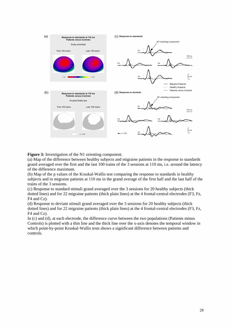
Figure 3: Investigation of the N1 orienting component.
(a) Map of the difference between healthy subjects and migraine patients in the response to standards
grand averaged over the first and the last 100 trains of the 3 sessions at 110 ms, i.e. around the latency
of the difference maximum.
(b) Map of the p values of the Kruskal-Wallis test comparing the response to standards in healthy
subjects and in migraine patients at 110 ms in the grand average of the first half and the last half of the
trains of the 3 sessions.
(c) Response to standard stimuli grand averaged over the 3 sessions for 20 healthy subjects (thick
dotted lines) and for 22 migraine patients (thick plain lines) at the 4 frontal-central electrodes (F3, Fz,
F4 and Cz).
(d) Response to deviant stimuli grand averaged over the 3 sessions for 20 healthy subjects (thick
dotted lines) and for 22 migraine patients (thick plain lines) at the 4 frontal-central electrodes (F3, Fz,
F4 and Cz).
In (c) and (d), at each electrode, the difference curve between the two populations (Patients minus
Controls) is plotted with a thin line and the thick line over the x-axis denotes the temporal window in
which point-by-point Kruskal-Wallis tests shows a significant difference between patients and
controls.
Page 30
29
MMNmean amplitude 115-145 ms at frontal sites
Am
pli
tu
de
(mic
ro
vo
lts)
- 2.0
- 1.6
- 1.2
- 0.8
- 0.4
0
S0 S1 S2
Migraineurs
Controls
Am
pli
tu
de
(mic
ro
vo
lts)
N2bmean amplitude 145-200 ms at Cz
P3amean amplitude 200-300 ms at Cz
(a) (b)
(d)(c)
- 1.4
- 1.2
- 1.0
- 0.8
- 0.6
- 0.4
- 0.2
0
S0 S1 S2
Migraineurs
Controls
Am
pli
tu
de
(mic
ro
vo
lts)
0
0.2
0.4
0.6
0.8
1.0
1.2
1.4
S0 S1 S2
Migraineurs
Controls
Am
pli
tu
de
(mic
ro
vo
lts)
0
0.2
0.4
0.6
0.8
1.0
1.2
1.4
S0 Peri-menses session.
Migraineurs
suffering headache during
one peri-menses session
Migraineurs without
headache during the
recording sessions
Controls
Am
pli
tu
de
(mic
ro
vo
lts)
P3aEffect of a migraine attack
Figure 4: Mean amplitude for each session of the different components measured in the deviance-
specific response (response to the generic deviant minus response to the generic standard). The black
lines denote the standard error of the mean.
In (a), (b), (c), the measures are displayed for each session in migraine patients and in controls, for: (a)
MMN (mean value in the 115 - 145 ms time-window at the 3 frontal electrodes), (b) N2b (mean value
in the 145 - 200 ms time-window at Cz) and (c) P3a (mean of positive values in the 200 - 300 ms
time-window at Cz). S0 = session 0, S1 = session 1, S2 = session 2.
In (d), P3a was measured in 3 groups of subjects: 10 patients who showed no migraine attack during
the recording sessions, 11 patients who suffered headache during one peri-menses session, and the 20
control subjects. The measures are displayed for the session between menses (S0) and for a peri-
menses session (the session with headache for the patients experiencing an attack during recording and
a randomly chosen peri-menses session (S1 or S2) for the other two groups).
Page 31
30
Figure 5: Long-term habituation of the response to deviance: difference response (generic deviant
minus generic standard) averaged for 22 patients (plain lines) and for 20 healthy subjects (dotted
lines), in the first (thick lines) and in the last (thin lines) 100 trains in each of the 3 recording sessions
(S0: Session 0, S1: Session 1, S2: Session 2). Top row: response averaged over the 3 frontal
electrodes, bottom row: response at Cz.
Page 32
31
Figure 6: “Short-term habituation” of the response to deviance: difference ERPs (deviant minus
standard) restricted to the first deviants in the blocks (thick lines) and to the deviants immediately
following the first deviant (repeated deviants, thin lines) averaged over the 3 recording sessions, for 22
patients (plain lines) and for 20 healthy subjects (dotted lines).
Page 33
32
Figure 7: Investigation of the “late N2b”.
(a) Map of the difference between healthy subjects and migraine patients in the deviance-specific
responses (generic deviants minus generic standards) in the first and the last 100 trains, averaged over
the 3 sessions at 200 ms, i.e. at the latency of the difference maximum.
(b) Map of the p values of the Kruskal-Wallis test comparing the deviance-specific responses in
healthy subjects and in migraine patients at 200 ms in the first half and the second half of the trains
averaged over the 3 sessions.
(c) Deviance-specific response grand averaged over the first 100 trains of the 3 sessions for 20 healthy
subjects (thick dotted lines) and for 22 migraine patients (thick plain lines) at the 3 frontal electrodes
(F3, Fz and F4). At each electrode, the difference curve between the two populations (Patients minus
Controls) is plotted with a thin line and the thick line over the x-axis denotes the temporal window in
which the point-by-point Kruskal-Wallis tests show a significant difference between patients and
controls.
(d) “Late N2b” amplitude (mean value and standard error of the mean in the 190 - 210 ms time-
window averaged over the 3 frontal electrodes) for each population and for the 100 first trains of each
session. S0 = session 0, S1 = session 1, S2 = session 2.
Page 34
33
Table 1
Table 1: Results of the four-way ANOVAs on the amplitudes of the N1 sub-components, with
Pathology as between-subject factor and with Session (S0, S1, and S2), Long-Term Habituation (LTH:
first half versus second half of the recording session) and Type of stimulus (standard versus deviant)
as within-subject factors. Degrees of freedom for each factor are indicated in parenthesis. Sensory N1
was measured at Cz in the 75-95 ms time-window where the maximal amplitude is observed in the
grand average response (see Figure 2) and orienting component was measured at the 4 frontal-central
electrodes in the 100-120 ms latency range, i.e., in the spatio-temporal window where a significant
between-group difference was observed with Kruskal-Wallis tests (see Figure 3). Results involving
Pathology are printed in italics. Significant p values (p ≤ 0.05) are printed in bold.
Page 35
34
Table 2
]
MMN N2b P3a
F3 + Fz + F4 (115-145 ms) Cz (145-200 ms) Cz (200-300 ms)
F ε - GG p F ε - GG p F ε - GG p
Pathology (1,40) 0.139 0.712 2.219 0.144 0.078 0.781
Session (2,80) 0.061 0.993 0.940 3.605 0.927 0 .0 3 5 0.355 0.921 0.685
Session x Pathology (2,80) 0.874 0.993 0.421 0.438 0.927 0.632 5.980 0.921 0 .0 0 5
LTH (1,40) 0.763 1.000 0.388 10.919 1.000 0 .0 0 2 13.401 1.000 0 .0 0 1
LTH x Pathology (1,40) 0.164 1.000 0.688 2.192 1.000 0.147 1.920 1.000 0.174
Session x LTH (2,80) 0.679 0.982 0.507 3.988 0.994 0 .0 2 3 1.008 0.991 0.369
Session x LTH x Pathology (2, 80) 0.626 0.982 0.534 1.753 0.994 0.180 0.579 0.991 0.561
Table 2: Results of the three-way ANOVAs on the amplitudes of the different components (MMN,
N2b, and P3a) of the difference response (deviant minus standard), with Pathology as between-subject
factor and with Session (S0, S1, and S2) and Long-Term Habituation (LTH: first half versus second
half of the session) as within-subject factors. Degrees of freedom for each factor are indicated in
parenthesis. MMN was assessed as the mean amplitude at the 3 frontal electrodes between 115 and
145 ms, N2b as the mean amplitude at Cz between 145 and 200 ms and P3a as the mean amplitude of
positive potentials at Cz between 200 and 300 ms. Results involving Pathology are printed in italics.
Significant p values (p ≤ 0.05) are printed in bold.
Page 36
35
Table 3
MMN N2b P3a
F3 + Fz + F4 (115-145 ms) Cz (145-200 ms) Cz (200-300 ms)
F ε - GG p F ε - GG p F ε - GG p
Pathology (1,40) 0.366 0.548 0.855 0.361 1.418 0.241
Session (2,80) 0.116 0.997 0.890 2.410 0.952 0.099 1.058 0.974 0.350
Session x Pathology (2,80) 0.282 0.997 0.755 0.205 0.952 0.805 1.572 0.974 0.215
STH (1,40) 12.375 1.000 0 .0 0 1 37.886 1.000 < 0 .0 0 1 49.620 1.000 < 0 .0 0 1
STH x Pathology (1,40) 0.675 1.000 0.416 4.144 1.000 0 .0 4 8 0.000 1.000 0.995
Session x STH (2,80) 0.130 0.834 0.842 0.620 0.994 0.540 0.664 0.873 0.499
Session x STH x Pathology (2, 80) 0.614 0.834 0.516 0.206 0.994 0.813 7.763 0.873 0 .0 0 1
Table 3: “Short-term habituation” of the main components of the difference response (this analysis
does not relate to short-term habituation in a classical sense, but rather examines how repeating a
deviant alters the deviant-specific response, see Introduction): results of the three-way ANOVAs on
the amplitudes of MMN, N2b and P3a, with Pathology as between-subject factor and with Session
(S0, S1, and S2) and Short-Term Habituation (STH, first deviant versus repeated deviant) as within-
subject factors. Degrees of freedom for each factor are indicated in parenthesis. MMN was assessed as
the mean amplitude at the 3 frontal electrodes between 115 and 145 ms, N2b as the mean amplitude at
Cz between 145 and 200 ms and P3a as the mean amplitude of positive potentials at Cz between 200
and 300 ms as in Table 2. Results involving Pathology are printed in italics. Significant p values (p ≤
0.05) are printed in bold.