+ All Categories
Home > Documents > August 1990 Thesis/Dissertation 1990 Thesis/Dissertation ... CHAPTER II Methods and Materials ......

August 1990 Thesis/Dissertation 1990 Thesis/Dissertation ... CHAPTER II Methods and Materials ......

Date post: 27-Apr-2018
Category:
Upload: vokhue
View: 220 times
Download: 2 times
Share this document with a friend
87
, 1 L 1. August 1990 Thesis/Dissertation Chromosomal Localization and Restriction Fragment Length !% Polymorphism Analysis of Annexins III, IV, and V N N S Donald Alan Frankenberry AFIT Student at: University of Washington AFIT/CI/CIA -90-072 AFIT/CI Wright-Patterson AFB OH 45433 Approved for Public Release lAW AFR 190-1 Distribution Unlimited ERNEST A. HAYGOOD, Ist Lt, USAF Executive Officer, Civilian Institution Programs DTIC f% ELECTE 82 UNCLASSIFIED
Transcript

, 1L

1. August 1990 Thesis/Dissertation

Chromosomal Localization and Restriction Fragment Length!% Polymorphism Analysis of Annexins III, IV, and VNN

S Donald Alan Frankenberry

AFIT Student at: University of Washington AFIT/CI/CIA -90-072

AFIT/CIWright-Patterson AFB OH 45433

Approved for Public Release lAW AFR 190-1

Distribution UnlimitedERNEST A. HAYGOOD, Ist Lt, USAF

Executive Officer, Civilian Institution Programs

DTICf% ELECTE

82

UNCLASSIFIED

GENERAL INSTRUCTIONS FOR COMPLETING SF 298The Report Documentation Page (RDP) is used in announcing and cataloging reports. It is importantthat this information be consistent with the rest of the report, particularly the cover and title page.Instructions for filling in each block of the form follow. It is important to stay within the lines to meetoptical scanning requirements.

Block 1. Agency Use Only (Leave Blank) Block 12a. Distribution/Availablity Statement,Denote public availability or limitation. Cite

Block 2. Report Date. Full publication date any availability to the public. Enter additionalincluding day, month, and year, if available (e.g. limitations or special markings in all capitals1 Jan 88). Must cite at least the year. (e.g. NOFORN, REL, ITAR)

Block 3. Type of Reoort and Dates Covered.State whether report is interim, final, etc. If DOD - See DoDD 5230.24, "Distributionapplicable, enter inclusive report dates (e.g. 10 Statements on TechnicalJun 87 - 30 Jun 88). Documents."

Block 4. Title and Subtitle. A title is taken from DOE - See authoritiesthe part of the report that provides the most NASA - See Handbook NHB 2200.2.meaningful and complete information. When a NTIS - Leave blank.report is prepared in more than one volume,repeat the primary title, add volume number,and include subtitle for the specific volume. On Block 12b. Distribution Code.classified documents enter the titleclassification in parentheses. DOD - DOD - Leave blank

DOE - DOE -'Enter DOE distribution categoriesBlock 5. FuningNumbers, To include contract from the Standard Distribution forand grant numbers; may include program Unclassified Scientific and Technicalelement number(s), project number(s), task Reportsnumber(s), and work unit number(s). Use the NASA - NASA - Leave blankfollowing labels: NTIS - NTIS - Leave blank.

C - Contract PR - ProjectG - Grant TA -TaskPE - Program WU - Work Unit Block 13. AbstracL Include a brief (Maximum

Element Accession No. 200 words) factual summary of the mostsignificant information contained in the report.

Block 6. Author(s). Name(s) of person(s)responsible for writing the report, performing Block 14. Subject Terms, Keywords or phrasesthe research, or credited with the content of the identifying major subjects in the report.report. If editor or compiler, this should followthe name(s). Block 15. Number of Pages. Enter the total

Block 7. Performing Organization Name(s) and number of pages.Addresses) Self-explanatory. Block 16. Price Code. Enter appropriate price

Block 8. Performino Organization Report code (NTIS only).Number, Enter the unique alphanumeric reportnumber(s) assigned by the organization Blocks 17. - 19. Security Classifications.performing the report. Self-explanatory. Enter U.S. Security

Classification in accordance with U.S. SecurityBlock 9. Sponsorina/Monitoring Agency Regulations (i.e., UNCLASSIFIED). If formNames(s) and Address(es). Self-explanatory. contains classified information, stamp

classification on the top and bottom of the page.Block 10. Soonsoring/Mon ito ring Agency,Report Number. (If known)

Block 20. Limitation of Abstract, This blockBlock 11. Sunolementary Notes. Enter must be completed to assign a limitation to theinformation not included elsewhere such as: abstract. Enter either UL (unlimited) or SARPrepared in cooperation with...; Trans. of ..., To (same as report) An entry in this block isbe published in .... When a report is revised, (sa as re antry in th block Iinclude a statement whether the new report necessary if the abstract is to be limited. Ifsupersedes or supplements the older report. blank, the abstract is andad Io Ba (Rli.i2e89

standard Form 298 Back (Rev. 2-89)

University of Washington

Abstract

Chromosomal Localization and Restriction Fragment LengthPolymorphism Analysis of Annexins III, IV, V

by Donald Alan Frankenberry

Chairperson of the Supervisory Committee:Assistant Professor Jonathan F. TaitDepartment of Laboratory Medicine

The annexins are a family of recently identified

calcium-dependent phospholipid binding proteins with

preference for anionic phospholipid. Chromosomal mapping of

the genes for annexins III, IV, and V was undertaken as part

of a study of their structure and function. The genes for

annexin III and annexin V were localized to human chromosome

4 by utilizing a panel of human-hamster somatic cell hybrids

and the polymerase chain reaction to amplify intron

containing regions of these genes. Verification of these

two localizations was performed using a human-mouse hybrid

cell line containing human chromosome 4 as its only human

DNA complement. Verification of the localization of annexin

III was also performed using Southern blot analysis of

genomic DNA specimens from the hybrid cell panel digested

with Hind III. The annexin IV gene was not definitively

localized in this study. Restriction mapping of these three

genes suggests that they are of moderate size, 20 kb to 50

kb. The following restriction enzymes were used to screen

for restriction fragment length polymorphisms at these loci:

Bam HI, Bgl II, Eco RI, Hind III, Msp I, Pst I, Pvu II,

90

Rsa I, Taq I. Restriction fragment length polymorphisms

were identified at the annexin V locus with Taq I and Pvu

II, and at the annexin III locus with Hind III and Bgl II.

Polymorphisms were not detected for annexin IV with Bam HI,

Bgl II, Eco RI, Hind III, Msp I, Pst I, Pvu II, Rsa I, and

Taq I.

Accession ForNTIS GRA&IDTIC TABUnannounced cJustificat o.

By

Availab11ity Codes

Dst

Chromosomal Localization and Restriction Fragment LengthPolymorphism Analysis of Annexins III, IV, and V

by

Donald Alan Frankenberry

A thesis submitted in partial fulfillmentof the requirements for the degree of

Master of Science

University of Washington

1990

Approved by + - --.(Chairman of Supervisory Committee)

j ,/ 1

Program Authorizedto Offer Degree g r e e

Date Z gwaz

In presenting this thesis in partial fulfillment of the

requirements for a Master's degree at the University of

Washington, I agree that the Library shall make its copies

freely available for inspection. I further agree that

extensive copying of this thesis is allowable only for

scholarly purposes, consistent with "fair use" as prescribed

in the U.S. Copyright Law. Any other reproduction for any

purposes or by any means shall not be allowed without my

written permission.

Signature "/ __ ___ __ _ ___

Date , /H

TABLE OF CONTENTS

Page

List of Figures............................................. iii

List of Tables............................................... iv

List of Abbreviations........................................ v

CHAPTER I Introduction.................................. 1

CHAPTER II Methods and Materials......................... 9

CHAPTER III Results...................................... 21

CHAPTER IV Discussion and Conclusions.................. 62

List of References........................................... 69

LIST OF FIGURES

Number Page

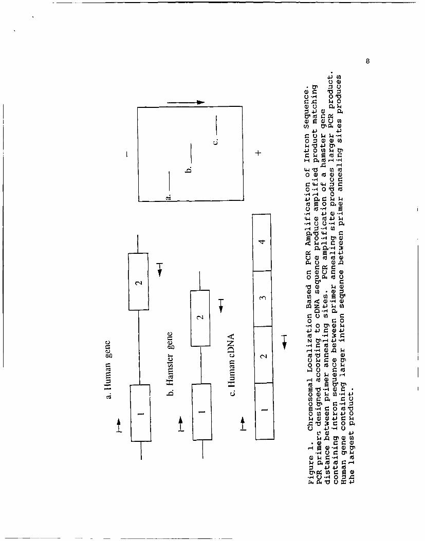
1. Chromosomal Localization based on PCRamplification of intron sequence ...................... 8

2. PCR Analysis of Somatic Cell Hybrid Panel

with Annexin V ...................................... 35

3. Southern Blot Analysis of Annexin V PCR Products .... 36

4. Discordancy Analysis for Annexins III and V ......... 37

5. PCR Analysis of Somatic Cell Hybrid Panelwith Annexin III .................................... 41

6. Southern Blot Analysis of Annexin III PCRProducts ............................................ 42

7. Chromosomal Localization of Annexin III bySouthern Blot of Human-Hamster Somatic CellHybrids ............................................. 43

8. PCR Analysis of the HA(4A) Cell Line ................ 44

9. Taq I RFLP at the Annexin V Locus .................... 59

10. Pvu II RFLP at the Annexin V Locus .................. 60

11. Bgl II RFLP at the Annexin III Locus ................ 61

iii

LIST OF TABLES

Number Page

1. Human Chromosome Content of the Human-Hamster

Hybrid Cell Lines ................................... 20

2. Annexin V PCR Primer Characteristics ................ 32

3. PCR Thermal Profiles used with Annexin V ............ 33

4. Annexin III PCR Primer Characteristics .............. 38

5. PCR Thermal Profiles used with Annexin III .......... 39

6. Annexin IV PCR Primer Characteristics ............... 45

7. PCR Thermal Profiles used with Annexin IV ........... 46

8. Restriction Map of Annexin III Gene ................. 50

9. Restriction Map of Annexin IV Gene .................. 52

10. Restriction Map of Annexin V Gene .................... 54

11. RFLP Screen Conditions for Annexins III, IV, V ...... 56

12. Annexin V RFLPs ..................................... 57

13. Annexin III RFLPs ................................... 58

iv

LIST OF ABBREVIATIONS

complementary DNA (DNA equivalent of final mRKA) ........ cDNA

deoxyribonucleic acid .................................... DNA

deoxynucleotide triphosphates .......................... dNTPs

ethylenediaminetetraacetic acid ......................... EDTA

placental anticoagulant protein .......................... PAP

polymerase chain reaction ................................ PCR

random hexanucleotide primers ......................... p(dN)6

restriction fragment length polymorphism ................ RFLP

v

ACKNOWLEDGEMENTS

Special thanks to Kathy Clayson for her support and

enthusiasm during the two years of this academic endeavor.

I would like to express particular appreciation to

Dr. Jonathan Tait for his unwavering guidance, infinite

patience, and impeccable expertise. A heartfelt thanks to

Don Gibson for preparation of oligonucleotides, Sheyla West

for DNA specimens, and the rest of the Genetics Laboratory

team for their assistance during my project. I would also

like to recognize Dr. Marie Coyle and Ms. Joyce Behrens for

serving on my thesis committee. Last but not least, my wife

Cindy deserves special thanks for her continual support

throughout this project.

vi

CHAPTER I

Introduction

Annexin Proteins

The annexins (Crumpton & Dedman, 1990) are a family of

recently identified calcium-dependent phospholipid binding

proteins with preference for anionic phospholipid (reviewed

by Klee, 1988; Crompton et al., 1988a; Burgoyne & Geisow,

1989). At the present time the annexin family contains a

minimum of eight members (Haigler et al., 1989; Hauptman

et al., 1989). Annexins I, II, III, IV, V, VII, and VIII

contain four copies of an 80-amino acid repeating unit

originally identified in lipocortin/annexin I (Wallner

et al., 1986). Annexin VI contains eight copies of the

80-amino acid repeating unit (Sudhof et al., 1988;

Crompton et al., 1988b).

Several groups have identified members of the annexin

family based on their in-vitro anticoagulant activity

(Reutelingsperger et al., 1985, 1988; Maurer-Fogy et al.,

1988; Hauptmann et al., 1989; Iwasaki et al., 1987;

Funakoshi et al., 1987a, 1987b; Tait et al., 1988, 1989).

At the University of Washington, four members of the annexin

2

family were isolated and named PAP I, II, III, and IV

(Funakoshi 't al., 1987a; Tait et al., 1988), corresponding

to annexins V, IV, III, and II. Annexins are potent

in-vitro anticoagulants and could be important in

physiologic or pharmacological control of blood coagulation.

However, the involvement of annexins in control of blood

coagulation in-vivo is so far unproven (Flaherty et al.,

1990). In addition to their anticoagulant properties, the

annexins have been identified by investigators as possible

mediators of exocytosis, regulators of inflammation,

substrates for protein tyrosine kinases, mediators of

membrane-cytoskeletal linkage, inhibitors of phospholipase

A2, and mediators of intracellular calcium signaling

(Haigler et al., 1989; Burns et al., 1989; Crompton et al.,

1988b; Kaetzel et al., 1989; Burgoyne & Geisow, 1989; Klee,

1988). Most recently, annexin III has been identified as

inositol 1,2-cyclic phosphate 2-phosphohydrolase, an enzyme

of inositol phosphate metabolism (Ross et al., 1990).

Previous Annexin Chromosomal Localization

At the start of this research project, two members of

this protein family had been localized. Annexin I was

localized to a single locus in the region 9qll-q22 (Huebner

et al., 1988). Annexin II was localized to four loci:

4q21-q31.l; 9pter-q34; lOq proximal to 10q24; and

3

15q21-15q22 (Huebner et al., 1988). During the course of

this project annexin VI has been localized to 5q32-q34

(Davies et al., 1989), and annexin V to 4q28-q32 (Modi

et al., 1989). The remaining members of the annexin family

have not yet been mapped to their chromosome of origin.

Chromosomal Localization

Determining the location of genes encoding proteins is

essential for the diagnosis of genetic disease and the

development of potential cures via gene therapy (Watson

et al., 1983). Localization of the annexins will add to the

stock of available genetic markers for general use in

genetic mapping. Chromosomal localization cL the genes for

the production of the annexins is also an integral part of

the study of their physiologic role and structure.

Somatic cell hybrids are commonly used in chromosomal

localization analysis (Ephrussi, 1972; Ringertz & Savage,

1976). Somatic cell hybrids are produced from the fusion of

human cells and cells from another species, commonly a

rodent species. Each somatic cell hybrid contains a full

complement of rodent chromosomes with the addition of one or

more human chromosomes. The complement of human chromosomes

in each hybrid cell line is normally identified by karyotype

(Harris & Watkins, 1965; Ephrussi & Weiss, 1965).

Chromosomal localization using somatic cell hybrids can

be performed utilizing DNA probes rpecific for the gene of

interest. This is usually done by Southern blot analysis.

Labelled DNA probe hybridizes to genomic DNA samples from

the hybrid cells digested with selected restriction enzymes.

The restriction enzymes are selected on the criteria that

they produce unique size human fragments significantly

different than the size of the fragments produced with the

rodent genomic DNA. The identification of the

characteristic human restriction fragment pattern in a panel

of hybrid cell lines, each containing one or more human

chromosomes, enables the investigator to determine the

chromosome(s) most likely to contain the gene of origin.

Another major method of chromosomal localization is

in-situ hybridization. This technique utilizes the

hybridization of labelled probe to metaphase chromosomal

spreads of human cells (Cannizzaro & Emanuel, 1984; Chandler

& Yunis, 1978; Gerhard et al., 1981). By examining the

hybridization pattern, investigators are able to determine

the chromosome(s) containing the gene(s) of interest and

often their location within the chromosome.

Very recently, the polymerase chain reaction (PCR) has

also been utilized in the performance of chromosomal

localization using hybrid cell lines (Iggo et al., 1989,

Abbott et al., 1989; Dionne et al., 1990). PCR is a

technique which allows the exponential amplification of a

specific nucleic acid sequence. PCR amplification utilizes

two oligonucleotide primers that span the DNA sequence to be

amplified. With repeated cycles of DNA denaturation,

annealing of primers to their complementary sequences, and

5

extension of the annealed primers with DNA polymerase, an

exponential increase in the target sequence is realized

(Saiki et al., 1985; Mullis & Faloona, 1987; Ehrlich, 1989).

Chromosomal localization studies using PCR and cell hybrids

utilize primers designed to be complementary to coding or

non-coding regions in or around the DNA sequence of

interest. Hybrid cell lines producing the characteristic

human PCR product contain the chromosome(s) containing the

gene(s) for the protein of interest. Chromosomal

localization by this technique can be performed by the

amplification of coding (exon) sequence (Dionne et al.,

1989). Somatic cell lines positive for the PCR product can

be inferred to contain the chromosome(s) of origin.

An alternative to chromosomal localization via

amplification of exon sequence is localization based on the

amplification of intron sequence (Iggo et al., 1989; Abbott

et al., 1989). PCR primers are designed complementary to

regions of the cDNA sequence that span intron sequence.

Since intron sequence is not translated into the final

protein, it is generally less highly conserved than exon

sequence through evolution. Primers amplifying PCR products

larger than the corresponding cDNA product are assumed to

span intron sequence (Figure 1). This difference in the

size of the amplified products between species is the basis

of chromosomal localization via amplification of intron

sequence (Iggo et al., 1989).

6

Restriction Fragment Length Polymorphisms (RFLPs)

RFLPs are a result of variation in the DNA sequence

among individuals. RFLPs are detected via Southern blot

analysis of human genomic DNA cleaved with restriction

enzymes and hybridized to DNA probes specific for the gene

product of interest (Kan & Dozy 1978; Donis-Keller et al.,

1986; Weatherall, 1985).

The identification of RFLPs serve an important function

in the process of mapping the entire human genome. It is

possible to arrange these RFLPs, representing specific loci,

by use of mathematical analysis and their physical location

into a human genomic map (Drayna, 1986; Donis-Keller et al.,

1987; Nakamura et al., 1987).

RFLPs closely associated with or linked to an abnormal

inheritable phenotype (DNA marker loci) are very useful

provided they can be followed through families afflicted by

the disease. The use of linkage analysis allows the mapping

of the gene(s) responsible for the disease with respect to

the DNA marker loci, without necessity for exact

characterization of the gene responsible (Botstein et al.,

1980). For example, the genes for Huntington's disease and

cystic fibrosis have been localized by this type of linkage

analysis (Gusella et al., 1983; Tsui et al., 1985; White

et al., 1985; Wainwright et al., 1985).

7

Thesis Objectives

The first objective of this research project was the

chromosomal localization of the genes for annexins III, IV,

and V (PAP III, II, I). The second objective was the

detection of possible RFLPs at the gene loci for these three

annexins. RFLPs discovered will be useful for general

genomic mapping and identification of genes causing disease.

In addition, discovery of a disease linked to an annexin

gene would be an important finding possibly clarifying the

physiologic role of the annexins.

40

04

04 040 0 02 4Sw~4J0 tO 4t

440 M 00 2

C44) 4 JC0 ~-4 0. -4

-,- r- -4

04 0

o 0 U- 0.r4 Z 0rq(

_____ _____ ___WO 44J 9 1i

1-4 4

-4 u ) 0

00 0 H

-4 0 4 3r

0 0 0 u

0 0) W fl -14-

~0 tp r_--0(J w ) w

r4 0 0

-4 ) ti

-r-r4 $4p 40

--1* 0 Q 1-i

CHAPTER II

Methods and Materials

Materials

Taq DNA polymerase (AmpliTaq, Perkin-Elmer Cetus,Emeryville, CA).

Proteinase K (Sigma Chemical Company, St. Louis, MO).

Ultrapure dNTP set (Pharmacia Technology Inc., Piscataway,NJ).

Mouse genomic DNA, prepared from male strain C57BL/6 (Giftfrom Dr. David Adler).

Human-hamster somatic cell hybrid DNA (Bios Corporation,New Haven, CT).

SeaKem GTG, ultra pure agarose (FMC BioProducts, Rockland,ME).

[alpha-32p]-dCTP, specific activity 3,000 Ci/mmol (product# NEG-013H, New England Nuclear Research Products, DuPont,Wilmington DE.)

Random hexanucleotide primers, p(dN)6 (PharmaciaBiotechnology Inc., Piscataway, NJ).

Klenow fragment of DNA Polymerase I (product # 104531,Boehringer Mannheim, Indianapolis, IN).

Restriction endonucleases (Boehringer Mannheim,Indianapolis, IN).

Sephadex G-50 column (Nick Column DNA grade, PharmaciaTechnology Inc., Piscataway, NJ).

Gene Screen Plus nylon transfer membranes (DuPontBiotechnology Systems, Boston, MA).

10X-ray film XAR-2 (Eastman Kodak, Rochester, NY).

DuPont Cronex Lightning Plus GL intensifying screens (DuPontBiotechnology Systems, Boston, MA).

Mineral oil (Sigma Chemical Company, St. Louis, MO).

Methods

PCR Primers

Oligonucleotides were synthesized using an Applied

Biosystems Model 381A DNA Synthesizer utilizing

phosphoramidite chemistry and reagents obtained from the

manufacturer. The stepwise yield was usually > 99.0% and

the oligonucleotides were used without further purification.

PCR Reactions

PCR reactions were performed as described by Mullis &

Faloona (1987) and Saiki et al. (1988). The reactions were

prepared in sterile 0.6 mL microcentrifuge tubes with

dedicated pipettes. Each 50 uL reaction contained

100-500 ng of genomic DNA, 1.0 U Taq polymerase, 200 uM of

each dNTP, 10 pmol of each primer, 10 mM Tris-HCl pH 8.3,

50 mM KCl, 0.1 g/L gelatin. Reactions contained 1.5 mM

MgCI2 except where noted otherwise. Each reaction was

overlayed with 40 uL of mineral oil. Amplification was

performed on the DNA Thermal Cycler (Perkin-Elmer) using

step cycle profile methodology. Each PCR reaction included

a 940C x 6 min denaturation step preceding amplification and

a 72°C x 10 min extension period following the

amplification.

1i

Electrophoresis of PCR Products

Amplified PCR product (15 uL) was electrophoresed for

70 V x 4 h on a (11 cm X 14 cm X 8 mm) 1.4% agarose gel in

0.089 M Tris base, 0.089 M boric acid, and 0.002 M EDTA,

pH 8.0. Molecular weight markers consisted of a Hind III

digest of phage lambda DNA and phi-X174 DNA digested with

Hae III. Agarose gels were stained with 1.2 ug/mL ethidium

bromide in H2 0 for 10 min, destained in H20 for 20 min,

and photographed under 302 nm transillumination.

DNA Preparation

Human genomic DNA was isolated from nuclei of white

cells following the method described by Kunkel et al. (1977)

and modified by Bell et al. (1981). Red blood cells were

lysed by mixing 3 to 5 mL of EDTA anticoagulated whole blood

with an equal volume of 2X lysis buffer containing 0.6 M

sucrose, 20 mM Tris base pH 7.0, 10 mM MgCI2 , and 3% Triton

X-100. An equal volume of 1X lysis buffer was added; the

tube was then mixed and placed on ice for 10 minutes. The

mixture was inverted several times during the incubation on

ice, centrifuged, and the supernatant was removed leaving

the white blood cell nuclei in pellet form.

Proteinase K digestion was performed by the addition of

3.5 mL of freshly prepared proteinase K lysis buffer

containing 0.01 M NaCI, 0.01 M Tris-HCl pH 7.6, 0.01 M EDTA,

0.5% sodium dodecyl sulfate, and proteinase K at a minimum

concentration of 200 ug/mL. The reaction mixture was

12

incubated at 370C overnight or until all visible clumps of

white blood cells were eliminated.

DNA extraction was performed with the addition of

2.0 mL of phenol saturated with 10 mM Tris-EDTA, pH 7.6, and

2.0 mL of chloroform. This procedure was repeated twice on

the aqueous layer. The last extraction was performed with

2 mL of chloroform.

Precipitation of the DNA from the aqueous layer

proceeded with the addition of 4.0 M NaCI to a concentration

of 0.4 M NaCl. The DNA was precipitated with the addition

of 2 to 2.5 volumes of 100% ethanol. The mixture was placed

at -200C overnight, centrifuged, and the ethanol removed.

The saved precipitate was dried under vacuum and resuspended

in 200 uL of buffer containing 10 mM Tris-HCl pH 7.6, 10 mM

NaCl, and 1 mM Na2-EDTA. The DNA concentration was

determined spectrophotometrically at 260 nm using a 1 cm

light path and an extinction coefficient of 0.02 mL/ug.

Purified genomic DNA samples from a panel of 25 human-

hamster hybrid cell lines were obtained from the Bios

Corporation. The cell lines were prepared by fusing normal

human B lymphocytes from unrelated donors with Chinese

hamster ovary-Kl cells. The cell lines were selected

according to Carlock et al. (1986). The chromosome content

of each cell line was determined by the supplier based on

cytogenetic evaluation of 20 metaphase spreads (Table 1).

13

The HA(4A) cell line, a mouse-human hybrid containing only

human chromosome 4, was obtained from Dr. Anne Killary

(Baylor University).

cDNA Probes

The cDNA probe for annexin V was the plasmid pPAPI-I.6,

which contains a 1459-bp insert in the EcoRI site of pUCI8

(Funakoshi et al., 1987b). This cDNA contains the complete

960-bp protein coding region as well as 12 bp of 5' non-

coding sequence and 487 bp of 3' non-coding sequence. The

cDNA sequence was confirmed by restriction mapping with

Eco RI, Hind III, and Pst I.

The cDNA probe for annexin IV was the plasmid

pPAPII-B6, which contains a 1915-bp insert in the EcoRI site

of pUCl8 (Miao, C. H., unpublished). This cDNA contains the

complete 963-bp protein coding region plus 12 bp of 5' non-

coding sequence and 940 bp of 3' non-coding sequence. The

sequence is identical to bases 74 to 1036 of the human PP4-X

cDNA (Grundman et al., 1988) with the exception of a G in

place of the A seen at position 366 in PP4-X (Grundman

et al., 1988). The cDNA sequence was confirmed by

restriction mapping with Dde I, Eco RI, Hind III, Pst I, and

Pvu II.

The cDNA probe for annexin III was the plasmid

pPAPIII-28A, which contains a 1268-bp insert in the Eco RI

site of pUCl8 (Miao, C.H., unpublished). This cDNA contains

the complete 969-bp protein coding region and 36 bp of 5'

14

non-coding sequence and 263 bp of 3' non-coding sequence.

The sequence of this clone is identical to bases 11 to 1278

of the human lipocortin III cDNA (Pepinsky et al, 1988) with

the exception of a T in place of the C seen at position 1057

in lipocortin III (Pepinsky et al., 1988). The cDNA

sequence was confirmed by restriction mapping with

Eco RI, Hinc II, Hinf I, Pst I, Pvu II, and Taq I.

Preparation of cDNA Inserts

The cDNA insert was removed from the polylinker region

of pUCI8 at the Eco RI cloning sites. 50 ug of plasmid was

digested for two hours at 370 C with 4 U of Eco RI per ug of

DNA in a total volume of 500 uL. The restriction buffer

contained 50 mM Tris-HCl pH 7.5, 10 mM MgCI 2 , 100 mM NaCI,

1 mM dithioerythritol. The restriction digest was loaded

with 0.2 volume Type II loading dye containing 25% Ficoll,

0.25% bromophenol blue, and 0.25% xylene cyanol. The sample

was run into an (11 cm X 14 cm X 8 mm) agarose gel

(SeaKem GTG) at 100 volts for 25 minutes. Electrophoresis

was performed using optimal conditions for identification of

the band of interest: annexin III, 1% gel, 22 V for 16 h;

annexin IV, 0.9% gel, 25 V for 18 h; annexin V, 0.9% gel,

25 V for 18 h, in 0.089 M Tris base, U.U89 M boric acid, and

0.002 M Na2-EDTA, pH 8.0. The gels were stained with

ethidium bromide as described above. The band of interest

was excised from the gels. The DNA was electro-eluted from

the agarose fragments using Model 422 Electro-Eluter (Bio-

15

Rad Laboratories). Current for elution of the three annexin

cDNA fragments was constant at 10 mA per elution tube.

Annexin III cDNA was eluted for 45 min, annexin IV for

58 min, and annexin V for 50 min. The DNA elution buffer

contained 40 mM Tris base, 20 mM glacial acetic acid, and

1 mM 0.5 M Na2-EDTA, pH 8.0.

The electroeluted DNA was extracted by the addition of

1 volume phenol saturated with 10 mM Tris-EDTA, pH 7.6,

followed by extraction with 0.5 volume phenol plus 0.5

volume chloroform. The last extraction was performed with

1 volume of chloroform. Each extract was micro-centrifuged

for 1 minute and the top layer removed. The DNA was

purified by ethanol precipitation by the addition of

0.1 volume 3 M sodium acetate pH 5.2, and mixed by

inversion; 2 volumes of 100% ethanol were then added, mixed,

and centrifuged for 10 minutes. The precipitate was washed

with 1000 uL of 70% ethanol. After a micro-centrifuge spin

for 10 minutes, the saved precipitate was dried under vacuum

and resuspended in 100 uL 10 mM Tris-HCl pH 8.0 and 1 mM

Na2-EDTA. The DNA concentration was determined

spectrophotometrically at 260 nm using an extinction

coefficient of 0.02 mL/ug and a 1 cm light path.

cDNA Probe Labelling

The cDNA fragments were then radioactively labelled

with [alpha 32p]-dCTP using random hexanucleotide primers and

Klenow fragment according to Feinberg and Vogelstein (1983).

16

The reaction mixture contained 50 ng of DNA probe, 2.5 uL of

primers (A260 = 50), and H20 to bring the final reaction

volume to 25 uL after addition of all reagents. The

reaction was heated in a boiling H20 bath for 3 minutes,

quickly centrifuged, and briefly placed on ice. The

remaining reagents were then added: 2.5 uL of a solution

containing dATP, dTTP, and dGTP each at a concentration of

0.5 mM; 2.5 uL of buffer containing 500 mM Tris-HCl pH 7.5,

100 mM MgCI 2 , 50 mM dithiothreitol, and 500 ug/mL of

nuclease-free BSA; 5 ul (50 uCi) or 10 ul (100 uCi) of

[alpha32p]-dCTP (specific activity of 3,000 Ci/mmol); and

1 uL of 5 U/uL Klenow fragment. This mixture was briefly

centrifuged and incubated at room temperature for 2 hours.

1 uL of 0.5 M Na2 -EDTA was added to stop the reaction.

Unincorporated [alpha32p]-dCTP was removed from the labelled

probe by gel filtration using a Sephadex G-50 column

measuring 2.5 cm x 1.2 cm. The probe was added to a column

pre-equilibrated with 3,000 uL of buffer and eluted with

800 uL of buffer. The radioactivity of a 5 uL aliquot of

the collected eluate was then quantitated on a radioisotope

counter and the specific activity calculated in dpm/ug.

The majority of labelling reactions produced specific

activities in excess of 1 x 10⁹ dpm/ug.

Southern Blot Technique 17

DNA was transferred from agarose gels to nylon

membranes using the alkaline-transfer method of Reed and

Mann (1985). Electrophoretic gels were depurinated with

gentle agitation for 15 to 30 min in 0.25 N HCI. DNA

denaturation was accomplished with gentle agitation during a

90 min wash in 0.4 M NaOH. A nylon membrane was placed on

top of each inverted gel followed by the addition of 10

pieces of Whatman 3 mm chromatography paper, the first four

saturated in 0.4 M NaOH. Approximately 5 cm of paper towels

were stacked on top with the addition of a glass plate and a

225-250 g weight. After blotting for a minimum of 3.5 hr

the DNA was fixed to the nylon membrane by washing for 3 min

in 0.4 M NaOH and 3 min in a solution of 0.03 M sodium

citrate and 0.3 M sodium chloride.

The membranes were pre-hybridized at 650 C for 30 min in

hybridization solution containing 1% sodium dodecyl sulfate,

0.05 g/mL dextran sulfate, 0.5% non-fat powdered milk, 0.9 M

NaCl, 0.005 M Na2-EDTA, 0.1 M Tris-HCl, and 0.1 M Tris base

adjusted to pH 8.0. Membranes were hybridized 18 hr in

hybridization solution containing 3 X 10⁶ dpm/mL of probe.

Following hybridization the membranes were washed down to

300-1000 cpm on a mini monitor in a solution containing

1.5 mM sodium citrate, 15.0 mM sodium chloride, and 1%

sodium dodecyl sulfate at 600 C.

18

X-ray film was subsequently exposed to the radio-

labelled membranes at -700 C, with or without intensifying

screens, for optimal exposure.

Stripping of the nylon membranes was performed to allow

investigation with mulitple cDNA probes. Membranes were

washed in 200 ml of 0.4 N NaOH at 420C for 30 minutes with

gentle agitation. Membranes were removed and immediately

washed at 420C for 30 minutes with gentle agitation in a

1.5 mM sodium citrate, 15.0 mM sodium chloride, 0.1% sodium

dodecyl sulfate, and 0.2 M Tris-HCL pH 7.6. Membranes were

then ready for rehybridization.

Genomic DNA Restriction Enzyme Digestion for Chromosomal

Localization Studies

Human genomic DNA (9 ug), hamster genomic DNA (9 ug),

and human-hamster hybrid cell lines (4 to 9 ug), were

digested with Hind III (9 U per ug of DNA) for two hours at

370C. Digested DNA samples were then electrophoresed at

25 V x 18 h on (11 cm x 14 cm x 8 mm) 0.6% agarose gel using

the same buffer and molecular weight markers listed in the

section "electrophoresis of PCR products". Gels were then

stained with ethidium bromide and DNA transferred to nylon

membranes via the Southern blot procedure listed in the

previous section.

Genomic DNA Restriction Enzyme Digestion for RFLP Studies

Depending upon the experimental conditions, 5 to 15 ug

of human genomic DNA was digested with 4 to 10 U of enzyme

19

per ug of DNA for 2 h in the buffer supplied by the

manufacturer. The enzymes used in the RFLP screen were

selected because they were likely to reveal RFLPs based on

the experience of previous investigators. All digestions

were incubated at 370 C with the sole exception of digestion

with Taq I at 650 C. Following digestion samples were

electrophoresed under desired conditions, stained with

ethidium bromide as outlined above, and inspected for

satisfactory digestion. Southern blots were then prepared

of electrophoretic gels and hybridized with labelled cDNA

probe specific for each annexin.

20

-h +1 ~

co

r-4 1

CL U

a + +

z1 44)

z O. +

R 44 441

F00

41 04 + F0

0 + ++q b

0

-. at0 , + +j

0) c00CD at Ae be)

0 10 +

01 0. .1.

44 a~.

.44

, F l 11 . . 4 $oioiii! ;

CHAPTER III

RESULTS

General Approach to Chromosomal Localization by PCR Analysisof Cell Hybrids

This localization procedure can be divided into five

basic steps: PCR primers were designed based on the known

cDNA structure of each human annexin. Since the annexin

gene structures are unknown, primers were designed to span

entire repeating units or portions of adjacent repeating

units. This was done in an attempt to produce primer pairs

that span human annexin gene intron-exon boundaries and

produce distinctive human-sized products in the presence of

homologous hamster genes. These larger amplified products

would provide initial evidence for amplification of intron

containing regions of the annexin genes.

PCR conditions were optimized for each primer set and

multiple human DNA specimens were tested to ensure uniform

production of amplified products.

PCR reactions were then performed with hamster genomic

DNA to ensure that the size of any amplified product(s) were

significantly different than the size of the human

product(s).

22

To verify that amplified products were of annexin gene

origin and rule out non-specific PCR amplification, high

stringency probing of Southern blots of PCR product was

performed with the appropriate labelled annexin cDNA.

PCR analysis of all 25 human-hamster hybrid cell lines

was then performed. This amplification was followed by

Southern blot verification of all cell lines producing the

human sized PCR products with labelled cDNA probe.

Chromosomal Localization of Annexin V

Four primer sets were tested for annexin V (Table 2).

PCR reactions were performed as described in Chapter II with

a multitude of thermal profiles (Table 3). Primers JT102

and JT104 amplified multiple products visible on ethidium

bromide stained gels with a major 1.5 kb band. This 1.5 kb

product was larger than the cDNA product of 232 bp. The

length of the cDNA products was determined according to the

distance between the primer annealing sites along the cDNA.

PCR reactions were then performed with the same conditions

using multiple human genomic DNA samples and hamster genomic

DNA. All human samples showed the same results and the

hamster DNA produced multiple amplified products, all less

than 1.5 kb. Longer primers were then designed to improve

the specificity of primer annealing to human DNA templates.

An additional 5 and 7 bp were added to the 3' end of primers

JT102 and JT104 to produce primers JT121 and JT122. PCR

conditions were then optimized using these new primers,

23

JT121 and JTl22, and 0.5 ug of template DNA. These

conditions produced a single human genomic product of 1.5 kb

and multiple hamster bands less than 1.5 kb.

Chromosomal localization was then attempted using the

25 human-hamster somatic cell hybrid lines. Results showed

one band of 1.5 kb amplified with human genomic DNA, a

single 1.5 kb product amplified strongly in somatic cell

lines 803 and 1006, and weakly in somatic cell line 867

(Figure 2). Several distinct products were amplified in

hamster DNA and the human-hamster somatic cell lines,

indicating amplification of hamster genes.

Southern blot verification of the amplified products

was performed using 2 uL of product from human genomic DNA,

hamster genomic DNA, cell lines containing the 1.5 kb band

(803, 1006, 867), and a sampling of the cell lines lacking

the 1.5 kb band (324, 811, 937). Annexin V cDNA (0.38 ng)

and annexin III cDNA (0.38 ng) were added to the gel as

positive and negative controls. Annexin V cDNA hybridized

to the 1.5 kb band seen on the ethidium bromide stained gels

for human genomic DNA and cell lines 803, 867, and 1006.

The hamster DNA and other cell lines did not show

hybridization with this probe in this region. Adequate

stringency of the wash was verified by the lack of cross-

hybridization with the related annexin III cDNA (Figure 3).

Analysis of the human chromosome complement in each

somatic ccll line indicates that the gene for annexin V is

located on chromosome 4. Results show 4% discordance for

24

chromosome 4 and at least 15% discordance for all other

chromosomes (Figure 4). This 4% discordancy with chromosome

4 comes from cell line 867 which produced weak amplification

of the 1.5 kb product but was negative for chromosome 4 by

cytogenetic analysis. The same discordancy has been

observed with this cell line for another gene localized to

chromosome 4 (Tabas & Zasloff, personal communication with

Dr. J. Tait). The most probable explanation is that cell

line 867 contains either a translocated portion of

chromosome 4 or a small percentage of cells containing

chromosome 4 which were not detected by cytogenetic

analysis.

Chromosomal localization of annexin V using Southern

blot analysis of Hind III digests of the somatic cell

hybrids did not prove as successful. No discernible human

specific bands were detectable in any of the cell lines and

therefore localization by this technique was impossible.

Localization of Annexin III

Two primer pairs were designed for annexin III

(Table 4). PCR reactions were performed with a multitude of

thermal profiles (Table 5). After optimizing PCR

conditions, primers JT114 and JTll5 produced a 480 bp band

from human genomic DNA larger than the corresponding cDNA

product of 230 bp. However, this PCR product failed to

hybridize with annexin III cDNA, indicating it was not

synthesized from the annexin III gene.

25

Primers JT130 and JT131 were then designed and

synthesized. PCR reactions produced a 2.0 kb band with

human genomic DNA and no visible bands with hamster genomic

DNA, presumably because the primer sequences did not match

the hamster gene closely enough to allow amplification.

Chromosomal localization of annexin III was performed using

these primers at optimized PCR conditions. The results

showed the presence of the 2.0 kb product with human genomic

DNA and cell lines 803 and 1006 (Figure 5). Hamster genomic

DNA and the other somatic cell lines did not show any bands.

High-stringency probing of a Southern blot to verify

the origin of the PCR products was performed with cell lines

containing the 2.0 kb band (803, 1006), cell lines lacking

the band (324, 811, 937), and the cDNA positive and negative

controls. Annexin III cDNA hybridized to the 2.0 kb band

seen on the ethidium bromide stained gels for human genomic

DNA, cell line 803, and 1006. Hamster genomic DNA and the

other cell lines did not show hybridization with this probe

in this region. Adequate stringency of the final wash was

verified by the lack of cross-hybridization with the related

annexin V probe (Figure 6).

Chromosomal localization of annexin III using Southern

blot analysis of Hind III digests of the somatic cell

hybrids confirmed results obtained with the PCR methodology.

Human-specific restriction fragments were seen of 9.4, 6.0,

5.6, 5.0, 4.2, 3.9, and 3.3 kb. These same human-specific

bands were seen in cell lines 803 and 1006. They were

26

absent in hamster genomic DNA and all other cell lines

(Figure 7).

Analysis of the human chromosome complement in each

somatic cell line indicates that the gene for annexin III

is located on chromosome 4. Results for annexin III show

0% discordance for chromosome 4 and at least 15% discordance

for all other chromosomes (Figure 4).

Additional Verification of Chromosomal Localization for

Annexins III and V

Verification of chromosome 4 as the location of the

genes coding for annexin III and V was performed utilizing

a mouse-human hybrid cell line containing chromosome 4 as

its only human chromosome (Figure 8). PCR reactions were

performed for both annexins as outlined earlier. An

ethidium bromide stained gel of PCR products amplified with

this cell line shows the characteristic 2.0 kb human-

specific band produced in annexin III PCR reactions, the

characteristic 1.5 kb human-specific band produced in

annexin V PCR reactions, and the amplification of a 0.9 kb

product in the mouse genomic specimen and the mouse-human

cell line with annexin V PCR reactions. This 0.9 kb product

is most likely produced from the mouse annexin V gene.

These results, using a different rodent species and

alternate source of hybrid DNA verify earlier results

obtained with the human-hamster somatic cell hybrids.

Chromosomal Localization of Annexin IV 27

Chromosomal localization of annexin IV via PCR

amplification of intronic sequences was initiated with the

design and synthesis of multiple primer pairs (Table 6).

Two primer pairs produced amplified products with a

denaturing temperature of 940C and 30 cycles (Table 7).

Initial PCR reactions with primers JT124 and JT140 generated

products of 190 bp and 560 bp. High-stringency probing of a

Southern blot containing the products with annexin IV cDNA

was performed with no hybridization to either product,

indicating that these two products were not of annexin gene

origin. Primers JT128 and JT129 produced very faint,

multiple bands less than 1.4 kb. Due to the small amount of

these products produced, as visualized on ethidium bromide

stained gels, these primers were not further tested. PCR

reactions were then performed using all designed primer

pairs with a denaturing temperature of 960C and 40 cycles.

A few small bands were produced with multiple primer pairs,

while primers JT124 and JT140 produced a 3.5 kb product

(Table 7). High-stringency probing of a Southern blot

containing the products with annexin IV cDNA was performed

with positive hybridization to the 3.5 kb band. Repeating

the above PCR reaction after adjusting the denaturing

temperature to 940C produced the same 3.5 kb product.

PCR was then performed with primers JT124 and JT140

with the 25 human-hamster somatic cell hybrid cell lines

using a thermal cycle of denaturing 940C x 1 min, annealing

2855C x 1 min, and extension 72

0C x 3 min. Results showed

the absence of the 3.5 kb product in any of the cell lines

and multiple hamster bands all significantly smaller than

3.5 kb. The absence of the band in the somatic cell hybrids

could be due to the absence of the gene, mutation of the

gene preventing correct primer annealing or amplification,

or inhibitors present in sufficient concentrations in the

DNA to inhibit the activity of Taq polymerase. A Mg2+

titration was performed repeating the PCR reaction with

human and hamster genomic DNA with Mg2+ concentrations of

1.5 mM, used in all previous work, and 2.0, 2.5, 3.0 and

4.0 mM. The 3.5 kb human band and all hamster bands were

greatly enhanced with 2.0 mM and more concentrated MgCI 2.

Chromosomal localization was again attempted using

2.0 mM MgCI 2 . Ethidium bromide stained gels indicated the

presence of a weak 3.5 kb product in cell line 854. High-

stringency probing of Southern blots of these PCR products

from all 25 cell lines was performed with appropriately

labelled annexin IV cDNA. Cell line 854 showed two

positive, closely spaced bands of 3.5 and 3.3 kb. Cell line

983 contained a positive band at 3.5 kb and cell line 904

displayed a weak band at 3.5 kb. Percent discordancy for

each of the chromosomes ranged from 8% to 76%. Chromsome 2

was 8% discordant, followed by chromosome 10 at 16%. No

chromosome of origin for the annexin IV gene can be

definitely assigned based on this set of PCR experiments.

29

Chromosomal localization of annexin IV using Southern

blot analysis of Hind III digests of the somatic cell

hybrids proved unsuccessful in determining the chromosome of

origin. Hind III was selected on the basis of producing,

when probed with annexin IV cDNA, strong human-specific

bands easily distinquished from the hamster pattern. Human-

specific bands were seen of 10.5, 2.9, 2.7, 2.2, 2.0 and

1.7 kb. These bands were only visible in human genomic DNA.

They were not present in hamster genomic DNA or any of the

hybrid cell lines. Two bands, 6.2 and 4.9 kb, were shared

between human DNA, hamster DNA, and the hybrid cell lines.

Since no human-specific bands were seen in any of the hybrid

cell lines, annexin IV was not localized by this method.

However, the absence of the signal suggests that the annexin

IV gene is not on a chromosome that is well represented in

the hybrid cell panel.

Restriction Mapping and RFLP Analysis of the Genes for

Annexin III, IV, & V

Southern blot analysis of human genomic DNA was

performed in order to detect possible RFLPs and to establish

the sizes and restriction maps of the annexin genes. In

each case the genes are of moderate size, approximately 20

to 50 kb (Tables 8, 9, & 10).

Human genomic DNA from approximately 10 unrelated

Caucasians was screened for the presence of RFLPs with 9

restriction enzymes commonly used in the genetics

30

laboratory. A complete listing of enzymes used and

restriction fragments produced with each annexin cDNA

probe is found in Table 8 (annexin III), Table 9

(annexin IV), and Table 10 (annexin V). A synopsis of the

conditions used in the RFLP screen is found in Table 11.

Screening with the cDNA for annexin V, RFLP systems were

detected using Taq I and Pvu II (Table 12). Review of the

annexin V, Eco RI restriction fragment pattern, shows a

probable additional RFLP. This RFLP was recently identified

and brought to our attention by another investigator

(Murray, J., personal communication with Dr. J. Tait). RFLP

systems were detected with annexin III using Bgl II and

Hind III (Table 13). No RFLP systems were detected with

annexin IV.

Inheritance patterns for three of the four RFLPs were

investigated by family studies. In each case, the RFLP was

inherited as a Mendelian trait. The annexin III-Hind III

RFLP was not extensively investigated due to the extremely

low frequency of the low molecular weight allele producing

the 0.9 kb fragment. A Southern blot of a representative

family, from the four families studied (12 meioses), is

included for the annexin V-Taq I RFLP (Figure 9). Five

families (24 meioses) were examined and a Southern blot is

included of the annexin V-Pvu II RFLP (Figure 10). A

Southern blot of four random individuals illustrates the

three possible allele combinations, seen in the three

31

families evaluated (14 meiosis), of the annexin III-Bgl II

RFLP (Figure 11).

32

oC4 z

0 H>4 Q a~ 4- >4

~ . ~ .~ t U ' a) 44

to~ ON LA %0 0 0 to %Da) a.4 r-4 - LA r, LA N N, N

LA U) a% N- r LA N ON N,(D 4 1 1 1 1 1 1 1 1 LO LA

0 0 %o m LA m~ 0 LA LA .4J)C) LA O'~N N r LA N O'~N r, ia)

o W MWLO

U E-4 U)I n 0 w 10

u 4I

> ~ U (u En U a

S E4 E4 E-4 ~E-4 0. U 4 (0Hcl E-4 U E-4 U e4 e4 P U H Ha

0 E-4 U 0~E U U 0. 4.v r4J4 I ~ p ~ E- u p E-4 r_*4 c il

LA n U E-4 U U H'- E-1 p Eu UM. - ~ U12 I

a)V U0 '0C. E- 4 0u E-1 Z 0 0

4 V. 4 p 3 #0 U E-' U H- H-- 3 * U E-4 E-o CD U i) u w

E-4 a E-4 U U '4 VD'0a) E-E~j4 aD C E-4 E4 U

C) E-4 U U E-4 P4 U E-4 -44 H 4 $4 H

84 UC 0* 0 UE4lD00

'4 E-4 p4 4 4. a

"~~0 4-4 Q- D ec D a 0) Q)adCD U CD U ~ to M- Ua~ ))

w .14 Ud) -4

r-4 N m 10 0 rq I4n 4JI .4 4JI0 0 0 H H- -r-4 H -H - - - -H N 0) m~aH H H- H P P P P P0I

Z -3 j l 4

~33

0 o 0 0 0 0 0 LO A 0

4.)

H 0 0 0 0 0 0 0 t

5.4 >4 rEn 0~41)

0 403

L) U.-d U". -I U.H U 0-q U .,4 U"d -Ix 0 0 0 00ra0IEr 0 0 0 0 w4

H1 0-44

04I0 .4.U U U U .

urn 0r u u u u C.) 41 S 0r- 0 0 0 0 0 0 0 0

It LO 0 L0Ln0 L n 0 L Ln 0 Lnc 0 c~ 003 1

444.

E-4~ S.r0

031C a C

CJ~ -4 U* r.~ U* -4 U. -4~ C03 0 0 0 R 0-4

Hw v~ O le O qH OH T'i OH -wH v~ rqa% -4 f- ar4 i r4 I r- af-I a% -4 % C4 04

In 1-1 tl4t1I tC04

r-4 v-4 (NI N CVf0lO 0 0 0 0 0 0 Hr-4 .

H H- H- r- H- P- H- H- - 41~w E-i E-0 -4 E- E-4 E-4 E4 E-

34

LA

010 0 0 0 LO LO LO

.- 44.1

0

4.11-4-4ot

$4, r. ,,v u l.

J ,, 00

(a CI WO 0l

to 4 . -4

Q) > 04

44 0 0 0 0 00 0 0 0 0 0) Wrg

o o> 020 J J ~ 4

N094.1 00

F--4~ r-4 Fa -4

44 oo oD .o o4J., ,- 0 u o oo u o l1 u

4) -4 Q.9w4) x0 Q4 04t uwU w4

o 0 0 oR 0 0o 0 0

on cn ,n m ,o r- ,W m, Lnm Lnm (0

44 W WO

0-)F- 0H ROH OHO O H

,-A E- 4) r

4100

H V0 V

.94-4

r-4 C0 m N 4) W0004 002 0-4 002 w0 w0

0 0c Lac~ O- Lac ~ 000c 0

0) 0U 024

a. 0f-4 OHI v-4 OH NH N0- r- - - - -4 4J41UEO-4 E-4 EI ~4O-4 u-E-4.-U

35

-4~ 4 4

-4 0 C 4JV0 a)-.-I C

4- U 4-)

660t 00 M J-)Z

900L XQ

Z9600 qt'06 0) --4 0.

P-99 0 -4

606 r).0 UQ

C86 1V 13 0 4 (

~~e9L 00~- 0)

r-4 Ln~~.~lZOg '-4 U 0~0) 0 -1 .44 0

19g 4-4 94 0

Crq ti) U 4 00 JV000 4JOsqq 44 U Q (A4- --

Coo U0a4) 10'4 .0 -44.4 0

.4CJ r- 44t~ 0

'- -4--4 .- 4 Ca J

0i V4r -4

UeW%, V 0) w it '-4 0r.~ .0 E- ~ 4 04J U

4 >4 0)a

0)4 0) .4 V. )*-0 1.4 ' 4 J r*-4 0) 44

r~ 4) - V Ca4 090 Q)4)

0-r- 4 r4 r- C. 0 04J rO 0 . 0 U

36

-4 Q)

4J- W r

OWW

• 0 wow M

A uuvA591V ZOmU4

.ff ~44 0 a

N I wZco U4.

Lie Z XLi

U VH U

0

-O H 4J.C

0 -4N9001 C

o .'0- Z.

o 44 -

0 H 0

UwnH 4n

r-I N eC 0

I 4.1 Q

4 A 1 4 X >-4 -

C0 M w -o 4) 0Li.r-4 -

, 0O

r-4 0 CQ) in u

37

4Ja 4) 4(

cn: 0

0H .

N N

a 00 ( 0r0

aN-4r-

> r-4 H('0

'- 0 0OOH 4 (4-4

> ao> ) >4 >4r4 r. U

0 00

0 U

U) 0 4J- -4 H

te) >1 ) 9: x0 )~

4 ~ d

~0 m0 0

o~ R 0 0U00 U. C 4

C4 0OH RIUPPIOOSIQI lu;)OJd 4-4H m~

Q) H U)

W JQw iQ) t N.> -W0 zr-

-'4 0 > c

rA ~ .rk H .

0 LO '- Q

IA c o o- U') r-41 0 0 I A I I0

'0 a ~ *o C -u O co m' ON

r- ONi LA %D cc~

0I

E- CD E-4(&4 4 & .441

U E4 C E-4 0 tC

4 E E4 Erl 4 41

0 D En H~ ~ OA

CD CD U 4.) 0

HE- P~ P- E-Uc%4) E- U 0. E-4 Iw A

H, 1t4 0uH1UE-4

0i I H

MA E- 4 U E- 4 'o

E-4 E-4 &' E4U0 E-4 C E-4 U W

S E-4 E4 '0 40 - U P 0i E-

0~~~ 04E-0D C

EeE-4 P- E- E4H -E-3 U- t-) t-34

39

0 4.1

01 0 0 0 0 0 0 0

a)4-)

-H

:% a)

or C. cU rqU ),

4-)

C.)0~ 44

F- 4 0R 0 0~ 0~ R0 0 0 O 3 0)LnC4 q r-4 -C-4 r-i C-( m -C Ln( m-Ha)

4)

- 4-C 4-

U- -4H UHq

In 0 . 0 0~ 0a 0~ 0 00 9i0 M 0

CAi V- %a Af4 %C4 m r 0 - %C4 4tH 4)

E-44 U

4.1

a) -H

HOH Or- OH f-4 r4 t-4 r-4 4.3

u -H

f- -4 r- -4 r-4 V-4 C-4E-4~- -H P &

40

0" 4. .r-4 v4 0M-0 10 oS 4oo oo

0 H 0 000 0Q) .4 0 co :1% *41 ... . a)

vN C N C%4N N 0

.,q

0w

En U 0 ..

H 0 0 0 0 OLO 0 (

00 --

rr

W 0 0 0 0~ 0 Ira 0 I9 0 ~i 0 0 • I

0 xu C 4.J 0

' 0. , "4 0

0~~ 04,-

0

E-4 0 0 Ai 0 0 0 0 F. 0) 0oo (n m C- 1w m U) fn Lnc O

V .. ,)044

0* CL'- E-4 CD- C"4 OH s(

.m 9: c r. c C 4J 0

41 ~ ~ ~ ~ ~ ~ ~ .. -4.,,4 .1 0,4 .1.1 :X-

"-44

'4- ( 00~~~4 U. it UO

CJU U* r- -4 OI -4W W 0~

H U 0 0 0 0 41

0 00 -,q0RU V-1' r-4 m m~c m m

-M. r- 4 - r . -

w E- 4 E 4 H E4 4 >

41

0

-4 IO 0 -4Ic E4N4.i

6 ON 4 U r-W660Lr=4-0Z6tO L w w290 0L W1

Z96 wr.00 0-

V'06C * n0V98 .40 - 4-4 4-)

4 tr 0 C H 36066606 3 o u w 0

Ln -4 0 -

0 04 uL86 c~ 0 0

LL8~ C14 wO 0ou(44 I4'

£89 iVr.4U

e~~e -H r-4 -4 4.4 r-4

-i U4 4-

o o098 w -4 - .- u

OG/~ m~ 0 -H~0 0 4 4.3

t~~~cz~ 0 - i

ct00 UW 0w

vj~c 0 uH u 0UMH~ t4 -

.1aswe. -14 4 E -4 4J -M~ 0 ~- -4

PIS 4'-40

-- 4 . 4-

C ~ 941L)0 002 -4 Z

(0A 04 00. z 0 U )

1 ) > : 0V 4) U -4 V

0 0 Rr-4 U4(

42

4.) 4) M

Q4 0

U U 4.JAuuv :1 -4

00H

w H V

x w

0 u

x- u -- 1 0,Zgo0 '0>NiH

r- r-4 , Ha9004 s< 0

W. 4 >>

44 4444- i co H(r.0H

C~JN ~ v 0 OH0

0 (a-WH

0 4 W

%40 0 4 c

O W4H

(0 C H

43

0

23-9

9.4- 4b,

6.6- LJ

0

4.4- -

Figure 7. Chromosomal Localization of Annexin III bySouthern Blot of Human-Hamster Somatic Cell Hybrids.Genomic DNA from somatic cell hybrids (4 to 9 ug), human(9 ug), or hamster (9 ug) was digested with Hind III(9 U/ug of DNA). Genomic Southern blot of human-hamsterhybrids was probed with annexin III cDNA. Note the human-specific bands in cell lines 803 and 1006. Other hybridcell lines not shown in the figure were negative for thehuman-specific bands.

44

2.0-

1.4-

0.9-

Annexin V Annexin III

Figure 8. PCR Analysis of the HA(4A) Cell Line. Thismouse-human hybrid cell line contains only humanchromosome 4. Note the 2.0 kb human-specific band withannexin III, the 1.5 k human-specific product with annexinV, and the 0.9 kb mouse-specific product with annexin V.Refer to Chapter II for electrophoretic conditions.

45

U)4"-4 0 m >4

w >4 H z- HDE~ -o4 H4 aD Pr pDU>-4U C) z r:1 ~0 >4

-~ - - - - - - - - -

41. (a r-4 Cl H - 00 ON 0 N4 L) CAul IQ V. o (N co (N CV - 0 H- ml

.Ir ON LA %D (N w (N %0 H-.4 '4 1 1 1 1 I I 1 1 1 I) Z %D IN N 1'- 0 co ON r. 9- LO

41 U) tA'. 0 %.0 r- 0 IV c 0 N'C) U r, 0'. LO %D (N 4e C LA t-Hv4

U)C)U*L U)

M. 4J.

w CD CD p 4 uQ

S C E-1 E-o E-1 4 E-14 H 4t (a

.E ' D ) E4 D '4C 4 CU C) E-l~ E-4 E-) E-4 E-4 E-4 N

CD U) ' E- i E-4 ) P4' 1 UCD

C) -4 E-4 CD C) CDE-DC4E CE-) U) CD E-4 E- E-4 U ~ E- CDQ.C'4

0 D E-4 E-4 P4 '4 0 E-4 E-4 0 -4

x E4 C) E-4 E4 0 0C) C) E4 E-4 4JI~* 0 E- U- E-4 U~ QD U 4 CD4 '1*4 -O E- OH P &

E- U4 E-4 E- 0 CD C -0D E-4

E- E- C) E- C) C) E-4 0o E- CDU A - E-' ' E-4 'U E-4 E-4 W

4 E-4 E-1 u -

r-4 (0 M~ u0 C) QE CU E-4 U E-o U E-4 &4 CD4U 0.'1

&H &0 &4 E4 H - - E1 E4 E- p~ r. -4

E4 0Eaa 4 a4 4 E E4

'4:-4 4

E- E4 -4 E- E4E-4 E-4 E-4 -'-E- 4

46

U 4 4

0 r 0 0 0 0 0 0 0 000 0a 00)

4)

(141

00

0 0

H -441

-)

40 *.-q (004 4.4 .4w0 4. 0u4

004 rq- Ud r-4 04) r.4 C- a) )4 0 0~ E 0 0i 0 o 0 0 0 0 R 0 0

(1 o 0 0 U p-C rH U'H NH Nl NN Nl oN 4

'4, - I a.-0.) 44 )

E--4 -4 En0

04 6-4 r0

Nj U- U-H .4 x - - -In 0 0 0 00F 0 9i 0 0 0 0~ 0 00Q

0w l 0 19 0 -qONH 471 O'I a% r O r- O r- ON f-I O H M f- 41-

410

ml ml m m LA LA LA LA )H- H- Hi H N N1 N N0Hi H- H- H- H Hi H- H 4)

E4 E4 E4 E-4 E-4 E-4 E-4 E-4

.,-4 ."q

R N -N- N N H'r- H- H- H- HH

E-4 H- H H H& H H

47

%D 0 %D%0 ) Q U) n

00 0 0 0 Lr_- 0 00 0 % .0 c

w L 0 L~ 0 0 U qc0'~ ~ 0 0 l 0) 00(n * L

HH Hr4 0 0 LA

U) H 41)

4J-

00ww

0. n OaO 0 Ln 0Ln O LA 0 0 0 t

0

00

.4 ".4

4)H.- .U.I U".H .,I U .I J 4 UH C.,I U -4 -

0Ox N N N N N N N 0w ) 0 -N r - -c ,L -c -4a)

r-4 r-4 V41

0

E-4 0 0 z 0a 00 0R 0 g 09 ZQ

S H 0LA LA 0 0 0 In .41 41V-1 cn ?-I V -4 W -4 LOAfH LOH LAH in0I c

-44 a)i 0'r..-4 0,

4.14.) U- rq- U..- rq- r.- C*I 0 x

9: In0 0 0~ 0a 0 r 0~ 0 1 0 0040 '.0 '.0 '.0 k"%D %D

V-) ONH a'-I 0)H I V- ON -4 a V- 4 C% a r4 .41

4J0

0 0 0 0 0 0 0 MflW

pa #a #a H 4.)

f-4~'- *,.I-4 r-

E- &4&

48

-4 0 04 0a 4) 00 Q0 w~ CV

01V OLn 0 0 Ln 4.j 0 - 0 0 0 (0r.0 r- r. r. -fH r 4-4- 1 4

-4-i400VN r -4

0 H41w

040

-if H 00 00 0 0 00 00 0 0

0 044~

V. r - l -I

Q)H 94 0

4---H F. -0 0 0 0 U-v- U0- U-- U-- 0 F. o(O0 N ~ N CN N N~ N CN N N

w ) .LO U') I'-Cm r- m r c-N -N C '-4 m , r Q - -H04 M

0 to0 0 00-4- COf -4

E- 4 0 F 0~ 0 P 0 0 0 0 0~ 0V - LC) 0 A n LA 00 LO LAO0 0 41 41 00 to Ln y-f t.o -4 LO r-4 vqcy L U) Ln d L m LO C-f to to

-44 f-) 00rV-- . 0 d--f 0

0 0 E-4 0 )ri ~4-i )

4-i 0--f U-- U--1 U-v- U-- CJ- U-X U-fr- I 0 0000 0~ 0~ 0~ R 0 0~ 0 0 0

0 %0i kD '0 --l v l %4 4-40 clHr OH r-4 a%-4 OH OH a% f -4 (A 04 O -4 ON r- -) MI

f 44

0 0 0 0) a % O % Mw z0qw q w N N CN Nq N 0) f4

E-4 v--f E- H- H- H H H- (a s

'-4F v -4M ca -a'- 0 0 VO

19 CN N~ Nl N1 N N N N 0-vH r- H4 C- H- H- v-f H- Hq0 0 -0-. 4 E4 - E- E-4 E- E4 -

49

01 0 J 4 0:

*HU -- r- 4 0 00V ro 0 0 0 0 0 coz 0 m .0

W4 44 -4 C4 0o 04

00Caq ., 4J

4-)

'0

H00 0 0 0 0 0 0 to

0) C.) 4-)C-

U 0)

44M 04

0 x C 1 .'0mH r l r -C4 -N r -4

wa 4.1

-4-4 00 0 0 00 0)0 0

00iE-4 4)-a

0H 4J 0 )0U1 U-fl UW U-4 U-H U-H U-

r. I 0 0 0 0 0e0 0 009000g0 rz 00 -w ui0 00 %~0 LO 10 10 0o .,q4

N0 0) r-4~ CA C-4 LA O -4 ON CH CN C-4 ANH 0. U

4J0

4.)

0a 60 0- -H& a a4

U4 &4~ &~ ~4 a- O- 4OH 'I 4.1WEtl t3 t) ?") -3 P300

50

tt (N H %~'0 ON co %.0 %0 (N 4.)4-) IV m~ V~ 00.

4.)

00

0

co w

1-4 - H r

H

tom( N *n Hm.4J .QH 0

0 (N'0 qi

o o00 CO '-4*'.

0~ *n * * (

~-4 0* . ** LA

04 )

N Fi LA N4 V O (a r4 4.1 .u U* IV a a' * (NWU Z E- H M ~ '0 34

51

(n C 0 0 C%4

0.qw q0.0

00

00

H4-J

45.

0 0

(4-1 %0 000

oc

- 0 U

H2 H Hq r-

rz H. H H U 4

0 u v 9-

-x 0 .

52

tz 0 LA r- 0 0) N, 0 0) LO H44-) J LO H4 N Ml (N H- N 4

00

0) 4-)

.44

0 C

0

0 0- r- 0h-4ss 1

0 0- 0- 0 4

(U 0% 00 .

-4HH H

04 * 0C ~(nH (n H .

-4 -(

cnC 24 N .

H 0 H H (N (N (N N

0~ 04 Nl (o LA * .0 L L*l m w .. t3 U. -

(U Er .4 H HA4 (

53

C% 0 LO c 0

4-) H ~ H cl) H 0

0 01E-4 01

010

x 41)01 U

00

z 4.)

0

410

LO 00)1

01000

01 )-Cl

c 00~ 0 N

01

E- LO

o c-

9 0

LA 0 '0 4- *U

54

0

- rC C N~ 0 Ln m ON '4

M U)MN lm0

0 CA

H4.1

40

004 1-4 0

x -44

1-1

0 %D--

* 0

0 0 r-

-4C1 cO 4

0 H w

-4 H-4

4.10

00 4 C 0

0 0 0 1 0 10 H)H * l to . ) UN * -4

0 ~ ~ E-4 ZO NM . 0 H r

55

41) N N NV N N

0

0

4-)u

x

4-4

o0

00

44-)

-4

-)U

r. N-

0uU

6 r-4 co 4-%0 co %

U) * 0E- C H

0 u0

0 0 01U Co - CZ

56

Table 11: RFLP Screen Conditions for Annexins III, IV, V

SmallestEnzyme Agarose Gel Observable Banda

Bam HI 0.9% 1.4 kb

Eco RI 0.7% 0.6 kb

Msp I 0.9, 1.2% 0.3 kb

Taq I 0.9, 1.4% 0.3 kb

Rsa I 0.9, 1.2% 1.4 kb

Bgl II 0.9% 1.4 kb

Pvu II 0.9% 1.4 kb

Pst I 0.7, 0.9% 0.9 kb

Hind III 0.9% 0.6 kb

Xba I 0.6, 0.9% 0.3 kb

Dde I 0.6, 0.9% 0.3 kb

Kpn I 0.6, 0.9% 0.3 kb

Nco I 0.6, 0.9% 0.3 kb

Sac I 0.6, 0.9% 0.3 kb

Stu I 0.6, 0.9% 0.3 kb

a detectable regions determined by the percent agarose,

voltage, and time of electrophoresis.

57

>4u

0\P 'P * 0 0 00 ~ 00\ 0\0 00 0 0\ N\U); U') C 0 N C

U)

>1 H N N A CN Ml N lC4 1

HN r-4 H H 4 N CN rn

U)

Q.4L4

H H0o 6p 0 OP dp

x LO~ IV 19 N NH H

4: (

-) -0

N 0lHv H w ,%0k

E-4 j0 0 CO N If

Ht .

00 (D

H (3)

U)i

4:4

58

>1

0\ \ \ \ U

004 W o o

N H Nt N

cq H o H ( H4

co C

H4 U) oW **

H Q N cN q

C) H H 4

0 R

0 ) 0

H~~ 4-~H ' ) V

cc *

9) 000)

59

2,2 1,3 2,3 1,2

*8.7*7.67.2 11oo

*5.6- O

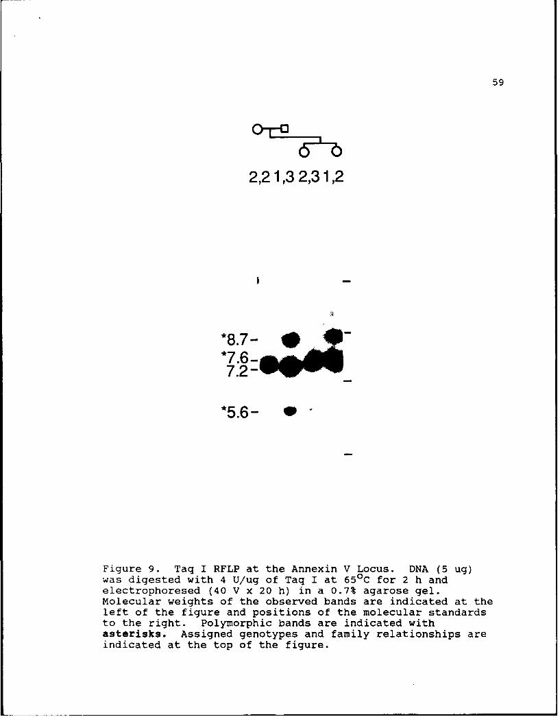
Figure 9. Taq I RFLP at the Annexin V Locus. DNA (5 ug)was digested with 4 U/ug of Taq I at 650 C for 2 h andelectrophoresed (40 V x 20 h) in a 0.7% agarose gel.Molecular weights of the observed bands are indicated at theleft of the figure and positions of the molecular standardsto the right. Polymorphic bands are indicated withasterisks. Assigned genotypes and family relationships areindicated at the top of the figure.

60

1,1 1,2 1,2 1,1

*10.1 -0@ .06.6:4*felt

*4.7- -_

1.5-0 0

Figure 10. PVU II RFLP at the Annexin V Locus. DNA (5 ug)was digested with 6 U/ug of Pvu II at 370 C for 2 h andelectrophoresed (25 V x 24 h) in a 0.9% agarose gel.Molecular weights of the observed bands are indicated at theleft of the figure and positions of the molecular standardsto the right. Polymorphic bands are indicated withasterisks. Assigned genotypes and family relationships areindicated at the top of the figure. Slight migrationvariation of 1.5 kb band due to composite figure of multiplegels.

61

2,21,11,21,1

*10.5-*9.0 -7.9-8 6

4.9-0000

3.788S

2.3-0000-

*2-11.9-4 00

1.6-* b e*1.4-e ..-

Figure 11. Bgl II RFLP at the Annexin III Locus. DNA(5 ug) was digested with 6 U/ug of Bgl II at 370 C for 2 hand electrophoresed (25 V x 25 h) in a 0.9% agarose gel.Molecular weights of the observed bands are indicated at theleft of the figure and positions of the molecular standardsto the right. Polymorphic bands are indicated withasterisks. Assigned genotypes are indicated at the top ofthe figure.

CHAPTER IV

Discussion and Conclusions

Chromosomal Localization

The gene for annexin III is located on chromosome 4.

The localization was identified by three independent

techniques: PCR of intron sequence using somatic cell

hybrids (Figure 5), Southern blot of DNA restriction enzyme

digests (Figure 7), and in-situ hybridization (performed in

collaboration with Drs. David Adler and Christine Disteche)

to chromosome 4q21.

The gene for Annexin V is located on chromosome 4.

This chromosome was identified by two independent

techniques: PCR of intron sequence using somatic cell

hybrids (Figure 2), and in-situ hybridization (performed in

collaboration with Drs. David Adler and Christine Disteche)

to 4q27. The localization of annexin V to chromosome 4

confirms previous localization of annexin V to 4q28-32 (Modi

et al., 1989). Their localization was performed using

Southern blot analysis of somatic cell hybrids and in-situ

hybridization.

63The chromosomal localization results for annexin IV

using PCR and Southern blot analysis were inconclusive.

However, in-situ hybridization (performed in collaboration

with Drs. David Adler and Christine Disteche) demonstrated

a unique locus at 2p13.

Failure to localize annexin IV using Southern blot

evaluation of the Hind III digests of human genomic DNA is

interesting since Hind III was selected as the enzyme of

choice for Southern blot analysis of annexin IV. Possible

reasons for the inability to identify the characteristic

human restriction pattern might be due to the relatively

small amount of DNA used or to a variable percentage of the

gene of interest present in each cell line. Hybrid cell

lines containing a low number of copies of the annexin IV

gene may have escaped detection with hybridization of the

cDNA probe.

The percent discordancy obtained with PCR analysis for

the annexin IV gene in the hybrid cell lines ranged from 8%

to 76%, with chromosome 2 showing the least discordance.

Repeat performance of the PCR reactions with the cell lines

produced inconsistent results. These sporadic results may

be due to the large size (3.5 kb) of the PCR product

amplified. Large PCR products (greater than 1.5 kb) are

less consistently amplified in the PCR reaction.

The PCR parameters used for localization of annexin IV

were similar to those used for annexin III and V with two

exceptions: 40 cycles for amplification of products instead

64

of 30, and 2.0 mM Mg2 + in place of 1.5 mM Mg2 +. The

amplified products seen in cell lines lacking chromosome 2

(983 and 904) could have been produced from genes closely

related to annexin IV. The additional 10 cycles of

amplification may have produced enough product, although

less product than the cell line containing chromosome 2

(854), to show weak hybridization on the Southern blot.

As mentioned in Chapter III, the increased MgCI 2

concentration was used to increase synthesis of the desired

product. However, increasing the Mg2+ concentration can

also decrease specificity of amplification (Ehrlich, 1989).

Quite possibly the optimal Mg2+ concentration was reached

and slightly exceeded, thereby allowing amplification of

other products from a related gene or an unrelated gene

oriented perfectly in cell lines 983 and 904 to allow

amplification.

Chromosome 2 may be present in low concentration in

cell lines 983 and 904. This would be similar to the

presence of chromosome 4 in cell line 867 discovered during

chromosomal localization of annexin V.

Failure to localize annexin V using Southern blot

evaluation of the Hind III digests of human genomic DNA is

interesting since annexin III localization results

correlated perfectly with the PCR results. Possible

explanations for this localization failure include the two

reasons stated for annexin IV, the fact that Hind III is not

the optimal enzyme for producing different sized

65

inter-species restriction fragments with annexin V, and the

fact that the annexin III and IV probes were stripped from

the membranes prior to the hybridization with annexin V cDNA

probe. stripping of the membrane may have removed just

enough DNA to prevent localization with hybridization of

the cDNA probe. However, the more sensitive technique of

chromosomal localization by PCR proved successful.

Restriction Fragment Length Polymorphism (RFLP) Study

As outlined in Chapter I, RFLPs serve a very important

function in the mapping of the human genome and diagnosis of

genetic disease. At the present time the RFLPs associated

with the annexin family do not have any direct clinical

application. However, as more is discovered about the

in-vivo role of the annexins, the chromosomal location of

all members, and the identification of genes in close

proximity to the annexins, the value of existing RFLPs will

increase. Nonetheless, the RFLPs identified in this

research project are by themselves very interesting

(Table 12 & Table 13).

Chromosomal Localization Techniques

Chromosomal localization based on PCR amplification of

somatic cell hybrids is a very new technique, with only a

handful of published studies so far (Iggo et al., 1989;

Dionne et al., 1990). Application in this project proved

very successful. This technique can be performed without

knowledge of specific gene structure (Iggo et al., 1989).

66

Two pieces of information were used to make educated guesses

about intron locations in the annexin genes: the location

of the repeating units in the proteins and the structure

(including intron-exon boundaries) of the murine annexin II

gene (Amiguet et al., 1990). Primers were designed with

certain specifics in mind: primer pairs should be of

similar length, primer pairs should have approximately the

same GC content, and primers should not have complementary

sequences at the 3' end. Once primer pairs successfully

amplified product larger than the product from the cDNA, PCR

conditions were optimized for each primer combination.

Multiple thermal profiles were tested with each primer pair

to ensure that the best intensity and specificity of

amplified products was obtained. The PCR parameters

adjusted in this study included amount of DNA template,

denaturation time and temperature, annealing time and

temperature, extension time and temperature, the

concentration of MgCI2 in the PCR buffer, and number of

amplification cycles.

The technique of chromosomal localization using hybrid

cell lines based on PCR amplification of intron sequence

holds some advantages over chromosomal localization via

amplification of exon sequence. The absence of the human-

specific PCR product using amplification of exon sequence

could be due to the absence of the gene(s) containing the

complementary sequence or due to failure of the PCR

reaction. Failure of the PCR reaction may be due to a

67

myriad of problems unrelated to the presence of the genetic

region of interest, thus producing false negative results.

Chromosomal localization utilizing intron amplification

relies on the presence of a human PCR product(s) of a

specific size, along with product(s) of different sizes

amplified from similar or homologous sequence in the hybrid

DNA, or no amplificiation of hybrid PCR products. Hybrid

cell lines lacking the human specific PCR products but

producing the hamster PCR products, almost certainly do not

contain the human gene of interest. Thus, false negative

results due to the absolute failure of the PCR reaction are

ruled out. Chromosomal localization based on PCR

amplification of intron sequence includes another level of

quality control. Since intron sequence is not highly

conserved from species to species, PCR products of identical

size are usually not amplified from two different species.

Therefore, production of human-sized product in hybrid cell

line suggests human origin.

As the PCR results with annexin V showed, primers will

sometimes amplify sequences of a different size in

dissimilar species (Figure 2). These amplified sequences

may be the hamster annexin V gene, other known annexin

genes, other unknown annexin genes, or totally unrelated

genes. By contrast, annexin III PCR reactions did not

amplify discernable PCR product with hamster genomic DNA

(Figure 5). This difference is most likely due to a

68

decreased degree of complementarity between the human

derived primers and hamster exon sequence.

Three virtues of chromosomal localization via PCR

amplification of intron sequence are: it is more rapid than

th- Southern blot technique, the procedure is non-

radioactive provided the product is visible on the ethidium

bromide stained gel, and it can be performed with very small

quantities of relatively impure template DNA (Iggo

et al., 1989).

Preparation time for the more conventional chromosomal

localization technique, Southern blots of restriction

digests of genomic DNA, is often as long as 3 to 5 days.

Chromosomal localization via PCR amplification of intron

sequence, at least the initial detection on ethidium bromide

stained gels, can be completed in one day.

PCR products indicating initial chromosomal

localization of annexin III and V were visible on the

etaidium bromide stained gels. Southern blotting was

utilized for confirmation of these PCR products for annexins

III and V. In comparison, Southern blots of PCR products

from all 25 hybrid cell lines were used for initial

detection of gene origin for annexin IV. These Southern

blots were performed for all cell lines since PCR products

were not visible on ethidium bromide stained agarose gels.

The use of PCR for chromosomal localization brings an

increased degree of sensitivity when compared to the more

traditional method of Southern blot evaluation of genomic

69

DNA digested with restriction enzymes. This increased

sensitivity, due to the exponential production of the

specific template sequence, affords the luxury of using

significantly less DNA (Saiki et al., 1988). 4 ug to 9 ug

of genomic DNA in preparing the Hind III Southern blots used

for localization of the three annexin proteins. In

contrast, the amount of template DNA used in the PCR

localization assays varied from 100 ng to 500 ng. The

amplified PCR product is largely target sequence including

any intron sequence located between the primer sites. The

genomic specimens cleaved with restriction enzyme contain an

infinitesimally (close to one in a million based on cDNA

size) small percentage of target sequence compared to total

DNA.

Future Work

Where does the project go from here? Future work on

this project lies in a few basic areas. In order to verify

that these proteins were localized to their chromosome of

origin, the exact base sequence of the PCR products could be

obtained. If correct, sequenced amplified products would

contain regions identical to the cDNA sequences separated by

intervening sequences, introns.

Verification of chromosome 2 as the origin of annexin

IV should be performed with an alternate hybrid cell line

containing chromosome 2 as its only human chromosome. This

chromosomal localization, using PCR amplification of intron

70

sequence, should clarify the discordant results seen with

the human-hamster somatic cell hybrids. An alternative plan

would be to design new PCR primers that would amplify a

product smaller than 3.5 kb. This smaller product should be

amplified more consistently.

Characterization of the entire genes coding for these

three annexins would be another worthwhile project.

Information about annexin function and regulation of gene

expression would undoubtedly be obtained.

The RFLP study could be expanded by the digestion of

human genomic DNA with additional enzymes. These additional

restriction patterns and possible RFLPs would provide

further insight into annexin gene structure and disease

linkage.

LIST OF REFERENCES

Abbott C, West L, Povey S, Jeremiah S, Murad Z, DiScipio R,Fey G. The gene for human complement component C9mapped to chromosome 5 by polymerase chain reaction.Genomics 1989;4:606-609.

Amiguet P, D'Eustachio P, Kristensen T, Wetsel RA,Saris CLM, Hunter T, Chaplin DD, Tack BF. Structureand chromosome assignment of the murine p36 (calpactinI heavy chain) gene. Biochemistry 1990;29:1226-1232.

Bell GI, Karam JH, Rutter WJ. Polymorphic DNA regionadjacent to the 5' end of the human insulin gene.Proc Natl Acad Sci USA 1981;78:5759-5763.

Botstein D, White RL, Skolnick M, Davis RW. Construction ofa genetic linkage map in man using restriction fragmentlength polymorphisms. Am J Hum Genet 1980;32:314-331.

Burgoyne RD, Geisow MJ. The annexin family of calcium-binding proteins. Cell Calcium 1989;10:1-10.

Burns AL, Magendzo K, Shirvan A, Srivastava M, Rojas E,Alijani MR, Pollard HB. Calcium channel activity ofpurified human synexin and structure of the humansynexin gene. Proc Natl Acad Sci USA 1989;86:3798-3802.

Cannizzaro LA, Emanuel BS. An improved method for G-bandingchromosomes after in situ hybridization. CytogenetCell Genet 1984;38:308-309.

Carlock LR, Smith D, Wasmuth JJ. Genetic counterselectiveprocedure to isolate interspecific cell hybridscontaining single human chromosomes: Construction ofcell hybrids and recombinant DNA libraries specific forhuman chromosomes 3 and 4. Somatic Cell and MolecularGenetics 1986; 12:163-174.

Chandler ME, Yunis JJ. A high resolution in situhybridization technique for the direct visualization oflabeled G-banded early metaphase and prophasechromosomes. Cytogenet Cell Genet 1978;22:352-356.

Crompton MR, Moss SE, Crumpton MJ. Diversity in thelipocortin/calpactin family. Cell 1988a;55:1-3.

Crompton MR, Owens RJ, Totty NF, Moss SE, Waterfield MD, 72Crumpton MJ. Primary structure of the human, membrane-associated Ca 2+-binding protein p68: a novel member ofa protein family. EMBO Journal 1988b;7:21-27.

Crumpton MJ, Dedman JR. Protein terminology tangle. Nature1990; 345:212.

Davies AA, Moss SE, Crompton MR, Jones TA, Spurr NK,Sheer D, Kozak C, Crumpton MJ. The gene coding for thep68 calcium-binding protein is localised to bandsq32-q34 of human chromosome 5, and to mouse chromosome11. Hum Genet 1989;82:234-238.

Dionne CA, Kaplan R, Seuanez H, O'Brien SJ, Jaye M.Chromosome assignment by polymerase chain reactiontechniques: Assignment of the oncogene FGF-5 to humanchromosome 4. BioTechnioues 1990;8:190-194.

Donis-Keller H, Barker DF, Knowlton RG, Schumm JW,Braman JC, Green P. Highly polymorphic RFLP probes asdiagnostic tools. Cold Spring Harbor Symposia onQuantitative Biology 1986;LI:317-324.

Donis-Keller H, Green P, Helms C, Cartinhour S,Weiffenbach B, Stephens K, Keith TP, Bowden DW,Smith DR, Lander ES, Botstein D, Akots G, Rediker KS,Gravius T, Brown VA, Rising MB, Parter C, Powers JA,Watt DE, Kauffman ER, Bricker A, Phippz P,Muller-Kahle H, Fulton TR, Ng S, Schumm JW, Braman JC,Knowlton RG, Barker DF, Crooks SM, Lincoln SE, Daly MJ,Abrahamson J. A genetic linkage map of the humangenome. Cell 1987;51:319-337.

Drayna D. Fine structure linkage mapping of the humangenome using RFLP analysis. BioTechniques 1986;4:412-418.

Erlich HA. PCR Technology. Principles and Applications forDNA Amplification. Stockton Press, New York, NY.1989.

Ephrussi B. Hybridization of Somatic Cells. PrincetonUniversity Press. Princeton, NJ. 1972.

Ephrussi B, Weiss MC. Interspecific hybriiization ofsomatic cells. Proc Natl Acad Sci USA 1965;53:1040-1042.

Feinberg AP, Vogelstein B. A technique for radiolabelingDNA restriction endonuclease fragments to high specificactivity. Anal Biochem 1983;132:6-13.

Flaherty MJ, West S, Heimark RL, Fujikawa K, Tait JF. 73Placental anticoagulant protein-I: Measurement inextracellular fluids and cells of the hemostaticsystem. J Lab clin Med 1990;115:174-181.

Funakoshi T, Heimark RL, Hendrickson LE, McMullen BA,Fujikawa K. Human placental anticoagulant protein:Isolation and characterization. Biochemistry 1987a;26:5572-5578.

Funakoshi T, Hendrickson LE, McMullen BA, Fujikawa K.Primary structure of human placental anticoagulantprotein. Biochemistry 1987b;26:8087-8092.

Gerhard DS, Kawasaki ES, Bancroft C, Szabo P. Localizationof a unique gene by direct hybridization in situ.Proc Natl Acad Sci USA 1981;78:3755-3759.

Gusella JF, Wexler NS, Conneally PM, Naylor SL, Anderson MA,Tanzi RE, Watkins PC, Ottina K, Wallace MR,Sakaguchi AY, Young AB, Shoulson I, Bonilla E,Martin JB. A polymorphic DNA marker genetically linkedto Huntington's disease. Nature 1983;306:234-238.

Grundmann U, Amann E, Abel K-J, Kupper HA. Isolation andexpression of cDNA coding for a new member of thephospholipase A2 inhibitor family. Behring Inst Mitt1988;82:59-67.

Haigler HT, Fitch JM, Jones JM, Schlaepfer DD. Twolipocortin-like proteins, endonexin II and anchorinCII, may be alternate splices of the same gene. TrendsBiochem Sci 1989;14:48-50.

Harris H, Watkins JF. Hybrid cells derived from mouse andman: Artificial heterokaryons of mammalian cells fromdifferent species. Nature 1965;205:640-646.

Hauptmann R, Maurer-Fogy I, Krystek E, Bodo G, Andree H,Reutelingsperger CPM. Vascular anticoagulant beta:a novel human Ca2+/phospholipid binding protein thatinhibits coagulation and phospholipase A2 activity.Eur J Biochem 1989;185:63-71.

Huebner K, Cannizzaro LA, Frey AZ, Hecht BK, Hecht F,Croce CM, Wallner BP. Chromosomal localization of thehuman genes for lipocortin I and lipocortin II.Oncogene Research 1988;2:299-310.

Iggo R, Gough A, Xu W, Lane DP, Spurr NK. Chromosomemapping of the human gene encoding the 68-kDa nuclearant .gen (p68) by using the polymerase chain reaction.Proc Natl Acad Sci USA 1989;86:6211-6214.

Iwasaki A, Suda m, Nakao H, Nagoya T, Saino Y, Arai K, 74Mizoguchi T, Sato F, Yoshizaki H, Nirata M, Miyata T,Shidara Y, Murata M, Maki M. Structure and expressionof cDNA for a inhibitor of blood coagulation isolatedfrom human placenta: A new lipocortin-like protein.J Biochem 1987;102:1261-1273.

Kaetzel MA, Hazarika P, Dedman JR. Differential tissueexpression of three 35-kDa annexin calcium-dependentphospholipid-binding proteins. J Biol Chem1989;264:14463-14470.

Kan YW, Dozy AN. Polymorphism of DNA sequence adjacent tohuman beta-globulin structural gene: Relationship tosickle mutation. Proc Natl Acad Sci USA 1978;75:5631-5635.

Klee CB. Ca2+-dependent phospholipid- (and membrane-)binding proteins. Biochemistry 1988;27:6647-6652.

Kunkel LM, Smith KD, Boyer SH, Borgaonkar DS, Wachtel SS,Miller OJ, Breg WR, Jones Jr HW, Rary JM. Analysis ofhuman Y chromosome-specific reiterated DNA inchromosome variants. Proc Natl Acad Sci USA1977;74:1245-1249.

Maurer-Fogy I, Reutelingsperger CPM, Pieters J, Bodo G,Stratowa C, Hauptmann R. Cloning and 5xpressin of cDNAfor human vascular anticoagulant, a Ca +-dependentphospholipid-binding protein. Eur J Biochem1988;174:585-592.

Modi WS, Seuanez HN, Jaye M, Haigler HJ, Kaplan R,O'Brien SJ. The human endonexin II (ENX2) gene islocated at 4q28-q32. CytoQenet Cell Genet 1989;52:167-169.

Mullis KB, Faloona FA. Specific synthesis of DNA in vitrovia a polymerase-catalyzed chain reaction. Methods inEnzymoloQy 1987;155:335-350

Nakamura Y, Leppert M, O'Connell P, Wolff R, Holm T,Culver M, Martin C, Fujimoto E, Hoff M, Kumlin E,White R. Variable number of tandem repeat (VNTR)markers for human gene mapping. Science 1987;235:1616-1622.

Pepinsky RB, Tizard R, Mattaliano RJ, Sinclair LK,Miller GT, Browning JL, Chow EP, Burne C, Huang KS,Pratt D, Wachter L, Hession C, Frey AZ, Wallner BP.Five distinct calcium and phospholipid binding proteinsshare homology with lipocortin I. J Biol Chem1988;263:10799-10811.

Reed KC, Mann DA. Rapid transfer fo DNA from agarose gels 75to nylon membranes. Nucleic Acids Research 1985;13:7207-7221.

Reutelingsperger CPM, Hornstra G, Hemker HC. Isolation andpartial purification of a novel anticoagulant fromarteries of human umbilical cord. Eur J Biochem1985;151:625-629.

Reutelingsperger CPM, Kop JMM, Hornstra G, Hemker HC.Purification and characterization of a novel proteinfrom bovine aorta that inhibits coagulation.Eur J Biochem 1988;173:171-178.

Ringertz NR, Savage RE. Cell Hybrids. Academic Press.New York, NY. 1976.

Saiki RK, Gelfand DH, Stoffel S, Scharf SJ, Higuchi R,Horn GT, Mullis KB, Erlich HA. Primer-directedenzymatic amplification of DNA with a thermostableDNA polymerase. Science 1988;239:487-491.

Sudhof TC, Slaughter CA, Leznicki I, Barjon P, Reynolds GA.Human 67-kDa calelectrin contains a duplication of fourrepeats found in 35-kDa lipocortins. Proc Natl AcadSci USA 1988;85:664-668.

Tait JF, Gibson D, Fujikawa K. Phospholipid bindingproperties of human placental anticoagulant protein-I,a member of the lipocortin family. J Biol Chem1989;264:7944-7949.

Tait JF, Sakata M, McMullen BA, Miao CH, Funakoshi T,Hendrickson LE, Fujikawa K. Placental anticoagulantproteins: Isolation and comparative characterizationof four members of the lipocortin family. Biochemistry1988;27:6268-6276.

Tsui LC, Buchwald M, Barker D, Braman JC, Knowlton R,Schumm JW, Eiberg H, Mohr J, Kennedy D, Plavsic N,Zsiga M, Markiewicz D, Akots G, Brown V, Helms C,Gravius T, Parker C, Rediker K, Donis-Keller H. Cysticfibrosis locus defined by a genetically linkedpolymorphic DNA marker. Science 1985;230:10541057.

Wainwright BJ, Scambler PJ, Schmidtke J, Watson EA, Law HY,Farrall M, Cooke HJ, Eiberg H, Williamson R.Localization of cystic fibrosis locus to humanchromosome 7cen-q22. Nature 1985;318:384-385.

Wallner BP, Mattaliano RJ, Hession C, Cate RL, Tizard R, 76Sinclair LK, Foeller C, Chow EP, Browning JL,Ramachandran KL, Pepinsky RB. Cloning and expressionof human lipocortin, a phospholipase Ai inhibitor withpotential anti-inflammatory activity. Nature 1986;320:77-81.

Watson J, Tooze J, Kurtz D. Recombinant DNA. A ShortCourse. Scientific American Books, New York, NY.1983.

Weatherall DJ. The New Genetics and Clinical Practice.Second Edition. Oxford University Press, New York, NY.1985.

White R, Woodward S, Leppert M, O'Connell P, Hoff M,Herbst J, Lalouel JM, Dean M, Vande Woude G. A closelylinked genetic marker for cystic fibrosis. Nature1985;318:382-385.


Recommended