ARTICLE AUNIP/C1orf135 directs DNA double-strand breaks towards the homologous recombination repair pathway Jiangman Lou 1 , Hongxia Chen 1,2 , Jinhua Han 1 , Hanqing He 1 , Michael S.Y. Huen 3 , Xin-hua Feng 1 , Ting Liu 4 & Jun Huang 1 DNA double-strand breaks (DSBs) are mainly repaired by either homologous recombination (HR) or non-homologous end-joining (NHEJ). Here, we identify AUNIP/C1orf135, a largely uncharacterized protein, as a key determinant of DSB repair pathway choice. AUNIP physi- cally interacts with CtIP and is required for efficient CtIP accumulation at DSBs. AUNIP possesses intrinsic DNA-binding ability with a strong preference for DNA substrates that mimic structures generated at stalled replication forks. This ability to bind DNA is necessary for the recruitment of AUNIP and its binding partner CtIP to DSBs, which in turn drives CtIP- dependent DNA-end resection and HR repair. Accordingly, loss of AUNIP or ablation of its ability to bind to DNA results in cell hypersensitivity toward a variety of DSB-inducing agents, particularly those that induce replication-associated DSBs. Our findings provide new insights into the molecular mechanism by which DSBs are recognized and channeled to the HR repair pathway. DOI: 10.1038/s41467-017-01151-w OPEN 1 Life Sciences Institute and Innovation Center for Cell Signaling Network, Zhejiang University, Hangzhou, Zhejiang 310058, China. 2 Huadong Research Institute for Medicine and Biotechniques, Nanjing, Jiangsu 210002, China. 3 School of Biomedical Sciences, Li Ka Shing Faculty of Medicine, The University of Hong Kong, Hong Kong Special Administrative Region, Pok Fu Lam, Hong Kong, China. 4 Department of Cell Biology, Zhejiang University School of Medicine, Hangzhou, Zhejiang 310058, China. Jiangman Lou and Hongxia Chen contributed equally to this work. Correspondence and requests for materials should be addressed to J.H. (email: [email protected]) NATURE COMMUNICATIONS | 8: 985 | DOI: 10.1038/s41467-017-01151-w | www.nature.com/naturecommunications 1
Transcript
ARTICLE
AUNIP/C1orf135 directs DNA double-strand breakstowards the homologous recombination repairpathwayJiangman Lou1, Hongxia Chen1,2, Jinhua Han1, Hanqing He1, Michael S.Y. Huen3, Xin-hua Feng1, Ting Liu4
& Jun Huang 1
DNA double-strand breaks (DSBs) are mainly repaired by either homologous recombination
(HR) or non-homologous end-joining (NHEJ). Here, we identify AUNIP/C1orf135, a largely
uncharacterized protein, as a key determinant of DSB repair pathway choice. AUNIP physi-
cally interacts with CtIP and is required for efficient CtIP accumulation at DSBs. AUNIP
possesses intrinsic DNA-binding ability with a strong preference for DNA substrates that
mimic structures generated at stalled replication forks. This ability to bind DNA is necessary
for the recruitment of AUNIP and its binding partner CtIP to DSBs, which in turn drives CtIP-
dependent DNA-end resection and HR repair. Accordingly, loss of AUNIP or ablation of its
ability to bind to DNA results in cell hypersensitivity toward a variety of DSB-inducing agents,
particularly those that induce replication-associated DSBs. Our findings provide new insights
into the molecular mechanism by which DSBs are recognized and channeled to the HR repair
pathway.
DOI: 10.1038/s41467-017-01151-w OPEN
1 Life Sciences Institute and Innovation Center for Cell Signaling Network, Zhejiang University, Hangzhou, Zhejiang 310058, China. 2 Huadong ResearchInstitute for Medicine and Biotechniques, Nanjing, Jiangsu 210002, China. 3 School of Biomedical Sciences, Li Ka Shing Faculty of Medicine, The University ofHong Kong, Hong Kong Special Administrative Region, Pok Fu Lam, Hong Kong, China. 4 Department of Cell Biology, Zhejiang University School of Medicine,Hangzhou, Zhejiang 310058, China. Jiangman Lou and Hongxia Chen contributed equally to this work. Correspondence and requests for materials should beaddressed to J.H. (email: [email protected])
DNA double-strand breaks (DSBs) are the most deleteriousform of DNA damage, which if unrepaired or repairedincorrectly, can contribute to various genetic disorders
including cancer, neurodegeneration, and immunodeficiency1.DSBs can arise as a result of errors during DNA replication, andcan be induced by exogenous DNA-damaging agents includingionizing radiation (IR) and various chemotherapeutic drugs1.DSBs are mainly repaired via two pathways–non-homologousend joining (NHEJ) and homologous recombination (HR), bothof which are highly conserved from yeast to human2–6. NHEJ is arelatively fast and simple process that involves direct end-to-endligation of the DSB ends, and this pathway is active throughoutinterphase7–9. The key players in NHEJ include the DNAend-binding Ku70/80 heterodimer, the DNA-dependent proteinkinase catalytic subunit (DNA-PKcs), X-ray cross-complement-ing protein 4 (XRCC4), XRCC4-like factor (XLF), DNA ligaseIV, and the newly identified PAXX (a paralog of XRCC4 andXLF)7, 8, 10, 11. In contrast to NHEJ, HR is a complex, multi-steprepair pathway that requires the sequential activity of a cohort ofproteins and occurs primarily in the S and G2 phases of the cellcycle2–6. HR relies on the presence of a sister chromatid as adonor template, and is initiated by nuclease-mediated extensive5′-3′ resection of DSB ends, resulting in long stretches of 3′single-stranded DNA (ssDNA) that subsequently invades the
homologous duplex DNA12–14. It is now well-established thatDSBs are resected in a two-step manner12–14. Initially, theevolutionarily-conserved MRE11-RAD50-NBS1/XRS2 (MRN/X)complex and its associated factor CtIP/Sae2 carry out limitedresection near the break site to generate a short 3′ overhang15–19.The partially-resected DNA is further processed by two parallelpathways; one that is dependent on the 5′-3′ exonuclease Exo1and the other dependent on the concerted action of the BLM/Sgs1helicase and the Dna2 endonuclease20–23.
While both NHEJ and HR machineries can repair DSBs, choiceof the more appropriate DSB repair pathway is key to main-tenance of genome stability, especially at the organismallevel24, 25. To date, a number of determinants have been reportedto influence the choice between the two pathways. One of these iscell cycle24, 26–28. Studies have shown that efficient DNA endprocessing is restricted to the S and G2 phases, and is regulated bycell cycle-dependent CDK activity24, 26–30. By preventing HRoutside of the S and G2 phases of the cell cycle, exchangesbetween homologous chromosomes are reduced, cells therebysuppress DSB-associated loss of heterozygosity and chromosomalrearrangements24, 26, 27. In addition to effects arising from cellcycle, the nature of DSBs also influences choice of repairpathways24, 31–33. Indeed, although both NHEJ and HR con-tribute to repair of X- or γ-ray-induced two-ended DSBs in the S
a b
e f
250 kD150 kD100 kD75 kD
50 kD
SFB-CtIP
37 kD
20 kD
25 kD
Mar
ker
Vecto
r
CtIP
CtIP
IP: S
bea
ds
SFB-CtIP
Myc-AUNIP
Inpu
t
WT
81–3
57
1–28
0
α-Flag
α-Myc
α-Flag
α-Myc
IgG
αA
UN
IP
αA
UN
IP
IPIn
put
CPT – – + – – +
MRE11
AUNIP
AUNIP
RAD50
RAD50
MRE11
CtIP
CtIP
g
CtIP purification
Protein Peptides Coverage (%)
CtIP
6
9
24
AUNIP
MRE11
RAD50
54
19.6
16.5
10
36
ATM 8 2.9
7
TopBP1 15 9.7
FANCD2 19 14.4
4.1
NBS1 3 4
BARD1 6.44
BRCA1
AUNIP purification
Protein Peptides Coverage (%)
AUNIP
20RAD50
13
20.5
30.5
DNA2 2 2.2
MRE11 7 11.5
CtIP 1 1
αC
tIP
αC
tIP
IgG
IPIn
put
CPT
MRE11
AUNIP
AUNIP
RAD50
RAD50
MRE11CtIP
CtIP
250 kD150 kD
100 kD75 kD
50 kDSFB-AUNIP
37 kD
20 kD
25 kD
Mar
ker
Vecto
r
AUNIPc d
GST beads pull down
MBP-CtIP
GST
+
+
+
5% In
put
+
–
–
GSTGST-281–357
GST-WTGST-1–280
250 kD150 kD100 kD75 kD
50 kD
37 kD
25 kD
+ +
GST-1–280
GST-281–357
–
–
– –
– –
+ ––
+ – –
Coomassie staining
MBP-CtIP
GST-WT
150 kD
150 kD 150 kD
75 kD 75 kD
150 kD 150 kD
37 kD 37 kD150 kD 150 kD
75 kD 75 kD
150 kD 150 kD
37 kD 37 kD
37 kD
150 kD
150 kD
50 kD
50 kD
37 kD
Fig. 1 Identification of AUNIP as a CtIP-associated protein. a, c CtIP (a) or AUNIP (c) protein complexes separated by SDS-PAGE were stained withCoomassie blue. b, d Proteins identified by mass spectrometric analyses of CtIP or AUNIP protein complexes are listed. Bait proteins are indicated in boldletters. e AUNIP interacts with CtIP and the MRN complex. HeLa cell lysates treated with Benzonase were precipitated with anti-AUNIP or anti-CtIPantibodies and were analyzed by immunoblotting with indicated antibodies. f AUNIP interacts with CtIP via its C-terminus. HEK293T cells transfected withindicated plasmids were lysed with NETN buffer 24 h post transfection. Cell lysates were then incubated with S-protein beads and immunoprecipitatedproteins were analyzed by immunoblotting experiments using indicated antibodies. g Direct in vitro binding between recombinant GST-AUNIP and MBP-CtIP purified from E. coli. GST served as a negative control for CtIP binding. Top: CtIP was detected by immunoblotting experiment. Bottom: purifiedproteins were visualized by Coomassie staining
and G2 phases in mammalian cells24, 31–33, HR-deficient cells aremuch more tolerant to irradiation, indicating that NHEJ likelyplays a more important role in the repair of two-endedDSBs33–36. By contrast, replication-associated one-ended DSBsare repaired almost exclusively by HR37, 38. In support of thisworking model, NHEJ is accountable for the genome instabilityand cell cytotoxicity phenotypes in HR-deficient cells whenchallenged with agents known to induce replication-associatedDSBs, including camptothecin (CPT, a DNA topoisomerase Iinhibitor) and poly(ADP-ribose) polymerase (PARP)inhibitors39–41. Nevertheless, it remains unclear how the nature ofDSB determines usage of DSB repair pathways.
In this study, we used an affinity purification approach toisolate CtIP-containing protein complexes, and have identifiedAUNIP/C1orf135 as a primary determinant of DSB repairpathway choice. We show that AUNIP is recruited to DNAdamage sites through a DNA-binding motif that shows a strongbinding preference for DNA substrates that mimick structuresgenerated at stalled replication forks. We further demonstratethat AUNIP physically interacts with CtIP and is required forefficient CtIP concentration at DNA lesions. Consequently, loss ofAUNIP, or ablation of its ability to bind to DNA or CtIP,impaired CtIP-dependent DNA-end resection, compromised HRrepair, and resulted in cell hypersensitivity toward a variety ofDSB-inducing agents, particularly those that induce replication-associated DSBs. Our results support a model in which AUNIPserves as a sensor of DNA damage, anchoring CtIP to DSB sitesto drive CtIP-dependent DNA end resection and ensuing HRrepair.
ResultsIdentification of AUNIP as a CtIP-associated protein. To fur-ther elucidate the role of CtIP in DNA end resection and DSBrepair pathway choice, we performed tandem affinity purification(TAP) using a HEK293T cell line that stably expresses SFB-tagged(S-protein tag, Flag epitope tag, and streptavidin-binding peptidetag) wild-type CtIP to isolate proteins that associate with CtIP(Fig. 1a). Mass spectrometric analysis identified a number ofpreviously reported CtIP-interacting proteins, including the MRN(MRE11-RAD50-NBS1) complex, FANCD2, TopBP1, ATM,BRCA1, and BARD1 (Fig. 1b and Supplementary Data 1).Intriguingly, we also reproducibly recovered AUNIP/C1orf13542,a largely uncharacterized protein, as a putative CtIP-interactingprotein (Fig. 1b and Supplementary Data 1). To determine ifAUNIP could indeed form a complex with CtIP, we conductedreverse TAP experimentations using a cell line engineered tostably express SFB-tagged AUNIP, and uncovered CtIP, MRE11,RAD50, and DNA2 as major AUNIP-associated proteins (Fig. 1c,d and Supplementary Data 2). These findings strongly suggestthat AUNIP and CtIP exist in the same protein complex in cells.AUNIP was originally identified as an Aurora-A-interactingprotein and has been shown to target Aurora-A to spindlepoles42. Interestingly, AUNIP does not contain any knownfunctional motifs, and human AUNIP orthologs are foundexclusively in vertebrates, such as Pan troglodytes, Bos Taurus,Mus musculus, Canis lupus familiaris, and Xenopus laevis.
To verify the TAP-mass spectrometry data, we first performedco-immunoprecipitation experiments using lysates derived fromHEK293T cells transiently transfected with plasmids that encodeSFB-tagged AUNIP together with expression constructs thatencode Myc-tagged CtIP, -MRE11, -RAD50, or -NBS1. As shownin Supplementary Fig. 1a, AUNIP interacted with CtIP and allthree components of the MRN complex, but not with theunrelated protein Morc3. We further carried out reciprocal co-immunoprecipitation experiments in HeLa cells and confirmed
that endogenous AUNIP and CtIP proteins formed a complexin vivo (Fig. 1e). The complex formation was not affected by CPTtreatment, and was resistant to benzonase treatment, arguingagainst the possibility that the observed interaction was mediatedby DNA/RNA (Fig. 1e). Notably, cross-species amino-acidsequence alignment revealed that both termini of the AUNIPpeptide are highly conserved (Supplementary Fig. 1b). We thustested whether these evolutionarily-conserved regions of AUNIPare involved in its interaction with CtIP. Co-immunoprecipitation experiments showed that AUNIP interactedwith CtIP through its conserved C-terminus, since a deletionmutant lacking the C-terminal 77 amino acids (1–280) wasunable to co-precipitate with CtIP (Fig. 1f).
To test whether the interaction between AUNIP and CtIP wasdirect, we performed GST pull-down assays using recombinantGST-tagged AUNIP and MBP-tagged CtIP purified from E. coli.As shown in Fig. 1g, wild-type, but not the 1–280 deletion mutantof AUNIP, directly interacted with CtIP in vitro. Interestingly, theC-terminal region alone (281–357) was also able to directlyinteract with CtIP (Fig. 1g). Altogether, these results suggestedthat the conserved C-terminal region of AUNIP is necessary andsufficient for its interaction with CtIP.
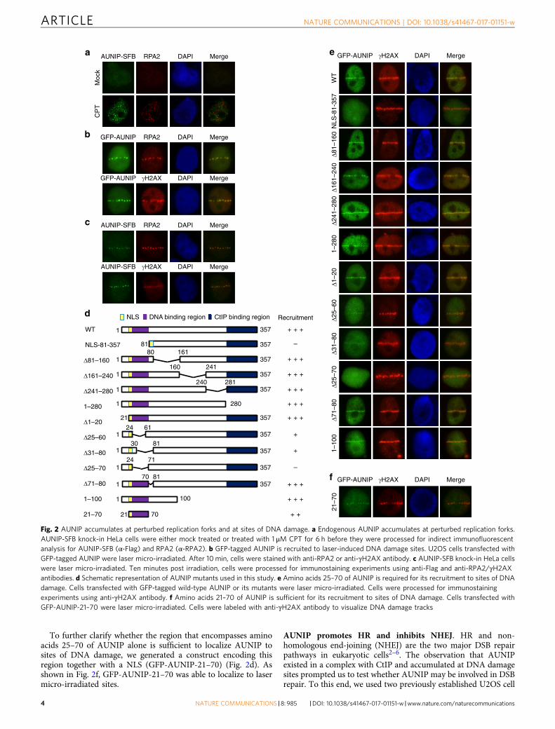
AUNIP accumulates at sites of DNA damage. Given thatAUNIP exists in a complex with CtIP, we examined whetherAUNIP may also be recruited to damaged replication forks or toDNA lesions. Because we were unable to detect AUNIP byindirect immunofluorescence experiments using our in-houseanti-AUNIP antibodies, we, therefore, utilized the CRISPR/Cas9-directed recombinant adeno-associated virus (rAAV)-mediatedgene targeting approach43 to integrate an SFB tag onto the C-terminus of the endogenous AUNIP gene (SupplementaryFig. 2a). DNA sequencing results confirmed that the tag wastargeted correctly, and expression of the AUNIP-SFB protein wasconfirmed on immunoblots probed with anti-Flag antibody(Supplementary Fig. 2b). As shown in Fig. 2a, in most untreatedcells, AUNIP showed a diffuse staining pattern. However, whencells were treated with the topoisomerase I inhibitor CPT, AUNIPconcentrated into distinct nuclear foci (Fig. 2a). Notably,although AUNIP foci only partially colocalized with RPA2 foci(Supplementary Fig. 2c), they were found exclusively in RPA2foci-positive cells (Supplementary Fig. 2d), indicating thatAUNIP foci may correspond to perturbed replication forks.
We next laser micro-irradiated U2OS cells that stably expressGFP-tagged AUNIP to determine whether AUNIP is recruited tosites of DNA damage. As shown in Fig. 2b, GFP-AUNIP wasreadily recruited to RPA2- and γH2AX-marked laser-generatedstripes. Moreover, similar to GFP-AUNIP, endogenous AUNIPcan also be detected at laser micro-irradiated tracks (Fig. 2c).
To determine which domain is responsible for the recruitmentof AUNIP to laser-irradiated sites, we generated a series ofAUNIP deletion mutants that span the entire AUNIP protein(Fig. 2d), and monitored the recruitment of each of these deletionmutants. Since the very end of the N-terminus of AUNIPcontained a functional nuclear localization signal (NLS)(K21RRK24) (Supplementary Fig. 2e), we added an NLS to theN-terminus of the 81–357 deletion mutant (lacking the N-terminal 80 amino acids) to ensure its proper nuclear localization.As shown in Fig. 2e, aside the NLS-81–357 deletion mutant, allother AUNIP mutants retained the ability to accumulate at laser-induced γH2AX-marked DNA damage tracks, indicating that theN-terminus of AUNIP is indispensable for its recruitment toDNA lesions. Further deletion analysis narrowed down the regionessential for AUNIP accumulation at DNA damage tracks toresidues 25–70 (Fig. 2e).
To further clarify whether the region that encompasses aminoacids 25–70 of AUNIP alone is sufficient to localize AUNIP tosites of DNA damage, we generated a construct encoding thisregion together with a NLS (GFP-AUNIP-21–70) (Fig. 2d). Asshown in Fig. 2f, GFP-AUNIP-21–70 was able to localize to lasermicro-irradiated sites.
AUNIP promotes HR and inhibits NHEJ. HR and non-homologous end-joining (NHEJ) are the two major DSB repairpathways in eukaryotic cells2–6. The observation that AUNIPexisted in a complex with CtIP and accumulated at DNA damagesites prompted us to test whether AUNIP may be involved in DSBrepair. To this end, we used two previously established U2OS cell
a
1–10
0N
LS-8
1-35
7W
TΔ2
5–70
Δ71–
80Δ2
5–60
Δ31–
80Δ1
–20
γH2AX DAPI MergeGFP-AUNIP
Δ81–
160
Δ161
–240
Δ241
–280
1–28
021
–70
d
NLS-81-357
WT
Δ81–160
Δ161–240
Δ241–280
1–280
Δ71–80
Recruitment
+
+
–
–
Δ1–20
Δ25–60
Δ31–80
Δ25–70
1–100
21–70 + +
1 357
81
180 161
1160 241
357
357
357
1240 281
357
1 280
21 357
24 61357
30 81
24 71
70 81
1 100
21 70
357
357
357
1
1
1
1
+ + +
+ + +
+ + +
+ + +
+ + +
+ + +
+ + +
+ + +
e
b
γH2AX DAPI MergeGFP-AUNIP
RPA2 DAPI MergeGFP-AUNIP
RPA2 DAPI Merge
Moc
k
AUNIP-SFB
CP
T
CtIP binding regionDNA binding regionNLS
AUNIP-SFB RPA2 DAPI Merge
AUNIP-SFB γH2AX DAPI Merge
c
γH2AX DAPI MergeGFP-AUNIPf
Fig. 2 AUNIP accumulates at perturbed replication forks and at sites of DNA damage. a Endogenous AUNIP accumulates at perturbed replication forks.AUNIP-SFB knock-in HeLa cells were either mock treated or treated with 1 μM CPT for 6 h before they were processed for indirect immunofluorescentanalysis for AUNIP-SFB (α-Flag) and RPA2 (α-RPA2). b GFP-tagged AUNIP is recruited to laser-induced DNA damage sites. U2OS cells transfected withGFP-tagged AUNIP were laser micro-irradiated. After 10 min, cells were stained with anti-RPA2 or anti-γH2AX antibody. c AUNIP-SFB knock-in HeLa cellswere laser micro-irradiated. Ten minutes post irradiation, cells were processed for immunostaining experiments using anti-Flag and anti-RPA2/γH2AXantibodies. d Schematic representation of AUNIP mutants used in this study. e Amino acids 25–70 of AUNIP is required for its recruitment to sites of DNAdamage. Cells transfected with GFP-tagged wild-type AUNIP or its mutants were laser micro-irradiated. Cells were processed for immunostainingexperiments using anti-γH2AX antibody. f Amino acids 21–70 of AUNIP is sufficient for its recruitment to sites of DNA damage. Cells transfected withGFP-AUNIP-21–70 were laser micro-irradiated. Cells were labeled with anti-γH2AX antibody to visualize DNA damage tracks
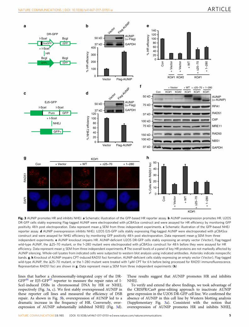
lines that harbor a chromosomally-integrated copy of the DR-GFP44 or EJ5-GFP45 reporter to measure the repair rates of I-SceI-induced DSBs in chromosomal DNA by HR or NHEJ,respectively (Fig. 3a, c). We first stably overexpressed AUNIP inthese reporter cell lines and measured the efficiency of DSBrepair. As shown in Fig. 3b, overexpression of AUNIP led to adramatic increase in the frequency of HR. Conversely, over-expression of AUNIP substantially inhibited NHEJ (Fig. 3d).
These results suggest that AUNIP promotes HR and inhibitsNHEJ.
To verify and extend the above findings, we took advantage ofthe CRISPR/Cas9 gene-editing approach to inactivate AUNIPgene expression in the U2OS DR-GFP cell line. We confirmed theabsence of AUNIP in this cell line by Western blotting analysis(Supplementary Fig. 3a). Consistent with the notion thatoverexpression of AUNIP promotes HR and inhibits NHEJ,
a b
+ I-SceI
HR
iGFPSceGFP
I-SceI BcgI
iGFPGFP+
BcgI BcgI
DR-GFP
e
% H
R e
ffici
ency
Con
KO#1
+ V
ecto
r
+ W
T
+ Δ
25–7
0
+ 1
–280
+ V
ecto
r
020406080
100120140
KO#1 KO#2
% H
R e
ffici
ency
AUNIP(α-Flag)
GAPDH
Vecto
r
Flag-A
UNIP
Vector Flag-AUNIP0
100
200
300
400
c d
EJ5-GFP
Puro GFP
GFP+
NHEJ
f
NBS1
CtIP
RAD50
MRE11
GAPDH
+ WT
Con KO#1 KO#2 KO#1
g
AUNIP(α-AUNIP)
Con
KO#1
+ V
ecto
r
+ W
T
+ Δ
25–7
0
h
+ Δ25–70
KO#1
I-SceII-SceI
+ I-SceI
RAD51
RPA1
+ Vector
10
20
30
40
50
+ 1
–280
Con + WT
KO#1
+ Vector + Δ25–70
RA
D51
DA
PI
+ 1–280
0% C
ells
with
RA
D51
foci
+ 1–280
KO#1
*
% N
HE
J ef
ficie
ncy
Vector Flag-AUNIP
AUNIP(α-Flag)
GAPDH
Vecto
r
Flag-A
UNIP
0
20
40
60
80
100
120
50 kD
150 kD
100 kD
75 kD
37 kD
75 kD
100 kD
37 kD
50 kD
37 kD
50 kD
37 kD
Fig. 3 AUNIP promotes HR and inhibits NHEJ. a Schematic illustration of the GFP-based HR reporter assay. b AUNIP overexpression promotes HR. U2OSDR-GFP cells stably expressing Flag-tagged AUNIP were electroporated with pCBASce construct and were assayed for HR efficiency by monitoring GFPpositivity 48 h post electroporation. Data represent mean± SEM from three independent experiments. c Schematic illustration of the GFP-based NHEJreporter assay. d AUNIP overexpression inhibits NHEJ. U2OS EJ5-GFP cells stably expressing Flag-tagged AUNIP were electroporated with pCBASceconstruct and were assayed for NHEJ efficiency by monitoring GFP positivity 48 h post electroporation. Data represent mean± SEM from threeindependent experiments. e AUNIP knockout impairs HR. AUNIP-deficient U2OS DR-GFP cells stably expressing an empty vector (Vector), Flag-taggedwild-type AUNIP, the Δ25–70 mutant, or the 1–280 mutant were electroporated with pCBASce construct for 48 h before they were assayed for HRefficiency. Data represent mean± SEM from three independent experiments. f The overall levels of a panel of key HR proteins are not markedly affected byAUNIP silencing. Whole-cell lysates from indicated cells were subjected to western blot analysis using indicated antibodies. Asterisks indicate nonspecificbands. g, h Knockout of AUNIP impairs CPT-induced RAD51 foci formation. AUNIP-deficient cells stably expressing an empty vector (Vector), Flag-taggedwild-type AUNIP, the Δ25–70 mutant, or the 1–280 mutant were treated with 1 μM CPT for 6 h before being processed for RAD51 immunofluorescence.Representative RAD51 foci are shown in g. Data represent mean± SEM from three independent experiments (h)
0Con KO#1 KO#2 Con KO#1 KO#2 Con KO#1 KO#2 Con KO#1 KO#2
20
40
60
80
% C
ells
with
RP
A2
foci
0
20
40
60
80
% C
ells
with
RP
A2
foci
0
20
40
60
80
0
20
40
60
80
+W
T+
Δ25–
70
+W
T+
Δ25–
70
KO KO
+–
+ Δ25–70
KO KO
+–
+ 1–280
+ 1
–280
p-RPA2
p-RPA2
+ 1
–280
37 kD
37 kD
37 kD
37 kD
37 kD
37 kD
37 kD
37 kD
37 kD
50 kD
Fig. 4 AUNIP stimulates DNA end resection. a–d Knockout of AUNIP impairs CPT-or X-ray-induced RPA2 foci formation. AUNIP-deficient HeLa cells weretreated with 1 μM CPT or 10Gy X-rays and were processed for RPA2 immunostaining experiment 1 h after. Representative RPA2 foci are shown in a, c. Datarepresent mean± SEM from three independent experiments (b, d). e, f AUNIP does not affect HU-induced RPA2 foci formation. AUNIP-deficient HeLa cells weretreated with 10mM HU for 1 h before being processed for immunofluorescence studies using anti-RPA2 antibody. Representative RPA2 foci are shown in e. Datarepresent mean± SEM from three independent experiments f. (g, h) Knockout of AUNIP impairs ssDNA formation. Wild-type or AUNIP-deficient U2OS cellswere labeled with BrdU for 24 h, and were subsequently treated with 1 μM CPT for 1 h. Cells were then stained with anti-BrdU antibody under non-denaturingconditions. Representative BrdU foci are shown in g. Data represent mean± SEM from three independent experiments (h). i Knockout of AUNIP impairs CPT-induced RPA2 phosphorylation. Cells were treated with 1 μM CPT for 1 h and were processed for western blot analysis. j–l The AUNIP mutants lacking the abilityto either localize to DNA damage sites or to bind to CtIP failed to restore RPA2 phosphorylation and RPA2/BrdU foci formation in AUNIP-deficient cells. AUNIP-deficient cells stably expressing vector control (Vector), wild-type AUNIP, the Δ25–70 mutant, or the 1–280 mutant were treated with 1 μM CPT for 1 h. Cellswere subsequently processed for western blot analysis (j) or for indirect immunofluorescence studies using anti-RPA2 (k) or anti-BrdU antibody (l)
knockout of AUNIP led to a dramatic decrease in the frequencyof HR (Fig. 3e, f). In addition, downregulation of AUNIPincreased the frequency of NHEJ (Supplementary Fig. 3b, c).Notably, these effects were not a simple consequence of thechanges in cell cycle phase distribution (Supplementary Fig. 3d).
To investigate how AUNIP facilitates the HR process, weexamined whether AUNIP deficiency may affect RAD51 fociformation in response to CPT. As shown in Fig. 3g, h, in theabsence of AUNIP, CPT-induced RAD51 foci formation wereseverely impaired. By contrast, AUNIP silencing had no effect onγH2AX and 53BP1 foci formation (Supplementary Fig. 3e–h).Moreover, expression of RAD51 and relevant HR factors were notaffected by loss of AUNIP (Fig. 3f). To ensure that the observedphenotypes were directly related to AUNIP inactivation, wereconstituted AUNIP-deficient cells with wild-type AUNIP andperformed rescue experiments. As shown in Fig. 3e–h, re-expression of wild-type AUNIP in AUNIP-deficient cells fullyrestored RAD51 focus formation and HR repair, stronglyindicating that these observed HR defects are associated withAUNIP deficiency. By contrast, re-introduction of AUNIPmutants that were either defective in accumulating atDNA damage sites or in interacting with CtIP did notreverse the AUNIP-associated deficits in HR repair repair(Fig. 3e–h). These results suggest that the ability of AUNIP tolocalize to DSBs and to bind to CtIP are both critical for itsfunction in DSB repair.
AUNIP stimulates DNA end resection. The key event thatcontrols the DSB repair pathway choice is DNA end resection,which prevents repair by NHEJ and commits cells to homology-dependent repair25. Because AUNIP promotes HR and inhibitsNHEJ, we speculated that AUNIP might regulate DNA endresection. We, therefore, determined whether knockout ofAUNIP in HeLa cells would affect RPA2 foci formation inresponse to CPT or X-ray treatment. As shown in Fig. 4a–d,RPA2 foci formation was markedly impaired in the absence ofAUNIP. Similar results were obtained in U2OS cells (Supple-mentary Fig. 4a–e). By contrast, AUNIP silencing had no obviouseffect on RPA2 foci formation induced by the replication inhi-bitor hydroxyurea (HU), a drug that can generate resection-independent exposure of ssDNAs by uncoupling the DNApolymerase and the MCM helicase at replication forks (Fig. 4e, f).These results suggest that the loss of AUNIP specifically impairedRPA2 foci formation in response to DNA damage. To furthersubstantiate the role of AUNIP in DNA end resection, we used aBrdU-staining method to monitor ssDNA levels under non-denaturing conditions. As shown in Fig. 4g, h, BrdU foci for-mation was dramatically reduced in AUNIP-deficient cells,indicating that AUNIP is required for the efficient generation ofRPA-coated ssDNA at resected DSBs.
The RPA subunit RPA2 becomes hyper-phosphorylated in cellswhen bound to ssDNAs and the inhibition of key resectionfactors leads to a decrease in this mark. In line with a proposedrole of AUNIP in stimulating DNA end resection, its deficiencyseverely impaired CPT-induced RPA2 hyper-phosphorylation onSer-4/Ser-8 and Thr-21 (Fig. 4i). Importantly, defects in RPA2and BrdU foci formation, as well as dampened RPA2 hyper-phosphorylation, were fully restored by re-expression of wild-typeAUNIP in AUNIP-deficient cells, but not by mutants that eitherlack the ability to localize to sites of DNA damage or bind to CtIP(Fig. 4j–l and Supplementary Fig. 4f, g). Moreover, re-expressionof the AUNIP mutant that lacks the NLS (ΔKRKK) was alsounable to reverse these defects (Supplementary Fig. 4h–j). Takentogether, these results indicate that AUNIP may control DSBrepair pathway choice by promoting DNA end resection.
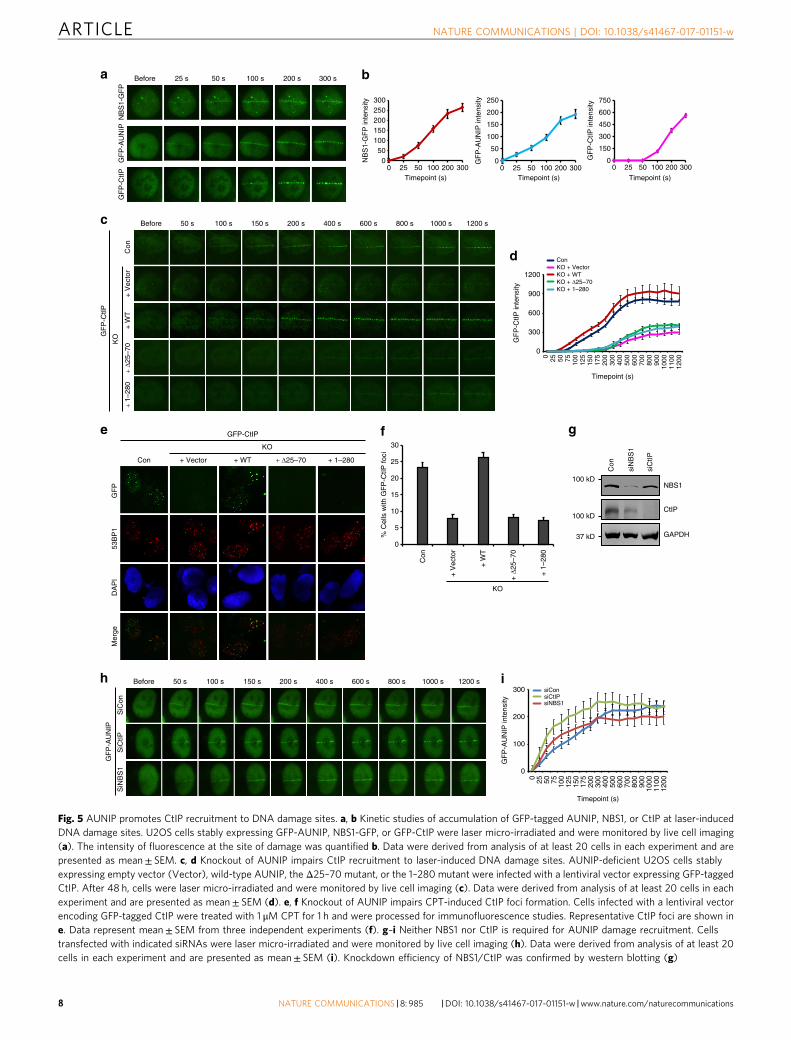
AUNIP promotes CtIP recruitment to sites of DNA damage.To obtain mechanistic insight into how AUNIP contributes toDNA end resection, we assessed whether AUNIP may affect DNAdamage-dependent recruitment of factors known to be involvedin this process. To this end, we first performed time-lapse ima-ging of micro-irradiated U2OS cells that stably express GFP-tagged AUNIP, -CtIP or -NBS1 (the GFP tag was added to the C-terminus of NBS1 to avoid unwanted functional deficits) tocompare the recruitment kinetics of AUNIP with that of CtIP andNBS1. As shown in Fig. 5a, b, the recruitment of NBS1 to sites ofDNA damage could be observed as early as 25 s after micro-irradiation. By contrast, we observed a substantial delay in therecruitment of GFP-CtIP to laser-induced DNA lesions, com-pared with that of NBS1-GFP (Fig. 5a, b). These findings are inagreement with previous observations where the MRN complex isrequired for efficient recruitment of CtIP to sites of DNAdamage19, 46, 47. Remarkably, GFP-AUNIP accumulated at sites oflaser-inflicted DNA damage tracks with kinetics similar to NBS1-GFP, indicating that AUNIP might also act upstream of CtIP inDNA end resection (Fig. 5a, b). Indeed, loss of AUNIP sig-nificantly and reproducibly hampered GFP-CtIP recruitment tolaser-generated DSB tracks (Fig. 5c, d). Consistently, loss ofAUNIP also impaired the recruitment of CtIP to CPT-inducedDNA damage foci (Fig. 5e, f). More importantly, wild-typeAUNIP, but not its mutants that either lack the ability to localizeto sites of DNA damage or in binding to CtIP, completelyrestored CtIP recruitment to DNA damage sites in AUNIP-deficient cells (Fig. 5c–f). These results, taken together with theobservation that CtIP depletion did not noticeably affect AUNIPaccumulation at sites of laser-induced DNA damage (Fig. 5g–i),argue in favor of the idea that CtIP functions downstream of bothMRN and AUNIP in DNA end resection.
We next examined whether the localization of AUNIP to sitesof DNA damage is dependent on the MRN complex or vice versa.As shown in Supplementary Fig. 5a–c, AUNIP deficiency did notaffect the accumulation of NBS1-GFP at sites of laser-inflictedDNA damage in both the presence and absence of H2AX.Similarly, depletion of NBS1 had no significant effect on therecruitment of AUNIP to sites of laser-induced DNA damage(Fig. 5g–i). These results suggest that, despite similar recruitmentkinetics, DNA damage-induced recruitment of AUNIP and theMRN complex may be regulated independently.
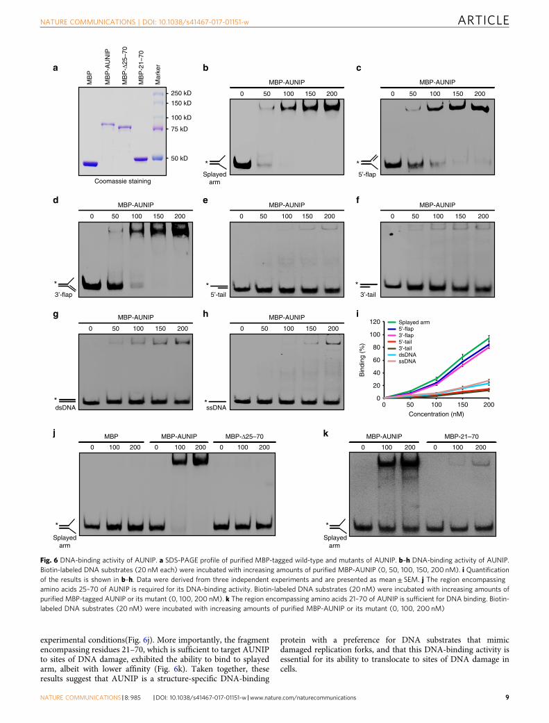
AUNIP is a structure-specific DNA-binding protein. Havingestablished that AUNIP accumulates at DNA lesions and facil-itates CtIP-dependent DNA-end resection, we next sought todetermine the mechanism by which AUNIP is recruited to sites ofDNA damage. We first examined whether AUNIP recruitment toDNA damage sites may be dependent on the ATM or ATR kinasepathway. As shown in Supplementary Fig. 6a, ATM inhibitiondid not noticeably affect AUNIP recruitment to sites of DNAdamage. In line with this notion, depletion of H2AX or RNF8 hadno effect on AUNIP concentration at DNA breaks (Supplemen-tary Fig. 6b, c). Moreover, there was also no significant change inthe localization of AUNIP to sites of DNA damage using an ATRinhibitor (Supplementary Fig. 6a). These findings prompted us tohypothesize that AUNIP might be recruited to sites of DNAdamage via direct interaction with DNA. To test this hypothesis,recombinant wild-type AUNIP was purified and analyzed inelectromobility shift assays against a variety of DNA substrates.As shown in Fig. 6a–i, AUNIP bound strongly to splayed arm, 5′-flap, and 3′-flap DNA substrates, but only weakly to double-stranded DNA (dsDNA), dsDNA with a tail, or ssDNA. Notably,the Δ25–70 deletion mutant, which had lost the ability to accu-mulate at DSB sites, failed to bind to splayed arm under the same
Before 100 s 150 s 200 s50 s 400 s 600 s 1000 s 1200 s800 s
GF
P-A
UN
IP in
tens
ity
i
Timepoint (s)
SiN
BS
1
Con
e
GF
P
53B
P1
Mer
geD
AP
I
f
siCon
siNBS1siCtIP
% C
ells
with
GF
P-C
tIP fo
ci
200
100
300
0
+ Vector + WT + Δ25–70
KO
0 25 50 75 100
125
150
175
200
300
400
500
600
700
800
900
1000
1100
1200
Con
KO
+ V
ecto
r
+ W
T
+ Δ
25–7
0
+ 1
–280
+ 1–280
0
5
10
15
20
25
30
GFP-CtIP
NBS1
CtIP
GAPDH
Con
siN
BS
1
siC
tIP
g
Con
+ V
ecto
r+
WT
+ Δ2
5–70
GF
P-C
tIP in
tens
ity
c
d
Before 100 s 150 s 200 s50 s 400 s 600 s 1000 s 1200 s800 s
GF
P-C
tIP
KO
+ 1–
280
Timepoint (s)
0 25 50 75 100
125
150
175
200
300
400
500
600
700
800
900
1000
1100
1200
1200
900
600
300
0
KO + VectorKO + WTKO + Δ25–70
Con
KO + 1–280
100 kD
37 kD
100 kD
Fig. 5 AUNIP promotes CtIP recruitment to DNA damage sites. a, b Kinetic studies of accumulation of GFP-tagged AUNIP, NBS1, or CtIP at laser-inducedDNA damage sites. U2OS cells stably expressing GFP-AUNIP, NBS1-GFP, or GFP-CtIP were laser micro-irradiated and were monitored by live cell imaging(a). The intensity of fluorescence at the site of damage was quantified b. Data were derived from analysis of at least 20 cells in each experiment and arepresented as mean± SEM. c, d Knockout of AUNIP impairs CtIP recruitment to laser-induced DNA damage sites. AUNIP-deficient U2OS cells stablyexpressing empty vector (Vector), wild-type AUNIP, the Δ25–70 mutant, or the 1–280 mutant were infected with a lentiviral vector expressing GFP-taggedCtIP. After 48 h, cells were laser micro-irradiated and were monitored by live cell imaging (c). Data were derived from analysis of at least 20 cells in eachexperiment and are presented as mean± SEM (d). e, f Knockout of AUNIP impairs CPT-induced CtIP foci formation. Cells infected with a lentiviral vectorencoding GFP-tagged CtIP were treated with 1 μM CPT for 1 h and were processed for immunofluorescence studies. Representative CtIP foci are shown ine. Data represent mean± SEM from three independent experiments (f). g–i Neither NBS1 nor CtIP is required for AUNIP damage recruitment. Cellstransfected with indicated siRNAs were laser micro-irradiated and were monitored by live cell imaging (h). Data were derived from analysis of at least 20cells in each experiment and are presented as mean± SEM (i). Knockdown efficiency of NBS1/CtIP was confirmed by western blotting (g)
experimental conditions(Fig. 6j). More importantly, the fragmentencompassing residues 21–70, which is sufficient to target AUNIPto sites of DNA damage, exhibited the ability to bind to splayedarm, albeit with lower affinity (Fig. 6k). Taken together, theseresults suggest that AUNIP is a structure-specific DNA-binding
protein with a preference for DNA substrates that mimicdamaged replication forks, and that this DNA-binding activity isessential for its ability to translocate to sites of DNA damage incells.
a c
MBP MBP-Δ25–70MBP-AUNIP
b
d
*
*Splayed
arm
3’-flap
250 kD
150 kD
100 kD
75 kD
50 kD
Coomassie staining
MB
P
MB
P-A
UN
IP
MB
P-Δ
25–7
0
MB
P-2
1–70
Mar
ker
*ssDNA
e f
g h
0
20
40
60
80
100
120
0 50 100 150 200
Bin
ding
(%
)
Concentration (nM)
Splayed arm5'-flap3'-flap
3'-taildsDNAssDNA
5'-tail
i
j
MBP-AUNIP
0 50 100 150 200
*5’-flap
MBP-AUNIP
0 50 100 150 200
*dsDNA
MBP-AUNIP
0 50 100 150 200
*3’-tail
MBP-AUNIP
0 50 100 150 200
MBP-AUNIP
0 50 100 150 200
MBP-AUNIP
0 50 100 150 200
*5’-tail
MBP-AUNIP
0 50 100 150 200
MBP-21–70MBP-AUNIP
*Splayed arm
k0 100 200 0 100 200
*Splayed
arm
0 100 200 0 100 200 0 100 200
Fig. 6 DNA-binding activity of AUNIP. a SDS-PAGE profile of purified MBP-tagged wild-type and mutants of AUNIP. b–h DNA-binding activity of AUNIP.Biotin-labeled DNA substrates (20 nM each) were incubated with increasing amounts of purified MBP-AUNIP (0, 50, 100, 150, 200 nM). i Quantificationof the results is shown in b–h. Data were derived from three independent experiments and are presented as mean± SEM. j The region encompassingamino acids 25–70 of AUNIP is required for its DNA-binding activity. Biotin-labeled DNA substrates (20 nM) were incubated with increasing amounts ofpurified MBP-tagged AUNIP or its mutant (0, 100, 200 nM). k The region encompassing amino acids 21–70 of AUNIP is sufficient for DNA binding. Biotin-labeled DNA substrates (20 nM) were incubated with increasing amounts of purified MBP-AUNIP or its mutant (0, 100, 200 nM)
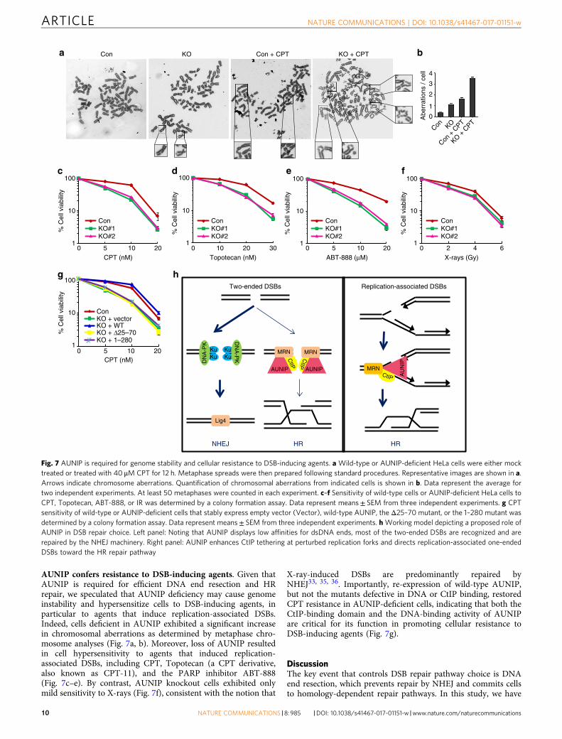
AUNIP confers resistance to DSB-inducing agents. Given thatAUNIP is required for efficient DNA end resection and HRrepair, we speculated that AUNIP deficiency may cause genomeinstability and hypersensitize cells to DSB-inducing agents, inparticular to agents that induce replication-associated DSBs.Indeed, cells deficient in AUNIP exhibited a significant increasein chromosomal aberrations as determined by metaphase chro-mosome analyses (Fig. 7a, b). Moreover, loss of AUNIP resultedin cell hypersensitivity to agents that induced replication-associated DSBs, including CPT, Topotecan (a CPT derivative,also known as CPT-11), and the PARP inhibitor ABT-888(Fig. 7c–e). By contrast, AUNIP knockout cells exhibited onlymild sensitivity to X-rays (Fig. 7f), consistent with the notion that
X-ray-induced DSBs are predominantly repaired byNHEJ33, 35, 36. Importantly, re-expression of wild-type AUNIP,but not the mutants defective in DNA or CtIP binding, restoredCPT resistance in AUNIP-deficient cells, indicating that both theCtIP-binding domain and the DNA-binding activity of AUNIPare critical for its function in promoting cellular resistance toDSB-inducing agents (Fig. 7g).
DiscussionThe key event that controls DSB repair pathway choice is DNAend resection, which prevents repair by NHEJ and commits cellsto homology-dependent repair pathways. In this study, we have
% C
ell v
iabi
lity
% C
ell v
iabi
lity
% C
ell v
iabi
lity
% C
ell v
iabi
lity
% C
ell v
iabi
lity
c d e f
g
CPT (nM) Topotecan (nM) ABT-888 (μM) X-rays (Gy)
1
10
100
0 5 10 201
10
100
0 10 20 301
10
100
0 5 10 20
ConKO#1KO#2
ConKO#1KO#2
ConKO#1KO#2
ConKO#1KO#2
1
10
100
0 2 4 6
a
h
0 5 10 20
ConKO + vectorKO + WTKO + Δ25–70KO + 1–280
CPT (nM)
1
10
100
0
1
2
3
Abe
rrat
ions
/ ce
ll
b
4
Con KO
Con +
CPT
KO + C
PT
Con KO Con + CPT KO + CPT
Two-ended DSBs
HR
Replication-associated DSBs
HR
AUNIP
Lig4
AUNIP
AU
NIP
CtIP
CtIP
CtIP
MRN
MRNMRN
NHEJ
KuKu
KuKu
DN
A-P
K
DN
A-P
K
Fig. 7 AUNIP is required for genome stability and cellular resistance to DSB-inducing agents. a Wild-type or AUNIP-deficient HeLa cells were either mocktreated or treated with 40 μM CPT for 12 h. Metaphase spreads were then prepared following standard procedures. Representative images are shown in a.Arrows indicate chromosome aberrations. Quantification of chromosomal aberrations from indicated cells is shown in b. Data represent the average fortwo independent experiments. At least 50 metaphases were counted in each experiment. c–f Sensitivity of wild-type cells or AUNIP-deficient HeLa cells toCPT, Topotecan, ABT-888, or IR was determined by a colony formation assay. Data represent means± SEM from three independent experiments. g CPTsensitivity of wild-type or AUNIP-deficient cells that stably express empty vector (Vector), wild-type AUNIP, the Δ25–70 mutant, or the 1–280 mutant wasdetermined by a colony formation assay. Data represent means± SEM from three independent experiments. hWorking model depicting a proposed role ofAUNIP in DSB repair choice. Left panel: Noting that AUNIP displays low affinities for dsDNA ends, most of the two-ended DSBs are recognized and arerepaired by the NHEJ machinery. Right panel: AUNIP enhances CtIP tethering at perturbed replication forks and directs replication-associated one-endedDSBs toward the HR repair pathway
provided several lines of evidence to show that AUNIP is a criticalregulator of DNA end resection. First, overexpression of AUNIPpromoted HR and inhibited NHEJ. Consistently, AUNIP silen-cing decreased the frequency of HR but increased the frequencyof NHEJ. Second, AUNIP physically interacted with CtIP and wasrequired for efficient CtIP recruitment to sites of DNA damageand the subsequent CtIP-dependent DNA end resection. Third,AUNIP possessed intrinsic DNA-binding activity, a feature thatwas required for its relocalization to sites of DNA damage and itsfunction in DNA end resection. Finally, AUNIP-deficient cellsshowed a marked increase in cellular sensitivity to DSB-inducingagents, particularly to those that induce replication-associatedDSBs. Our results provide novel insights into how DSBs arerecognized and may be channeled to specific repair pathways.
A scenario is emerging in which the nature of DSBs caused bystalled replication forks is distinct from that caused by X- or γ-rays. Indeed, ample evidence now indicate that DSBs arising fromstalled replication forks are predominantly repaired by HR,whereas those that are induced by X- or γ-rays are mostly pro-cessed by NHEJ33, 35, 36. However, the manner in which thesereplication-associated DSBs are distinguished from two-endedDSBs in cells is largely unclear.
In this study, we found that AUNIP preferentially bound toDNA substrates that mimicked structures generated at stalledreplication forks and was critical for CtIP recruitment to sites ofDNA damage. We proposed that AUNIP may serve as a mole-cular sensor for stalled replication forks, and by tethering CtIP atDNA intermediates generated during replication fork stalling,directs replication-associated DSBs toward the HR repair path-way (Fig. 7h). On the other hand, since AUNIP displayed lowaffinities for dsDNA ends, it is tempting to speculate that the CtIPprotein may not be fully activated at two-ended DSBs, includingthose that are generated by X- or γ-rays (Fig. 7h). This model thusoffers an explanation to address and to further experimentallyexamine why two-ended DSBs are primarily repaired by NHEJ.Importantly, this model is supported by our observations whereAUNIP-deficient cells were hypersensitive to genotoxins thatinduce replication-associated DSBs, but were only mildly sensitiveto X-rays. Thus, the identification of AUNIP represents animportant step toward understanding how cells channel repairprocesses to resolve specific problems associated with DNAdamage.
CtIP and its homologs are well-known molecular switches thatregulate DNA end resection, and their function in this process isstrictly dependent on their appropriate recruitment to sites ofDNA damage. However, exactly how CtIP is recruited to DSBs isnot fully understood. Studies have shown that the MRN complexphysically interacts with CtIP and helps retain CtIP atDSBs19, 46, 47. In addition to its direct involvement in CtIPrecruitment, the MRN complex also facilitates CtIP recruitmentthrough tethering broken DNA ends and in promoting ATMactivation19, 46. It has been suggested that ATM-mediated CtIPphosphorylation may unmask the DNA-binding motif in CtIP,which then binds to DNA at DSBs, resulting in CtIP recruitmentto DSBs19. In resemblance to the MRN complex, AUNIP alsobinds to DNA, forms a complex with CtIP, and is required forefficient CtIP recruitment to sites of DNA damage. However,AUNIP is not involved in ATM activation (SupplementaryFig. 7). Moreover, DNA damage-induced recruitment of AUNIPand the MRN complex are independent of each other. Theseresults indicate that AUNIP may serve as a DNA-binding modulefor the AUNIP-CtIP-MRN complex to target specific DNAlesions, and that the multiple DNA-binding modules on theAUNIP-CtIP-MRN complex might play coordinated roles tosupport full activation of CtIP at sites of DNA damage. Inaddition to AUNIP, the MRN complex, and ATM, several other
proteins including BRCA1, LEDGF/p75, SRCAP, and USP4, arealso involved directly or indirectly in recruiting CtIP to sites ofDNA damage48–52. However, the mechanistic details with whichthese proteins cooperate to support optimal recruitment of CtIPis still not clear and warrants further investigation.
In summary, the identification and biochemical characteriza-tion of the AUNIP protein described here provides new insightsinto the molecular mechanism by which DSBs, in particularreplication-associated DSBs, may be recognized and repaired inhuman cells. Understanding the detailed mechanisms and reg-ulation of this process will help in the design of more efficientanti-cancer therapeutics.
MethodsAntibodies. Polyclonal anti-AUNIP antibodies (1:200 dilution) were generated byimmunizing rabbits with MBP-AUNIP (residues 81–357) or MBP-AUNIP (resi-dues 220–357) fusion proteins expressed and purified from E. Coli. Polyclonal anti-RAD51 (1:2000 dilution) and anti-RNF8 (1:1000 dilution) antibodies were gen-erated by immunizing rabbits with GST-RAD51 (residues 1–339), or GST-RNF8(residues 1–485) fusion proteins expressed and purified from E. Coli, respectively.Antisera were affinity-purified against the immunized antigens using the Amino-Link plus Immobilization and purification kit (Pierce). Polyclonal anti-phospho-RPA2 (S4/S8) (A300-245A, 1:3000 dilution) and anti-NBS1 (A301-284A, 1:1000dilution) antibodies were purchased from Bethyl Laboratories. Anti-CtIP (61141,Clone: 14-1, 1:1000 dilution), anti-MRE11 (GTX70212, 1:1000 dilution), and anti-Myc (9E10, 1:1000 dilution) antibodies were purchased from Active Motif, GeneTex, and Covance, respectively. Anti-RPA2 (ab2175, 1:2000 dilution), anti-RAD50(ab89, 1:1000 dilution), anti-ATM (ab199726, 1:1000 dilution), anti-53BP1(ab175188, 1:100 dilution), anti-H2AX (ab11175, 1:2000 dilution), and anti-phospho-ATM (S1981) (ab81292, 1:1000 dilution) antibodies were purchased fromAbcam. Anti-GAPDH (MAB374, 1:5000 dilution) and anti-phospho-H2AX(Ser319) (Clone JBW301, 1:1000 dilution) antibodies were purchased from Milli-pore. Anti-Flag (Clone M2, 1:10000 dilution) antibody was purchased from Sigma.Uncropped immunoblots are shown in Supplementary Fig. 8.
Constructs. All complementary DNAs were amplified by polymerase chain reac-tion (PCR) and cloned into either pDONR201 or pDONR221 vector according tothe Gateway cloning procedure (Invitrogen). Entry clones were then recombinedinto Gateway-based destination vectors for the expression of N- or C-terminal-tagged fusion proteins. AUNIP deletion mutants were generated using theQuickChange Site-directed mutagenesis kit (Stratagene). All constructs used in thisstudy were confirmed by DNA sequencing.
Cell culture and transfection. HeLa, U2OS, and HEK293T cells purchased fromATCC were cultured in Dulbecco’s modified essential medium containing 10%fetal bovine serum and 1% penicillin and streptomycin and maintained at 37 °C in5% CO2. U2OS DR-GFP and U2OS EJ5-GFP cell lines were kindly provided by Dr.Maria Jasin (Memorial Sloan-Kettering Cancer Center) and Dr. Jeremy Stark(Beckman Research Institute of the City Hope), respectively. All these cell linesused in this study were confirmed to be free of mycoplasma contamination beforeuse. For transient transfection experiments, cells were transfected with expressionvectors using Lipofectamine 2000 (Invitrogen) according to the manufacturer’sinstructions. Small-interfering RNA (siRNA) duplexes targeting AUNIP (#1:CUUGUUU GCUAGACCGAAAUU; #2: CCAUUUGAUCCCAGGCUUAUU),CtIP (GCUAAAACAGGAACGAAUCdTdT), NBS1 (CCAACUAAAUUGCCAA-GUAU U), H2AX (CAACAAGAAGACGCGAAUCdTdT), RNF8 (ACUCAGU-GUCCAACUU GCUdTdT), and a nontargeting control siRNA(UUCAAUAAAUUCUUGAGGUUU) were purchased from Thermo Fisher Sci-entific. For siRNA-mediated depletion experiments, cells were transfected twicewith siRNAs (100 nM) using Lipofectamine RNAiMAX (Invitrogen) according tothe manufacturer’s instructions.
Tandem affinity purification (TAP). HEK293T cells were transfected with plas-mids encoding SFB-tagged CtIP or AUNIP and were selected with medium con-taining 2 μg ml−1 puromycin. Cell lines that stably express SFB-tagged CtIP orAUNIP were confirmed by immunoblotting and immunostaining experiments. Toaffinity purify CtIP- and AUNIP-protein complexes, engineered cells were lysedwith NETN buffer (20 mM Tris-HCl [pH 8.0], 100 mM NaCl, 1 mM EDTA, and0.5% Nonidet P-40) containing protease inhibitors (1 μg ml−1 aprotinin, 1 μg ml−1
leupeptin, and 100 mM PMSF) at 4 °C for 30 min. After centrifugation, the pelletwas resuspended and sonicated in buffer (20 mM HEPES [pH 7.8], 0.4 M NaCl,1 mM EDTA, 1 mM EGTA, and protease inhibitors) for 40 s to extract chromatin-bound proteins. The supernatants were then cleared by centrifugation at 15,000×gfor 10 min at 4 °C to remove debris, and were subsequently incubated in thepresence of with 150 μl of streptavidin-conjugated beads (GE Healthcare) for 2 h at4 °C with gentle rocking. The immunocomplexes were washed three times with
NETN buffer and were eluted with 1 mgml−1 biotin (Sigma-Aldrich) for 1 h at4 °C. The eluates were then incubated with 60 μl of S-protein Agarose beads (EMDMillipore) for 1 h at 4 °C. Proteins bound to S-protein Agarose were washed threetimes with NETN buffer, separated by sodium dodecyl sulfate polyacrylamide gelelectrophoresis (SDS-PAGE), and analyzed by mass spectrometry.
GST pull-down assays. The coding sequence of wild-type CtIP was subcloned intopCold-MBP vector and transformed into BL21 E. Coli cells. At OD600 0.6, cellswere induced with 0.1 mM IPTG at 16 °C for 16 h. Cells were then pelleted,resuspended in lysis buffer (20 mM Tris-HCl, 300 mM NaCl, 1% Triton X-100, and1 μg ml−1 each of leupeptin, aprotinin, and pepstatin) and sonicated on ice. The celllysates were cleared by centrifugation at 40,000×g for 40 min at 4 °C and wereincubated with Amylose resin for 2 h at 4 °C. After washing the beads with washingbuffer (20 mM Tris-HCl, 500 mM NaCl, 0.5% NP-40, 1 mM DL-Dithiothreitol(DTT), and 1 μg ml−1 each of leupeptin, aprotinin, and pepstatin), bound proteinswere eluted with lysis buffer containing 10 mM Maltose. The coding sequences ofwild-type and mutant AUNIP were subcloned into pCold-GST vector and trans-formed into E. Coli BL21 cells. At OD600 0.6, cells were induced with 0.2 mM IPTGat 16 °C for 16 h. Cells were then harvested, resuspended in lysis buffer (20 mMTris-HCl, 300 mM NaCl, 1% Triton X-100, and 1 μg ml−1 each of leupeptin,aprotinin, and pepstatin) and sonicated on ice. The cell lysates were cleared bycentrifugation at 40,000×g for 40 min at 4 °C and incubated with glutathione-Sepharose resins for 2 h at 4 °C. After washing the beads with washing buffer(20 mM Tris-HCl, 500 mM NaCl, 0.5% NP-40, 1 mM DTT, and 1 μg ml−1 each ofleupeptin, aprotinin, and pepstatin), the bound proteins were used for pull-downassays. For in vitro pull-down assays, MBP-CtIP were incubated with GST or GST-AUNIP in NETN buffer for 2 h at 4 °C. The beads-bound proteins were thenwashed five times with NETN buffer and resolved on SDS-PAGE. MBP-CtIP wasdetected by a monoclonal anti-CtIP antibody.
Co-immunoprecipitation and western blotting. Cells were lysed with NETNbuffer containing 20 mM NaF and protease inhibitors (1 μg ml−1 aprotinin andleupeptin) on ice for 20 min. After centrifugation, the supernatants were incubatedwith either S-protein Agarose beads (EMD Millipore) or Protein A-Sepharosecoupled with 2 μg of indicated antibodies for 4 h at 4 °C with gentle rocking. Thebeads-bound proteins were then washed three times with NETN buffer and wereresolved on SDS-PAGE. Immunoblotting was performed according to standardprocedures.
HR and NHEJ reporter assays. In all, 0.5 × 10⁶ U2OS DR-GFP or U2OS EJ5-GFPcells were seeded in 6-well plates and were electroporated with 3 μg I-SceIexpression plasmid (pCBASce) 24 h after. 48 h post pCBASce electroporation, cellswere harvested and were subjected to flow cytometry analysis for GFP expression.Means were obtained from three independent experiments.
CRISPR/Cas9 gene editing. For CRISPR/Cas9-mediated knockout of AUNIP, thefollowing guide RNAs (gRNAs) were used: AUNIP#1: GTGCTAAGCCTGG-GATCAAA and AUNIP#2: GGAGGCCTGCGGCGTG TGGC. The gRNAsequences were cloned into the pX330-U6-Chimeric_BB-CBh-hSpCas9 vector(kindly provided by Dr. Feng Zhang) according to standard protocols53, 54. Cellswere transfected with the gRNA/Cas9 expression construct and were selected inmedium containing 2 μg ml−1 puromycin. AUNIP expression was analyzed bywestern blotting with anti-AUNIP antibody.
Immunofluorescence staining. Cells were cultured on coverslips and were treatedwith 1 μM CPT or 10 Gy X-rays for the indicated times. Cells were then washedwith PBS, permeabilized with PBS buffer containing 0.5% Triton X-100 for 5 min atroom temperature, and were subsequently fixed with 3% paraformaldehyde for10 min at room temperature. Coverslips were then blocked with 5% milk for10 min before incubation with primary antibodies for 20 min at room temperature.Coverslips were washed three times with PBS before they were incubated withsecondary antibodies for an additional 20 min at room temperature. Coverslipswere then incubated with 4′,6-diamidino-2-phenylindole (DAPI) to visualizenuclear DNA. Images were captured with use of a fluorescence microscope (Eclipse80i; Nikon) equipped with a Plan Fluor 60× oil objective lens (NA 0.5–1.25; Nikon)and a camera (CoolSNAP HQ2; PHOTOMETRICS).
Detecting ssDNA lesions by BrdU incorporation. Forty-eight hour post siRNAtransfection, U2OS cells were incubated with 10 μM BrdU (Sigma) for 24 h, fol-lowed by treatment with 1 μM CPT for 1 h. After pre-extraction for 5 min in PBScontaining 0.5% Triton X-100, cells were fixed with 3% paraformaldehyde solutionfor 10 min at room temperature. Subsequently, fixed cells were immunostainedwith anti-BrdU antibodies for 20 min at room temperature. Following three 5-minwashes with PBS, cells were incubated with secondary antibodies for 20 min atroom temperature. DAPI staining was then performed to visualize nuclear DNA.
Laser micro-irradiation and live-cell imaging. Laser micro-irradiation was per-formed as described previously55. Briefly, cells cultured on glass-bottomed 35-mm
dishes were micro-irradiated using a computer-controlled MicroPoint laser Abla-tion System (Photonics Instruments; 365 nm, 20 Hz) coupled to a Nikon EclipseTi-E inverted microscope (63× oil-immersion objective). Time-lapse images of livecells were taken under the same microscope with the MetaMorph MicroscopeAutomation & Image Analysis software.
Recombinant protein expression and purification. The coding sequences ofwild-type and mutant AUNIP were subcloned into pCold-MBP vector andtransformed into BL21 E. Coli cells. At OD600 0.6, cells were induced with 0.1 mMIPTG at 16 °C for 16 h. Cells were then harvested, resuspended in lysis buffer(20 mM Hepes [PH 7.5], 300 mM NaCl, 1 mM DTT, and 1 μg ml−1 each of leu-peptin and aprotinin) and sonicated on ice. The cell lysates were cleared by cen-trifugation at 40,000×g for 40 min at 4 °C and incubated with Amylose resin for 4 hat 4 °C. After washing the beads with washing buffer (20 mM Hepes [PH 7.5],500 mM NaCl, 1 mM DTT, and 1 μg ml−1 each of leupeptin and aprotinin),bound proteins were eluted with lysis buffer containing 10 mM Maltose. Elutedproteins were dialyzed in dilution buffer (20 mM Hepes [PH 7.5], 1 mM DTT, and1 μg ml−1 each of leupeptin and aprotinin) and were loaded on pre-equilibrated1 ml Hitrap Q HP column (GE Healthcare). Column was washed with 8 ml bufferA (20 mM Hepes [PH 7.5], 1 mM DTT and 1 μg ml−1 each of leupeptin andaprotinin) and proteins were then eluted with a 50 ml gradient of buffer A con-taining 0–500 mM NaCl. Peak protein fractions were pooled and were con-centrated with a 30-kDa Amicon Ultra centrifugal filter device (Millipore).
Electrophoretic mobility shift assay (EMSA). The EMSA assay was conductedusing the Lightshift chemiluminescent EMSA kit (20148, Pierce) according to themanufacturer’s instructions. Briefly, 20 nM biotin-labeled DNA substrates wereincubated with indicated concentrations of AUNIP protein in 1× binding buffercontaining 10 mM MgCl2, 2.5% glycerol, and 0.05% Nonidet P-40 for 20 min atroom temperature. Reactions were then stopped by the addition of 5 μl of gelloading buffer, resolved on a 10% native polyacrylamide gel, and transferred to aPVDF (polyvinylidene fluoride) membrane on ice. After UV (120 mJ per cm2)cross-linking of the DNA to membrane, biotin-labeled DNA was detected using theChemiluminescent Nucleic Acid Detection Module. The splayed arm substrate wasgenerated by annealing oligo 1 (5′-GACGCTGCCGAATTCTACC AGTGCCTTGCTAGGACATCTTTGCCCACCTGCAG GTTCAC-3′) with oligo 2 (5′-ATAGTCGGATCCTCTAGACAGCTCCATGTAGCAAGGCACTGGTAGAATTCGGCAGCGTC-3′). The blunt-ended DNA substrate was generated by annealing oligo1 with oligo 3 (5′-GTGAACCTGCAGGTGGGCAAAGATGTCCTAGCAAGGCACTGGTAG AATTCG GCAGCGTC-3′). The 3′ tail DNA substrate was generatedby annealing oligo 1 with oligo 4 (5′-TAGCAAGGCACTG GTAGAATTCGGCAGCGTC-3′). The 5′-flap structure was generated by annealing oligo 1 with oligo 2and oligo 5 (5′-GTGAACCTG CAGGTGGGCAAAGATGTCC-3′). The 3′-flapstructure was generated by annealing oligo 1 with oligo 2 and oligo 6 (5′-CATGGAGCTGTCTAGAGGATC CGACTAT-3′). Oligo 1 was 5′-biotin-labeled and theannealing reaction contains 250 nM oligo1 and 750 nM each of oligo 2, oligo 3,oligo 4, oligo 5, and oligo 6. All DNA substrates were purified by gelelectrophoresis.
Lentivirus packaging and infection. AUNIP, CtIP, and NBS1 entry cloneswere transferred into a lentivirus-based, Gateway-compatible destination vectorwith N- or C-terminal GFP fusion tag. Lentiviruses were produced inHEK293T cells by co-transfection of the lentiviral-based construct with thepackaging plasmids pMD2G and pSPAX2 (kindly provided by Dr. Songyang Zhou,Baylor College of Medicine). Forty-eight hour after transfection, infectious lenti-viruses were harvested and were used for the transduction of HeLa or U2OS cells inthe presence of 8 μg ml−1 polybrene (Sigma). Stable cell pools were selected inmedium containing 500 μg ml−1 G418 (Calbiochem).
Retrovirus production and infection. Wild-type and mutant AUNIP entry cloneswere transferred into a Gateway-compatible retroviral destination vector pEF1A-HA-Flag. Retroviruses were produced in HEK293T cells by co-transfection of theretroviral plasmid with the packaging plasmids pCL-ECO and VSV-G. Forty-eighthour after transfection, viral supernatant were harvested and were used for thetransduction of HeLa cells in the presence of 8 μg ml−1 polybrene (Sigma). Stablepools were selected in medium containing 2 μg ml−1 puromycin (Calbiochem).
BrdU incorporation assays. AUNIP knockout HeLa cells were split and trans-ferred onto 60 mm dishes. Twenty-four hours later, cells were incubated with 100μM BrdU for 1 h. Harvested cells were then washed with PBS, fixed in ice-cold 70%ethanol, and stored at 4 °C. DNA was subsequently denatured by using 2.5 M HClfor 1 h at room temperature. After three washes with PBS, cells were incubated withanti-BrdU antibody (Roche) diluted 1:100 in blocking buffer (PBS + 0.1% Triton X-100 + 5% BSA) for 12 h followed by incubation with the secondary FITC-conjugated anti-mouse antibody (1:100, Jackson immunoresearch) for 4 h at roomtemperature. Finally, cells were stained at 37 °C for 20 min with propidium iodide(20 μg ml−1) and RNase A (200 μg ml−1), and were analyzed on a FACScan flowcytometer (Beckman).
Analysis of chromosomal aberrations. HeLa cells were either mock treated ortreated with 40 μM CPT for 12 h. Cells were then exposed to 1 μg ml−1 colcemid for4 h and were swollen using 75 mM KCl for 15 min at 37 °C. After fixing inmethanol/acetic acid (3:1) (vol/vol) for 20 min, cells were dropped onto ice-coldwet slides, air dried, and were stained with 5% Giemsa for 5 min. The number ofchromosome aberrations were scored in 50 metaphases per sample.
Cell survival assays. HeLa cells (1 × 10³) were seeded onto 60-mm dish in tri-plicates. At 24 h after plating, cells were treated with CPT, Topotecan, ABT-888, orIR at indicated doses. Twenty-four hours later, drug-containing medium wasreplaced with fresh medium and cells were cultured for additional 14 days at 37 °Cto allow colony formation. Resulting colonies were then stained with Coomassieblue and counted.
Data availability. The data that support the findings of this study are availablefrom the corresponding author upon request.
Received: 7 March 2017 Accepted: 21 August 2017
References1. Jackson, S. P. & Bartek, J. The DNA-damage response in human biology and
disease. Nature 461, 1071–1078 (2009).2. Hartlerode, A. J. & Scully, R. Mechanisms of double-strand break repair in
somatic mammalian cells. Biochem. J. 423, 157–168 (2009).3. Pardo, B., Gomez-Gonzalez, B. & Aguilera, A. DNA repair in mammalian cells:
DNA double-strand break repair: how to fix a broken relationship. Cell. Mol.Life Sci. 66, 1039–1056 (2009).
4. San Filippo, J., Sung, P. & Klein, H. Mechanism of eukaryotic homologousrecombination. Ann. Rev. Biochem. 77, 229–257 (2008).
5. Moynahan, M. E. & Jasin, M. Mitotic homologous recombination maintainsgenomic stability and suppresses tumorigenesis. Nat. Rev. Mol. Cell Biol. 11,196–207 (2010).
6. Liu, T. & Huang, J. Quality control of homologous recombination. Cell. Mol.Life Sci. 71, 3779–3797 (2014).
7. Chiruvella, K. K., Liang, Z. & Wilson, T. E. Repair of double-strand breaks byend joining. Cold Spring Harbor. Perspect. Biol. 5, a012757 (2013).
8. Lieber, M. R. The mechanism of double-strand DNA break repair by thenonhomologous DNA end-joining pathway. Ann. Rev. Biochem. 79, 181–211(2010).
9. Orthwein, A. et al. Mitosis inhibits DNA double-strand break repair to guardagainst telomere fusions. Science 344, 189–193 (2014).
10. Ochi, T. et al. DNA repair. PAXX, a paralog of XRCC4 and XLF, interacts withKu to promote DNA double-strand break repair. Science 347, 185–188 (2015).
11. Xing, M. et al. Interactome analysis identifies a new paralogue of XRCC4 innon-homologous end joining DNA repair pathway. Nat. Commun. 6, 6233(2015).
12. Bernstein, K. A. & Rothstein, R. At loose ends: resecting a double-strand break.Cell 137, 807–810 (2009).
13. Huertas, P. DNA resection in eukaryotes: deciding how to fix the break. Nat.Struct. Mol. Biol. 17, 11–16 (2010).
14. Garcia, V., Phelps, S. E., Gray, S. & Neale, M. J. Bidirectional resection of DNAdouble-strand breaks by Mre11 and Exo1. Nature 479, 241–244 (2011).
15. Clerici, M., Mantiero, D., Lucchini, G. & Longhese, M. P. The Saccharomycescerevisiae Sae2 protein promotes resection and bridging of double strand breakends. J. Biol. Chem. 280, 38631–38638 (2005).
16. Limbo, O. et al. Ctp1 is a cell-cycle-regulated protein that functions with Mre11complex to control double-strand break repair by homologous recombination.Mol. Cell 28, 134–146 (2007).
17. Sartori, A. A. et al. Human CtIP promotes DNA end resection. Nature 450,509–514 (2007).
18. Nicolette, M. L. et al. Mre11-Rad50-Xrs2 and Sae2 promote 5′ strand resectionof DNA double-strand breaks. Nat. Struct. Mol. Biol. 17, 1478–1485 (2010).
19. You, Z. et al. CtIP links DNA double-strand break sensing to resection. Mol.Cell 36, 954–969 (2009).
20. Mimitou, E. P. & Symington, L. S. Sae2, Exo1 and Sgs1 collaborate in DNAdouble-strand break processing. Nature 455, 770–774 (2008).
21. Zhu, Z., Chung, W. H., Shim, E. Y., Lee, S. E. & Ira, G. Sgs1 helicase and twonucleases Dna2 and Exo1 resect DNA double-strand break ends. Cell 134,981–994 (2008).
22. Gravel, S., Chapman, J. R., Magill, C. & Jackson, S. P. DNA helicases Sgs1 andBLM promote DNA double-strand break resection. Genes Dev. 22, 2767–2772(2008).
23. Cejka, P. et al. DNA end resection by Dna2-Sgs1-RPA and its stimulation byTop3-Rmi1 and Mre11-Rad50-Xrs2. Nature 467, 112–116 (2010).
24. Chapman, J. R., Taylor, M. R. & Boulton, S. J. Playing the end game: DNAdouble-strand break repair pathway choice. Mol. Cell 47, 497–510 (2012).
25. Symington, L. S. & Gautier, J. Double-strand break end resection and repairpathway choice. Ann. Rev. Genet. 45, 247–271 (2011).
26. Daley, J. M. & Sung, P. 53BP1, BRCA1, and the choice between recombinationand end joining at DNA double-strand breaks. Mol. Cell. Biol. 34, 1380–1388(2014).
27. Panier, S. & Boulton, S. J. Double-strand break repair: 53BP1 comes into focus.Nat. Rev. Mol. Cell Biol. 15, 7–18 (2014).
28. Huertas, P., Cortes-Ledesma, F., Sartori, A. A., Aguilera, A. & Jackson, S. P.CDK targets Sae2 to control DNA-end resection and homologousrecombination. Nature 455, 689–692 (2008).
29. Steger, M. et al. Prolyl isomerase PIN1 regulates DNA double-strand breakrepair by counteracting DNA end resection. Mol. Cell 50, 333–343 (2013).
30. Wang, H. et al. The interaction of CtIP and Nbs1 connects CDK and ATM toregulate HR-mediated double-strand break repair. PLoS Genet. 9, e1003277(2013).
31. Wyman, C. & Kanaar, R. DNA double-strand break repair: all’s well that endswell. Ann. Rev. Genet. 40, 363–383 (2006).
32. Helleday, T., Lo, J., van Gent, D. C. & Engelward, B. P. DNA double-strandbreak repair: from mechanistic understanding to cancer treatment. DNA Repair6, 923–935 (2007).
33. Shibata, A. et al. Factors determining DNA double-strand break repair pathwaychoice in G2 phase. EMBO J. 30, 1079–1092 (2011).
34. Beucher, A. et al. ATM and Artemis promote homologous recombination ofradiation-induced DNA double-strand breaks in G2. EMBO J. 28, 3413–3427(2009).
35. Wang, H. et al. Nonhomologous end-joining of ionizing radiation-inducedDNA double-stranded breaks in human tumor cells deficient in BRCA1 orBRCA2. Cancer Res. 61, 270–277 (2001).
36. Wang, H. et al. Efficient rejoining of radiation-induced DNA double-strandbreaks in vertebrate cells deficient in genes of the RAD52 epistasis group.Oncogene 20, 2212–2224 (2001).
37. Helleday, T. Pathways for mitotic homologous recombination in mammaliancells. Mutat. Res. 532, 103–115 (2003).
38. Sakasai, R. & Iwabuchi, K. The distinctive cellular responses to DNA strandbreaks caused by a DNA topoisomerase I poison in conjunction with DNAreplication and RNA transcription. Genes Genet. Syst. 90, 187–194 (2016).
39. Bunting, S. F. & Nussenzweig, A. End-joining, translocations and cancer. Nat.Rev. Cancer 13, 443–454 (2013).
40. Adachi, N., So, S. & Koyama, H. Loss of nonhomologous end joining conferscamptothecin resistance in DT40 cells. Implications for the repair oftopoisomerase I-mediated DNA damage. J. Biol. Chem. 279, 37343–37348(2004).
41. Patel, A. G., Sarkaria, J. N. & Kaufmann, S. H. Nonhomologous end joiningdrives poly(ADP-ribose) polymerase (PARP) inhibitor lethality in homologousrecombination-deficient cells. Proc. Natl Acad. Sci. USA 108, 3406–3411 (2011).
42. Lieu, A. S. et al. Functional characterization of AIBp, a novel Aurora-A bindingprotein in centrosome structure and spindle formation. Int. J. Oncol. 37,429–436 (2010).
43. Kaulich, M. et al. Efficient CRISPR-rAAV engineering of endogenous genes tostudy protein function by allele-specific RNAi. Nucleic Acids Res. 43, e45(2015).
44. Weinstock, D. M., Nakanishi, K., Helgadottir, H. R. & Jasin, M. Assayingdouble-strand break repair pathway choice in mammalian cells using a targetedendonuclease or the RAG recombinase. Methods Enzymol. 409, 524–540(2006).
45. Bennardo, N., Cheng, A., Huang, N. & Stark, J. M. Alternative-NHEJ is amechanistically distinct pathway of mammalian chromosome break repair.PLoS Genet. 4, e1000110 (2008).
46. Yuan, J. & Chen, J. N terminus of CtIP is critical for homologousrecombination-mediated double-strand break repair. J. Biol. Chem. 284,31746–31752 (2009).
47. Chen, L., Nievera, C. J., Lee, A. Y. & Wu, X. Cell cycle-dependent complexformation of BRCA1.CtIP.MRN is important for DNA double-strand breakrepair. J. Biol. Chem. 283, 7713–7720 (2008).
48. Yu, X., Fu, S., Lai, M., Baer, R. & Chen, J. BRCA1 ubiquitinates itsphosphorylation-dependent binding partner CtIP. Genes Dev. 20, 1721–1726(2006).
49. Daugaard, M. et al. LEDGF (p75) promotes DNA-end resection andhomologous recombination. Nat. Struct. Mol. Biol. 19, 803–810 (2012).
50. Dong, S. et al. The human SRCAP chromatin remodeling complex promotesDNA-end resection. Curr. Biol. 24, 2097–2110 (2014).
51. Liu, H. et al. The deubiquitylating enzyme USP4 cooperates with CtIP in DNAdouble-strand break end resection. Cell Rep. 13, 93–107 (2015).
52. Wijnhoven, P. et al. USP4 Auto-Deubiquitylation Promotes HomologousRecombination. Mol. Cell 60, 362–373 (2015).
53. Cong, L. et al. Multiplex genome engineering using CRISPR/Cas systems.Science 339, 819–823 (2013).
54. Mali, P. et al. RNA-guided human genome engineering via Cas9. Science 339,823–826 (2013).
55. Mu, Y. et al. SLFN11 inhibits checkpoint maintenance and homologousrecombination repair. EMBO Rep. 17, 94–109 (2016).
AcknowledgementsWe thank M. Jasin and J. Stark for U2OS DR-GFP and EJ5-GFP cell lines, and all ourcolleagues in the Huang laboratory for insightful discussions. This work was supported inpart by the Key Program of the National Natural Science Foundation of China(31730021), National Natural Science Funds for Distinguished Young Scholar, NationalProgram for Special Support of Eminent Professionals, National Basic Research Programof China Grant 2013CB911003, National Natural Science Foundation of China Grant81661128008, 31571397 and 31071243, and the China’s Fundamental Research Funds forthe Central Universities.
Author contributionsJ.H., T.L., X.-H.F., M.S.Y.H., and J.L. designed the experiments; J.L., H.C., J.H.H., andH.H. performed the experiments; J.L., H.C., J.H.H., T.L., and J.H. analyzed the data; J.H.and T.L. wrote the manuscript.
Additional informationSupplementary Information accompanies this paper at doi:10.1038/s41467-017-01151-w.
Competing interests: The authors declare no competing financial interests.
Reprints and permission information is available online at http://npg.nature.com/reprintsandpermissions/
Publisher's note: Springer Nature remains neutral with regard to jurisdictional claims inpublished maps and institutional affiliations.
Open Access This article is licensed under a Creative CommonsAttribution 4.0 International License, which permits use, sharing,
adaptation, distribution and reproduction in any medium or format, as long as you giveappropriate credit to the original author(s) and the source, provide a link to the CreativeCommons license, and indicate if changes were made. The images or other third partymaterial in this article are included in the article’s Creative Commons license, unlessindicated otherwise in a credit line to the material. If material is not included in thearticle’s Creative Commons license and your intended use is not permitted by statutoryregulation or exceeds the permitted use, you will need to obtain permission directly fromthe copyright holder. To view a copy of this license, visit http://creativecommons.org/licenses/by/4.0/.


![Promotionof DNA Strand Breaks in … · (CANCER RESEARCH 48, 3094-3099, June 1, 1988] Promotionof DNA Strand Breaks in CoculturedMononuclearLeukocytes by Protein Kinase C-dependentProoxidative](https://static.documents.pub/doc/80x56/5e5db883e6c6855edb662cb6/promotionof-dna-strand-breaks-in-cancer-research-48-3094-3099-june-1-1988-promotionof.jpg)


