19
Australian Hip Surveillance Guidelines for Children with Cerebral Palsy 2014 Wynter M, Gibson N, Kentish M, Love SC, Thomason P, Willoughby K, Graham HK

Australian Hip Surveillance Guidelines for Children with Cerebral Palsy 2014Wynter M, Gibson N, Kentish M, Love SC, Thomason P, Willoughby K, Graham HK
Australian Hip Surveillance Guidelines for Children with Cerebral Palsy: 2014 | 01
Every child should be referred for hip surveillance1 at the time cerebral palsy (CP)2 is identified.
The reported rates of hip displacement3 and hip dislocation3 in children with CP2 vary widely and have been reported from 2% to 75% (Bagg et al, 1993). More recent population studies have identified the rate of hip displacement3 to be around 30%. Hip displacement3 is not related to the movement disorder but is related directly to gross motor function as determined by the Gross Motor Function Classification System (GMFCS)4 (Soo et al, 2006, Hagglund et al, 2007, Connelly et al, 2009, Kentish et al, 2011). Hip dislocation3 is preventable through early identification and intervention.
Hip surveillance1 is the process of identifying and monitoring the critical early indicators of progressive hip displacement3. Early identification is an essential part of the strategy for prevention of hip displacement3 and its sequelae3. These Hip Surveillance1 Guidelines document the recommended process for screening, monitoring and triaging to orthopaedic services as part of the overall prevention of hip dislocation3 (Kentish et al, 2011, Terjesen, 2012). Surgical recommendations and management guidelines do not form part of this document.
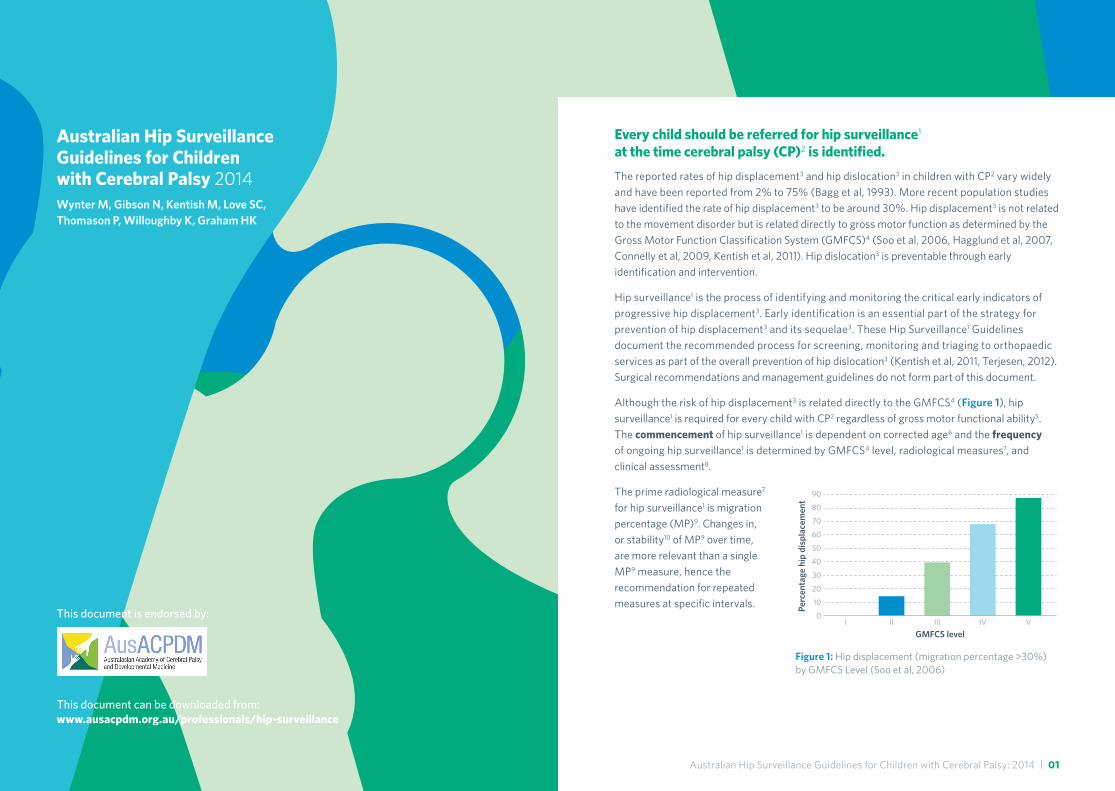
Although the risk of hip displacement3 is related directly to the GMFCS4 (Figure 1), hip surveillance1 is required for every child with CP2 regardless of gross motor functional ability5. The commencement of hip surveillance1 is dependent on corrected age6 and the frequency of ongoing hip surveillance1 is determined by GMFCS4 level, radiological measures7, and clinical assessment8.
The prime radiological measure7 for hip surveillance1 is migration percentage (MP)9. Changes in, or stability10 of MP9 over time, are more relevant than a single MP9 measure, hence the recommendation for repeated measures at specific intervals.
Figure 1: Hip displacement (migration percentage >30%) by GMFCS Level (Soo et al, 2006)
90
80
70
60
50
40
30
20
10
II III IV VI0
Perc
enta
ge h
ip d
ispl
acem
ent
GMFCS level
Australian Hip Surveillance Guidelines for Children with Cerebral Palsy 2014Wynter M, Gibson N, Kentish M, Love SC, Thomason P, Willoughby K, Graham HK
This document is endorsed by:
This document can be downloaded from: www.ausacpdm.org.au/professionals/hip-surveillance

02 | Australian Hip Surveillance Guidelines for Children with Cerebral Palsy: 2014 Australian Hip Surveillance Guidelines for Children with Cerebral Palsy: 2014 | 03
GMFCS II
• Initial clinical assessment8 and AP pelvic radiograph11 at twelve to twenty-four months of age6 (or at identification if older than twenty-four months)
• Review twelve months later
– Verify GMFCS4 level
~ If GMFCS4 II confirmed13, repeat clinical assessment8 and AP pelvic radiograph11
~ If GMFCS4 level has changed, ongoing surveillance1 according to confirmed13 classification
– If MP9 is abnormal15 and/or unstable10, continue twelve monthly surveillance1 until stability10 is established
– When MP9 is stable10, review at four to five years of age6
• Review at four to five years of age6
– Verify GMFCS4 level
~ If GMFCS4 level II confirmed13, repeat clinical assessment8 and AP pelvic radiograph11
~ If GMFCS4 level has changed, or if identified as WGH IV12 hemiplegia (Figure 2), ongoing surveillance1 according to confirmed13 classification
– If MP9 is stable10, review at eight to ten years of age6
– If MP9 is abnormal15 and/or unstable10, continue twelve monthly surveillance1 until stability10 is established
• Review at eight to ten years of age6, prepuberty16
– Verify GMFCS4 level
~ If GMFCS4 II confirmed13, repeat clinical assessment8 and AP pelvic radiograph11
~ If GMFCS4 level has changed, or if identified as WGH IV12 hemiplegia (Figure 2), ongoing surveillance1 according to confirmed13 classification1
– If MP9 is stable10, discharge14 from surveillance1
– If MP9 is abnormal15 and/or unstable10, continue twelve monthly surveillance1 until stability10 is established or skeletal maturity17
• In the presence of pelvic obliquity19, leg length discrepancy19 or deteriorating gait20, continue twelve monthly surveillance1
GMFCS I
• Initial clinical assessment8 and antero-posterior (AP) pelvic radiograph11 at twelve to twenty-four months of age6 (or at identification if older than twenty-four months)
• Review at three years of age6
– Verify GMFCS4 level
~ If GMFCS4 I is confirmed13, repeat clinical assessment8. AP pelvic radiograph11 is NOT required
~ If GMFCS4 level has changed, ongoing surveillance1 according to confirmed13 classification
– If identified as group IV hemiplegia as described by Winters, Gage and Hicks (WGH)12, 1987 in Figure 2, ongoing surveillance1 according to WGH IV12 classification
• Review at five years of age6
– Verify GMFCS4 level
~ If GMFCS4 I is confirmed13, repeat clinical assessment8. AP pelvic radiograph11 is NOT required and if nil other significant signs, discharge14 from surveillance1
~ If GMFCS4 level has changed, ongoing surveillance1 according to confirmed13 classification
– If identified as WGH IV12 hemiplegia (Figure 2), ongoing surveillance1 according to WGH IV12 classification
Recommended frequency of hip surveillance
GMFCS I
GMFCS II

04 | Australian Hip Surveillance Guidelines for Children with Cerebral Palsy: 2014 Australian Hip Surveillance Guidelines for Children with Cerebral Palsy: 2014 | 05
GMFCS IV
• Initial clinical assessment8 and AP pelvic radiograph11 at twelve to twenty-four months of age6
• Review six months later
– Verify GMFCS4 level
~ If GMFCS4 IV confirmed13, repeat clinical assessment8 and AP pelvic radiograph11
~ If GMFCS4 level has changed, ongoing surveillance1 according to confirmed13 classification
– If MP9 is abnormal15 and/or unstable10, continue six monthly surveillance1 until MP9 stability10 is established
– When MP9 is stable10, reduce frequency of surveillance1 to twelve monthly
• Review at seven years of age6
– If MP9 is stable10, below 30% and gross motor function5 is stable, surveillance1 may be discontinued until prepuberty16
– Twelve monthly AP pelvic radiographs11 must resume prepuberty16 and continue until skeletal maturity17
• Independent of MP9, when clinical8 and/or radiographic evidence of scoliosis18 or pelvic obliquity19 is present, six monthly surveillance1 is required until skeletal maturity17
• At skeletal maturity17, if MP9 is abnormal15 and progressive scoliosis18 or significant pelvic obliquity19 is present, continue twelve monthly surveillance1
GMFCS III
• Initial clinical assessment8 and AP pelvic radiograph11 at twelve to twenty-four months of age6
• Review six months later
– Verify GMFCS4 level
~ If GMFCS4 III confirmed13, repeat clinical assessment8 and AP pelvic radiograph11
~ If GMFCS4 level has changed, ongoing surveillance1 according to confirmed13 classification
– If MP9 is abnormal15 and/or unstable10, continue six monthly surveillance1 until MP9 stability10 is established
– When MP9 is stable10, reduce frequency to twelve monthly surveillance1
• Review at seven years of age6
– Verify GMFCS4 level
~ If GMFCS4 III confirmed13, repeat clinical assessment8 and AP pelvic radiograph11
~ If GMFCS4 level has changed, ongoing surveillance1 according to confirmed13 classification
– If MP9 is abnormal15 and/or unstable10, continue six monthly surveillance1 until MP9 stability10 is established
– If MP9 is stable10, below 30%, and gross motor function5 is stable, AP pelvic radiographs11 may be discontinued until prepuberty16
– Twelve monthly AP pelvic radiographs11 must resume prepuberty16 and continue until skeletal maturity17
• At skeletal maturity17, in the presence of pelvic obliquity19, leg length discrepancy19 or deteriorating gait20, continue twelve monthly surveillance1
GMFCS III
GMFCS IV
06 | Australian Hip Surveillance Guidelines for Children with Cerebral Palsy: 2014 Australian Hip Surveillance Guidelines for Children with Cerebral Palsy: 2014 | 07
Winters, Gage and Hicks hemiplegia group IV (WGH IV)12
WGH IV12 gait20 pattern clearly declares itself by four to five years of age6.
The child with a classification of WGH IV12 has the potential for late onset progressive hip displacement3 regardless of GMFCS4 level.
• Review at five years of age6
– Verify WGH12 and GMFCS4
~ If WGH I–III12, ongoing hip surveillance1 according to confirmed13 GMFCS4
~ If WGH IV12 and MP9 stable10, review ten years of age6
– If MP9 is abnormal15 and/or unstable10, continue twelve monthly surveillance1 until MP9 stability10 established
• Review at ten years of age6
– Verify WGH IV12
~ If WGH IV12 confirmed13, repeat clinical assessment8 and AP pelvic radiograph11
~ Continue twelve monthly surveillance1 until skeletal maturity17
• At skeletal maturity17 if significant scoliosis18, pelvic obliquity19, leg length discrepancy19 or deteriorating gait20, continue twelve monthly surveillance1
GMFCS V
• Initial clinical assessment8 and AP pelvic radiograph11 at twelve to twenty-four months of age6
• Review six months later
• Repeat clinical assessment8 and AP pelvic radiograph11
– Verify GMFCS4 level
~ If GMFCS4 V confirmed13, continue six monthly surveillance1 until seven years of age6 or until MP9 stability10 is established
~ If GMFCS4 level has changed, ongoing surveillance1 according to confirmed13 classification
• Review at seven years of age6
– If MP9 is stable10, below 30% and gross motor function5 is stable, continue twelve monthly surveillance1 until skeletal maturity17
• Independent of MP9, when clinical8 and/or radiographic evidence of scoliosis18 or pelvic obliquity19 is present, six monthly surveillance1 is required until skeletal maturity17
• At skeletal maturity17, if MP9 is abnormal15 and progressive scoliosis18 or significant pelvic obliquity19 is present, continue twelve monthly surveillance1
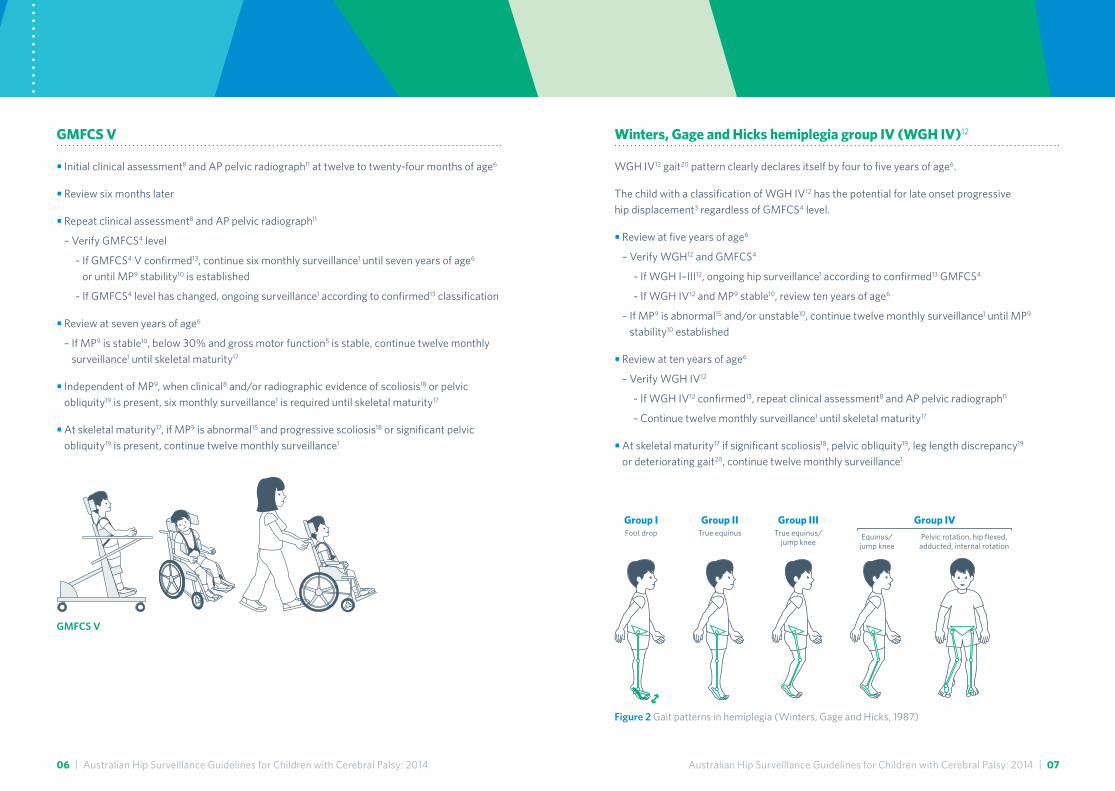
Group IFoot drop
Group IITrue equinus
Group IIITrue equinus/
jump knee
Group IVPelvic rotation, hip flexed,adducted, internal rotation
Equinus/jump knee
Figure 2 Gait patterns in hemiplegia (Winters, Gage and Hicks, 1987)
GMFCS V

08 | Australian Hip Surveillance Guidelines for Children with Cerebral Palsy: 2014 Australian Hip Surveillance Guidelines for Children with Cerebral Palsy: 2014 | 09
Hip Surveillance after skeletal maturity and transition into adulthood
• As part of transition29 the hip should be classified according to the Melbourne Cerebral Palsy Hip Classification Scale (MCPHCS)30 (Figure 8)
– if MCPHCS30 hip classification IV or V, refer for ongoing orthopaedic review27
– if MCPHCS30 II or III advise regarding future hip health31
• Ongoing referral for orthopaedic review27 should occur in the presence of pain25, progressive scoliosis18, significant pelvic obliquity19 and/or deteriorating function5
Increased frequency of hip surveillance will be required when:
• Deterioration in function5 including altered gait20, decreased ability or tolerance of sitting or standing
• Presence of scoliosis18, pelvic obliquity19, or significant leg length discrepancy19
• Deterioration in musculoskeletal measures21 relating to the hip
– change in muscle tone22, including, but not limited to, increasing levels of spasticity23
– reduced range of movement21, reduced muscle length21, development of, or increased asymmetry24 of range of movement21
• Increased difficulty with perineal care/hygiene
• Onset of, or increase in pain25 related to the hip
Referral to orthopaedic consultant should occur when:
• MP9 is unstable10 and/or progresses to greater than 30%
• There is pain25 related to the hip
• Other orthopaedic conditions26 are identified
The intention of hip surveillance1 is that orthopaedic review27 occurs at the appropriate time. Every child referred to orthopaedic services should be managed with an individual treatment plan27 which may include ongoing hip surveillance1.
Referral back to hip surveillance should occur following:
• The postoperative period for any child who has undergone surgery for hip management27
• An unplanned break in surveillance1 for any other medical reason
• Neurosurgical interventions28 such as selective dorsal rhizotomy28, or intrathecal baclofen (ITB)28
Increased frequency of hip surveillance
10 | Australian Hip Surveillance Guidelines for Children with Cerebral Palsy: 2014 Australian Hip Surveillance Guidelines for Children with Cerebral Palsy: 2014 | 11
2. Cerebral palsy
The term cerebral palsy (CP) refers to cerebral palsy and like conditions, where clinical signs or descriptions are most relevant, not aetiology.
In line with the decision made by the Surveillance of Cerebral Palsy in Europe (SCPE, 2000) and methodology adopted in 2003 by The Australian Cerebral Palsy Register Group, (Blair et al, 2007), for the purposes of this document any definition of CP is accepted that includes the following five key elements (Mutch et al, 1992):
1. CP is a group of disorders i.e. it is an umbrella term
2. It involves a disorder of movement and/or posture and of motor function
3. It is due to a non-progressive interference/lesion/abnormality; and
4. This interference/lesion/abnormality is in the developing/immature brain
5. It is permanent but not unchanging
An international review of “The Definition and Classification of Cerebral Palsy” in 2006 defines CP as:
“ A group of permanent disorders of the development of movement and posture, causing activity limitation, that are attributed to non-progressive disturbances that occurred in the developing foetal or infant brain. The motor disorders of cerebral palsy are often accompanied by disturbances of sensation, perception, cognition, communication, and behaviour, by epilepsy, and by secondary musculoskeletal problems.” Rosenbaum et al, 2007
This definition was annotated in an attempt to provide better clarification of the classification and description of CP. The definition is now widely accepted internationally.
In conditions other than CP, where there is no evidence for the natural history of hip displacement3, the risk seems likely to also relate to functional ability5. It is posited that the more clinically similar a child’s condition is to CP, the more likely that these guidelines will be effective in identifying hips at risk.
For the purposes of these guidelines, like conditions refers to those conditions where motor dysfunction results from genetic and metabolic aetiologies, including clearly recognised syndromes or recognisable progressive brain disorders (Badawi et al, 1998), or from brain injury acquired in childhood within the first two to three years of life.
1. Hip surveillance
Hip surveillance is the process of monitoring and identifying the critical early indicators of progressive hip displacement3. These early indicators include GMFCS4, age6, gait classification (WGH IV)12 and MP9. The information gathered from the clinical assessment8 and radiological review11 are vital components of hip surveillance and are required to capture often silent displacement3 of the hip while minimising radiation exposure. Hip surveillance cannot be based on clinical assessment8 alone.
Hip surveillance will assist identification of prognosis for the hip; inform planning for ongoing hip management; support education and assist clear communication. Surgical recommendations and management guidelines are beyond the scope of this document.
Hip surveillance is an ongoing process that continues for every child until skeletal maturity17 or discharge14. Hip surveillance should recommence: following the post operative period for any child who has undergone surgery for hip management32, following neurosurgical interventions28 such as selective dorsal rhizotomy28, or intrathecal baclofen28, or following an unplanned break in surveillance for any other medical reason.
All children with CP2 or like conditions should be referred for hip surveillance even if classification and determination of GMFCS4 are not yet confirmed13.
A body of evidence supports the implementation of hip surveillance as an effective means towards prevention of hip dislocation3. A systematic review of the evidence for children with CP2 (Gordon and Simkiss, 2006) identified six studies where results showed support for hip surveillance programs. All studies used radiological measures7 to monitor hip displacement3, with MP9 (Reimers, 1980) most frequently used. The monitoring of MP9 enabled identification of children for surgery at a younger age6, thus reducing the need for later salvage surgery32.
The initial Consensus Statement on Hip Surveillance for Children with Cerebral Palsy: Australian Standards of Care 2008 (Wynter et al, 2011) documented the commencement and frequency of hip surveillance, where surveillance is based on risk relative to GMFCS4 level. Since the development and implementation of these guidelines in 2008, two population based studies (Kentish et al, 2011, Terjesen, 2012) have demonstrated the effectiveness of hip surveillance programs at identifying progressive hip displacement3 in children with CP2.
The 2008 Consensus Statement has been reviewed and updated to incorporate new evidence in this area.
Annotations and references

12 | Australian Hip Surveillance Guidelines for Children with Cerebral Palsy: 2014 Australian Hip Surveillance Guidelines for Children with Cerebral Palsy: 2014 | 13
The GMFCS has five levels for describing differences in severity of motor abilities5. Distinctions between levels are based on functional limitations, the need for hand-held mobility devices or wheeled mobility, and to a much lesser extent, quality of movement. Since classification of motor function5 is dependent on age6, separate descriptions are provided for several age6 bands within each level. The age6 ranges described are as follows: before 2nd birthday, from 2nd to 4th birthday, from 4th to 6th birthday, from 6th to 12th birthday, and from 12th to 18th birthday. There is a tendency for children classified prior to six years of age6 to be reclassified after six years of age6 (Palisano et al, 2006) hence the need to confirm GMFCS level at each occasion of clinical presentation.
The distinctions between Levels I and II are not as pronounced as the distinctions between the other levels, particularly for infants less than two years of age6. Emphasis is on what the child can do (usual performance in home, school, and community settings), rather than what the child may be able to achieve at their best (capability). It is therefore important to classify current performance in gross motor function5 and not to include judgments about the quality of movement or prognosis for improvement. Generally it takes only a few minutes to assign a GMFCS classification.
The GMFCS (Palisano et al, 1997,) and the GMFCS: Expanded and Revised (E & R) (Palisano et al, 2007) can be downloaded free of charge from the website: motorgrowth.canchild.ca/en/gmfcs/resources/gmfcs-er.pdf
5. Gross motor functional ability
Gross motor functional ability refers to the gross motor activities that the child is able to accomplish in his/her own environment (performance) rather than what he/she may be able to achieve in a testing situation (capability). Gross motor functional ability includes the achievement of developmental milestones.
6. Corrected age
Assessment for hip surveillance1 takes into consideration corrected age for prematurity up to two years of age. Pre-term or premature is defined as a gestational age less than 36 weeks. To calculate corrected age subtract the expected date of birth (i.e. not actual date of birth) from the date of evaluation.
In the absence of natural history data for children with acquired brain injury, early and frequent surveillance1 is recommended, as clinical experience indicates a high prevalence of hip displacement3 in this group.
Motor disorders of spinal, peripheral nerve, muscular or mechanical origin are not considered like conditions.
Disorders of impaired cognition with no gross motor impairment are not considered like conditions.
3. Progressive hip displacement, dislocation and sequelae
Progressive hip displacement refers to the gradual displacement of the femoral head laterally out of the acetabulum. This displacement is expressed as a migration percentage (MP)9.
Hip Subluxation defines the state of the hip joint and can be used interchangeably with hip displacement where MP9 is between 10% and 99%.
Hip Dislocation is defined when the femoral head is completely displaced laterally out of the acetabulum (MP9 = 100%).
The sequelae of progressive hip displacement are variable (Cornell, 1995). Progressive displacement can result in asymmetric pressure that may deform the femoral head and or acetabulum (also termed acetabular dysplasia). Hip dysplasia may lead to degeneration of articular cartilage and pain25. Problems with limited range of movement21 and pain25 can interfere with function5, ability to be positioned, hygiene and personal care. In a large subset of children the progressive displacement can develop into dislocation of one or both hips (Cooke et al, 1989).
4. The Gross Motor Function Classification System (GMFCS)
The Gross Motor Function Classification System (GMFCS) is used to describe the gross motor function5 of children with CP2. The GMFCS was published in 1997 and expanded and revised in 2007. When referring to GMFCS in these guidelines the authors are referring to the expanded and revised version of the GMFCS.
The GMFCS classifies the gross motor function5 of children and youth with CP2 on the basis of their self-initiated movement with particular emphasis on sitting, walking, and wheeled mobility (Palisano et al, 1997, Palisano et al, 2007, Palisano et al, 2008).
14 | Australian Hip Surveillance Guidelines for Children with Cerebral Palsy: 2014 Australian Hip Surveillance Guidelines for Children with Cerebral Palsy: 2014 | 15
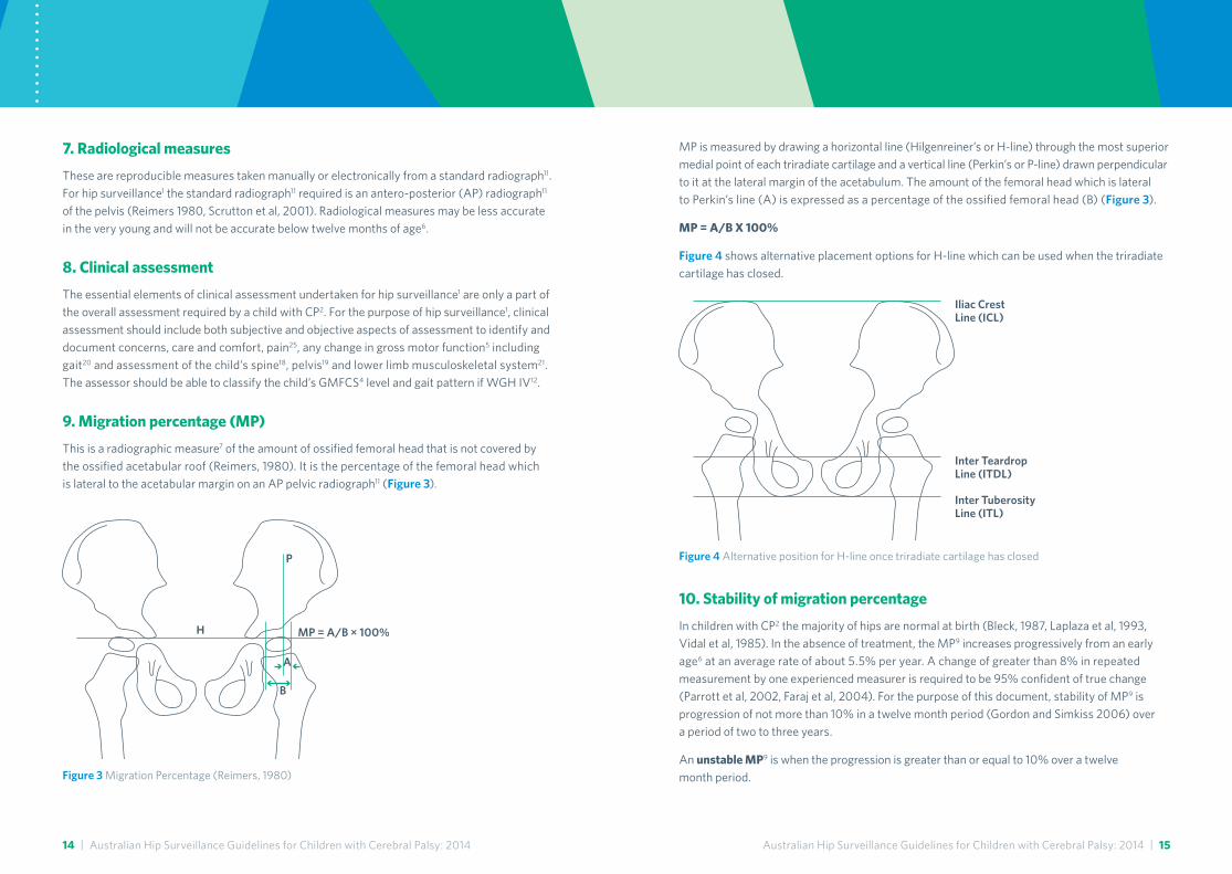
MP is measured by drawing a horizontal line (Hilgenreiner’s or H-line) through the most superior medial point of each triradiate cartilage and a vertical line (Perkin’s or P-line) drawn perpendicular to it at the lateral margin of the acetabulum. The amount of the femoral head which is lateral to Perkin’s line (A) is expressed as a percentage of the ossified femoral head (B) (Figure 3).
MP = A/B X 100%
Figure 4 shows alternative placement options for H-line which can be used when the triradiate cartilage has closed.
10. Stability of migration percentage
In children with CP2 the majority of hips are normal at birth (Bleck, 1987, Laplaza et al, 1993, Vidal et al, 1985). In the absence of treatment, the MP9 increases progressively from an early age6 at an average rate of about 5.5% per year. A change of greater than 8% in repeated measurement by one experienced measurer is required to be 95% confident of true change (Parrott et al, 2002, Faraj et al, 2004). For the purpose of this document, stability of MP9 is progression of not more than 10% in a twelve month period (Gordon and Simkiss 2006) over a period of two to three years.
An unstable MP9 is when the progression is greater than or equal to 10% over a twelve month period.
7. Radiological measures
These are reproducible measures taken manually or electronically from a standard radiograph11. For hip surveillance1 the standard radiograph11 required is an antero-posterior (AP) radiograph11
of the pelvis (Reimers 1980, Scrutton et al, 2001). Radiological measures may be less accurate in the very young and will not be accurate below twelve months of age6.
8. Clinical assessment
The essential elements of clinical assessment undertaken for hip surveillance1 are only a part of the overall assessment required by a child with CP2. For the purpose of hip surveillance1, clinical assessment should include both subjective and objective aspects of assessment to identify and document concerns, care and comfort, pain25, any change in gross motor function5 including gait20 and assessment of the child’s spine18, pelvis19 and lower limb musculoskeletal system21. The assessor should be able to classify the child’s GMFCS4 level and gait pattern if WGH IV12.
9. Migration percentage (MP)
This is a radiographic measure7 of the amount of ossified femoral head that is not covered by the ossified acetabular roof (Reimers, 1980). It is the percentage of the femoral head which is lateral to the acetabular margin on an AP pelvic radiograph11 (Figure 3).
P
A
B
MP = A/B × 100%H
Figure 3 Migration Percentage (Reimers, 1980)
Iliac Crest Line (ICL)
Inter Teardrop Line (ITDL)
Inter Tuberosity Line (ITL)
Figure 4 Alternative position for H-line once triradiate cartilage has closed
16 | Australian Hip Surveillance Guidelines for Children with Cerebral Palsy: 2014 Australian Hip Surveillance Guidelines for Children with Cerebral Palsy: 2014 | 17
12. Winters, Gage and Hicks classification
Winters, Gage and Hicks (WGH) classification of hemiplegic gait20 describes four types of gait20 pattern based on the sagittal plane kinematics of the ankle, knee, hip and pelvis (Winters et al, 1987). The characteristic of each group is as follows:
Group I foot drop in the swing phase of gait20, normal dorsiflexion range in stance phase of gait20
Group II excessive plantarflexion of the ankle in both stance and swing phase of gait20
Group III Group II deviations as above plus limited flexion/extension range of motion at the knee during stance and swing phases of gait20
Group IV Group III deviations as above plus limited flexion/extension range of motion at the hip during stance and swing phases of gait20
This is represented diagrammatically in Figure 2.
There are limitations in using this classification as it is based only on sagittal plane kinematics (Dobson et al, 2006). Many children with hemiplegia will present with coronal and transverse plane gait20 deviations that may predispose them to a higher risk of hip displacement3 than those with only sagittal plane deviations. Hence children with coronal or transverse plane abnormalities particularly at the hip level should also be considered in this group for the purposes of hip surveillance1. While this classification is based on three dimensional gait analysis kinematic data, visual observation of gait20 and musculoskeletal measures21 relating to the hip are sufficient for classification of WGH IV for the purpose of hip surveillance1. Children classified as WGH IV are those at risk of progressive hip displacement3. Children with WGH IV develop displacement3 later than children with bilateral CP2 and the hip MP9 progresses slowly until puberty16. Presentation at puberty16 may be characterised by pain25, rapid increasing leg length discrepancy19, apparent leg length discrepancy19 and/or pelvic obliquity19.
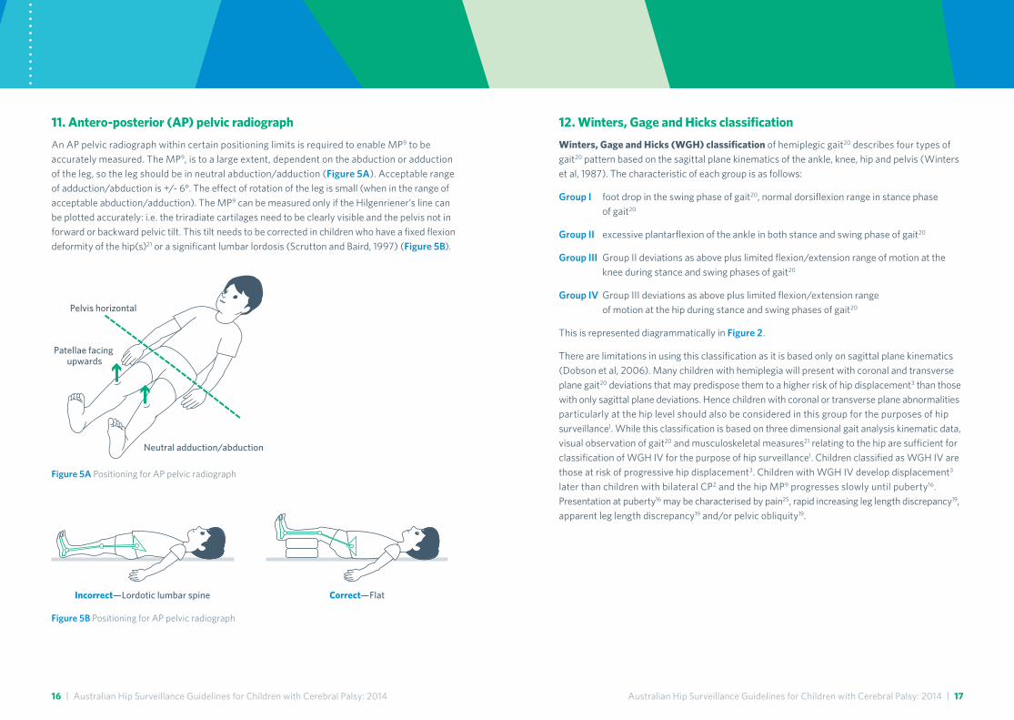
11. Antero-posterior (AP) pelvic radiograph
An AP pelvic radiograph within certain positioning limits is required to enable MP9 to be accurately measured. The MP9, is to a large extent, dependent on the abduction or adduction of the leg, so the leg should be in neutral abduction/adduction (Figure 5A). Acceptable range of adduction/abduction is +/- 6°. The effect of rotation of the leg is small (when in the range of acceptable abduction/adduction). The MP9 can be measured only if the Hilgenriener’s line can be plotted accurately: i.e. the triradiate cartilages need to be clearly visible and the pelvis not in forward or backward pelvic tilt. This tilt needs to be corrected in children who have a fixed flexion deformity of the hip(s)21 or a significant lumbar lordosis (Scrutton and Baird, 1997) (Figure 5B).
Figure 5A Positioning for AP pelvic radiograph
Figure 5B Positioning for AP pelvic radiograph
Neutral adduction/abduction
Pelvis horizontal
Patellae facing upwards
Incorrect—Lordotic lumbar spine Correct—Flat

18 | Australian Hip Surveillance Guidelines for Children with Cerebral Palsy: 2014 Australian Hip Surveillance Guidelines for Children with Cerebral Palsy: 2014 | 19
17. Skeletal maturity
There are a number of operational definitions of skeletal maturity from radiographic parameters which may be selected according to the patient population. One of the earliest is closure of the triradiate cartilage (Dimeglio, 2006) which is followed by closure of the growth plate of the olecranon apophysis at the elbow, followed by progressive capping and closure of the iliac apophysis, also known as the Risser sign (Risser 1958) (Figure 6).
The closure of the triradiate cartilage (Acheson, 1957) can be a useful marker if the radiograph11 does not include the iliac crests. For adolescents who are GMFCS4 I–III this may suffice. However, for adolescents at GMFCS4 IV and V, the prevalence of scoliosis18 and pelvic obliquity19 is high and it is suggested that skeletal maturity should be judged using the Risser sign which requires an AP radiograph11 of the pelvis including the iliac crests.
13. Confirmed GMFCS
For the purpose of this document confirmed is defined as the GMFCS4 level which best fits on today’s assessment. GMFCS4 levels may not always be distinct or easily apparent, particularly for the younger child and between the higher levels (Palisano et al, 1997, Gorter et al, 2009). It is important to reassess for the correct GMFCS4 level on each occasion of hip surveillance1.
14. Discharge
Discharge is the cessation or release from continuing surveillance1. Children will most often be involved with other management programs including spasticity23 management or orthopaedic gait20 corrective surgery according to best practise and evidence based medicine. Gait20 corrective surgery may simultaneously address displacement3 of the femoral head whilst correcting other bony alignment.
15. Normal/abnormal migration percentage
A normal MP9 is considered to be zero or even negative as displacement3 should not occur in a normal hip (Perkins, 1928). Reimers (1980) found that among children with normal motor development, the 90th centile for hip migration at four years of age6 was 10%. For the purpose of these guidelines, normal MP9 is less than 10% after the corrected age6 of four years. A MP9 above 30% is high and should be considered at risk/abnormal.
16. Puberty
Puberty can be recognised by a combination of growth acceleration, development of secondary sexual characteristics, chronological age6 and bone age. Bone age can be assessed with a range of radiological investigations of which radiograph of the wrist or elbow are the most widely used. In typically developing children, girls will experience the onset of puberty at eleven years (bone age) and boys at 13 years (bone age) but there is wide variation in both typically developing children and even more so in children with CP2. In typically developing children, about 50% have a bone age that is significantly different from their chronological age6 and in CP2 the percentage is even higher (Dimeglio, 2006). Delayed bone age is particularly common in severe CP2 (GMFCS4 IV and V) and it is probable that the pattern of skeletal maturation varies by GMFCS4 level. Although hip displacement3 may occur in children with CP2 from early childhood, the pubertal growth spurt is a period of particular risk for both progression of existing hip displacement3, the development of hip displacement3 in previously stable10 hips, as well as the development of pelvic obliquity19 and scoliosis18.
0 1
3
2
4 5
Figure 6 Risser’s sign
20 | Australian Hip Surveillance Guidelines for Children with Cerebral Palsy: 2014 Australian Hip Surveillance Guidelines for Children with Cerebral Palsy: 2014 | 21
It is important to determine the contributions of both real and apparent shortening in the evaluation of leg length discrepancy as well as the contribution of suprapelvic and infrapelvic factors. This is done by careful clinical examination21 of real and apparent leg length with interpretation of this information with radiographs of the pelvis and/or spine. Although unilateral hip subluxation3 and dislocation3 may result in a real leg length discrepancy, there is frequently a combination of real and apparent discrepancy.
20. Gait
Gait describes the particular manner or way of moving on foot. It is the description of locomotion style. Alterations in gait that may necessitate increased frequency of hip surveillance1 may include increasing asymmetry24 of the pelvis with retraction or pelvic obliquity19, increased hip adduction21 or internal rotation21, changes or increased asymmetry24 of step length. This is by no means inclusive of all possible gait deviations.
18. Scoliosis
In CP2 most spinal deformities involve neuromuscular scoliosis although sagittal plane deformities such as kyphosis (thoracic spine) and lordosis (lumbar spine) are also common. Spinal deformities in children with CP2 are related to the severity of involvement and are most common in GMFCS4 IV and V (Miller, 2005). Initially the problems are postural but tend to progress rapidly and become fixed24 during puberty16.
Radiographic surveillance for spinal deformity should include antero-posterior11 and lateral radiographs of the whole spine including the pelvis. These radiographs should be taken with the least amount of support required i.e. standing independently for children and adolescents at GMFCS4 I and II, standing with the usual support for children and adolescents who function5 at GMFCS4 III, and sitting with support for children and adolescents who function5 at GMFCS4 IV and V. For some children and adolescents with severe fixed deformities, supine radiographs are sometimes the only feasible technique.
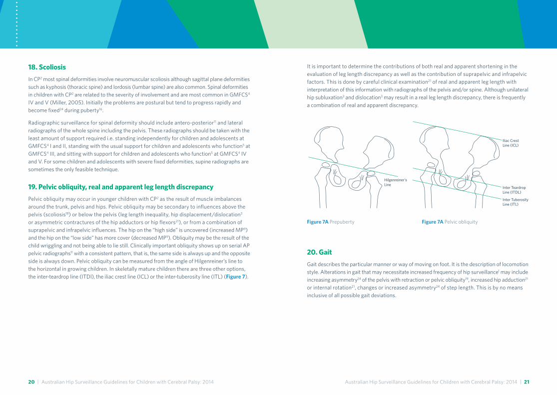
19. Pelvic obliquity, real and apparent leg length discrepancy
Pelvic obliquity may occur in younger children with CP2 as the result of muscle imbalances around the trunk, pelvis and hips. Pelvic obliquity may be secondary to influences above the pelvis (scoliosis18) or below the pelvis (leg length inequality, hip displacement/dislocation3 or asymmetric contractures of the hip adductors or hip flexors21), or from a combination of suprapelvic and infrapelvic influences. The hip on the “high side” is uncovered (increased MP9) and the hip on the “low side” has more cover (decreased MP9). Obliquity may be the result of the child wriggling and not being able to lie still. Clinically important obliquity shows up on serial AP pelvic radiographs11 with a consistent pattern, that is, the same side is always up and the opposite side is always down. Pelvic obliquity can be measured from the angle of Hilgenreiner’s line to the horizontal in growing children. In skeletally mature children there are three other options, the inter-teardrop line (ITDl), the iliac crest line (ICL) or the inter-tuberosity line (ITL) (Figure 7).
Hilgenreiner's Line
Inter Teardrop Line (ITDL)
Inter Tuberosity Line (ITL)
Iliac Crest Line (ICL)
Figure 7A Prepuberty Figure 7A Pelvic obliquity

22 | Australian Hip Surveillance Guidelines for Children with Cerebral Palsy: 2014 Australian Hip Surveillance Guidelines for Children with Cerebral Palsy: 2014 | 23
22. Muscle tone
Muscle tone refers to the normal resting tension or the change in the resistance of the muscle to passive movement or muscle lengthening. It excludes resistance as a result of joint, ligament, or skeletal properties such as those that may occur with fixed deformities, including contracture (Sanger et al, 2003). An abnormal increase in resistance to passive movement is termed hypertonia. Hypertonia may be the result of a number of factors, one of which is spasticity23.
23. Spasticity
Spasticity is a disorder of the motor system characterised by a velocity dependent increase in muscle tone22 with exaggerated tendon jerks, resulting from hyperexcitabilty of the stretch reflex. It is one component of the upper motor neuron syndrome, along with released flexor reflexes, weakness, and loss of dexterity (Mayer, 2002). When spasticity is present, the resistance to externally imposed movement rises rapidly above a threshold speed or joint angle (Delgado and Albright 2003, Sanger et al, 2003). Spasticity does not worsen with age but its manifestation of movement such as the paucity of variety of movement, may result in worsening of secondary effects of spasticity e.g. contractures (Delgado and Albright, 2003).
The Modified Tardieu Scale (MTS) is a rating of spasticity that measures the intensity of muscle reaction at maximal velocity movement through range (Boyd and Graham, 1999). The quality of the muscle response is noted if there is a “catch” in motion and the angle at which the “catch” occurs is measured. The “catch” is sometimes referred to as R1, the first resistance to rapid passive movement. It is described as the clinical estimate of the threshold angle of spasticity (Boyd and Graham, 1999). A lowering of the “threshold” for R1 (i.e. an earlier “catch”), may be an indication that there is increasing spasticity. Spasticity can be graded using the Modified Ashworth Scale (MAS) (Bohannon and Smith, 1987).
21. Musculoskeletal measures relating to the hip
Musculoskeletal measures relating to the hip should include assessment of the spine18, pelvis19, leg length discrepancy19 and physical examination of the lower limbs including passive and dynamic range of movement (Boyd and Graham, 1999), muscle strength, and measures of spasticity23.
Assessment of musculoskeletal measures around the hip should include;
• Passive range of movement
– Hip abduction with hips at 90 degrees of flexion
– Hip abduction with hips at 0 degrees of flexion
– Thomas test
– Hip flexion
– Hip extension (Staheli)
– Hip internal rotation
– Hip external rotation
– Femoral neck anteversion (FNA)
– Popliteal angle
– Pelvic obliquity19
• Real and apparent leg length
• Dynamic contracture as measured by Modified Tardieu Scale23 (Boyd and Graham, 1999)
– Hip adductors
– Hamstrings
• Modified Ashworth Scale23 (Bohannon and Smith, 1987)
– Hip adductors
– Hamstrings
– Hip flexors
• Functional mobility
– Functional Mobility Scale (FMS) (Graham et al, 2004)
• Assessment of pain25 about the hip

24 | Australian Hip Surveillance Guidelines for Children with Cerebral Palsy: 2014 Australian Hip Surveillance Guidelines for Children with Cerebral Palsy: 2014 | 25
26. Other orthopaedic conditions
Other orthopaedic conditions include, but are not limited to, developmental dysplasia of the hip, muscle contracture that is not able to be managed conservatively, an inflammatory reaction, such as transient or toxic synovitis, a slipped capital epiphyses, Perthes Disease, excessive femoral anteversion, juvenile idiopathic arthritis, septic arthritis or bursitis, osteomyelitis, other unusual bone or joint anomalies and in rare cases, bone tumours.
27. Individualised management plan
Individualised management plan is the adaptation of a standard management plan in response to individual clinical presentation and need. This management plan may include ongoing hip surveillance1, altered frequency of surveillance1 and/or intervention including surgical intervention32.
28. Neurosurgical interventions
Neurosurgical interventions include those directed at the central nervous system to modulate spasticity23 and movement disorders. Selective dorsal rhizotomy (SDR) is a neurosurgical procedure used in children with CP2 to reduce spasticity23 in the lower limb by surgically interrupting the afferent input of the monosynaptic stretch reflex. The procedure involves dividing the dorsal root into separate rootlets and only a portion of these are transected, leaving the others intact, thereby preserving sensory function and minimising sphincter dysfunction (Grunt et al, 2013).
Continuous intrathecal Baclofen transfusion (ITB) involves the administration of Baclofen directly to the cerebrospinal fluid, by way of a surgically implanted pump with a catheter directed into the intrathecal space. The continuous administration of Baclofen acts directly at the level of the spinal cord to reduce spasticity23 and abnormal posturing.
Referral back to hip surveillance1 should occur following neurosurgical interventions.
24. Fixed posture and asymmetry
Fixed posture describes structural changes to the posture/mobility of the trunk and/or limbs that cannot be voluntarily or passively corrected. This can be assessed clinically8 and/or radiologically11 and is differentiated from non-structural postural changes which may be fully corrected.
Asymmetry is dissimilarity in corresponding parts on opposite sides of the body which are normally alike. Fixed asymmetry describes structural changes to the trunk18, pelvis19 and/or limbs characterised by the lack or absence of symmetry which cannot be voluntarily or passively corrected. This can be assessed clinically and/or radiologically11 and is differentiated from non-structural postural changes which may be fully corrected.
Newly developed is a clinical sign or measure of recent onset which was not apparent at the previous assessment8, or is subjectively described by the patient/caregiver as having recently appeared.
25. Pain
Pain in the hip region for children with CP2 is variably reported in the literature and may or may not be associated with hip displacement3 or dislocation3. In some cases pain may be clinically expressed in the knee or leg but be referred from the hip. The relationship between hip pain and displacement3 or dislocation3 remains elusive in children and adults. Chronic musculoskeletal pain is a complaint in up to 73% of children (Parkinson et al, 2010) and up to 67% of adults with CP2 (Engel et al, 2003), most commonly in the low back, hip, (Engel et al, 2003, Jahnsen et al, 2004, Opheim et al, 2009) and leg (Engel et al, 2003, Parkinson et al, 2013).
In non-ambulatory adolescents with CP2 pain has been reported at rest, with certain positions, or with such movements as passive abduction21 (Hodgkinson et al, 2001). Identifying the source of pain in the region of the hip is a challenge. In children with limited communication, the clinician must rely on the perception of the parents or caregivers to help identify the source. Pain may originate in the skin or subcutaneous tissues, the musculature surrounding the hip, the osteoarticular structures, or may be referred from another location (Spiegel and Flynn 2006).
Pain should be measured and recorded as part of the clinical assessment8 for hip surveillance1.

26 | Australian Hip Surveillance Guidelines for Children with Cerebral Palsy: 2014 Australian Hip Surveillance Guidelines for Children with Cerebral Palsy: 2014 | 27
29. Transition
Transition is defined as “the purposeful planned movement of adolescents and young adults with chronic physical and medical conditions from child-centred to adult-oriented health care systems” (Blum et al, 1993).
Transition from hip surveillance1 will occur at the point of discharge14 from surveillance1 or at the conclusion of paediatric services. Young people with CP2 with a risk related to future pain25 or progressive hip displacement3 require advice, information, and at times referral to adult services to ensure optimal hip health31 in the future.
Classification of The Melbourne Cerebral Palsy Hip Classification Scale (MCPHCS)30 at skeletal maturity17 is required to identify hips at risk of future progressive displacement3, pain25 associated with arthritic changes, or dislocation3. The presentation of MCPHCS30 III or IV in young people GMFCS4 II or III and/or WGH IV12, may benefit from counselling on the possibility of future interventions for optimising hip health31. The presentation of MCPHCS30 IV or V in young people with progressive scoliosis18 and/or pelvic obliquity19 requires ongoing hip surveillance1 as hip dislocation3 in this population remains an ongoing risk.
Summary documentation at transition, must include details of orthopaedic interventions32 for the hip.
30. The Melbourne Cerebral Palsy Hip Classification Scale (MCPHCS)
The Melbourne Cerebral Palsy Hip Classification Scale (MCPHCS) (Robin et al, 2009) is a five level ordinal grading system, which was designed to describe hip morphology at skeletal maturity17 for young people with CP2 across all GMFCS4 levels. The classification covers a wide range of radiographic features, from a Grade I (normal15 hip), through to a Grade V (dislocated hip3). The classification includes sub-classifications for femoral head deformity, acetabular deformity and pelvic obliquity19. For detail of the sub-classifications refer to the published paper (Robin et al, 2009). A Grade VI was added to denote that the hip joint has been lost to some form of salvage surgery32. The utilisation of MP9 in the MCPHCS ensures backwards compatibility with data from hip surveillance1 in childhood. It is recommended as a simple way of classifying the outcomes of hip development, hip surveillance1 and management in children with CP2 at skeletal maturity17. The MCPHCS is valid, (based on the MP9) and has been shown to be reliable (Murnaghan et al, 2010).
Figure 8 Melbourne Cerebral Palsy Hip Classification Scale (Robin et al, 2009)
Grade I: Normal Hip – Migration Percentage <10%
1. Shenton’s arch intact
2. Femoral head round (within 2mm using Mose circles)
3. Acetabulum – normal acetabular development with a normal horizontal sourcil, an everted lateral margin and normal tear drop development
4. Pelvic obliquity less than 10 degrees
Grade II: Near Normal Hip – Migration Percentage –>10% –<15%
1. Shenton’s arch intact
2. Femoral head round or almost round
3. Acetabulum – normal or near normal development
4. Pelvic obliquity less than 10 degrees

28 | Australian Hip Surveillance Guidelines for Children with Cerebral Palsy: 2014 Australian Hip Surveillance Guidelines for Children with Cerebral Palsy: 2014 | 29
31. Hip health
The hip should be a flexible pain25 free joint that does not limit function5. The femoral head should be well covered by the acetabulum.
32. Orthopaedic interventions
Management options for the hip include both non-operative and operative measures. Non-operative interventions include postural systems, seating and standing systems and bracing. Orthopaedic surgical interventions include preventive, reconstructive and salvage surgery. These include both soft tissue and bony procedures. Discussion of surgical recommendations and management guidelines are beyond the scope of this document.
Grade III: Dysplastic Hip – Migration Percentage >15% –<30%
1. Shenton’s arch intact or broken by less than or equal to 5mm
2. Femoral head round or mildly flattened
3. Acetabulum normal or mildly dysplastic including blunting of the acetabular margin and a widened tear drop
4. Pelvic obliquity less than 10 degrees
Grade IV: Subluxated Hip – Migration Percentage >30% <100%
1. Shenton’s arch broken by more than 5mm
2. Femoral head variable deformity
3. Acetabulum variable deformity
4. Pelvic obliquity variable
Grade V: Dislocated Hip – Migration Percentage –>100%
1. Shenton’s arch completely disrupted
2. Femoral head variable deformity
3. Acetabulum variable deformity
4. Pelvic obliquity variable
Grade VI: Salvage Surgery
1. Valgus osteotomy
2. Arthrodesis
3. Excision arthroplasty (Castle) ± valgus osteotomy (McHale)
4. Replacement arthroplasty

30 | Australian Hip Surveillance Guidelines for Children with Cerebral Palsy: 2014 Australian Hip Surveillance Guidelines for Children with Cerebral Palsy: 2014 | 31
DisclaimerThese guidelines are based on review of the current medical literature and current knowledge of natural history of CP and data from established hip surveillance programs in Australia. None of the published studies include complete data on a population of children with CP.
These guidelines are based on careful and considered analysis of expert opinion and the evidence to date. There may well be a range of unknown factors yet to be determined in hip surveillance for children with CP. Clinical judgement can and should over-ride these guidelines when clinical or carer concerns are noted, and appropriate action should be taken.
AcknowledgementThe authors extend appreciation and thanks to all our colleagues for their valuable input and comments.
This document was prepared and submitted for endorsement in 2014 by an Australian National Hip Surveillance Working Group. It is due for review by December 2019.
Australian National Hip Surveillance Working Group 2013–14
Felicity Baker, South Australia
Noula Gibson, Western Australia
Kerr Graham, Victoria
Megan Kentish, Queensland
Kelly Kerr, South Australia
Ann Lancaster, New South Wales
Sarah Love, Western Australia
Katherine Stannage, Western Australia
Pam Thomason, Victoria
Kate Willoughby, Victoria
Leisl Wylie, Tasmania
Meredith Wynter, Queensland
Reference listAcheson RM. (1957) The Oxford method of assessing skeletal maturity. Clin Orthop 10: 19-39.
Badawi N, Watson L, Petterson B, Blair E, Slee J, Haan E, Stanley F. (1998) What constitutes cerebral palsy? Dev Med Child Neurol 40(8): 520-527.
Bagg MR, Farber J, Miller F. (1993) Long-term follow-up of hip subluxation in cerebral palsy patients. J Pediatr Orthop 13(1): 32-36.
Blair E, Badawi N, Watson L. (2007) Definition and classification of the cerebral palsies: the Australian view. Dev Med Child Neurol 49(Suppl 109): 33-34.
Bleck EE. (1987) Orthopaedic management of cerebral palsy. Clinics in Dev Med 99/100 London: McKeith Press.
Blum RW, Garell D, Hodgman CH, Jorissen TW, Okinow NA, Orr DP, Slap GB. (1993) Transition from child-centered to adult health-care systems for adolescents with chronic conditions: a position paper of the Society for Adolescent Medicine. J Adolesc Health 14(7): 570-576.
Bohannon RW, Smith MB. (1987) Interrater reliability of a modified Ashworth scale of muscle spasticity. Phys Ther 67(2): 206-207.
Boyd RN, Graham HK. (1999) Objective measurement of clinical findings in the use of botulinum toxin type A for the management of children with cerebral palsy. Eur J Neurol 6(Suppl 4) 23-35.
Connelly A, Flett P, Graham HK, Oates J. (2009) Hip surveillance in Tasmanian children with cerebral palsy. J Paediatr Child Health 45(7-8):437-443
Cooke PH, Cole W, Carey R. (1989) Dislocation of the hip in cerebral palsy. Natural history and predictability. J Bone Joint Surg Br 71(3): 441-446.
Cornell M. (1995) The hip in cerebral palsy. Dev Med Child Neurol 37(1): 3-18.
Delgado MR, Albright AL. (2003) Movement disorders in children: Definitions, classifications, and grading systems. J Child Neurol 18(Suppl 1):1-8.
Dimeglio A. (2006) Growth in Pediatric Orthopaedics in Lovell, Winter eds. Pediatric Orthopaedic 6th ed. Lippincott, Williams, Wilkins: Philadelphia Ch 2:35-65.
Dobson F, Morris M, Baker R, Wolfe R, Graham HK. (2006) Clinician agreement on gait pattern ratings in children with spastic hemiplegia. Dev Med Child Neurol 48(6): 429-435.
Engel JM, Jensen, MP, Hoffman AJ, Kartin D. (2003) Pain in persons with cerebral palsy: extension and cross validation. Arch Phys Med Rehabil 84(8): 1125-1128.
Faraj S, Atherton W, Stott N. (2004) Inter-and intra-measurer error in the measurement of Reimers’ hip migration percentage. J Bone Joint Surg Br 86(3): 434-437.
Gordon G, Simkiss DE. (2006) A systematic review of the evidence for hip surveillance in children with cerebral palsy. J Bone Joint Surg Br 88(11): 1492-1496.
Gorter JW, Ketelaar M, Rosenbaum P, Helders PJ, Palisano R. (2009) Use of the GMFCS in infants with CP: the need for reclassification at age 2 years or older. Dev Med Child Neurol 51(1):46-52.
Graham HK, Harvey A, Rodda J, Nattrass GR, Pirpiris M. (2004) The functional mobility scale (FMS). J Pediatr Orthop 24(5): 514-520.
Grunt S, Fieggen AG, Vermeulen RJ, Becher JG, Langerak NG. (2013) Selection criteria for selective dorsal rhizotomy in children with spastic cerebral palsy: a systematic review of the literature. Dev Med Child Neurol Pub online DOI:10.1111/dmcn.12277
Hagglund G, Lauge-Pedersen H, Wagner P. (2007) Characteristics of children with hip displacement in cerebral palsy. BMC Musculoskelatal Disorders 8: 101.
Hodgkinson I, Jindrich M, Duhaut P, Vadot J, Metton G, Berard C. (2001) Hip pain in 234 non ambulatory adolescents and young adults with cerebral palsy: a cross sectional multicentre study. Dev Med Child Neurol 43(12): 806-808.

32 | Australian Hip Surveillance Guidelines for Children with Cerebral Palsy: 2014
Jahnsen R, Villien L, Egeland T, Stanghelle JK, Holm I. (2004) Locomotion skills in adults with cerebral palsy. Clin Rehabil 18(3): 309-316.
Kentish M, Wynter M, Snape N, Boyd R. (2011) Five-year outcome of statewide hip surveillance of children and adolescents with cerebral palsy. J Pediatr Rehabil Med 4(3): 205-217.
Laplaza F, Root L, Tassanawipas A, Glasser D. (1993) Femoral torsion and neck-shaft angles in cerebral palsy. J Pediatr Orthop 13(2): 192-199.
Mayer N, Simpson D. (2002) Spasticity: Etiology, evaluation, management and the role of botulinum toxin. We Move, Worldwide Education and Awareness for Movement Disorders: 1.
Miller F. (2005). Cerebral palsy: Musculoskeletal management Section 1 cerebral palsy management: 433-522. New York: Springer.
Murnaghan M, Simpson P, Robin J, Shore B, Selber P, Graham HK. (2010) The cerebral palsy hip classification is reliable. An inter and intra-observer reliability study. J Bone Joint Surg Br 92(3): 436-441.
Mutch L, Alberman E, Hagberg B, Kodama K, Perat MV. (1992) Cerebral palsy epidemiology: where are we now and where are we going? Dev Med Child Neurol 34(6): 547-551.
Opheim A, Jahnsen R, Olsson E, Stanghelle JK. (2009) Walking function, pain, and fatigue in adults with cerebral palsy: a 7 year follow up study. Dev Med Child Neurol 51(5): 381-388.
Palisano R, Rosenbaum P, Walter S, Russell D, Wood E. Galuppi B. (1997) Development and reliability of a system to classify gross motor function in children with cerebral palsy. Dev Med Child Neurol 39(4): 214-223.
Palisano RJ, Cameron D, Rosenbaum, PL, Walter SD, Russell, D. (2006) Stability of the gross motor function classification system. Dev Med Child Neurol 48(6): 424-428.
Palisano P, Rosenbaum P, Bartlett D, Livingston M. (2007) Gross Motor Function Classification System – Extended & Revised © CanChild Centre for Childhood Disability Research, McMaster University.
Palisano RJ, Rosenbaum P, Bartlett D, Livingston MH. (2008) Content validity of the expanded and revised Gross Motor Function Classification System. Dev Med Child Neurol 50(10): 744-750.
Parkinson KN, Gibson L, Dickinson H, Colver AF. (2010) Pain in children with cerebral palsy: a cross-sectional multicentre European study. Acta Paediatric 99(3): 446-451.
Parkinson KN, Dickinson HO, Arnaud C, Lyons A, Colver A, Beckung E, Marcelli M. (2013) Pain in young people aged 13 to 17 years with cerebral palsy: cross-sectional, multicentre European study. Arch Dis Child 98(6): 434-440.
Parrott J, Boyd RN, Dobson F, Lancaster A, Love S, Oates J, Graham HK. (2002) Hip displacement in spastic cerebral palsy: repeatability of radiologic measurement. J Pediatr Orthop 22(5): 660-667.
Perkins G. (1928) Signs by which to diagnose congenital dislocation of the hip. Lancet 211: 648-650.
Reimers, J. (1980) The stability of the hip in children. A radiological study of the results of muscle surgery in cerebral palsy. Acta Orthop Scand (Suppl) 184: 1-100.
Risser JC. (1958) The Iliac apophysis; an invaluable sign in the management of scoliosis. Clin Orthop 11: 111-119.
Robin J, Graham HK, Baker R, Selber P, Simpson P, Symons S, Thomason P. (2009) A classification system for hip disease in cerebral palsy. Dev Med Child Neurol 51(3): 183-192.
Rosenbaum P, Paneth N, Leviton A, Goldstein M, Bax M, Damiano D, Jacobsson B. (2007) A report: the definition and classification of cerebral palsy April 2006. Dev Med Child Neurol (Suppl) 109: 8-14.
Sanger TD, Delgado MR, Gaebler-Spira D, Hallett M, Mink JW. (2003) Classification and definition of disorders causing hypertonia in childhood. Pediatrics 111(1): e89-e97.
Scrutton D, Baird G. (1997) Surveillance measures of the hips of children with bilateral cerebal palsy. Arch Disease Child 76: 381-384.
Scrutton D, Baird G, Smeeton N. (2001) Hip dysplasia in bilateral cerebral palsy: incidence and natural history in children aged 18 months to 5 years. Dev Med Child Neurol 43(9): 586-600.
Soo B, Howard JJ, Boyd RN, Reid SM, Lanigan A, Wolfe R, Graham HK. (2006). Hip displacement in cerebral palsy. J Bone Joint Surg Am 88(1): 121-129.
Spiegel DA, Flynn JM. (2006) Evaluation and treatment of hip dysplasia in cerebral palsy. Orthop Clin North Am 37(2): 185-196.
Surveillance of Cerebral Palsy in Europe (SCPE). (2000) Surveillance of cerebral palsy in Europe: a collaboration of cerebral palsy surveys and registers. Dev Med Child Neurol 42(12):816-824.
Terjesen T. (2012). The natural history of hip development in cerebral palsy. Dev Med Child Neurol 54(10): 951-957.
Vidal J, Deguillaume P, Vidal M. (1985) The anatomy of the dysplastic hip in cerebral palsy related to prognosis and treatment. Int Orthop 9(2): 105-110.
Winters T, Gage J, Hicks R. (1987) Gait patterns in spastic hemiplegia in children and young adults. J Bone Joint Surg Am 69(3): 437-441.
Wynter M, Gibson N, Kentish M, Love S, Thomason P, Graham HK. (2011) The Consensus Statement on Hip Surveillance for Children with Cerebral Palsy: Australian Standards of Care. J Pediatr Rehabil Med 4(3): 183-195.

160
698
Oct
ober
20
16
These hip surveillance guidelines for children with cerebral palsy were endorsed by the Australasian Academy of Cerebral Palsy and Developmental Medicine (AusACPDM) on 17th February 2014. Endorsement by AusACPDM is granted for a period not exceeding five years, at which date the approval expires. The AusACPDM expects that these guidelines will be reviewed no less than once every five years.
These Australian Hip Surveillance Guidelines are due for review by December 2019.


