Author: A. Kent Christensen, Ph.D., 2009 License: Unless otherwise noted, this material is made available under the terms of the Creative Commons Attribution – Share Alike 3.0 License: http://creativecommons.org/licenses/by-sa/3.0/ We have reviewed this material in accordance with U.S. Copyright Law and have tried to maximize your ability to use, share, and adapt it. The citation key on the following slide provides information about how you may share and adapt this material. Copyright holders of content included in this material should contact [email protected]with any questions, corrections, or clarification regarding the use of content. For more information about how to cite these materials visit http://open.umich.edu/education/about/terms-of-use. Any medical information in this material is intended to inform and educate and is not a tool for self-diagnosis or a replacement for medical evaluation, advice, diagnosis or treatment by a healthcare professional. Please speak to your physician if you have questions about your medical condition. Viewer discretion is advised: Some medical content is graphic and may not be suitable for all viewers.
Transcript
Author: A. Kent Christensen, Ph.D., 2009
License: Unless otherwise noted, this material is made available under the terms of the Creative Commons Attribution – Share Alike 3.0 License: http://creativecommons.org/licenses/by-sa/3.0/
We have reviewed this material in accordance with U.S. Copyright Law and have tried to maximize your ability to use, share, and adapt it. The citation key on the following slide provides information about how you may share and adapt this material.
Copyright holders of content included in this material should contact [email protected] with any questions, corrections, or clarification regarding the use of content.
For more information about how to cite these materials visit http://open.umich.edu/education/about/terms-of-use.
Any medical information in this material is intended to inform and educate and is not a tool for self-diagnosis or a replacement for medical evaluation, advice, diagnosis or treatment by a healthcare professional. Please speak to your physician if you have questions about your medical condition.
Viewer discretion is advised: Some medical content is graphic and may not be suitable for all viewers.
Citation Keyfor more information see: http://open.umich.edu/wiki/CitationPolicy
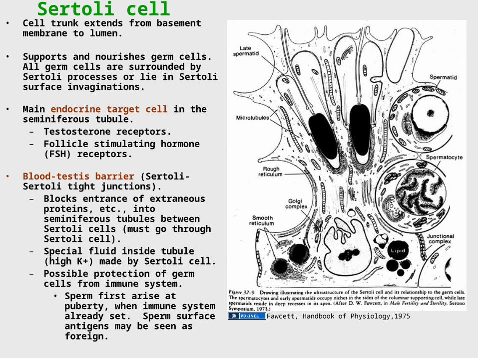
– Blocks entrance of extraneous proteins, etc., into seminiferous tubules between Sertoli cells (must go through Sertoli cell).
– Special fluid inside tubule (high K+) made by Sertoli cell.
– Possible protection of germ cells from immune system.
• Sperm first arise at puberty, when immune system already set. Sperm surface antigens may be seen as foreign. Fawcett, Handbook of Physiology,1975
Blood-testis barrier (= Sertoli-Sertoli junction)
(actin)
Fawcett, Handbook of Physiology,1975
Tight junctions of the blood-testis barrier, freeze-fracture, EM
Fawcett, Handbook of Physiology,1975
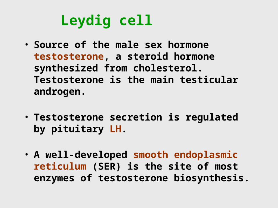
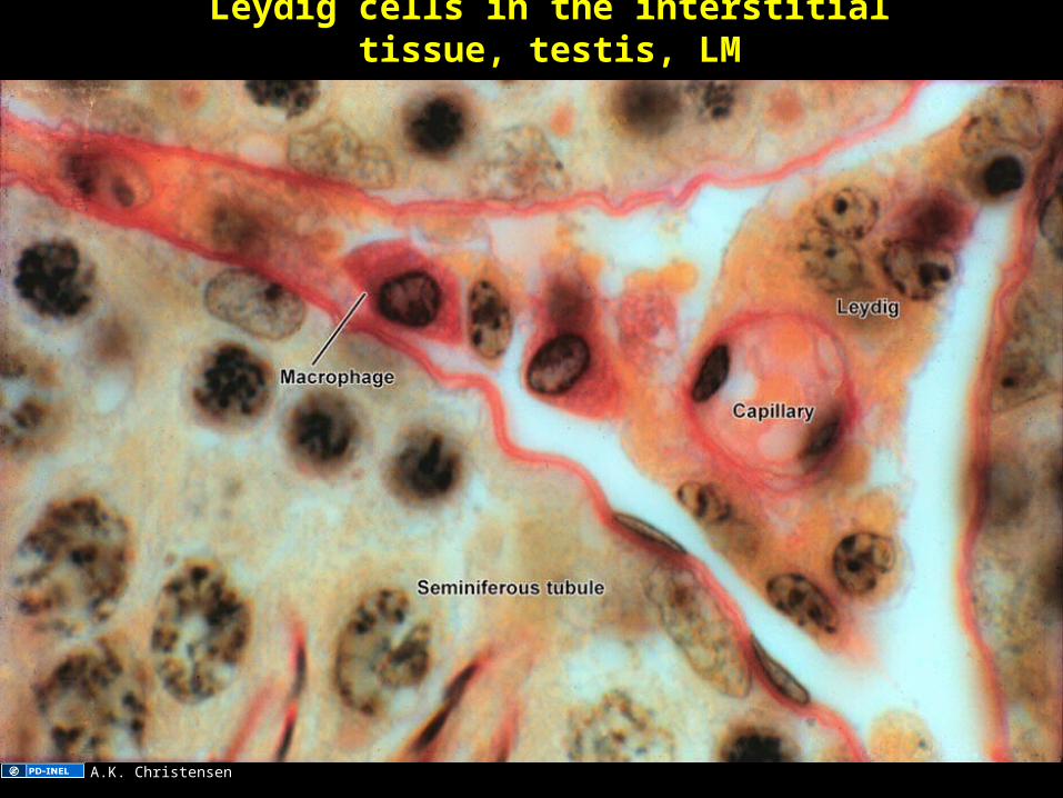
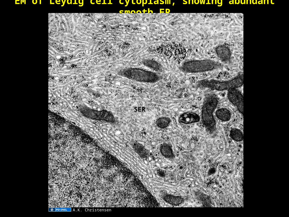
Leydig cell
• Source of the male sex hormone testosterone, a steroid hormone synthesized from cholesterol. Testosterone is the main testicular androgen.
• Testosterone secretion is regulated by pituitary LH.
• A well-developed smooth endoplasmic reticulum (SER) is the site of most enzymes of testosterone biosynthesis.
Leydig cells in the interstitial tissue, testis, LM
A.K. Christensen
EM of Leydig cell cytoplasm, showing abundant smooth ER
SER
A.K. Christensen
Actions of androgen• On Seminiferous tubules
– Testosterone is the main hormone regulating spermatogenesis. It acts on Sertoli cells, which have androgen receptors.
• On the male reproductive tract– Androgen regulates development and maintenance of
most of the tract. Dihydrotestosterone(DHT).
• On male secondary sexual characteristics, which arise at puberty– Muscle tone and strength.
– Lower voice.
– Axillary and pubic hair.
– Beard and dense body hair, acne.
– Receding hairline and baldness (also genetic).
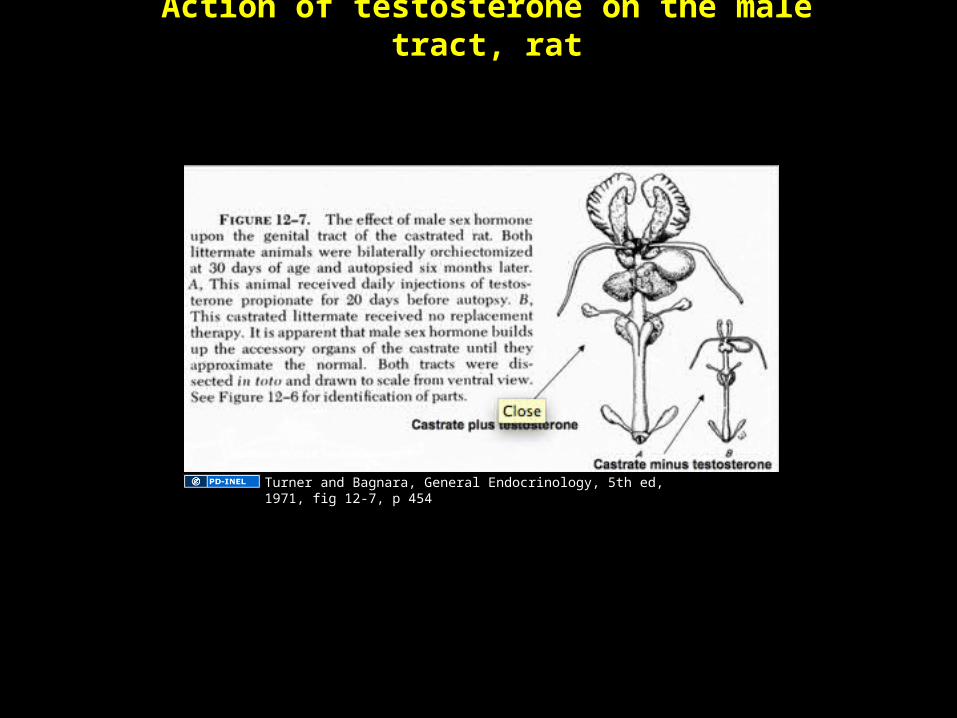
Action of testosterone on the male tract, rat
Castrate minus testosterone
Turner and Bagnara, General Endocrinology, 5th ed, 1971, fig 12-7, p 454
Pituitary regulation of male reproduction
• Luteinizing hormone (LH)– Regulates androgen secretion by Leydig cells. The androgen
then regulates spermatogenesis, the male tract and male secondary sexual characteristics.
– LH receptors are on the plasma membrane of Leydig cells.
• Follicle stimulating hormone (FSH)– Regulates the establishment of spermatogenesis at puberty.– FSH receptors are on the plasma membrane at the basal
surface of Sertoli cells.– Although Sertoli cells in the mature testis have FSH
receptors that probably have some functions, FSH does not appear to be essential for spermatogenesis in the adult testis.
Testis of a rat from which pituitary removed (= hypophysectomy), LM
A.K. Christensen
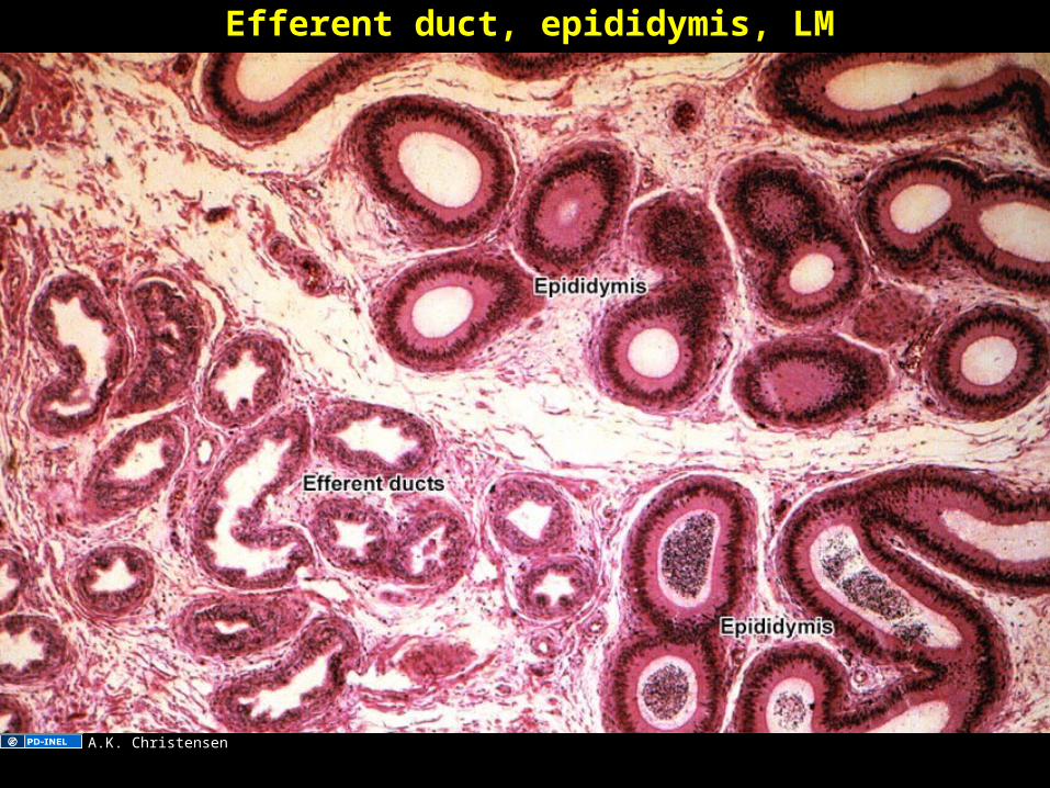
Sperm pathway through the male tract• Mediastinum of the testis.
Straight tubules. Rete testis.
• Efferent ducts. Passageway from testis to epididymis.There are 15-30 efferent ducts.
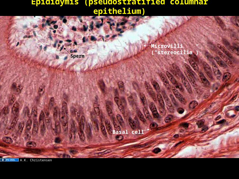
• Epididymis. Head, body, tail. Single long coiled duct (~6 m long). Sperm mature during passage, and are stored in the tail of the epididymis (for ejaculation).
• Ejaculation– Ductus deferens conducts
sperm from epididymal tail.– Seminal vesicles usually
furnish most of seminal fluid.
– Prostate gland contributes to seminal fluid.
– Semen passes through prostatic and penile urethra.
Ross and Pawlina, 5th ed, 2007, fig 22.4a, pg 732.
Semen Normally about 3.5 ml per ejaculate in humans.
• Sperm– About 100 million sperm per ml. Concentrations lower
than about 20 million/ml may cause fertility problems.
• Seminal fluid– Mainly from seminal vesicle (usually about 70%),
prostate and epididymis.
Seminal vesicle, human, low power LM
The secretion includes fructose, ascorbic acid, prostaglandins.
Kirkman histological slide collection
Seminal vesicle, LM
Smoothmuscle
Mucosal folds
Secretoryepithelium
From Japanese 35mm histological slide set (Mizoguti), slide 689
Prostate gland
Drawing of glands in a fetal prostate. Compound
tubuloalveolar glands, each emptying separately into
the prostatic urethra.Below is a cross section of
the fetal prostate.
Image of fetal prostate glands
removed. Original here: Campbell M.F. & Harrison
J.H., 1970, Urology, vol. 1, ed.
3, page 141
Adult prostate gland, cross section, low power LM. There are 30-50 compound tubuloalveolar glands. The secretion is
expelled into the semen during ejaculation. The contents include acid phosphatase, citric acid, fibrinolysin, and prostate-specific antigen (PSA).
Glands
Ejaculatory ducts
Urethra
Utricle
University of Michigan Virtual Slide Collection
Prostate Gland
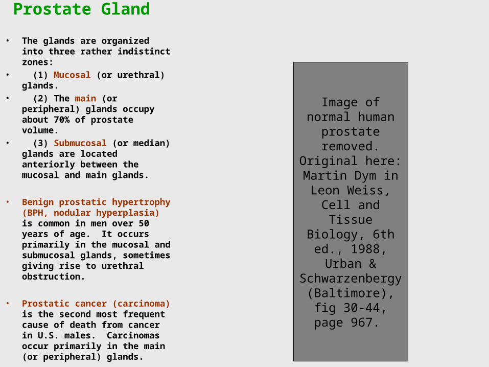
• The glands are organized into three rather indistinct zones:
• (1) Mucosal (or urethral) glands.
• (2) The main (or peripheral) glands occupy about 70% of prostate volume.
• (3) Submucosal (or median) glands are located anteriorly between the mucosal and main glands.
• Benign prostatic hypertrophy (BPH, nodular hyperplasia) is common in men over 50 years of age. It occurs primarily in the mucosal and submucosal glands, sometimes giving rise to urethral obstruction.
• Prostatic cancer (carcinoma) is the second most frequent cause of death from cancer in U.S. males. Carcinomas occur primarily in the main (or peripheral) glands.
Image of normal human prostate
removed. Original here: Martin Dym
in Leon Weiss, Cell and Tissue
Biology, 6th ed., 1988, Urban &
Schwarzenbergy (Baltimore), fig 30-
44, page 967.
Detail of prostate glands, LM
Gland
Smooth muscle
A.K. Christensen
Prostate gland, concretions (= amyloid bodies),composed of calcified glycoproteins
Concretion
Gland
Smoothmuscle
Kirkman histological slide collection
Penis cross section, LM drawing
Gray’s Anatomy, wikibooks
Fertilization• Semen is deposited in the female tract.• Cervix: Ability of sperm to pass depends on the
consistency of cervical mucus.• Lumen of uterus and oviducts: Sperm undergo
"capacitation," an induced change that will allow sperm to undergo subsequent acrosome reaction.
• Events in vicinity of ovum (usually in the ampulla of the oviduct):– Secretion from the ovum induces sperm to undergo an
acrosome reaction, releasing acrosomal hydrolytic enzymes that may facilitate sperm entry through the cumulus and zona pellucida of the ovum.
– A sperm enters the ovum. Subsequent sperm are excluded.– Fusion of the female and male pronuclei yields a nucleus with
23 pairs of chromosomes, the beginning of a new individual.
Fertilization: acrosome reaction releases hydrolytic enzymes that help sperm reach the surface of the egg
LadyofHats, wikimedia commons
Additional Source Informationfor more information see: http://open.umich.edu/wiki/CitationPolicy
Slide 5: LadyofHats, Wikimedia Commons, http://commons.wikimedia.org/wiki/File:Human_spermatozoa.pngSlide 6: Bloom and Fawcett Histology, 11th ed, fig 31-20, p 813.Slide 7: Bloom and Fawcett Histology, 11th ed, fig 31-20, p 813.Slide 8: LadyofHats, Wikimedia Commons, http://commons.wikimedia.org/wiki/File:Complete_diagram_of_a_human_spermatozoa.svgSlide 9: Visual atlas of human sperm structure and function for assisted reproductive technology, fig 3, p 397. Slide 10: Elf Sternberg, Wikimedia Commons, http://commons.wikimedia.org/wiki/File:Male_anatomy.png, CC:BY-SA http://creativecommons.org/ licenses/by-sa/3.0/Slide 12: Gray’s Anatomy, Answers, http://www.answers.com/topic/efferent-ductSlide 13: A. Kent ChristensenSlide 14: A. Kent ChristensenSlide 16: Source UndeterminedSlide 17: Heller and Clermont, 1964Slide 18: A. Kent ChristensenSlide 19: A. Kent ChristensenSlide 20: A. Kent ChristensenSlide 21: Junqueira and Carneiro, 10th ed., 2003, page436, fig. 22-9Slide 22: A. Kent ChristensenSlide 23: Fawcett, Handbook of Physiology,1975Slide 24: Fawcett, Handbook of Physiology, fig 29, 1975Slide 25: Fawcett, Handbook of Physiology, fig 29, 1975Slide 27: A. Kent ChristensenSlide 28: A. Kent ChristensenSlide 30: Turner and Bagnara, General Endocrinology, 5th ed, 1971, fig 12-7, p 454Slide 32: A. Kent ChristensenSlide 33: Ross and Pawlina, 5th ed, 2007, fig 22.4a, pg 732. Slide 34: A. Kent ChristensenSlide 35: A. Kent ChristensenSlide 36: A. Kent ChristensenSlide 37: A. Kent ChristensenSlide 39: Elf Sternberg, Wikimedia Commons, http://commons.wikimedia.org/wiki/File:Male_anatomy.png, CC:BY-SA http://creativecommons.org/ licenses/by-sa/3.0/Slide 40: Kirkman slide collectionSlide 42: Kirkman histological slide collectionSlide 43: From Japanese 35mm histological slide set (Mizoguti), slide 689
Slide 45: University of Michigan Virtual Slide CollectionSlide 46: Regents of the University of MichiganSlide 47: A. Kent ChristensenSlide 48: Kirkman histological slide collection Slide 49: Gray’s Anatomy, Wiki Books, http://en.wikibooks.org/wiki/Human_Physiology/The_male_reproductive_systemSlide 51: LadyofHats, Wikimedia Commons, http://commons.wikimedia.org/wiki/File:Acrosome_reaction_diagram.svg