48
Autoimmune Causes of Epilepsy? Manu Hegde, MD, PhD ECoE, VAMC-SF May 7, 2014
Autoimmune Causes of
Epilepsy?
Manu Hegde, MD, PhD
ECoE, VAMC-SF
May 7, 2014
Objectives
1) Understand the relationship between autoimmune
encephalitides and seizures.
2) Understand the role of autoantibodies in “uncomplicated”
epilepsy.
3) Understand rational approaches to autoantibody testing in
the epilepsy population.
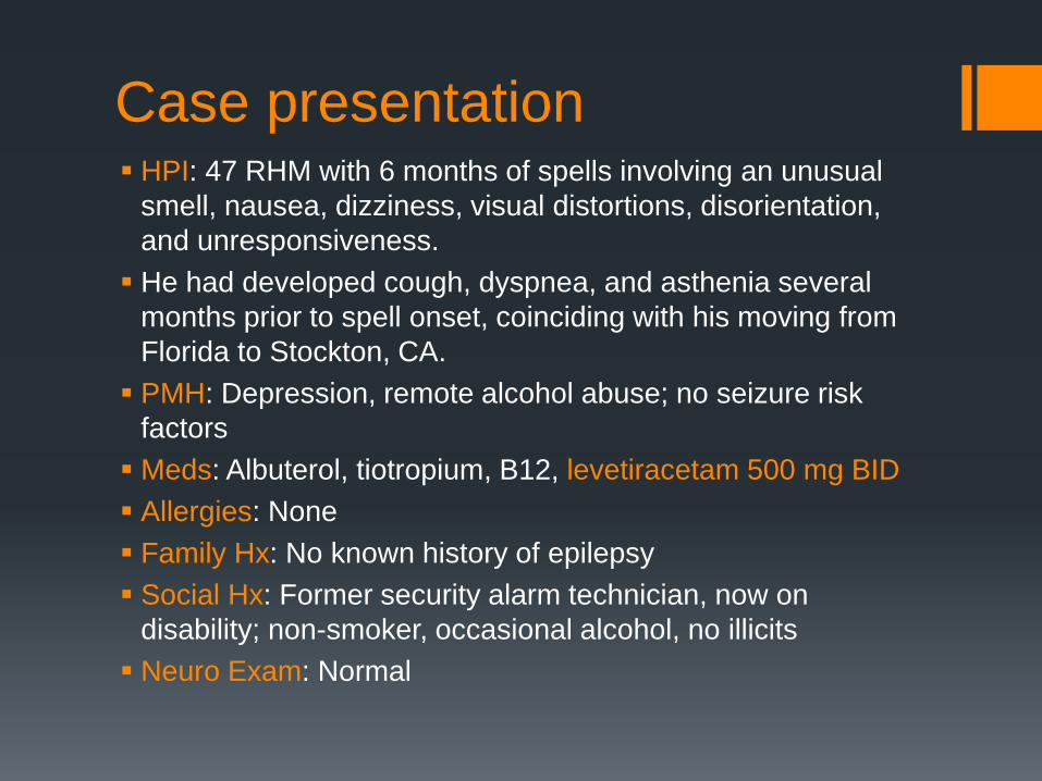
Case presentation HPI: 47 RHM with 6 months of spells involving an unusual
smell, nausea, dizziness, visual distortions, disorientation,
and unresponsiveness.
He had developed cough, dyspnea, and asthenia several
months prior to spell onset, coinciding with his moving from
Florida to Stockton, CA.
PMH: Depression, remote alcohol abuse; no seizure risk
factors
Meds: Albuterol, tiotropium, B12, levetiracetam 500 mg BID
Allergies: None
Family Hx: No known history of epilepsy
Social Hx: Former security alarm technician, now on
disability; non-smoker, occasional alcohol, no illicits
Neuro Exam: Normal
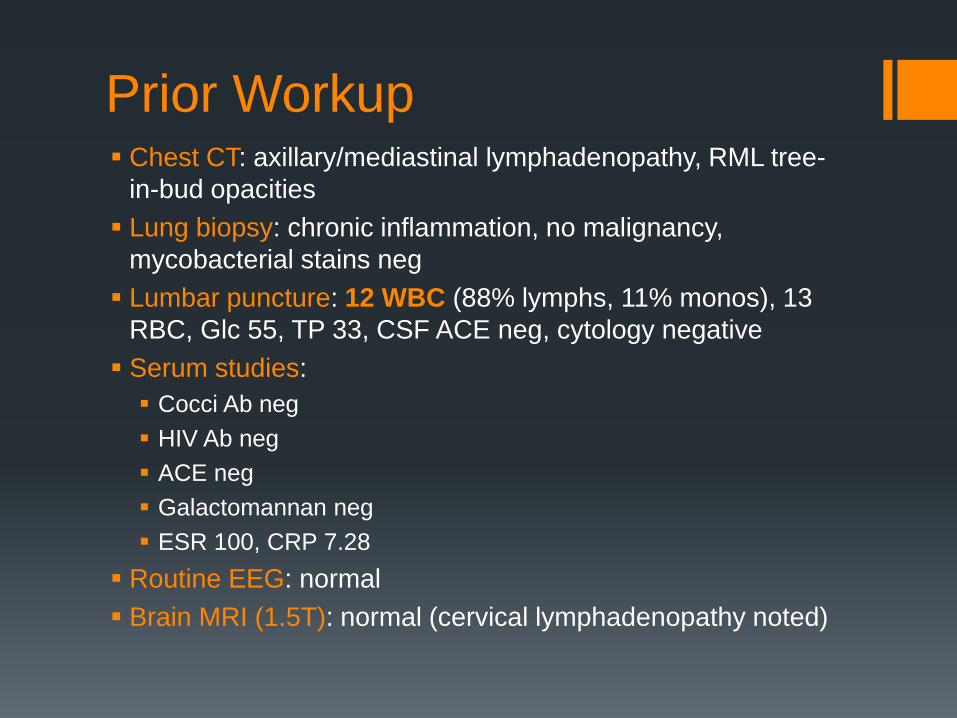
Prior Workup Chest CT: axillary/mediastinal lymphadenopathy, RML tree-
in-bud opacities
Lung biopsy: chronic inflammation, no malignancy,
mycobacterial stains neg
Lumbar puncture: 12 WBC (88% lymphs, 11% monos), 13
RBC, Glc 55, TP 33, CSF ACE neg, cytology negative
Serum studies:
Cocci Ab neg
HIV Ab neg
ACE neg
Galactomannan neg
ESR 100, CRP 7.28
Routine EEG: normal
Brain MRI (1.5T): normal (cervical lymphadenopathy noted)
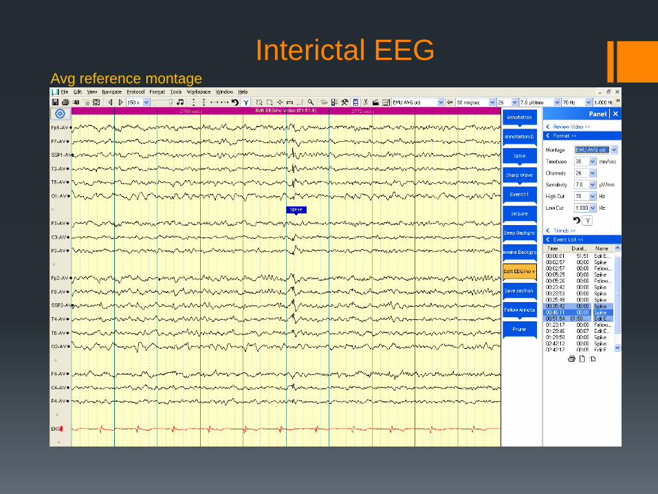
Interictal EEG Avg reference montage
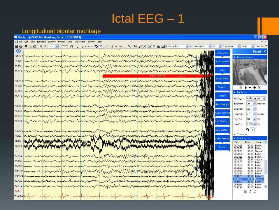
Ictal EEG – 1 Longitudinal bipolar montage
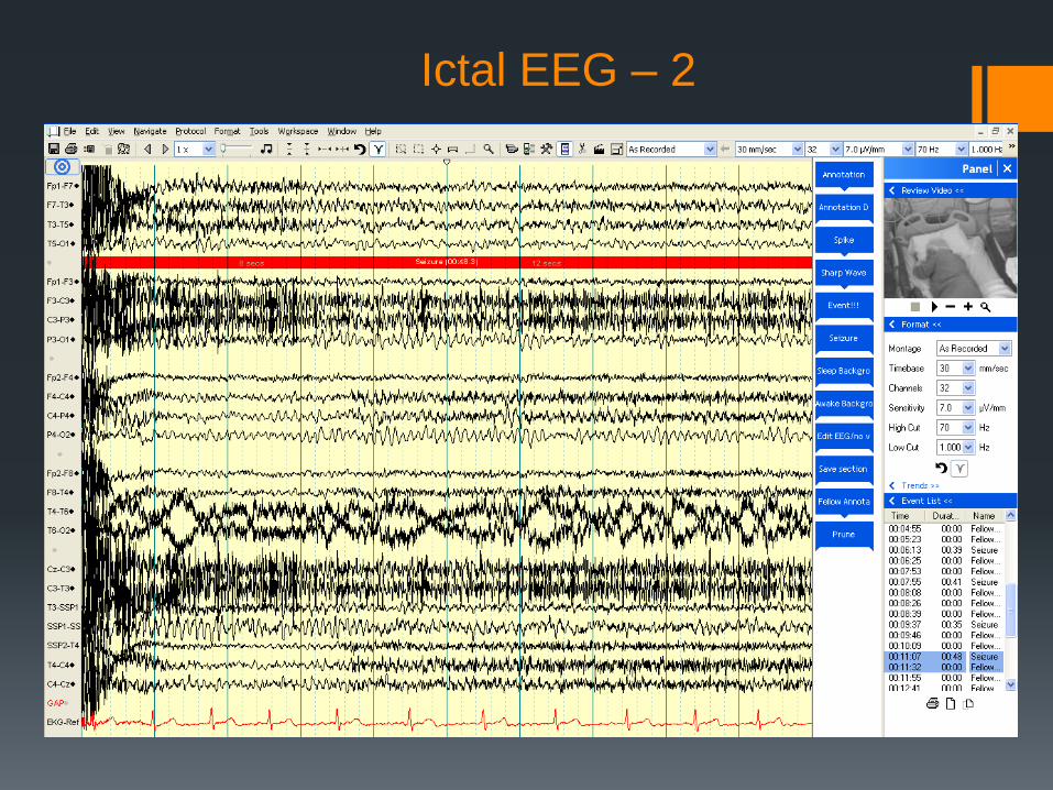
Ictal EEG – 2
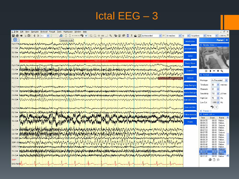
Ictal EEG – 3
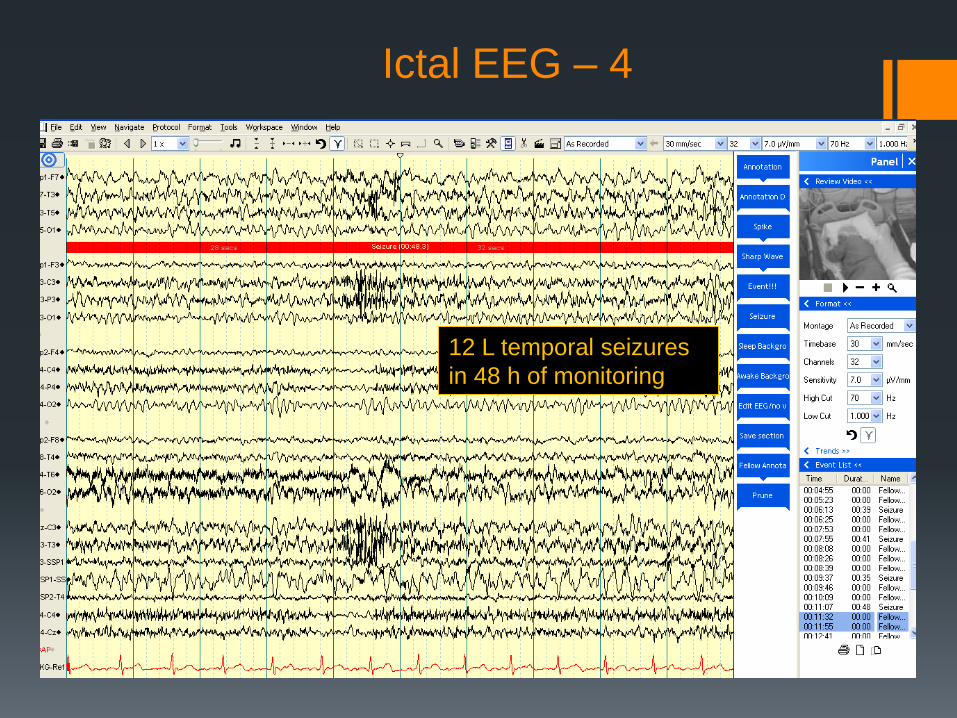
Ictal EEG – 4
12 L temporal seizures
in 48 h of monitoring
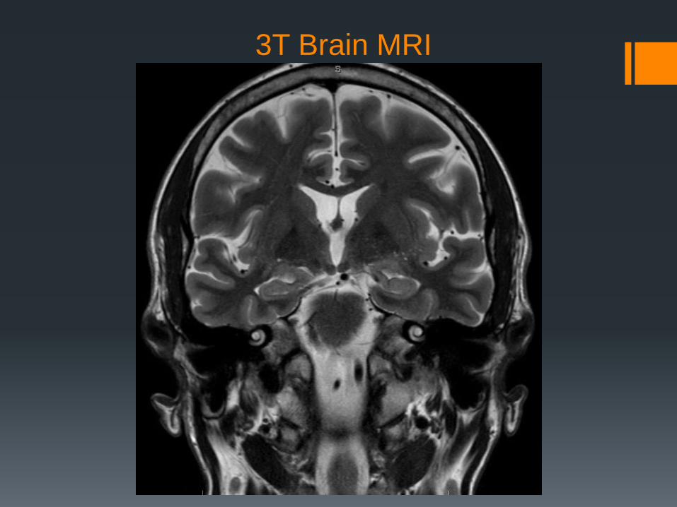
3T Brain MRI
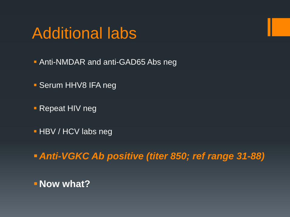
Additional labs
Anti-NMDAR and anti-GAD65 Abs neg
Serum HHV8 IFA neg
Repeat HIV neg
HBV / HCV labs neg
Anti-VGKC Ab positive (titer 850; ref range 31-88)
Now what?
Overview
Clinicians and scientists have long debated
whether immune or inflammatory factors are
important in epilepsy
Autoimmune neurological disease is often
unrelated to any particular antibody (i.e. MS)
However, autoantibodies to neuronal antigens
have been associated with a number of clinical
syndromes, especially limbic encephalitides (LE),
in the past decade
These syndromes often have seizures as a
prominent clinical feature
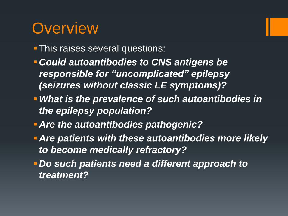
Overview This raises several questions:
Could autoantibodies to CNS antigens be
responsible for “uncomplicated” epilepsy
(seizures without classic LE symptoms)?
What is the prevalence of such autoantibodies in
the epilepsy population?
Are the autoantibodies pathogenic?
Are patients with these autoantibodies more likely
to become medically refractory?
Do such patients need a different approach to
treatment?
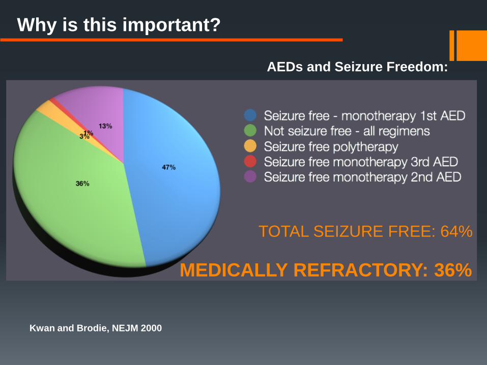
Why is this important?
Kwan and Brodie, NEJM 2000
AEDs and Seizure Freedom:
TOTAL SEIZURE FREE: 64%
MEDICALLY REFRACTORY: 36%
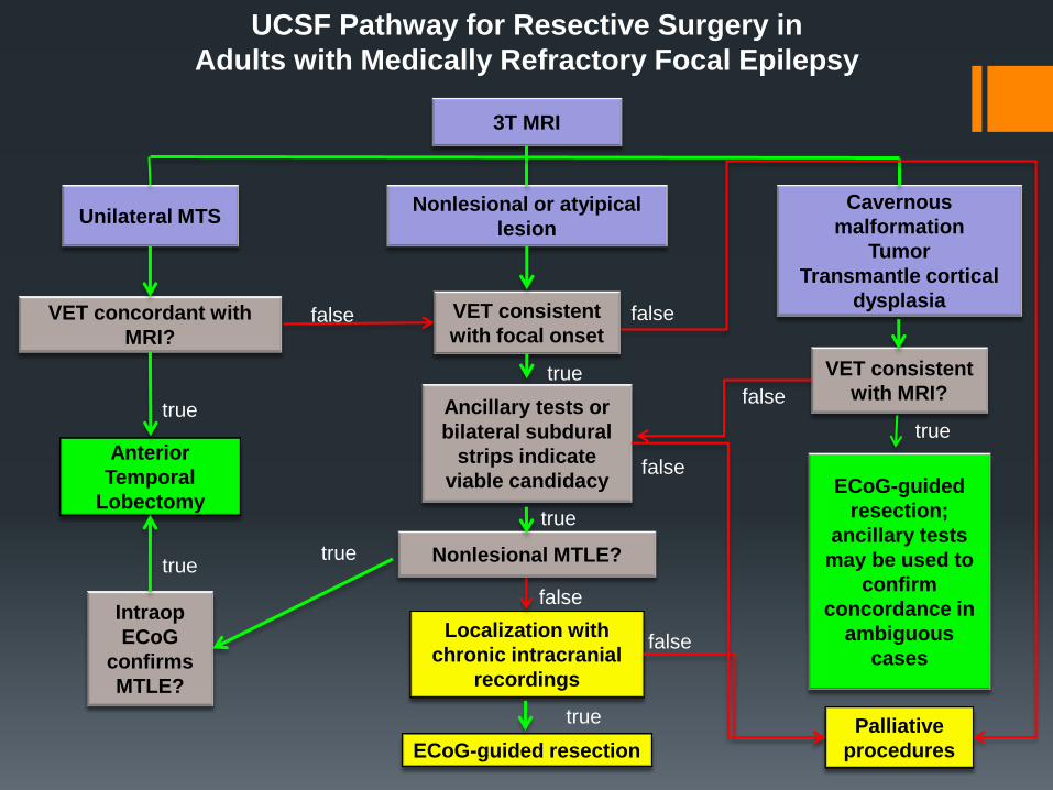
UCSF Pathway for Resective Surgery in
Adults with Medically Refractory Focal Epilepsy
Unilateral MTS Nonlesional or atyipical
lesion
Cavernous
malformation
Tumor
Transmantle cortical
dysplasia
3T MRI
Anterior
Temporal
Lobectomy
VET concordant with
MRI?
Intraop
ECoG
confirms
MTLE?
Localization with
chronic intracranial
recordings
Ancillary tests or
bilateral subdural
strips indicate
viable candidacy
ECoG-guided resection
VET consistent
with focal onset
Nonlesional MTLE?
ECoG-guided
resection;
ancillary tests
may be used to
confirm
concordance in
ambiguous
cases
VET consistent
with MRI?
Palliative
procedures
false
true
true
true
true
true true
false
false
false
false
true
false
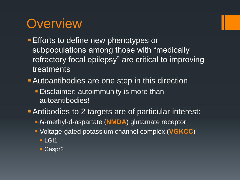
Overview Efforts to define new phenotypes or
subpopulations among those with “medically
refractory focal epilepsy” are critical to improving
treatments
Autoantibodies are one step in this direction
Disclaimer: autoimmunity is more than
autoantibodies!
Antibodies to 2 targets are of particular interest:
N-methyl-d-aspartate (NMDA) glutamate receptor
Voltage-gated potassium channel complex (VGKCC)
LGI1
Caspr2

Prevalence I Several series have studied autoantibody prevalence
in epilepsy populations
Marked differences in inclusion criteria
New onset vs chronic vs medically refractory
Age, gender
Selection biases (“suspected autoimmune epilepsy”)
In a study of 139 epilepsy patients McKnight et al
2005 found that 12% had VGKCC antibodies, and
4% had GAD antibodies
Subgroup (n=67) with medically refractory epilepsy:
2: VGKCC
3: GAD
1: ganglioside GM1
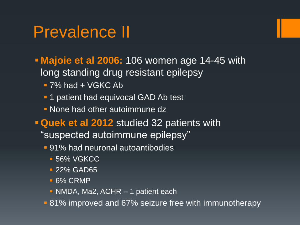
Prevalence II
Majoie et al 2006: 106 women age 14-45 with
long standing drug resistant epilepsy
7% had + VGKC Ab
1 patient had equivocal GAD Ab test
None had other autoimmune dz
Quek et al 2012 studied 32 patients with
“suspected autoimmune epilepsy”
91% had neuronal autoantibodies
56% VGKCC
22% GAD65
6% CRMP
NMDA, Ma2, ACHR – 1 patient each
81% improved and 67% seizure free with immunotherapy
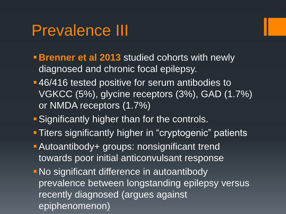
Prevalence III
Brenner et al 2013 studied cohorts with newly
diagnosed and chronic focal epilepsy.
46/416 tested positive for serum antibodies to
VGKCC (5%), glycine receptors (3%), GAD (1.7%)
or NMDA receptors (1.7%)
Significantly higher than for the controls.
Titers significantly higher in “cryptogenic” patients
Autoantibody+ groups: nonsignificant trend
towards poor initial anticonvulsant response
No significant difference in autoantibody
prevalence between longstanding epilepsy versus
recently diagnosed (argues against
epiphenomenon)
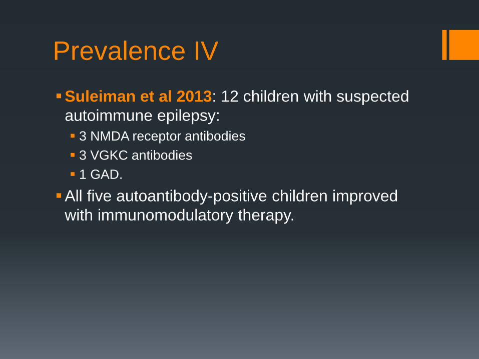
Prevalence IV
Suleiman et al 2013: 12 children with suspected
autoimmune epilepsy:
3 NMDA receptor antibodies
3 VGKC antibodies
1 GAD.
All five autoantibody-positive children improved
with immunomodulatory therapy.
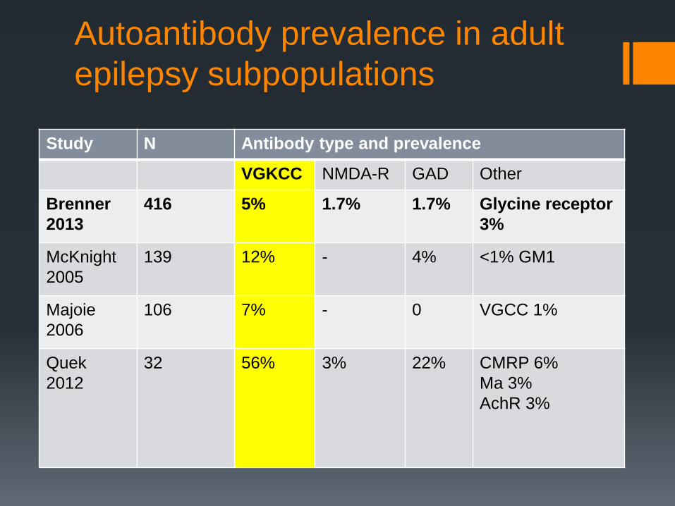
Autoantibody prevalence in adult
epilepsy subpopulations
Study N Antibody type and prevalence
VGKCC NMDA-R GAD Other
Brenner
2013
416 5% 1.7% 1.7% Glycine receptor
3%
McKnight
2005
139 12% - 4% <1% GM1
Majoie
2006
106 7% - 0 VGCC 1%
Quek
2012
32 56% 3% 22% CMRP 6%
Ma 3%
AchR 3%
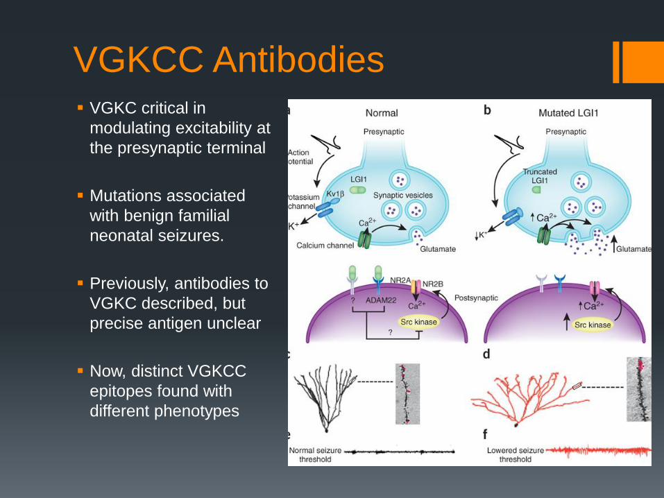
VGKCC Antibodies VGKC critical in
modulating excitability at
the presynaptic terminal
Mutations associated
with benign familial
neonatal seizures.
Previously, antibodies to
VGKC described, but
precise antigen unclear
Now, distinct VGKCC
epitopes found with
different phenotypes
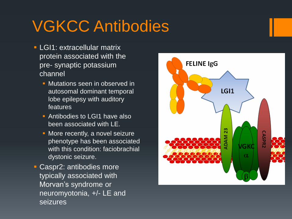
VGKCC Antibodies LGI1: extracellular matrix
protein associated with the
pre- synaptic potassium
channel
Mutations seen in observed in
autosomal dominant temporal
lobe epilepsy with auditory
features
Antibodies to LGI1 have also
been associated with LE.
More recently, a novel seizure
phenotype has been associated
with this condition: faciobrachial
dystonic seizure.
Caspr2: antibodies more
typically associated with
Morvan’s syndrome or
neuromyotonia, +/- LE and
seizures
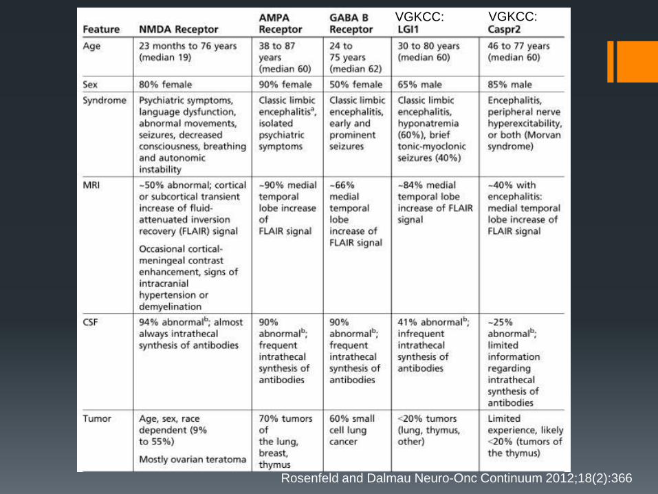
Rosenfeld and Dalmau Neuro-Onc Continuum 2012;18(2):366
VGKCC: VGKCC:

Faciobrachial Dystonic Seizures

FBDS and LGI1 antibodies
Irani et al 2011 found 29 patients with this syndrome
All were positive for VGKCC antibodies
89% had antibodies specific for LGI1
77% went on to develop typical LE symptoms
Irani et al 2013: follow-up study of 10 of these patients
9 were refractory to AEDs
All had favorable response to steroids
Outcome correlated with time to immunotherapy, but not time
to AED therapy
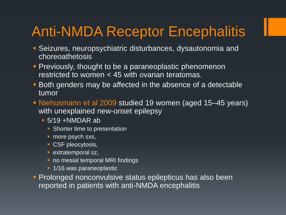
Anti-NMDA Receptor Encephalitis Seizures, neuropsychiatric disturbances, dysautonomia and
choreoathetosis
Previously, thought to be a paraneoplastic phenomenon restricted to women < 45 with ovarian teratomas.
Both genders may be affected in the absence of a detectable tumor
Niehusmann et al 2009 studied 19 women (aged 15–45 years) with unexplained new-onset epilepsy
5/19 +NMDAR ab
Shorter time to presentation
more psych sxs,
CSF pleocytosis,
extratemporal sz,
no mesial temporal MRI findings
1/16 was paraneoplastic
Prolonged nonconvulsive status epilepticus has also been reported in patients with anti-NMDA encephalitis

Clinical features of anti-NMDA receptor encephalitis
Two stages:
Early: neuropsychiatric sx (psychosis, behavior change, amnesia, dysphasia), seizures in 70%
Late: dyskinesias, altered consciousness, dysautonomia, central hypoventilation
Tumor association:
Paraneoplastic in 38% (46% of women, 6% of men), usu. ovarian teratoma (94%)
Diagnostic workup:
Clinical syndrome, MRI, EEG, serum and CSF (intrathecal Ab production) for anti-NMDAR Ab
Irani et al. Brain (2010) & Titulaer et al. Lancet Neurol (2013)
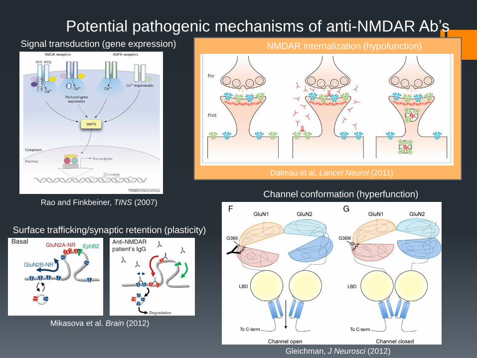
Rao and Finkbeiner, TINS (2007)
Dalmau et al, Lancet Neurol (2011)
Gleichman, J Neurosci (2012)
Potential pathogenic mechanisms of anti-NMDAR Ab’s Signal transduction (gene expression) NMDAR internalization (hypofunction)
Channel conformation (hyperfunction)
Mikasova et al. Brain (2012)
Surface trafficking/synaptic retention (plasticity)
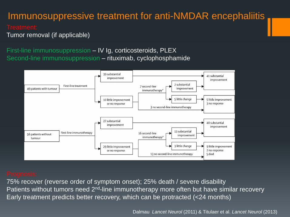
Immunosuppressive treatment for anti-NMDAR encephaliitis Treatment:
Tumor removal (if applicable)
First-line immunosuppression – IV Ig, corticosteroids, PLEX
Second-line immunosuppression – rituximab, cyclophosphamide
Prognosis:
75% recover (reverse order of symptom onset); 25% death / severe disability
Patients without tumors need 2nd-line immunotherapy more often but have similar recovery
Early treatment predicts better recovery, which can be protracted (<24 months)
Dalmau Lancet Neurol (2011) & Titulaer et al. Lancet Neurol (2013)
Anti-GABAA Receptor Encephalitis
HOT OFF THE PRESSES!
Anti-GABAA Receptor Encephalitis
Petit Pedrol et al, 2014 studied serum and CSF from 140
patients with encephalitis, seizures or status epilepticus, and
antibodies to unknown neuropil antigens
6/140 had high titers of antibodies to the GABAA receptor
Another 12 had low titers
Clinical presentation ranged from seizures alone (rarely) to a
typical limbic encephalitis picture
Some had features of Stiff-Person or Opsoclonus-Myoclonus
syndromes
12/15 responded to a mix of AED and immunomodulatory
therapies; 3 died
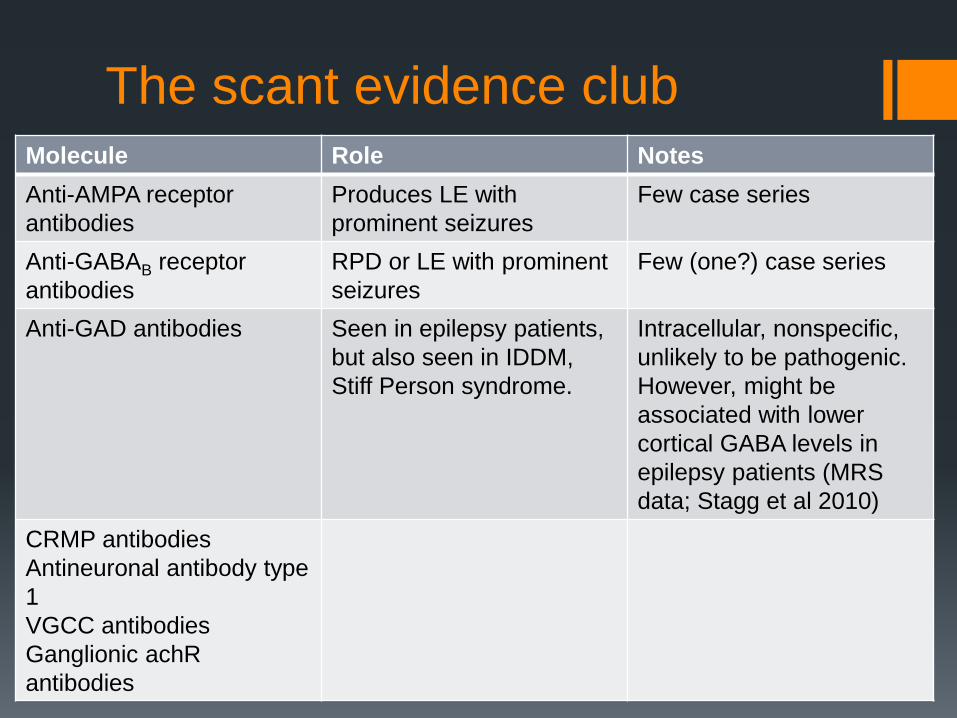
The scant evidence club Molecule Role Notes
Anti-AMPA receptor
antibodies
Produces LE with
prominent seizures
Few case series
Anti-GABAB receptor
antibodies
RPD or LE with prominent
seizures
Few (one?) case series
Anti-GAD antibodies Seen in epilepsy patients,
but also seen in IDDM,
Stiff Person syndrome.
Intracellular, nonspecific,
unlikely to be pathogenic.
However, might be
associated with lower
cortical GABA levels in
epilepsy patients (MRS
data; Stagg et al 2010)
CRMP antibodies
Antineuronal antibody type
1
VGCC antibodies
Ganglionic achR
antibodies

Treatment
EVEN HOTTER OFF THE PRESSES!
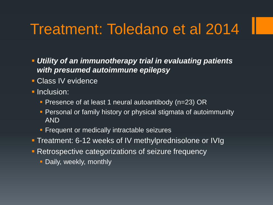
Treatment: Toledano et al 2014
Utility of an immunotherapy trial in evaluating patients
with presumed autoimmune epilepsy
Class IV evidence
Inclusion:
Presence of at least 1 neural autoantibody (n=23) OR
Personal or family history or physical stigmata of autoimmunity
AND
Frequent or medically intractable seizures
Treatment: 6-12 weeks of IV methylprednisolone or IVIg
Retrospective categorizations of seizure frequency
Daily, weekly, monthly

Treatment: Toledano et al 2014
Treatment: Toledano et al 2014
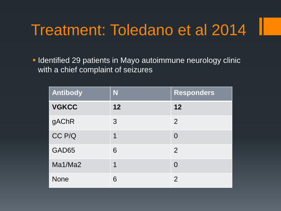
Identified 29 patients in Mayo autoimmune neurology clinic
with a chief complaint of seizures
Antibody N Responders
VGKCC 12 12
gAChR 3 2
CC P/Q 1 0
GAD65 6 2
Ma1/Ma2 1 0
None 6 2

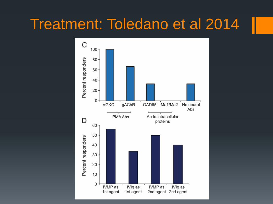
Treatment: Toledano et al 2014
62% favorable response
10/18 -> seizure free
Remainder had >50% reduction in seizure
frequency
Half of initial non-responders responded to
second immunotherapy agent
All remained on AEDs
89% of responders took long-term
maintenance immunotherapy
Treatment: Toledano et al 2014
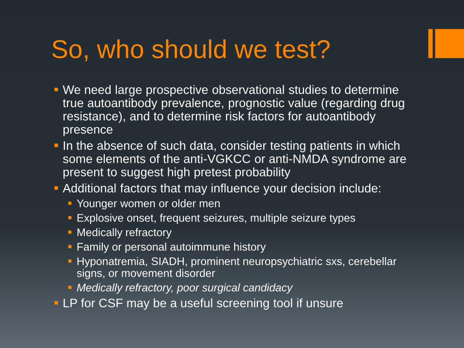
So, who should we test?
We need large prospective observational studies to determine true autoantibody prevalence, prognostic value (regarding drug resistance), and to determine risk factors for autoantibody presence
In the absence of such data, consider testing patients in which some elements of the anti-VGKCC or anti-NMDA syndrome are present to suggest high pretest probability
Additional factors that may influence your decision include:
Younger women or older men
Explosive onset, frequent seizures, multiple seizure types
Medically refractory
Family or personal autoimmune history
Hyponatremia, SIADH, prominent neuropsychiatric sxs, cerebellar signs, or movement disorder
Medically refractory, poor surgical candidacy
LP for CSF may be a useful screening tool if unsure

Back to our case…
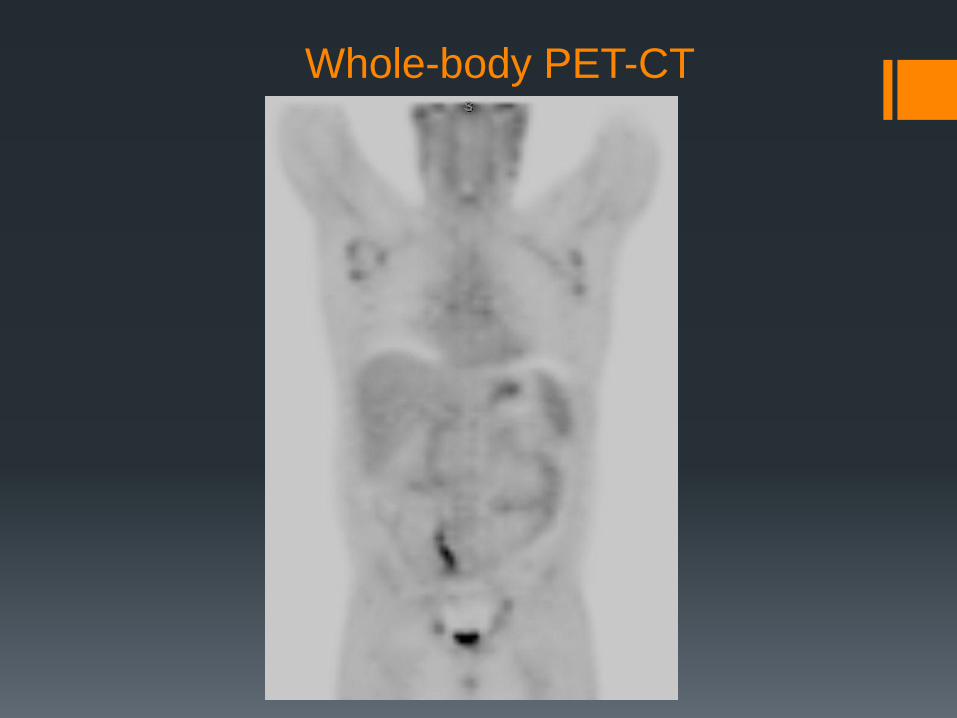
Whole-body PET-CT

Cervical Lymph Node Biopsy
Histology: nodular pattern of ‘onion-skinned’ follicles, abundance of
interfollicular CD138+ plasma cells, no lymphoma, HHV8 stain
negative
Flow cytometry: no clonal lymphoid populations
Dx: Multicentric Castleman’s Disease, plasma cell variant
N.B. images not from patient, c.f. Schulte and Talat (2010) Nat Rev Clin Oncol
Castleman’s Disease Angiofollicular lymph node hyperplasia: Rare lymphoproliferative disorder
with increased risk of lymphoma; unicentric (UCD) and multicentric
(MCD) forms; UCD associated with paraneoplastic pemphigus; MCD often
associated with HIV and HHV8
Clinical:
-UCD: young adult, often asymptomatic, mass can cause compression sx
-MCD: middle-aged patient, non-specific ‘B’ symptoms, peripheral
lymphadenopathy, cough/dyspnea related to pulmonary infiltrates
Diagnosis: imaging, labs (anemia, hypoalbuminemia, ↑ESR), lymph node bx
(pathology: hyaline vascular variant (90%), plasma cell variant, mixed)
Treatment:
-Resection curative in unicentric disease and prognosis favorable
-MCD is more aggressive, course variable but prognosis usu. worse (median
survival 8-14 mo); rituximab first-line, chemo if fails (etoposide vs. CHOP)
Significance:
<15 reported cases of CD with CNS involvement (all UCD with mass lesions,
MRI similar to meningiomas), 8 patients had seizures
Paraneoplastic limbic encephalitis has been rarely reported with malignant
hemopathies (AML, HL, NHL), sometimes with anti-VGKC Ab’s, but no
reports in Castleman’s
Follow-up
Seizures well-controlled on Depakote XR 1000 mg BID
Rituximab treatment initiated
Plan to follow with serial PET-CT and anti-VGKC Ab titers

Conclusions
Think about autoantibody testing in patients with new-onset
refractory focal epilepsy without a clear etiology
Antibodies to the VGKCC, NMDA receptor, and possibly
GABAA receptor are likely highest-yield
The full spectrum of these phenotypes is not yet known
More data is needed to determine autoantibodies’ prognostic
significance in epilepsy
A number of other immune/inflammatory mediators are under
active investigation for their roles in epilepsy
Acknowledgments
Vikram Rao, MD PhD
Sarah Shalev, MD
Siddharth Kharkar, MD
Yana Kriseman, MD
Susannah Cornes, MD
Nina Garga, MD
Karen Parko, MD
Sarosh Irani, MD, DPhil
References
Brenner et al, Epilepsia 54, 1028–1035 (2013).
Hegde & Lowenstein, Biomarkers Med. 8(3), 413–427 (2014)
Irani et al, Ann. Neurol. 69(5), 892–900 (2011).
Irani et al, Brain. 2013 Oct;136(Pt 10):3151-62
McKnight et al, Neurology 65(11), 1730–1736 (2005).
Niehusmann et al, Arch. Neurol. 66(4), 458–464 (2009).
Petit-Pedrol et al, Lancet Neurol 2014; 13: 276–86
Quek et al, Arch. Neurol. 69(5), 582 (2012).
Suleiman et al. Epilepsia 54(6),1036–1045 (2013)
Toledano et al. Neurology, published online April 4, 2014


