11
AUTOMATED SPECIMEN PROCESSING AND DIGITAL MICROBIOLOGY

AUTOMATED SPECIMEN PROCESSING AND DIGITAL MICROBIOLOGY

1 2
WASP® ADVANTAGE
WASP® uses traditional, reusable metal loops ranging in size from 1 µL, 10 µL and 30µl, allowing users to plant a variety of samples and volumes. Other automated systems use disposable pipet tips which cannot transfer volumes less than 10 µL.
Classic streaking patterns with reusable loops: Minimize disposable cost per sample. No staff retraining.
Automatic Gram slide preparation including permanent inkjet labeling directly onto the glass slide.
Automatic broth inoculation and labeling of tubes: No manual pre-labeling required.
Automatic loop check guarantees delivery of sample onto culture plate, as well as the integrity and accuracy of the loop.
Reduce repetitive stress: WASP® automatically opens and closes most specimen container types.
Automatic Kirby Bauer and ID disk application.
WASP® uses a liquid based approach, transforming samples to liquid for automatic processing. Additionally, WASP® can be programmed to process standard swabs and solid samples with minimal manual interaction.
WASP® manages all specimen types without batching: Continuous feeding of any sample type and automatic loop and tool change station mean longer walk away time and less user intervention.
No cross contamination: WASP® processes samples individually, no racks or open containers.
Two SCARA Robots
WASP®: WALK-AWAY SPECIMEN PROCESSOR is a total solution for pre-analytical Microbiology specimen processing. WASP® is the only instrument on the market that addresses all aspects of automated Microbiology specimen processing:
Planting and Streaking
Gram Slide Preparation with Automatic Inkjet Label Printing
Enrichment Broth Inoculation
Kirby Bauer and ID Disk Application
By maintaining a close relationship with the Microbiology community, COPAN continues to make innovative strides, and has designed WASP® as an open platform, modular instrument for the seamless addition of new features and capabilities.
WALK-AWAY SPECIMEN PROCESSOR
Spinner
Warehouse Carousel (Broth Inoculation and Disk Dispensing)
Gram SlidePrepVortex
Plate Exit Conveyor
Sample Entry and Exit Conveyors9 Silo Carousel
(holds up to 378 plates)
Label Printer
FRONT VIEW
TOP VIEW
3 4
15
2
3
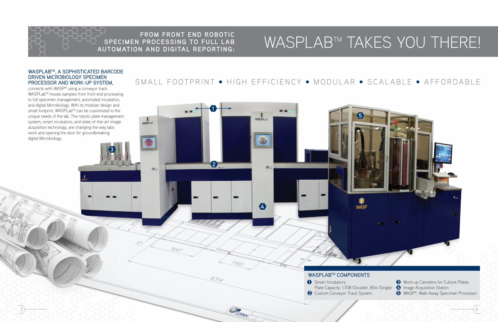
WASPLABTM TAKES YOU THERE!FROM FRONT END ROBOTIC
SPECIMEN PROCESSING TO FULL LAB AUTOMATION AND DIGITAL REPORTING:
S M A L L F O OT P R I N T H I G H E F F I C I E N C Y M O D U L A R S C A L A B L E A F F O R D A B L EWASPLABTM, A SOPHISTICATED BARCODE DRIVEN MICROBIOLOGY SPECIMEN PROCESSOR AND WORK-UP SYSTEM, connects with WASP® using a conveyor track. WASPLabTM moves samples from front end processing to full specimen management, automated incubation, and digital Microbiology. With its modular design and small footprint, WASPLabTM can be customized to the unique needs of the lab. The robotic plate management system, smart incubators, and state-of-the-art image acquisition technology, are changing the way labs work and opening the door for groundbreaking digital Microbiology.
1 Smart Incubators Plate Capacity: 1,708 (Double), 854 (Single)2 Custom Conveyor Track System
3 Work-up Canisters for Culture Plates4 Image Acquisition Station5 WASP®: Walk-Away Specimen Processor
WASPLABTM COMPONENTS
4

5
3
2
1 1
CUSTOM TRACK SYSTEM ENSURES FLEXIBILITY AND A SMALL FOOTPRINT
WASPLabTM is customized for each lab, using flexible conveyors that are designed to fit any lab, regardless of space restrictions, so there is no need to demolish your current laboratory space.
Plates travel automatically from the WASP® to the smart incubators on the track.
Manually loaded plates can be placed onto the track for image acquisition and incubation.
SCALABLE WORK-UP CANISTER SYSTEM
COMPACT, HIGH EFFICIENCY SMART INCUBATORS
Depending on user defined protocols, smart incubators can automatically invert each plate prior to incubation preventing condensation from falling onto the media.
Every plate is assigned a unique location, based on barcode, allowing for random and rapid retrieval.
Incubator shelves ensure homogeneous environmental conditions and excellent thermal conductivity to bring plates up to the appropriate temperature and atmospheric conditions quickly and efficiently.
Compact incubator shelves are easily removed and autoclaved to maintain the most sanitary conditions. Easy to clean!
Single incubators hold 854 plates and double incubators hold 1,708 plates.
Single Incubator
Double Incubator
12
3
Plates are automatically sent from incubators into easy-to-remove canisters for further work-up at the direction of laboratory staff.
Convenient work-up canisters can be designated for a particular specimen type or by operator for optimal workflow.
The canister system gives labs the flexibility to grow and add more stations without having to add additional conveyor track.

7
COMPOSITE IMAGE
The WASPLabTM Trilinear Camera sensor acquires the plate image slice by slice as the plate sweeps laterally beneath the camera in the plate shuttle carrier. Four thousand slices are captured and merged together to form a single 27 megapixel image of the 100mm plate.
MICROBIOLOGY IN A DIGITAL AGE27 MEGAPIXEL, LARGER THAN LIFE IMAGES
Plate photos taken with WASPLabTM Image Acquisition cameras are so sharp that only a plate microscope can boast such high resolution and clarity.
Each photo is 27 Megapixels, comprising of layered red, green and blue colors creating a bright vivid image. The WASPLabTM software can detect and differentiate colonies as small as 0.1 mm in diameter and present images to the operator.
In addition to the sharpest image in the industry, the WASPLabTM camera optics have an enormous 9mm depth of field, ensuring that both small, low colonies and large, high colonies are always in focus, so you will not miss discrete growth of a pathogen.
THREE LIGHTING SYSTEMS TO COLLECT OPTIMAL PLATE IMAGES
Not all plated media is the same. The WASPLabTM Image Acquisition system uses different lighting for photography depending on the media color or opacity.
Top light with background — Bench view, transparent agar
Top light no background — Bench view, opaque agar
Bottom light no background — Simulates holding to the light to see hemolysis
UNDISTORTED IMAGE
WASPLabTM uses telecentric camera optics and software vital when inspecting three-dimensional objects where image size and shape accuracy are critical. A Telecentric lens uses constant magnification; eliminating perspective angle error, so that the image on the screen is true to life with no distortion. This important feature enables the precise location and picking of colonies using the original image: no need to re-scan.
Conventional Telecentric
Optics
THE WASPLABTM IMAGE ACQUISITION TECHNOLOGY uses a highly sophisticated lighting and camera system so that each plate photo is clear and accurate. It’s like using a plate microscope with every plate, allowing you to make the most accurate work-up decision.
Camera
Front Light
White Panel
Back Light
LIGHTING ILLUSTRATION
UNIQUE COMPARATIVE DIFFERENTIAL IMAGE ANALYSIS FOR THE MOST PRECISE READING
A critical time zero reading of every culture plate is recorded in order to identify and eliminate any existing artifacts associated with each media plate.
Time zero is crucial to true comparative differential image analysis, allowing the software to ignore the noise and focus on the growth.
16 hours – 0 hours = True Growth
- =
16 hours 0 hours True Growth– =
9 10
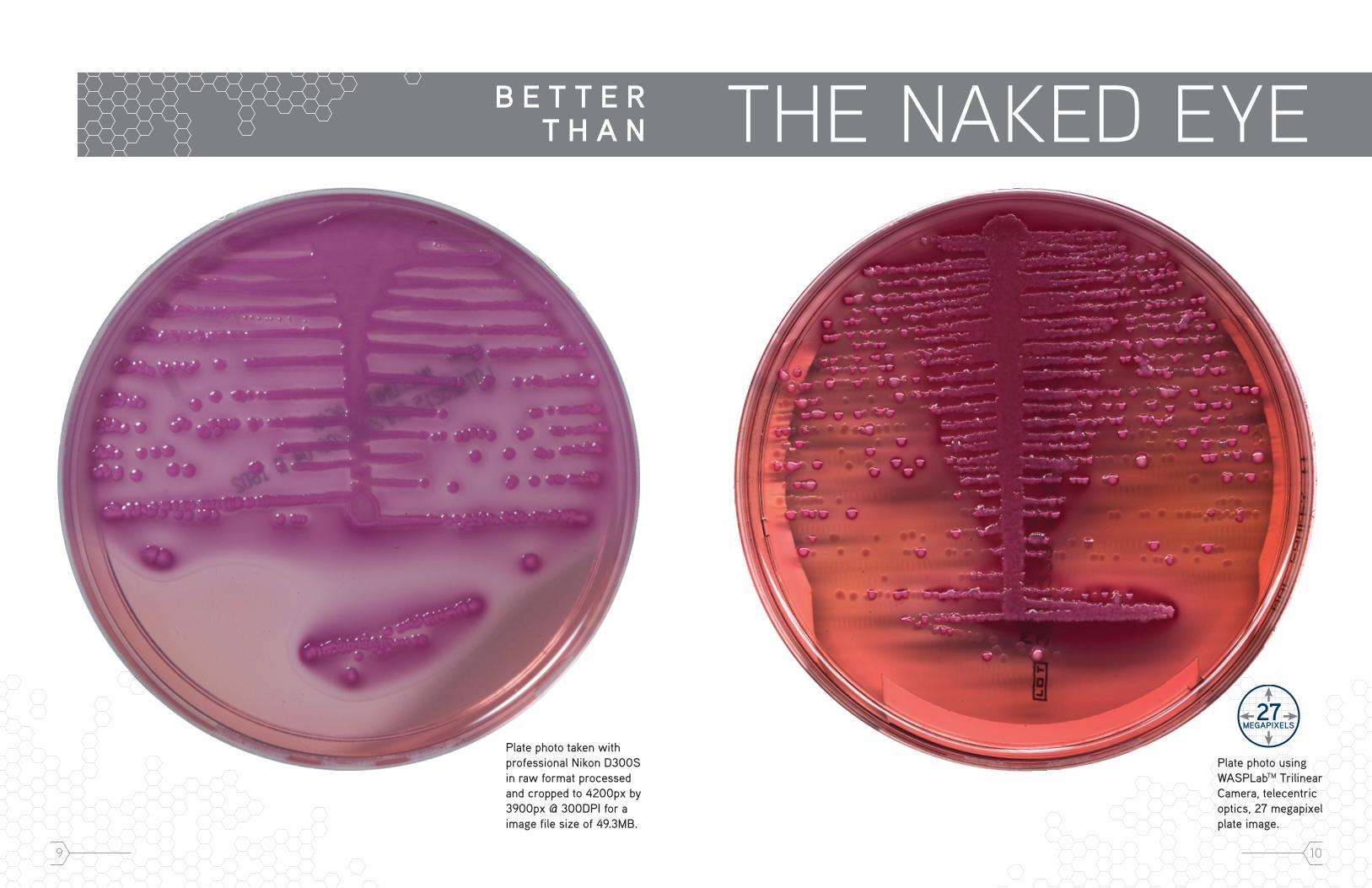
B E T T E R T H A N THE NAKED EYE
Plate photo taken with professional Nikon D300S in raw format processed and cropped to 4200px by 3900px @ 300DPI for a image file size of 49.3MB.
Plate photo using WASPLabTM Trilinear Camera, telecentric optics, 27 megapixel plate image.

11
READ, PICK, AND REPORTSCREEN,
Individual dashboards show the operator the workload for the shift: what has been done and what needs to be done.
Administrator dashboards provide real time snapshots of the laboratory workload and allow managers to reallocate the workload to prevent bottlenecks.
Key performance indicators and efficiency levels can be easily measured using the dashboards.
0 hours 6 hours 9 hours
16 hours 12 hours
DIGITAL MICROBIOLOGY IN PRACTICE
Incubation protocols can be set to scan and automatically record images as often as needed. Early scans of sterile body or joint fluids may allow for faster detection of growth and early intervention in patient treatment.
All plate images are presented to the technologist for review at the screening station. Images can be grouped based on colony counts from most growth to no growth or vice versa.
Plate images requiring further investigation are sent to the reading station. Users may zoom in to scrutinize and tag colonies creating work-up tickets for tasks such as AST, ID’s, Subculture, Gram Stain, or Spot Biochemical tests.
User-defined Presumptive ID’s are assigned to each tagged colony.
Plates are then automatically sent to a designated work-up canister for picking.
After scanning the barcode at the picking station, a plate image is automatically loaded with all digitally tagged colonies and Presumptive ID’s.
Users can also view a digital image of the Gram stain along with the plate image and work-up ticket.
After picking is complete, operators acknowledge that they have completed the tasks requested by the Readers, and close out the work-up ticket.
When screening culture plates the operator can use the toggle buttons to quickly go back and forth and review and compare growth on the same culture plate at different incubation time points.
EASY TO READ DIGITAL DASHBOARDS PROVIDE INSTANT VISIBILITY OF LAB PERFORMANCE
13 14
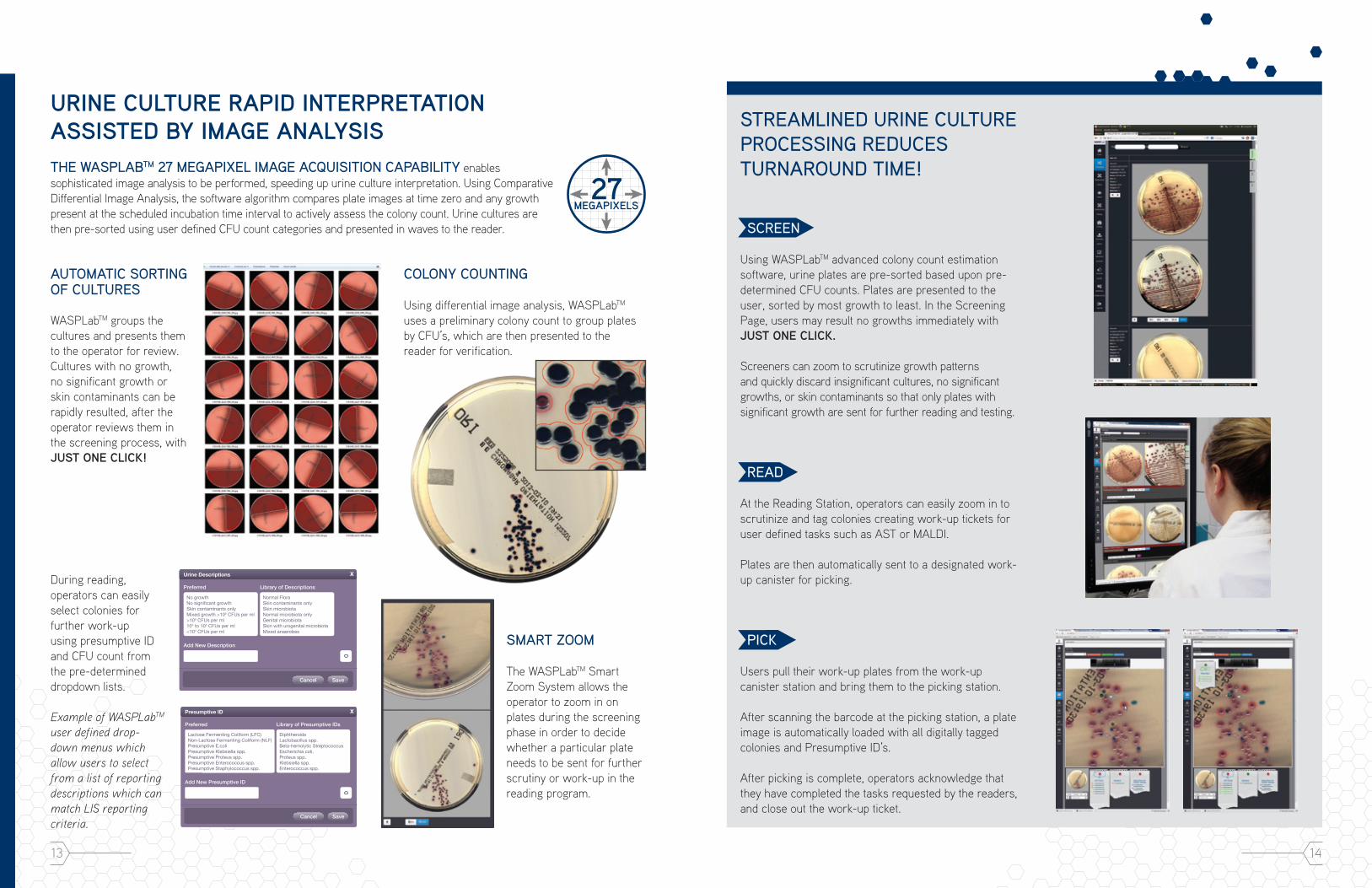
STREAMLINED URINE CULTURE PROCESSING REDUCES TURNAROUND TIME!
SCREEN
Using WASPLabTM advanced colony count estimation software, urine plates are pre-sorted based upon pre-determined CFU counts. Plates are presented to the user, sorted by most growth to least. In the Screening Page, users may result no growths immediately with JUST ONE CLICK.
Screeners can zoom to scrutinize growth patterns and quickly discard insignificant cultures, no significant growths, or skin contaminants so that only plates with significant growth are sent for further reading and testing.
READ
At the Reading Station, operators can easily zoom in to scrutinize and tag colonies creating work-up tickets for user defined tasks such as AST or MALDI.
Plates are then automatically sent to a designated work-up canister for picking.
PICK
Users pull their work-up plates from the work-up canister station and bring them to the picking station.
After scanning the barcode at the picking station, a plate image is automatically loaded with all digitally tagged colonies and Presumptive ID’s.
After picking is complete, operators acknowledge that they have completed the tasks requested by the readers, and close out the work-up ticket.
URINE CULTURE RAPID INTERPRETATION ASSISTED BY IMAGE ANALYSIS
AUTOMATIC SORTING OF CULTURES
WASPLabTM groups the cultures and presents them to the operator for review. Cultures with no growth, no significant growth or skin contaminants can be rapidly resulted, after the operator reviews them in the screening process, with JUST ONE CLICK!
COLONY COUNTING
Using differential image analysis, WASPLabTM uses a preliminary colony count to group plates by CFU’s, which are then presented to the reader for verification.
SMART ZOOM
The WASPLabTM Smart Zoom System allows the operator to zoom in on plates during the screening phase in order to decide whether a particular plate needs to be sent for further scrutiny or work-up in the reading program.
During reading, operators can easily select colonies for further work-up using presumptive ID and CFU count from the pre-determined dropdown lists.
Example of WASPLabTM user defined drop-down menus which allow users to select from a list of reporting descriptions which can match LIS reporting criteria.
Urine Descriptions
Preferred
Add New Description
X
No growthNo signi�cant growthSkin contaminants onlyMixed growth >105 CFUs per ml>105 CFUs per ml104 to 105 CFUs per ml<104 CFUs per ml
Cancel Save
Library of Descriptions
Normal FloraSkin contaminants onlySkin microbiotaNormal microbiota onlyGenital microbiotaSkin with urogenital microbiotaMixed anaerobes
Presumptive ID
Preferred
Add New Presumptive ID
X
Lactose Fermenting Coliform (LFC)Non-Lactose Fermenting Coliform (NLF)Presumptive E.coliPresumptive Klebsiella spp.Presumptive Proteus spp.Presumptive Enterococcus spp.Presumptive Staphylococcus spp.
Cancel Save
Library of Presumptive IDs
DiphtheroidsLactobacillus spp.Beta-hemolytic StreptococcusEscherichia coli,Proteus spp.Klebsiella spp.Enterococcus spp.
THE WASPLABTM 27 MEGAPIXEL IMAGE ACQUISITION CAPABILITY enables sophisticated image analysis to be performed, speeding up urine culture interpretation. Using Comparative Differential Image Analysis, the software algorithm compares plate images at time zero and any growth present at the scheduled incubation time interval to actively assess the colony count. Urine cultures are then pre-sorted using user defined CFU count categories and presented in waves to the reader.
15 16
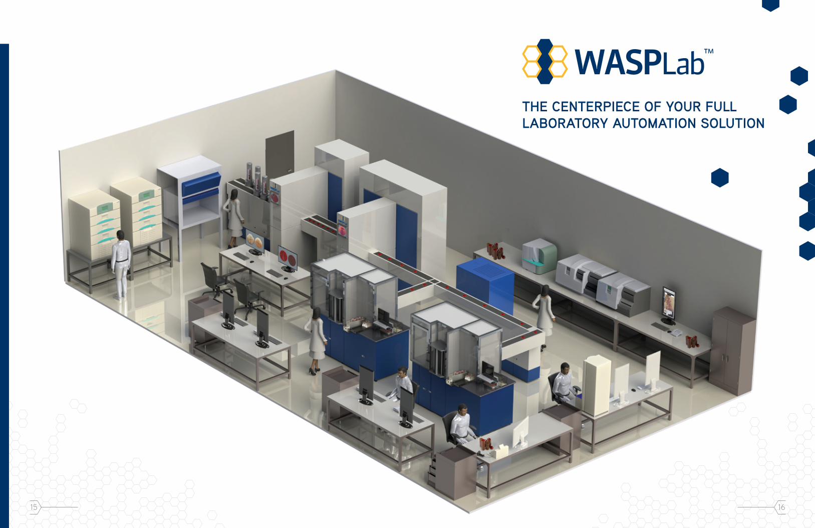
THE CENTERPIECE OF YOUR FULL LABORATORY AUTOMATION SOLUTION

17
PRODUCT SPECIFICATIONS
18
WASP®:
Dimensions: 43.5 inches wide x 81.5 inches long x 76 inches high
Weight: Approximately 1,300 lbs
Input Voltage: 220V, 20Amps
Network Ethernet: 100 MB
Interface: LIS interface available upon request
Peripherals: Touch screen monitor, external barcode reader, label printer
Certifications: CE, UL, CSA
Electrical Receptacle Plug: HBL2321 250V / 20A (for USA and Canada)
GRAM SLIDEPREPTM:
Dimensions: 28 inches wide x 23 inches long x 49.5 inches high
Weight: Approximately 221 lbs
INCUBATORS:
Dimensions Single: 45.1 inches wide x 33.7 inches long x 91.1 inches high
Dimensions Double: 68.5 inches wide x 33.7 inches long x 91.1 inches high
Weight: Approximately 1,000 lbs (Single) Approximately 2,000 lbs (Double)
Input Voltage: 220V, 20Amps
Atmospheric Conditions: CO2 and Aerobic
Capacity Single: 854 plates
Capacity Double: 1,708 plates
Electrical Receptacle Plug: HBL2321 250V / 20A (for USA and Canada)
IMAGE ANALYSIS AND DIGITAL MICROBI0LOGY
Zero hour reading of each culture plate allows for comparative differential analysis
Users can view Gram slide images on screen beside the plate image
Consists of PC, monitor, keyboard
Screening: Operator chooses plates with growth for further analysis.
Reading: Operators assign presumptive ID and further work-up for the picking bench
Consists of PC, monitor, barcode reader, printer and micro hood
Picking: Operators perform further plate work for identification or susceptibility
INTERPRETATION WORKSTATION
Incubation protocols determine scan times
Image Analysis consists of: Screening, Reading and Picking
PICKING/WORK-UP WORKSTATION
INTEGRATED GRAM SLIDES
WASPLABTM INTERFACES SEAMLESSLY with Gram slide photography microscopes to incorporate Gram slide photos into the patient record allowing users to compare plate growth with Gram stains.

COPAN DIAGNOSTICS, INC.26055 JEFFERSON AVENUEMURRIETA, CA 92562 USATL. FREE 800.216.4016TEL [email protected]
WASPLAB0414MP©2014 Copan Diagnostics, Inc. All rights reserved.
Brochure may not include the latest updates and developments. Please refer to COPAN Diagnostics website (www.copanusa.com) for the most recent version. Brochures are intended for marketing purposes. Always consult product inserts, instructions for use and manuals for the appropriate use of the products.


