Available online at www.sciencedirect.com Autonomous head-mounted electrophysiology systems for freely behaving primates Vikash Gilja 1,6 , Cindy A Chestek 2,6 , Paul Nuyujukian 3,4 , Justin Foster 2 and Krishna V Shenoy 2,3,5 Recent technological advances have led to new light-weight battery-operated systems for electrophysiology. Such systems are head mounted, run for days without experimenter intervention, and can record and stimulate from single or multiple electrodes implanted in a freely behaving primate. Here we discuss existing systems, studies that use them, and how they can augment traditional, physically restrained, ‘in-rig’ electrophysiology. With existing technical capabilities, these systems can acquire multiple signal classes, such as spikes, local field potential, and electromyography signals, and can stimulate based on real-time processing of recorded signals. Moving forward, this class of technologies, along with advances in neural signal processing and behavioral monitoring, have the potential to dramatically expand the scope and scale of electrophysiological studies. Addresses 1 Dept. of Computer Science, Stanford University, Stanford, CA 94305, United States 2 Dept. of Electrical Engineering, Stanford University, Stanford, CA 94305, United States 3 Dept. of Bioengineering, Stanford University, Stanford, CA 94305, United States 4 Medical Scientist Training Prog., Stanford University, Stanford, CA 94305, United States 5 Neurosciences Prog., Stanford University, Stanford, CA 94305, United States Corresponding author: Shenoy, Krishna V ([email protected]) 6 These authors contributed equally to this work. Current Opinion in Neurobiology 2010, 20:676–686 This review comes from a themed issue on New technologies Edited by Erin Schuman and Xiaowei Zhuang Available online 23rd July 2010 0959-4388/$ – see front matter # 2010 Elsevier Ltd. All rights reserved. DOI 10.1016/j.conb.2010.06.007 Introduction Electrophysiology methods pioneered by Evarts and Mountcastle have provided the ability to measure elec- trical activity from individual neurons in the nonhuman primate brain during sensory, motor, and cognitive tasks [1,2]. Such measurements are essential for uncovering fundamental scientific principles and enabling next gener- ation neurological treatments, including brain–machine interfaces (BMIs) (e.g. [3–9]). Unfortunately the current, widely available electrophysiology methods limit the range and complexity of studied behavioral tasks. Recent advances in miniature, low-power recording, processing, and stimulation technology are beginning to release these limitations. These technologies open the possibility for investigating an increasingly broad and naturalistic set of behaviors in nonhuman primates. Recordings traditionally use one or more penetrating elec- trodes to observe changes in the extracellular potential caused by the firing of nearby neurons (e.g. [10–20]). Each individual electrode requires at least one wired connection to a rack-mounted amplifier system. The signal path is very sensitive to micro-motion of the wires and to sources of electrical noise. Therefore studies typically constrain head position, body posture, and range of motion; consequently, experiments are limited to a few hours. This effectively prevents the study of a broad range of natural, real-world behaviors such as locomotion, navigation, object manip- ulation, vocalization, and social interaction. Importantly, it also limits the ability to study relatively simple behaviors, such as arm movements, across a broad range of behavioral contexts, often considered essential for elucidating the true underpinnings of neural control. New technology and techniques loosen these constraints. Some examples in- clude: studies of neurally or behaviorally contingent elec- trical stimulation that are typically limited to a few hours in a constrained setting [21] can be broadened to free-beha- vior for days [22 ]; multiday studies of the neural corre- lates of simple arm movements or of neural prosthetic systems become possible [23 ,24 ]; measures of neural tuning stability can be extended from hours to days by tracking neuron identities [25,26,27 ]. The history of electrophysiology is punctuated with heroic experiments that broke contemporary conventions using clever design and new technology to study ever wider ranges of behaviors. In smaller animals, tethered and cantilevered recording systems can be effectively applied if the range of motion is limited and the exper- imental field is carefully designed, such as a rat navigating a planar maze [28–31]. These experimental techniques have led to many breakthrough studies [32–35]. Such approaches have been applied in primates [36,37], but monkeys possess a high degree of dexterity and inquisi- tiveness that limits the applicability of such techniques to short timescale tasks with carefully designed geometries [38]. As Sun et al. [39] note humorously: ‘failure to Current Opinion in Neurobiology 2010, 20:676–686 www.sciencedirect.com
Transcript
Available online at www.sciencedirect.com
Autonomous head-mounted electrophysiology systems for freelybehaving primatesVikash Gilja1,6, Cindy A Chestek2,6, Paul Nuyujukian3,4, Justin Foster2 andKrishna V Shenoy2,3,5
Recent technological advances have led to new light-weight
battery-operated systems for electrophysiology. Such systems
are head mounted, run for days without experimenter
intervention, and can record and stimulate from single or
multiple electrodes implanted in a freely behaving primate.
Here we discuss existing systems, studies that use them, and
how they can augment traditional, physically restrained, ‘in-rig’
electrophysiology. With existing technical capabilities, these
systems can acquire multiple signal classes, such as spikes,
local field potential, and electromyography signals, and can
stimulate based on real-time processing of recorded signals.
Moving forward, this class of technologies, along with
advances in neural signal processing and behavioral
monitoring, have the potential to dramatically expand the
scope and scale of electrophysiological studies.
Addresses1Dept. of Computer Science, Stanford University, Stanford, CA 94305,United States2Dept. of Electrical Engineering, Stanford University, Stanford, CA94305, United States3Dept. of Bioengineering, Stanford University, Stanford, CA 94305,United States4Medical Scientist Training Prog., Stanford University, Stanford, CA94305, United States5Neurosciences Prog., Stanford University, Stanford, CA 94305, UnitedStates
Corresponding author: Shenoy, Krishna V ([email protected])6 These authors contributed equally to this work.
Current Opinion in Neurobiology 2010, 20:676–686
This review comes from a themed issue onNew technologiesEdited by Erin Schuman and Xiaowei Zhuang
Available online 23rd July 2010
0959-4388/$ – see front matter# 2010 Elsevier Ltd. All rights reserved.
DOI 10.1016/j.conb.2010.06.007
IntroductionElectrophysiology methods pioneered by Evarts andMountcastle have provided the ability to measure elec-trical activity from individual neurons in the nonhumanprimate brain during sensory, motor, and cognitive tasks[1,2]. Such measurements are essential for uncoveringfundamental scientific principles and enabling next gener-ation neurological treatments, including brain–machine
interfaces (BMIs) (e.g. [3–9]). Unfortunately the current,widely available electrophysiologymethods limit the rangeand complexity of studied behavioral tasks. Recentadvances in miniature, low-power recording, processing,and stimulation technology are beginning to release theselimitations. These technologies open the possibility forinvestigating an increasingly broad and naturalistic set ofbehaviors in nonhuman primates.
Recordings traditionally use one or more penetrating elec-trodes to observe changes in the extracellular potentialcaused by the firing of nearby neurons (e.g. [10–20]). Eachindividual electrode requires at least one wired connectionto a rack-mounted amplifier system.The signal path is verysensitive to micro-motion of the wires and to sources ofelectrical noise. Therefore studies typically constrain headposition, body posture, and range ofmotion; consequently,experiments are limited to a few hours. This effectivelyprevents the study of a broad range of natural, real-worldbehaviors such as locomotion, navigation, object manip-ulation, vocalization, and social interaction. Importantly, italso limits the ability to study relatively simple behaviors,such as armmovements, across a broad range of behavioralcontexts, often consideredessential for elucidating the trueunderpinnings of neural control. New technology andtechniques loosen these constraints. Some examples in-clude: studies of neurally or behaviorally contingent elec-trical stimulation that are typically limited to a few hours ina constrained setting [21] can be broadened to free-beha-vior for days [22!!]; multiday studies of the neural corre-lates of simple arm movements or of neural prostheticsystems become possible [23!,24!]; measures of neuraltuning stability can be extended from hours to days bytracking neuron identities [25,26,27!].
The history of electrophysiology is punctuated withheroic experiments that broke contemporary conventionsusing clever design and new technology to study everwider ranges of behaviors. In smaller animals, tetheredand cantilevered recording systems can be effectivelyapplied if the range of motion is limited and the exper-imental field is carefully designed, such as a rat navigatinga planar maze [28–31]. These experimental techniqueshave led to many breakthrough studies [32–35]. Suchapproaches have been applied in primates [36,37], butmonkeys possess a high degree of dexterity and inquisi-tiveness that limits the applicability of such techniques toshort timescale tasks with carefully designed geometries[38]. As Sun et al. [39] note humorously: ‘failure to
Current Opinion in Neurobiology 2010, 20:676–686 www.sciencedirect.com
appreciate the frustration of an unattended monkey canlead to the destruction of expensive equipment.’
Wireless technologies have been applied to eliminatethe restrictions of wired tethers. Delgado was an earlypioneer of such technologies, most famous for his 1963bullfighting demonstration. By switching on a wirelessneural stimulator in caudate nucleus he stopped a bullcharging toward him. Starting in the 1950s, Delgadoused wireless neural stimulation to modify free-behaviorin cats, monkeys, apes, and humans [40]. In addition tostimulation, technology for transmitting single-unitactivity was developed as early as the 1960s [41–43].These systems relied on analog signal transmission, inwhich even highly amplified signals can be contami-nated with noise during transmission; they are sensitiveto device orientation, occlusion, and interference. Suchsensitivities can compromise data as posture and pos-ition of the animal can alter the properties of thereceived signal. More recently, digital systems withonboard data storage or digital wireless transmissionwith error checking have been employed across animalmodels, from insect [44,45!] to rodent [46,47] to primate[48!,24!,49!!,50].
New tools and technologyIn the last five years, a variety of devices enablingelectrophysiological from freely behaving primates havebeen developed. Figure 1a is a concept sketch summar-izing, and expanding upon, the composite functionality ofthese systems, providing signal input, processing, andoutput capabilities. Many of these systems use a head-mounted form factor as shown in Figure 1b. In Figure 1b,a head-mounted electrophysiology system continuouslyrecords and wirelessly telemeters neural activity from amacaque in his home cage. At the moment captured bythe sketch, the macaque has decided to recover a treatplaced on a foraging board.
On the input side, systems have been developed forintracortical (spikes and local field potential) electrodes[24!,49!!,50,48!], electromyography [48!], and behavioralinputs [51,24!]). Signals are processed with methodsemployed in conventional electrophysiological setups.For example, intracortical signals can be amplified, fil-tered, digitized, and processed with fidelity equivalent tocommercial rack systems [24!]. These signals are eitherretained in full broadband form [24!,49!!,50,48!], as wave-form snippets [48!], or by applying a voltage threshold tothe spike band to isolate action potentials [49!!,52].Processed signals are stored to onboard memory[48!,24!], telemetered via radio link [49!!,50], or usedto control electrical stimulation output [48!]. Additionaloutputs, such as optical stimulation for use with optoge-netics [35,53–55] or electronically controlled drug deliv-ery could be integrated. The power requirements foroptical stimulation are well below the power consumption
of current systems and could be enabled with boardmountable super-luminescent LEDs or semiconductorlaser diodes.
For primate research, systems must be durable, light-weight, and should run without requiring servicing, suchas battery or memory changes, for long periods of time tominimize disruptions to free-behavior. Researchers haveaddressed these constraints and developed fully enclosedwireless neural recording systems that store data toonboard memory [48!,24!]. Systems with onboard mem-ory have proven fruitful in early studies of electrodeinterface stability, properties of motor cortical neurons,and BMI applications [24!,22!!,23!]. However, the datacannot be accessed until the onboard memory isretrieved. Thus, these systems lack real-time access toneural data, making synchronization with stimuli presen-tation or behavioral measurement, and the study of closedloop neural control difficult or impossible. Fortunately,recent technological advances allow wireless data tele-metry [49!!,50], enabling a broader class of studies.
Battery life tradeoffs can affect experimental design. Forexample, the HermesD [50] system is capable of con-tinuous operation for up to two days, recording anddigitally telemetering 32 channels of broadband neuraldata sampled with 12 bits of resolution at 30 kS/s. Withthe same battery system, HermesC [49!!] can telemeterone channel of broadband with 10 bits of resolution at15.7 kS/s and 20 channels of 1 kS/s threshold crossings for6.8 days. The HermesD system provides higher fidelitydata, but limits experimental duration relative to Her-mesC. HermesD is a better solution if the experimentallows daily servicing of equipment; however, if the studyrequires longer uninterrupted recording, HermesC maybe the better solution. The form factor of these recordingsystems is small enough to sit on top of an adult rhesusmacaques head without any notable changes in behavior;complete systems (chassis, electronics, and batteries)weigh 56–250 g (Neurochip 56 g [48!], HermesC 114 g[49!!], HermesB 250 g [24!]) and adult rhesus macaquestypically weigh approximately 10 kg.
Many other systems using application specific integratedcircuits (ASICs) have been developed, and these could beused for freely moving monkey experiments, if appro-priately modified, in the future [56,57!!,58–71]. ASICsoffer potential benefits to head-mounted systems, in-cluding substantial reductions in size and power con-sumption with simultaneous increases in channel countand signal bandwidth. They could also introduce newmodalities such as multichannel stimulation [67,60],onboard spike sorting [61,72], and chemical sensing usingvoltammetry [69]. Advances in this field are largelymotivated by the need for fully implantable systems thatmove toward the requirements for clinically viable neuralprostheses [59,70,58,64,71]. While some requirements for
Freely-behaving Primate Electrophysiology Gilja et al. 677
www.sciencedirect.com Current Opinion in Neurobiology 2010, 20:676–686
these clinically oriented devices share common goalswith head-mounted electrophysiology systems, fullyimplantable systems are subject to different designconstraints, ultimately distinguishing them from head-mounted systems.
Studies to dateAutonomous head-mounted electrophysiology hasdemonstrated utility in both the neural engineeringand basic neuroscience domains. On the neural engin-eering side, the HermesB system [24!] has documented
678 New technologies
Figure 1
Autonomous head-mounted electrophysiology systems overview: (a) shows a system diagram summarizing potential autonomous head-mountedelectrophysiology system designs. (b) Shows a sketch of how a wireless telemetry recording system operates and provides a general sense of scale ofthese systems. This is the recording setup used in HermesC [49!!] studies, in which one broadband channels and multiple threshold channels arerecorded and synchronized to recorded video.
Current Opinion in Neurobiology 2010, 20:676–686 www.sciencedirect.com
neural recording stability, and instability, with a Utahelectrode array [73] over several days. Understanding theproperties of electrode arrays is critical to the develop-ment of robust neural prosthetic systems and couldenable multiday learning studies requiring the trackingof single neurons. Figure 2a shows action potential wave-forms from a single channel across 29.5 hours. Note thatthe amplitudes of the two sorted units, from the sameelectrode, are flexing relative to each other. This neuralrecording system can monitor head movement continu-ously with an onboard accelerometer. This study alsoshowed large shifts in waveform amplitude coincidentwith >3 g acceleration events, see Figure 2b. Using headacceleration data as a proxy for overall level of movement,HermesB also demonstrates how a BMI can use local fieldpotential energy to reliably detect periods of physicalactivity and inactivity. HermesC [49!!], shown inFigure 2c,d, can track more neural channels for longerperiod of time, by multiplexing its broadband channel;
Figure 2e summarizes data from a nearly continuous 12.5-day dataset. We see that on the multiday timescalerecorded action potentials can be relatively stable(Figure 2f, left), but can also change dramatically(Figure 2f, right).
Studies with theNeurochip system have allowed for novelsystems neuroscience studies. Besides neural recordingcapability, Neurochip [48!] as shown in Figure 3a,b, canrecord from EMG electrodes and can provide neuralstimulation. Jackson et al. [22!!] utilized this capabilityto demonstrate plasticity in the motor cortex. Figure 3cshows a schematic of the recording and stimulation para-digm.Duringphysically constrained in-rig experiments, ananimalwas trained toperformawrist flexion/extension taskinwhich the tuning direction of an individual neuron couldbe measured. While the animal was freely moving in thehome cage, spikeswere recorded from one neuron,Nrec infigure, with characteristic tuning. When that neuron
Freely-behaving Primate Electrophysiology Gilja et al. 679
Figure 2
The Hermes system and single-unit stability across days. (a) Shows the 95% confidence intervals of sorted action potentials from a single channelrecorded with HermesB [24!] across 29.5 hours, note that the action potentials of the units are flexing relative to one another. (b) Shows a large shift inpeak-to-peak waveform amplitude coincident with a large accelerometer event. (c) A drawing of the HermesC system [49!!] from implant toelectronics. (d) Two pictures of the actual system: on the left is the chassis base, Utah array connector, and board stack; on the right is the fullyenclosed system with transmitting antenna. (e) Shows the 95% peak-to-peak envelope of waveforms observed for 12.5 days for channels 2–20 withHermesC. The voltage is averaged across three hour bins, normalized to the average voltage on that channel. Variations fell within 65–215% of themean, or 9.3–220 mV. As shown in (f), channels can remain static; although single units often appear and disappear.
www.sciencedirect.com Current Opinion in Neurobiology 2010, 20:676–686
spiked, small stimulation currents were delivered at asecond site, Nstim. Over a period of two days, the tuningdirection atNrec becamemore like that ofNstim, as shownin Figure 3d, suggesting that connections between the twoareas had been strengthened in some way. The multidaytimescales required for this particular study underscore theimportance of long-term recording and stimulation using a
wireless device. Previous plasticity experiments have beenlimited to shorter timescales, and perturbations that theanimal can adapt to within minutes. Some mechanisms oflearningmay simply be inaccessible to the experimenter attraditional timescales. Also, a wide range of new neuro-logical therapeutics might become possible with such amethod of encouraging cortical plasticity.
680 New technologies
Figure 3
The Neurochip system and multiday stimulation induced plasticity. (a) A drawing of the Neurochip system [48!] and (b) is a picture of the Neurochipboard. (c) A schematic of the paradigm employed by Jackson et al. in [22!!], in which an electrode with a sortable unit, Nstim, was stimulated after adelay from when Nrec, a unit recorded on another electrode, spiked. The paradigm was run continuously for two days with the Neurochip. After the twodays, tuning curves for Nstim, Nrec, and a control unit were recorded with a wrist flexion task. (d) Shows the tuning curves plotted relative to theaxis labels shown. Note that the tuning of Nstim after stimulation is more similar to Nrec than before stimulation and that the tuning of the control cell isrelatively unchanged. (Figure 3(b) is reprinted from the Journal of Neuroscience Methods, 148, Jaideep Mavoori, Andrew Jackson, Chris Diorio,Eberhard Fetz, An autonomous implantable computer for neural recording and stimulation in unrestrained primates, p. 73, copyright 2005, withpermission from Elsevier B.V. Figure 3(c) is adapted from Nature (22), copyright 2006, by permission from Macmillan Publishers Ltd.).
Current Opinion in Neurobiology 2010, 20:676–686 www.sciencedirect.com
A second study using Neurochip [23!] measured thecorrelation between the activity of single neurons andEMG during an in-rig wrist flexion/extension task, as wellas freely moving in the cage. They observe similar cor-relations across the day during free-behavior, but notduring sleep. These correlations could vary greatly be-tween free-behavior and a trained task, although thepreferred direction of a neuron tended to be preserved.Also, neural recordings during sleep show evidence of thetransition between slow wave and REM sleep.
Novel hippocampal studies are also enabled by autonom-ous head-mounted electrophysiology. Many studies ofhippocampus rely on the ability of an animal to navigatean environment. By developing a wireless system [39],Sun and colleagues have successfully recorded fromhippocampal neurons while monkeys were navigating a15 " 15 room to obtain treats in remembered locations.Interestingly, they record place fields, summarizing thecorrelation between spatial location and neural firing, thatchange between different task definitions in the same
environment. Using a monkey model for hippocampalstudies may enable more complex memory and naviga-tion tasks than are currently possible with commonlyexercised rodent models.
Expanding the scope of studyThe experiments above highlight a few specific appli-cations of this new class of technology, but why focus onsuch techniques when complex questions can be studiedwith carefully controlled, constrained, and measuredbehaviors in a traditional experimental rig? In the caseof motor control, we believe that the study of free-behavior can augment traditional experiments in animportant way.
As an example, consider the hypothetical progression ofnonhuman primate motor control studies shown inFigure 4a. The traditional setup is depicted in subpanel1, where a monkey is engaged in a highly constrained armmovement task, with his head position and body posturefixed. While this task design allows the experimenter to
Freely-behaving Primate Electrophysiology Gilja et al. 681
Figure 4
Expanding the scope of motor system studies: (a) shows a progression of potential motor control studies enabled by head-mounted electrophysiologysystems. (1) The traditional wired in-rig setup in which the animal is engaged in a reaching task with a highly constrained posture. (2) Utilizes a wirelesstelemetry system and the animal is able to move to many different postures as it plays with a sensorized manipulandam. (3) Shows the possibility ofstudying complex movements, such as leaping. (4) The ability to decode motor intentions. (b) A sketch of a potential computer vision system forrecording detailed body posture simultaneously with wireless neural data. The images from all cameras are used to construct a model of all jointpositions at every moment in time.
www.sciencedirect.com Current Opinion in Neurobiology 2010, 20:676–686
test hypotheses about the motor system with a great dealof control and precision, including the ability to acquiretens to hundreds of repeated trials per behavioral con-dition (e.g. [74]), it is a small subset of the full capabilityof the motor system. Dominated by simple point-to-pointarmmovement tasks, these careful studies have seminallyadvanced the field over the past few decades. However,even small deviations from the restricted regimes of armmovements studied in these highly constrained tasks (e.g.isolated or enforced shoulder posture, arm orientation,movement speed, and force) can lead to a breakdown inprevailing models (e.g. [75,76,14–20]).
Work by Aflalo and Graziano [77,78] demonstrates thediversity of neural responses during unconstrained armmovement in rig. In this study, the monkey’s head andipsilateral arm are constrained, but the contralateral arm isrecorded during free movement. Thus, the only differ-ence from the traditional setup is the lack of a definedbehavioral task. They show that existing models, devel-oped with constrained behavioral task data, do not explainthe majority of neural variability in their free movementdata. This result exposes a weakness in the prevailingcourse of study: the limits of task design can lead toimpoverished models of the observed neural system.Thus, we believe that data recorded during increasinglycomplex behavior are necessary to augment the under-standing developed from physically constrained in-rigstudies.
With head-mounted recording technologies, the field canbegin to explore an increasingly complicated range ofbehaviors with nonhuman primates, as in Figure 4a sub-panel 2, in which a monkey is manipulating a toy frommultiple postures. Suchmanipulanda can be sensorized toallow arbitrarily complicated behaviors to be preciselymeasured. Multiple infrared reflective fiducial markers,have long enabled researchers to study 3D position of thejoints of the arm and/or hand (e.g. ‘Polaris’, ‘Optotrak’,and ‘Vicon’ systems; also [79–81]). Unfortunately, the useof fiducial markers during free-behavior is difficult,requiring the use of ‘jackets’ and even then are likelyto be destroyed quickly due to innate monkey curiosity.Ideally, we need a system capable of tracking posturefrom markerless images. Fortunately, computer vision ismeeting such demands for the entertainment industry[82!]. These computer vision systems analyze markerlessimages from multiple camera views, allowing for 3Dreconstruction of the subject’s pose from which limb jointangles can be inferred. Figure 4b provides a sketch ofsuch a system in operation, with multiple camera viewsallowing estimation of all joint positions as shown to theright. Such systems, adapted for nonhuman primates,could enable the collection of 24 hours continuous data-sets of simultaneous full-body posture and correspondingneural data. These potential datasets will test the limits ofcurrent models of motor control, by allowing the study of
quite complex movements, as in subpanel 3. As ourunderstanding of the motor system advances, so shouldour ability to develop prosthetic systems that operate incomplex environments and are capable of decoding themere ‘thought’ of jumping, climbing, or other naturalisticmovements, as illustrated in subpanel 4.
With the ability to record neural and behavioral data for24 hours a day/seven days a week from a freely behavingmonkey, a new class of experiments can emerge. Bycollecting extremely long and precise datasets, exper-imental questions can be answered by data mining asis commonly explored in bioinformatic studies of thegenome. For example, an experimenter interested instudying the neural correlates of limb motor controlcan query long datasets of free-behavior for relevantmovements and corresponding neural data.
A growing number of proposed clinical treatments ofneurological diseases, including epilepsy [83], Parkinsons[84], and dystonia [85], rely on electrical stimulation ofneural tissue. Stimulation-based treatments of depressionand obsessive compulsive disorder are also currentlyunder study [86]. Wireless recording and stimulationsystems for primates can allow for rapid design and testingof such approaches. More generally, continuous neuralrecording from animal models of neurological disorderscould help guide drug discovery and implantable medicaldevice development.
Single trial analysis and decoding with parallelrecordingsIn-rig experiments allow the experimenter to repeat trialconditions through training and behavioral control. Thisrepetition allows the experimenter to focus on variabilityof interest by trial-averaging to remove undesired varia-bility, such as Poisson spiking noise [87]. As studies moveincreasingly toward free-behavior, such repetitionbecomes increasingly unlikely. We recognize that a majorchallenge is that every trial is effectively different, thusprecluding traditional trial-averaging which is necessarywhen recording from one neuron at a time. All analysesessentially become single trial analysis; we, and others,have been working in recent years to produce turn-keymethods for creating ‘single trial neural trajectories’, withmillisecond time resolution, which is possible when sim-ultaneously recording from tens to hundreds of neuronsand applying advanced analytical methods (for relevantreviews see [88!,89]; see also [90,87]).
Besides using techniques to denoise simultaneouslyrecorded neural data, we can limit the variables understudy by filtering for a smaller subset of behaviors. Suchan approach can be applied to long datasets; for example,in motor studies one can query for a limited range ofmovements and postures based upon a question of in-terest. Alternatively, the use of in cage ‘smart toys’ such as
682 New technologies
Current Opinion in Neurobiology 2010, 20:676–686 www.sciencedirect.com
button push panels, manipulandams, and treadmills, pro-vides methods for creating more constrained sets ofrepeated behavior. These methods allow for a continuumof study complexity, bridging highly constrainedtraditional in-rig tasks, and completely unconstrainedfree-behavior, as illustrated in Figure 4a.
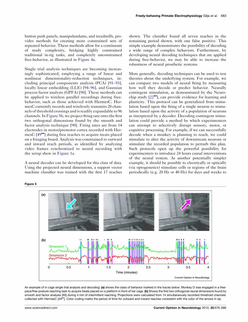
Single trial analysis techniques are becoming increas-ingly sophisticated, employing a range of linear andnonlinear dimensionality-reduction techniques, in-cluding principal components analysis (PCA) [91–93],locally linear embedding (LLE) [94–96], and Gaussianprocess factor analysis (GPFA) [90]. These methods canbe applied to wireless parallel recordings during free-behavior, such as those achieved with HermesC. Her-mesC currently records and wirelessly transmits 20 chan-nels of threshold crossings and is readily expandable to 96channels. In Figure 5b, we project firing rate onto the firsttwo orthogonal dimensions found by the smooth andfactor analysis technique [90]. Firing rates are from 14electrodes in motor/premotor cortex recorded with Her-mesC [49!!] during free reaches to acquire treats placedon a foraging board. Analysis was constrained to outwardand inward reach periods, as identified by analyzingvideo frames synchronized to neural recording withthe setup show in Figure 1a.
A neural decoder can be developed for this class of data.Using the projected neural dimensions, a support vectormachine classifier was trained with the first 17 reaches
shown. The classifier found all seven reaches in theremaining period shown, with one false positive. Thissimple example demonstrates the possibility of decodinga wide range of complex behaviors. Furthermore, bydeveloping neural decoding techniques that are appliedduring free-behavior, we may be able to increase therobustness of neural prosthetic systems.
More generally, decoding techniques can be used to testtheories about the underlying system. For example, wecan compare two models of neural firing by measuringhow well they decode or predict behavior. Neurallycontingent stimulation, as demonstrated by the Neuro-chip study [22!!], can provide evidence for learning andplasticity. This protocol can be generalized from stimu-lation based upon the firing of a single neuron to stimu-lation based upon the activity of a population of neuronsas interpreted by a decoder. Decoding contingent stimu-lation could provide a method by which experimenterscan attempt to selectively disrupt sensory, motor, orcognitive processing. For example, if we can successfullydecode when a monkey is planning to reach, we couldstimulate to alter the activity of downstream neurons orstimulate the recorded population to perturb this plan.Such protocols open up the powerful possibility forexperimenters to introduce 24 hours causal interventionsof the neural system. As another potentially simplerexample, it should be possible to electrically or optically(via optogenetics) stimulate cells or regions of the brainperiodically (e.g. 20 Hz or 40 Hz) for days and weeks to
Freely-behaving Primate Electrophysiology Gilja et al. 683
Figure 5
An example of in cage single trial analysis and decoding: (a) shows the class of behavior marked in the traces below. Monkey D was engaged in a free-pace/free-posture reaching task to acquire treats placed on a platform in front of her cage. (b) Shows the first two orthogonal neural dimensions found bysmooth and factor analysis [90] during 4 min of intermittent reaching. Projections were calculated from 14 simultaneously recorded threshold channelscollected with HermesC [49!!]. Color coding marks the period of time for outward and inward reaches consistent with the color of the arrows in (a).
www.sciencedirect.com Current Opinion in Neurobiology 2010, 20:676–686
test theories regarding the causal relationship betweenrhythmicity and behavior [97–99].
ConclusionsAutonomous head-mounted electrophysiology systemshave begun to make an impact on both basic neuro-science and neural engineering. As these systemsadvance and becomemore accessible to the electrophysi-ology community, they will augment existing studieswith the potential to expand experimental context.Experimenters can begin to release the physical con-straints necessary for using wired rack-mounted systemsand can expand studies from a few hours to several days.The utility of these techniques will rest heavily uponadvances in single trial neural signal processing andmethods for behavioral measurement, such asmarkerlessmotion tracking.
AcknowledgementsThis work was supported by NSF Graduate Research Fellowships(V.G., C.A.K.), an NDSEG graduate fellowship (V.G.), Stanford GraduateFellowships (C.A.K, J.F.), the MSTP program (P.N.), HHMI (P.N.), theSoros Foundation (P.N.), and the following to K.V.S.: Burroughs WellcomeFund, McKnight Foundation, NIH-NINDS EUREKA, and NIH Director’sPioneer Award.
References and recommended readingPapers of particular interest, published within the period of review,have been highlighted as:
! of special interest!! of outstanding interest
1. Evarts EV: Relation of pyramidal tract activity to forceexerted during voluntary movement. J Neurophysiol 1968,31:14-27.
2. Mountcastle VB, Lynch JC, Georgopoulos A, Sakata H, Acuna C:Posterior parietal association cortex of the monkey:command functions for operations within extrapersonalspace. J Neurophysiol 1975, 38:871-908.
3. Taylor D, Tillery SH, Schwartz A: Direct cortical control of 3Dneuroprosthetic devices. Science 2002, 296:1829-1832.
4. Serruya M, Hatsopoulos N, Paninski L, Fellows M, Donoghue J:Instant neural control of a movement signal. Nature 2002,416:141-142.
5. Musallam S, Corneil B, Greger B, Scherberger H, Andersen R:Cognitive control signals for neural prosthetics. Science 2004,305:258-262.
6. Hochberg LR, Serruya MD, Friehs GM, Mukand JA, Saleh M,Caplan AH, Branner A, Chen D, Penn RD, Donoghue JP:Neuronalensemble control of prosthetic devices by a human withtetraplegia. Nature 2006, 442:164-171.
8. Kim S, Simeral JD, Hochberg LR, Donoghue JP, Black MJ: Neuralcontrol of computer cursor velocity by decoding motorcortical spiking activity in humans with tetraplegia. J NeuralEng 2008, 5:455-476.
9. Velliste M, Perel S, Spalding MC, Whitford AS, Schwartz AB:Cortical control of a prosthetic arm for self-feeding. Nature2008, 453:1098-1101.
10. Georgopoulos A, Kalaska J, Caminiti R, Massey J: On therelations between the direction of two-dimensional armmovements and cell discharge in primate motor cortex.J Neurosci 1982, 2:1527-1537.
11. Georgopoulos A, Schwartz A, Kettner R: Neuronal populationcoding of movement direction. Science 1986, 233:1416-1419.
12. Ashe J, Georgopoulos A: Movement parameters and neuralactivity in motor cortex and area 5. Cereb Cortex 1994,4:590-600.
14. Scott S, Kalaska J: Reaching movements with similar handpaths but different arm orientations. I. Activity of individualcells in motor cortex. J Neurophysiol 1997, 77:826-852.
15. Scott SH, Sergio LE, Kalaska JF: Reaching movements withsimilar hand paths but different arm orientations. II. activity ofindividual cells in dorsal premotor cortex and parietal area 5.J Neurophysiol 1997, 78:2413-2426.
16. Sergio L, Kalaska J:Changes in the temporal pattern of primarymotor cortex activity in a directional isometric force versuslimb movement task. J Neurophysiol 1998, 80:1577-1583.
17. Cabel DW, Cisek P, Scott SH: Neural activity in primary motorcortex related tomechanical loads applied to the shoulder andelbow during a postural task. J Neurophysiol 2001,86:2102-2108.
18. Kakei S, Homan D, Strick P: Muscle and movementrepresentations in the primary motor cortex. Science 1999,285:2136-2139.
19. Cheney PD, Fetz EE: Functional classes of primatecorticomotoneuronal cells and their relation to active force.J Neurophysiol 1980, 44:773-791.
20. Fetz EE, Finocchio DV, Baker MA, Soso MJ: Sensory and motorresponses of precentral cortex cells during comparablepassive and active joint movements. J Neurophysiol 1980,43:1070-1089.
21. Li CS, Padoa-Schioppa C, Bizzi E: Neuronal correlates of motorperformance and motor learning in the primary motor cortexof monkeys adapting to an external force field. Neuron 2001,30:593-607.
22.!!
Jackson A, Mavoori J, Fetz EE: Long-term motor cortexplasticity induced by an electronic neural implant.Nature 2006,444:56-60.
This paper describes a long-term causal plasticity experiment in whichneural tuning is shifted as a result of neural stimulation in a freely movingprimate; it is enabled by the Neurochip system.
23.!
Jackson A, Mavoori J, Fetz EE: Correlations between the samemotor cortex cells and armmuscles during a trained task, freebehavior, and natural sleep in the macaque monkey.J Neurophysiol 2007, 97:360-374.
This study compares the relationship between EMG and neural activitywith a primate in-rig, freely moving, and during sleep. EMG and neuralrecordings were made with the Neurochip system.
24.!
Santhanam G, Linderman MD, Gilja V, Afshar A, Ryu SI, Meng TH,Shenoy KV, Hermes B: A continuous neural recording systemfor freely behaving primates. IEEE Trans Biomed Eng 2007,54:2037-2050.
The HermesB system records 2 of 16 channels of neural data, which canbe multiplexed during recording, and three-axis accelerometer data toflash memory from a freely moving primate.
25. Chestek C, Batista A, Santhanam G, Yu B, Afshar A,Cunningham J, Gilja V, Ryu S, Churchland M, Shenoy K: Single-neuron stability during repeated reaching in macaquepremotor cortex. J Neurosci 2007, 27:10742-10750.
26. Chestek CA, Cunningham JP, Gilja V, Nuyujukian P, Ryu SI,Shenoy KV: Neural prosthetic systems: current problems andfuture directions. Engineering in Medicine and Biology Society,2009. EMBC 2009. Annual International Conference of the IEEE(2009). 2009:3369-3375.
27.!
Dickey AS, Suminski A, Amit Y, Hatsopoulos HG: Single-unitstability using chronically implanted multielectrode arrays.J Neurophysiol 2009, 102:1331-1339.
This work describes a technique for tracking units across days for thepurpose of studying learning and plasticity.
684 New technologies
Current Opinion in Neurobiology 2010, 20:676–686 www.sciencedirect.com
28. Komisaruk BR, Olds J: Neuronal correlates of behavior in freelymoving rats. Science 1968, 161:810-813.
29. O’Keefe J, Dostrovsky J: The hippocampus as a spatial map.preliminary evidence fromunit activity in the freely-moving rat.Brain Res 1971, 34:171-175.
30. Wilson MA, McNaughton BL: Dynamics of the hippocampalensemble code for space. Science 1993, 261:1055-1058.
31. Wilson MA, McNaughton BL: Reactivation of hippocampalensemblememories during sleep. Science (New York, NY) 1994,265:676-679.
32. O’Keefe J, Recce ML: Phase relationship betweenhippocampal place units and the EEG theta rhythm.Hippocampus 1993, 3:317-330.
33. O’Keefe J, Burgess N: Geometric determinants of the placefields of hippocampal neurons. Nature 1996, 381:425-428.
34. Talwar SK, Xu S, Hawley ES, Weiss SA, Moxon KA, Chapin JK:Behavioural neuroscience: rat navigation guided by remotecontrol. Nature 2002, 417:37-38.
35. Airan RD, Thompson KR, Fenno LE, Bernstein H, Deisseroth K:Temporally precise in vivo control of intracellular signaling.Nature 2009, 458:1025-1029.
36. Ludvig N, Botero JM, Tang HM, Gohil B, Kral JG: Single-cellrecording from the brain of freely moving monkeys. J NeurosciMethods 2001, 106:179-187.
37. Jurgens U, Hage SR: Telemetric recordings of neuronal activity.Methods 2006, 38:195-201.
44. Takeuchi S, Shimoyama I: A radio-telemetry system with ashape memory alloy microelectrode for neural recording offreely moving insects. IEEE Trans Biomed Eng 2004, 51:133-137.
45.!
Harrison R, Fotowat H, Chan R, Kier R, Leonardo A, Gabbiani F: Awireless neural/EMG telemetry system for freely movinginsects. In Proceedings of the IEEE International SymposiumCircuit Systems (ISCAS) 2010 (in press)
This paper describes the design of a <1 g system for recording neuralactivity, EMG, and acceleration in freely behaving dragonflies.
46. Farshchi S, Nuyujukian PH, Pesterev A, Mody I, Judy JW: Atinyos-enabled mica-2 based wireless neural interface. IEEETrans Biomed Eng 2006, 53:1416-1424.
47. Cheney D, Goh A, Gugel K, Harris J, Sanchez JC, Principe JC:Wireless, in vivo neural recording using a custom integratedbioamplifier and the pico system. In Proceedings of the IEEEEMBS. 2007:4387-4391.
48.!
Mavoori J, Jackson A, Diorio C, Fetz EE: An autonomousimplantable computer for neural recording and stimulation inunrestrained primates. J Neurosci Methods 2005, 148:71-77.
The Neurochip chip system is described in detail. The system can recordEMG and neural activity as well as deliver stimulation autonomouslybased on spiking activity in a freely moving primate.
49.!!
Chestek CA, Gilja V, Nuyujukian P, Kier R, Solzbacher F, Ryu SI,Harrison RR, Shenoy KV, Hermes C: Low-power wireless neuralrecording system for freely moving primates. IEEE Trans NeuralSyst Rehabil Eng 2009, 17:330-338.
HermesC is a radio frequency (RF) wireless system for recording 1 of 20channels of neural data, which can be multiplexed during recording. Thissystem has been expanded to also include 20 channels of thresholdcrossings.
50. Miranda H, Gilja V, Chestek CA, Shenoy KV, Meng TH, Hermes D:A high-rate long-range wireless transmission system forsimultaneous multichannel neural recording applications.IEEE Trans Biomed Circ Syst 2010, 4:181–191.
51. Papailiou A, Sullivan E, Cameron JL: Behaviors in rhesusmonkeys (macaca mulatta) associated with activity countsmeasured by accelerometer. Am J Primatol 2008, 70:185-190.
52. Harrison RR, Kier RJ, Chestek CA, Gilja V, Nuyujukian P, Ryu S,Greger B, Solzbacher F, Shenoy KV: Wireless neural recordingwith single low-power integrated circuit. IEEE Trans Neural SystRehabil Eng 2009, 17:322-329.
53. Paralikar K, Cong P, Santa W, Dinsmoor D, Hocken B, Munns G,Giftakis J, Denison T: An implantable 5 mw/channel dual-wavelength optogenetic stimulator for therapeuticneuromodulation. IEEE ISSCC 2010 (in press).
54. Han X, Qian X, Bernstein JG, Zhou HH, Franzesi GT, Stern P,Bronson RT, Graybiel AM, Desimone R, Boyden ES: Millisecondtimescale optical control of neural dynamics in the nonhumanprimate brain. Neuron 2009, 62:191-198.
55. Diester I, Kaufman M, Pashaie R, Mogri M, Deisseroth K,Shenoy KV: An optogenetic toolbox for primates. Soc Neurosci2010. (Abstract).
56. Song YK, PattersonWR, Bull CW, Hwang NJ, Deangelis AP, Lay C,McKay JL, Nurmikko AV, Donoghue JP, Connors BW:Development of an integrated microelectrode,microelectronic device for brain implantableneuroengineering applications. In Proceedings of the IEEEEMBS. 2004:4053-4056.
57.!!
Nurmikko AV, Donoghue JP, Hochberg LR, Patterson WR,Song YK, Bull CW, Borton DA, Laiwalla F, Park S,Ming Y, Aceros J:Listening to brain microcircuits for interfacing with externalworld — progress in wireless implantable microelectronicneuroengineering devices. Proc IEEE 2010, 98:375-388.
This review describes advances in implantable multichannel neuralrecording devices.
58. Borton DA, Song YK, Patterson WR, Bull CW, Park S, Laiwalla F,Donoghue JP, Nurmikko AV: Wireless, high-bandwidthrecordings from non-human primate motor cortex using ascalable 16-ch implantable microsystem. IEEE EMBS.2009:5531-5534.
59. Harrison RR,Watkins PT, Kier RJ, Lovejoy RO, Black DJ, Greger B,Solzbacher F:A low-power integrated circuit for awireless 100-electrode neural recording system. IEEE J Solid State Circuits2007, 42:123-133.
60. Thurgood BK, Ledbetter NM, Warren DJ, Clark GA, Harrison RR:Wireless integrated circuit for 100-channel neural stimulation.In Proceedings of the IEEE Biomedical Circuit and Systems(BioCAS). 2008:129-132.
61. ChaeMS, Yang Z, YuceMR, Hoand L, LiuW:A 128-channel 6mwwireless neural recording IC with spike feature extraction andUWB transmitter. IEEE Trans Neural Syst Rehabil Eng 2009,17:312-321.
62. Mohseni P, Najafi K, Eliades SJ, Wang X: Wireless multichannelbiopotential recoding using an integrated FM telemetrycircuit. IEEE Trans Neural Syst Rehabil Eng 2005, 13:263-271.
63. Song HJ, Allee DR, Speed KT: Single chip system for bio-dataacquisition, digitization, and telemetry. In Proceedings of theIEEE International Symposium Circuit and Systems (ISCAS), vol. 3.1997:1848-1851.
65. Sodagar AM, Wise KD, Najafi K: A fully integrated mixed-signalneural processor for implantable multichannel corticalrecording. IEEE Trans Biomed Eng 2007, 54:1075-1088.
Freely-behaving Primate Electrophysiology Gilja et al. 685
www.sciencedirect.com Current Opinion in Neurobiology 2010, 20:676–686
66. Yin M, Field R, Ghovanloo M: A 15-channel wireless neuralrecording system based on time division multiplexing of pulsewidth modulated signals. In Proceedings of the InternationalConference on Microtechnology in Medicine and Biology.2006:297-300.
67. Ghovanloo M, Najafi K: A modular 32-site wireless neuralstimulation microsystem. IEEE J Solid State Circuits 2004,39:2457-2466.
68. Obeid I, Nicolelis MAL, Wolf P: Amultichannel telemetry systemfor single unit neural recordings. J Neurosci Methods 2003,133:33-38.
69. Roham M, Halpern JM, Martin HB, Chiel HJ, Mohseni P:Wirelessamperometric neurochemical monitoring using an integratedtelemetry circuit. IEEE Trans Biomed Eng 2008, 55:2628-2634.
70. Kim S, Bhandari R, Klein M, Negi S, Rieth L, Tathireddy P,Toepper M, Oppermann H, Solzbacher F: Integrated wirelessneural interface based on the utah electrode array. BiomedMicrodevices 2009, 11:453-466.
71. Stanslaski S, Cong P, Carlson D, Santa W, Jensen R, Molnar G,Marks WJ, Shafquat A, Denison T: An implantable bi-directionalbrain–machine interface system for chronic neuroprosthesisresearch. IEEE EMBS. 2009:5495-5497.
72. O’Driscoll S, Meng TH, Shenoy KV, Kemere C:Neurons to silicon:implantable prosthesis processor. International Solid StateCircuits Conference (ISSCC). 2006:552-553.
73. Nordhausen CT, Maynard EM, Normann RA: Single unitrecording capabilities of a 100 microelectrode array. Brain Res1996, 726:129-140.
74. Churchland MM, Afshar A, Shenoy KV: A central source ofmovement variability. Neuron 2006, 52:1085-1096.
75. Churchland M, Shenoy K: Temporal complexity andheterogeneity of single-neuron activity in premotor and motorcortex. J Neurophys 2007, 97:4235-4257.
76. Graziano M, Cooke D, Taylor C, Moore T: Distribution of handlocation in monkeys during spontaneous behavior. Exp BrainRes 2004, 155:30-36.
77. Aflalo TN, Graziano MSA: Relationship between unconstrainedarm movements and single-neuron firing in the macaquemotor cortex. J Neurosci 2007, 27:2760-2780.
78. Graziano M: The Intelligent Movement Machine: An EthologicalPerspective on the Primate Motor System Oxford, UK: OxfordUniversity Press; 2008.
80. Fujii N, Hihara S, Iriki A: Dynamic social adaptation of motion-related neurons in primate parietal cortex. PLoS ONE 2007,2:e397.
81. Zhuang J, Truccolo W, Vargas-Irwin C, Donoghue JP: Decoding3-d reach and grasp kinematics from high-frequency localfield potentials in primate primary motor cortex. IEEE TransBiomed Eng 2010, 57:1774-1784.
82.!
Sigal L, Balan A, Black M: Humaneva: synchronized video andmotion capture dataset and baseline algorithm for evaluationof articulated human motion. Int J Comput Vis 2010, 87:4-27.
This paper defines the markerless motion tracking problem and includesa detailed review of existing methods.
83. Fisher R, Salanova V, Witt T, Worth R, Henry T, Gross R,Oommen K, Osorio I, Nazzaro J, Labar D et al.: Electricalstimulation of the anterior nucleus of thalamus for treatmentof refractory epilepsy. Epilepsia 2010, 51:899-908.
84. Kleiner-Fisman G, Herzog J, Fisman DN, Tamma F, Lyons KE,Pahwa R, Lang AE, Deuschl G: Subthalamic nucleus deep brainstimulation: summary and meta-analysis of outcomes. MovDisord 2006, 21:S290-S304.
85. Aziz TZ, Green AL: Dystonia: a surgeon’s perspective.Parkinsonism Relat Disord 2009, 15:S75-S80.
86. Lakhan S, Callaway E: Deep brain stimulation for obsessive–compulsive disorder and treatment-resistant depression:systematic review. BMC Res Notes 2010, 3:60.
87. Churchland MM, Yu BM, Cunningham JP, Sugrue LP, Cohen MR,Corrado GS, Newsome WT, Clark AM, Hosseini P, Scott BB et al.:Stimulus onset quenches neural variability: a widespreadcortical phenomenon. Nat Neurosci 2010, 13:369-378.
88.!
Churchland MM, Yu BM, Sahani M, Shenoy KV: Techniques forextracting single-trial activity patterns from large-scale neuralrecordings. Curr Opin Neurobiol 2007, 17:609-618.
A review of dimensionality-reduction techniques applied to neural data.
89. Brown EN, Kass RE, Mitra PP: Multiple neural spike train dataanalysis: state-of-the-art and future challenges. Nat Neurosci2004, 7:456-461.
90. Yu BM, Cunningham JP, Santhanam G, Ryu SI, Shenoy KV,Sahani M: Gaussian-process factor analysis for low-dimensional single-trial analysis of neural population activity.J Neurophysiol 2009, 102:614-635.
91. Levi R, Varona P, Arshavsky YI, Rabinovich MI, Selverston AI: Therole of sensory network dynamics in generating a motorprogram. J Neurosci 2005, 25:9807-9815.
92. Mazor O, Laurent G: Transient dynamics versus fixed points inodor representations by locust antennal lobe projectionneurons. Neuron 2005, 48:661-673.
93. Nicolelis MA, Baccala LA, Lin RC, Chapin JK: Sensorimotorencoding by synchronous neural ensemble activity at multiplelevels of the somatosensory system. Science 1995,268:1353-1358.
95. Brown SL, Joseph J, Stopfer M: Encoding a temporallystructured stimulus with a temporally structured neuralrepresentation. Nat Neurosci 2005, 8:1568-1576.
96. Stopfer M, Jayaraman V, Laurent G: Intensity versus identitycoding in an olfactory system. Neuron 2003, 39:991-1004.
97. de Solages C, Hill B, Koop M, Henderson J, Bronte-Stewart H:Bilateral symmetry and coherence of subthalamic nuclei betaband activity in Parkinson’s disease. Exp Neurol 2010,221:260-266.
98. Bronte-Stewart H, Barberini C, Koop M, Hill B, Henderson J,Wingeier B: The stn beta-band profile in Parkinson’s disease isstationary and shows prolonged attenuation after deep brainstimulation. Exp Neurol 2009, 215:20-28.
99. Cardin J, Carlen M, Meletis K, Knoblich U, Zhang F, Deisseroth K,Tsai L, Moore C: Driving fast-spiking cells induces gammarhythm and controls sensory responses. Nature 2009,459:663-667.
686 New technologies
Current Opinion in Neurobiology 2010, 20:676–686 www.sciencedirect.com