REVIEW ARTICLE published: 14 March 2012 doi: 10.3389/fimmu.2012.00043 Autophagosomal protein dynamics and influenza virus infection Verónica I. Dumit 1,2 and Jörn Dengjel 1,2,3 * 1 School of Life Sciences – LifeNet, Freiburg Institute forAdvanced Studies, University of Freiburg, Freiburg, Germany 2 ZBSA Center for Biological Systems Analysis, University of Freiburg, Freiburg, Germany 3 BIOSS Center for Biological Signalling Studies, University of Freiburg, Freiburg, Germany Edited by: Christian Munz, University of Zurich, Switzerland Reviewed by: Laura Santambrogio, Albert Einstein College of Medicine, USA Loredana Saveanu, Institut National de la Sante et de la Recherche Medicale, France *Correspondence: Jörn Dengjel, School of Life Sciences – LifeNet, Freiburg Institute for Advanced Studies, University of Freiburg, Albertstr. 19, 79104 Freiburg, Germany. e-mail: joern.dengjel@ frias.uni-freiburg.de Autophagy is a constitutive, catabolic process leading to the lysosomal degradation of cytosolic proteins and organelles. However, it is also induced under stress conditions, remodeling the eukaryotic cell by regulating energy, protein, and lipid homeostasis. It is likely that the autophagosomal/lysosomal pathway evolved primordially to recycle cell com- ponents, but further functionally developed as to become part of the immune system to defend against invading pathogens. Likewise, pathogenic, foreign agents developed strate- gies to fight back and even to employ the autophagy machinery to their own benefit. Hence, the regulation of autophagy has many implications on human health and disease. This review summarizes the molecular dynamics of autophagosome formation, maturation, and target selection. Membrane dynamics, as well as protein–protein and protein–membrane interactions are particularly addressed. In addition, it recapitulates current knowledge of the influences of influenza virus infection on the process. Keywords: autophagy, influenza, organelle, autophagosome, protein interaction, ubiquitin SO IT BEGINS Autophagy comprises several diverse lysosomal degradation path- ways. Chaperone-mediated autophagy involves the direct translo- cation of cytosolic proteins across the lysosomal membrane (Dice, 2007). During microautophagy the lysosomal membrane invagi- nates or protrudes to sequester and deliver portions of cytoplasm directly into lysosomes (Klionsky et al., 2007). Macroautophagy, hereafter referred to as autophagy, is a lysosomal degradation pathway mediated by specialized organelles, autophagosomes, and will be the focus of this review. Autophagosomes enclose part of the cytoplasm destined for recycling. The exact mechanism of autophagosome formation remains still unknown. However, growing evidence suggests that a subdomain of the endoplasmic reticulum (ER) is crucial for autophagosome biogenesis (Hayashi- Nishino et al., 2009, 2010; Ylä-Anttila et al., 2009). Particularly, it has been observed by electron microscopy that ER cisternae often associate with early autophagic structures (Hayashi-Nishino et al., 2010). Nevertheless, the origin of the autophagosomal membrane and how it is formed is still under debate (Chen and Klionsky, 2011). Next to the ER, mitochondria (Hailey et al., 2010), and the plasma membrane (Ravikumar et al., 2010) have been discussed as membrane sources. Almost two decades ago, complementa- tion screening of yeast genes allowed identification of minimally 15 genes responsible for autophagosome formation (Tsukada and Ohsumi, 1993). Lately this list has grown to 33 entries of which 17 are required for all autophagy subtypes (Inoue and Klionsky, 2010). These genes are named ATG (autophagy-related) and their orthologs are essentially conserved in all eukaryotes (Noda et al., 2009). In this work, Atg refers to autophagy genes in yeast and ATG is reserved for their mammalian orthologs. Although autophagy is a constitutive process, it can also be induced by different stress conditions, e.g., amino acid starva- tion or growth factor deprivation (Figure 1A). These treatments induce autophagy through the inhibition of the mammalian tar- get of rapamycin (mTOR), a serine–threonine kinase central in autophagy regulation. mTOR exists as part of at least two com- plexes: complex 1 (mTORC1) is sensitive to nutrient abundance and is made-up of mTOR along with the subunits Raptor, mLST8 (also known as G protein beta subunit-like) and PRAS40. When activated, mTORC1 stimulates cell growth by promoting protein translation and ribosome synthesis, while it inhibits cellular degra- dation by autophagy (Chan, 2009). mTORC2, containing Rictor, mSin1, mLST8, and Protor next to mTOR, is discussed as acting on the cytoskeleton through other kinases, such as the serine– threonine kinase Akt and SGK1 kinase (Chan, 2009; Kim and Guan, 2011; Zoncu et al., 2011). In Drosophila melanogaster a third TOR complex was identified, dTTT (Drosophila TOR, TELO2, TTI1) which is required for dTORC1/dTORC2 activity and cell growth (Glatter et al., 2011). It is known from yeasts, that inhibition of TORC1 by rapamycin, starvation, or other stresses induces formation of an activated Atg1 complex along with the cofactors Atg13 and Atg17, both needed for maximal Atg1 catalytic activity (Mizushima, 2010). The Atg1–Atg13–Atg17 complex has serine–threonine kinase activity and its formation leads to autophagy induction. In contrast, when TORC1 is active, it leads to Atg13 phosphory- lation and subsequent destabilization of the complex and effec- tive Atg1 inactivation (Chan, 2009). It remains unclear, how- ever, whether TOR directly phosphorylates Atg13 (Mizushima, 2010). www.frontiersin.org March 2012 |Volume 3 | Article 43 | 1
Transcript
REVIEW ARTICLEpublished: 14 March 2012
doi: 10.3389/fimmu.2012.00043
Autophagosomal protein dynamics and influenzavirus infectionVerónica I. Dumit 1,2 and Jörn Dengjel 1,2,3*
1 School of Life Sciences – LifeNet, Freiburg Institute for Advanced Studies, University of Freiburg, Freiburg, Germany2 ZBSA Center for Biological Systems Analysis, University of Freiburg, Freiburg, Germany3 BIOSS Center for Biological Signalling Studies, University of Freiburg, Freiburg, Germany
Edited by:
Christian Munz, University of Zurich,Switzerland
Reviewed by:
Laura Santambrogio, Albert EinsteinCollege of Medicine, USALoredana Saveanu, Institut Nationalde la Sante et de la RechercheMedicale, France
*Correspondence:
Jörn Dengjel , School of LifeSciences – LifeNet, Freiburg Institutefor Advanced Studies, University ofFreiburg, Albertstr. 19, 79104Freiburg, Germany.e-mail: [email protected]
Autophagy is a constitutive, catabolic process leading to the lysosomal degradation ofcytosolic proteins and organelles. However, it is also induced under stress conditions,remodeling the eukaryotic cell by regulating energy, protein, and lipid homeostasis. It islikely that the autophagosomal/lysosomal pathway evolved primordially to recycle cell com-ponents, but further functionally developed as to become part of the immune system todefend against invading pathogens. Likewise, pathogenic, foreign agents developed strate-gies to fight back and even to employ the autophagy machinery to their own benefit. Hence,the regulation of autophagy has many implications on human health and disease. Thisreview summarizes the molecular dynamics of autophagosome formation, maturation, andtarget selection. Membrane dynamics, as well as protein–protein and protein–membraneinteractions are particularly addressed. In addition, it recapitulates current knowledge ofthe influences of influenza virus infection on the process.
Keywords: autophagy, influenza, organelle, autophagosome, protein interaction, ubiquitin
SO IT BEGINSAutophagy comprises several diverse lysosomal degradation path-ways. Chaperone-mediated autophagy involves the direct translo-cation of cytosolic proteins across the lysosomal membrane (Dice,2007). During microautophagy the lysosomal membrane invagi-nates or protrudes to sequester and deliver portions of cytoplasmdirectly into lysosomes (Klionsky et al., 2007). Macroautophagy,hereafter referred to as autophagy, is a lysosomal degradationpathway mediated by specialized organelles, autophagosomes, andwill be the focus of this review. Autophagosomes enclose partof the cytoplasm destined for recycling. The exact mechanismof autophagosome formation remains still unknown. However,growing evidence suggests that a subdomain of the endoplasmicreticulum (ER) is crucial for autophagosome biogenesis (Hayashi-Nishino et al., 2009, 2010; Ylä-Anttila et al., 2009). Particularly, ithas been observed by electron microscopy that ER cisternae oftenassociate with early autophagic structures (Hayashi-Nishino et al.,2010). Nevertheless, the origin of the autophagosomal membraneand how it is formed is still under debate (Chen and Klionsky,2011). Next to the ER, mitochondria (Hailey et al., 2010), and theplasma membrane (Ravikumar et al., 2010) have been discussedas membrane sources. Almost two decades ago, complementa-tion screening of yeast genes allowed identification of minimally15 genes responsible for autophagosome formation (Tsukada andOhsumi, 1993). Lately this list has grown to 33 entries of which17 are required for all autophagy subtypes (Inoue and Klionsky,2010). These genes are named ATG (autophagy-related) and theirorthologs are essentially conserved in all eukaryotes (Noda et al.,2009). In this work, Atg refers to autophagy genes in yeast and ATGis reserved for their mammalian orthologs.
Although autophagy is a constitutive process, it can also beinduced by different stress conditions, e.g., amino acid starva-tion or growth factor deprivation (Figure 1A). These treatmentsinduce autophagy through the inhibition of the mammalian tar-get of rapamycin (mTOR), a serine–threonine kinase central inautophagy regulation. mTOR exists as part of at least two com-plexes: complex 1 (mTORC1) is sensitive to nutrient abundanceand is made-up of mTOR along with the subunits Raptor, mLST8(also known as G protein beta subunit-like) and PRAS40. Whenactivated, mTORC1 stimulates cell growth by promoting proteintranslation and ribosome synthesis, while it inhibits cellular degra-dation by autophagy (Chan, 2009). mTORC2, containing Rictor,mSin1, mLST8, and Protor next to mTOR, is discussed as actingon the cytoskeleton through other kinases, such as the serine–threonine kinase Akt and SGK1 kinase (Chan, 2009; Kim andGuan, 2011; Zoncu et al., 2011). In Drosophila melanogaster a thirdTOR complex was identified, dTTT (Drosophila TOR, TELO2,TTI1) which is required for dTORC1/dTORC2 activity and cellgrowth (Glatter et al., 2011).
It is known from yeasts, that inhibition of TORC1 byrapamycin, starvation, or other stresses induces formation of anactivated Atg1 complex along with the cofactors Atg13 and Atg17,both needed for maximal Atg1 catalytic activity (Mizushima,2010). The Atg1–Atg13–Atg17 complex has serine–threoninekinase activity and its formation leads to autophagy induction.In contrast, when TORC1 is active, it leads to Atg13 phosphory-lation and subsequent destabilization of the complex and effec-tive Atg1 inactivation (Chan, 2009). It remains unclear, how-ever, whether TOR directly phosphorylates Atg13 (Mizushima,2010).
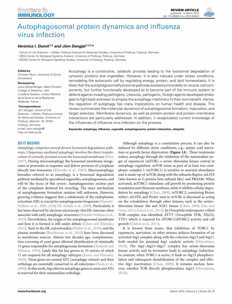
FIGURE 1 | Autophagosome formation and underlying signaling
events. (A) Autophagy is a cellular stress response that can be inducedinter alia by nutrition and growth factor deprivation. Active mTORC1 inhibitsautophagosome generation, whereas active ULK- and VPS34 class IIIPI3K-complexes are prerequisites for autophagosome formation. Thephagophore enraptures cytoplasm and matures to an autophagosome, alsofusing with endosomes. Acidic hydrolases degrade the constituents inautolysosomes enabling a recycling of bio-molecules (green: stimulatorysignals; pink: inhibitory signals; figure not complete). (B) Under growthconditions mTOR is active inhibiting the ULK-complex and by thisautophagy. (C) Under starvation conditions mTOR is inactive and ULKactive, phosphorylating itself and its binding partners and translocating topre-autophagosomal structures.
The functional counterparts to this complex in mammals areULKs, ATG13 and FIP200 (orthologs of Atg1, Atg13, and Atg17,respectively). ULK1 is the best characterized Atg1 homolog. Therole of the other isoforms ULK2 and ULK3 in autophagy are yetless clear. Although it seems likely that ULK2 is partially redundantto ULK1 (Chan, 2009), ULK3 may not have equivalent functions(Mizushima, 2010). Under non-stress conditions, mTORC1
associates with the ULK1–ATG13–FIP200–ATG101 complex bya direct interaction between Raptor and ULK1 (Chan, 2009;Mizushima, 2010), and phosphorylates ULK1 and ATG13, inhibit-ing their activity (Figure 1B). On the other hand, when mTORC1is inactive, it dissociates from the ULK1 complex, leading to ULK1activation. In its active state, ULK1 undergoes autophosphoryla-tion and phosphorylates ATG13 and FIP200 (Figure 1C). ULK1,ATG13, FIP200, and ATG101 accomplish their function by translo-cating from the cytosol to subdomains of the ER, and are thusessential for initiation of autophagosome formation (Mizushima,2010). These proteins lead to the isolation of membrane subdo-mains by recruitment of a class III phosphatidylinositol-3-OHkinase (PI3K) complex to the ER. The PI3K complex includesVPS34 (also known as PIK3C3), VPS15 (PIK3R4 and p150),Beclin-1 (ATG6), ATG14, and AMBRA1 (Levine et al., 2011). Atthis point of autophagy induction, not only protein componentshave decisive functions in autophagosome formation, also therole of lipids is crucial in its regulation. In the following section,we focus on the hinge role of lipids within protein dynamics inautophagy.
THE LIPID CONNECTIONPhosphatidylinositols (PI) are negatively charged phospholipidspresent as minor component at the cytosolic side of eukaryotic cellmembranes (Leevers et al., 1999). PI can be phosphorylated on itsinositol ring to form PI-phosphate (PIP), PI-bisphosphate (PIP2),and PI-trisphosphate (PIP3; Burman and Ktistakis, 2010). PIP,PIP2, and PIP3 are collectively called phosphoinositides (Leeverset al., 1999).
In general, PI3Ks are responsible for phosphorylating the3′OH-position of the inositol ring of PI, yielding PI3P (Bur-man and Ktistakis, 2010). Synthesis of PI3P is a strictly necessaryrequirement for all organisms undergoing autophagy (Burmanand Ktistakis, 2010). The function of PI3P is to gather signalingproteins containing specific lipid-binding domains to the mem-brane. Particularly in autophagy, such effectors are the doubleFYVE-containing protein 1 (DFCP1) and WD40-repeat domainphosphoinositide-interacting (WIPI, homolog to Atg18 in yeast)family proteins (Levine et al., 2011). DFCP1, in contrast to mostFYVE domain proteins that localize to endosomes, is locatedmainly at the ER, where PI3P is usually absent until autophagyis induced (Noda et al., 2010). Then, DFCP1 translocates to theautophagosome formation site, drawn by PI3P, to produce ER-associated Ω-like structures called omegasomes (Axe et al., 2008).The other effector of PI3P during autophagy, WIPI/Atg18, func-tions downstream of DFCP1 and was suggested to help the devel-opment of omegasomes into autophagosomes. WIPI2 is the majorisoform among the four WIPI isoforms in most mammalian cells(Polson et al., 2010).
The FYVE domain is named after the four proteins in which ithas been found: Fab1p (yeast ortholog of PIKfyve), YOTB, Vac1p(vesicle transport protein), and EEA1 (early endosome antigen1). It is characterized by having two zinc ions and eight poten-tial zinc coordinating cysteine residues. Additionally, several basicamino acids are localized around the cysteines. FYVE domains arepart of cysteine-rich proteins, which bind PI3P in a way depen-dent on their metal ion coordination and interaction of their basic
Frontiers in Immunology | Antigen Presenting Cell Biology March 2012 | Volume 3 | Article 43 | 2
amino acids with the negative charged PI3P head group (Kraussand Haucke, 2007). Figure 2A shows the tertiary structure of aFYVE domain and the coordination mode of the two metal ions tothe eight cysteine residues. Figure 2B shows EEA1 and its bindingmode to inositol-1,3-diphosphate, as a representative example oftheir interaction. It has been suggested that the binding causes con-formational changes regulating protein–protein or lipid–proteininteractions (Leevers et al., 1999). Most pathways regulated byPI, including autophagy, depend on their generation and like-wise on their consumption. A situation where PI persist longerthan the lipid signal is needed will result in loss of homeostasis.Jumpy is a PI3P phosphatase, which inhibits recruitment of WIPIto the autophagic membranes, and thus is in charge of PI3P signaltermination (Vergne et al., 2009).
Independently of the conditions triggering the catabolic path-way, autophagy begins with activation of the class III PI3KBeclin-1-complex in mammals, necessary to target membranesfor autophagosome generation and posterior maturation. Beclin-1, homolog of yeast Atg6, will be treated in detail below as targetof various viruses to abort autophagy and to use the autophagymachinery for their own infectious purposes.
ONE WAY TICKET TO THE AUTOPHAGOSOMETwo ubiquitin-like conjugation systems (UBL), ATG12, and LC3,have been implicated in biogenesis and membrane expansion ofautophagosomes (Münz, 2011a; Weidberg et al., 2011a). Modifi-cation of proteins with ubiquitin-like proteins follows a similarmechanism like modification with ubiquitin itself. For this rea-son, it is worthwhile to briefly summarize the general process(Figure 3A): Ubiquitin is a 76 amino acid-residue polypeptide,whose role is to direct proteins to the proteasome for degradation,among other regulatory functions. Ubiquitin is activated by an E1enzyme and, subsequently, E2 enzymes pick up activated ubiquitinby transthiolation and together with E3 enzymes catalyze ubiquiti-nation of substrates. E3 enzymes function to recognize substratesand are also capable of interacting with E2, allowing conjuga-tion of ubiquitin to target proteins. There are two major typesof E3 enzymes in eukaryotes, defined by the presence of either aHECT or a RING domain (Deshaies and Joazeiro, 2009). HECTand RING E3s catalyze ubiquitin transfer by different mechanisms
(Figure 3A). HECT-domain containing E3s bind themselves ubiq-uitin from an E2 protein before transferring it to its target protein.RING E3s function as a bridge between an activated E2 and atarget protein. Ubiquitination can be a repetitive process leadingto the generation of polyubiquitin chains or multiple mono-ubiquitinations. It is also a dynamic process. Deubiquitinatingenzymes are able to trim polyubiquitin chains or to remove singlemoieties allowing their recycling. Although eukaryotic cells haveonly one or few E1 enzymes, they encode more than 40 isoformsof E2 and more than 600 E3 enzymes (Grabbe et al., 2011). Asit is to be expected, the number of E2 and E3 isoforms show ahigher degree of complexity of the ubiquitin conjugation systemsin human cells relative to yeast (Hicke et al., 2005), enabling to rec-ognize diverse proteins in a highly specific manner (Hochstrasser,2009).
Figure 3B shows the two UBL systems operating in autophagy.Firstly, the ATG12–ATG5 conjugation is generated by ATG7 (E1-like) and ATG10 (E2-like). ATG12–ATG5 bind to ATG16L1 andpromote autophagosome formation (Fujita et al., 2008a; Weidberget al., 2011a). It has been shown in yeast, that it is the Atg12–Atg5 complex itself that catalyzes the transfer of Atg8 from Atg3to the substrate, phosphatidylethanolamine (PE), thus, behav-ing as a ubiquitin–protein ligase E3-like enzyme (Hanada et al.,2007). Secondly, LC3s, mammalian Atg8 homologs, are synthe-sized as precursors with an extra sequence at the C-terminus,which must be cleaved by the protease ATG4, resulting in theLC3 form I (LC3-I; Mizushima et al., 2011). LC3-I is then readilyconjugated to PE (forming LC3-II). PE is a lipid found in bio-logical membranes and lipidation of LC3/Atg8 during autophagyanchors this protein to the autophagosomal membrane. LC3/Atg8may serve different purposes. Yeast Atg8 has been shown tobe important for phagophore membrane elongation (Abeliovichet al., 2000). In addition, LC3/Atg8 functions as a membraneanchor enabling the targeting of substrates to the autophago-some (Münz, 2011a). It was also shown that it is importantfor membrane tethering and fusion (Nakatogawa et al., 2007;Weidberg et al., 2011b). However, these studies were performedin vitro and in a recent in vivo study it was suggested thatsoluble NSF attachment protein receptor (SNARE) proteins arerequired for membrane fusion, given that Atg8 is not able to
FIGURE 2 | (A) Structure of protein FYVE domain of the RUN and FYVEdomain containing protein 1; Zn atoms are shown in red and coordinatingcysteins in yellow (PDB code 2yw8). (B) Homodimer of EEA1’s
C-terminal FYVE domain bound to inositol-1,3-diphosphate (PDB code1joc). The picture was prepared using the program VMD (Humphreyet al., 1996).
FIGURE 3 | Ubiquitin and ubiquitin-like conjugation systems. (A) Theubiquitination system. E1–E2–E3 enzymatic cascades are depicted. (B) Thewhole set of ubiquitin-like reactions taking part in autophagy involves the E1-,E2-, and E3-like enzymes ATG7, ATG3/ATG10, and ATG12–ATG5/ATG16L1,respectively. Crystallographic structure of (C) LC3 (PDB code 1ugm), (D)
GABARAP (PDB code 1kjt), (E) GABARAPL2 (PDB code 1eo6), (F) ubiquitin(PDB code 1aar), and (G) superimposition of ubiquitin (yellow) and LC3(green) crystallographic structures. MultiProt Server was employed for proteinalignment based on their structures (Shatsky et al., 2004). The picture wasprepared using the program VMD (Humphrey et al., 1996).
mediate membrane fusion under physiological PE levels (Nairet al., 2011).
Yeast has a single Atg8 protein while mammals have sev-eral paralogs: three MAP1 light chain three (LC3A, LC3B, andLC3C) and four gamma-aminobutyrate receptor associated pro-tein (GABARAP) and GABARAP-like proteins (GABARAPL1-3),collectively referred to as LC3s. The roles of LC3s remained unclearfor a long time, since the other isoforms partially compensatedthe loss of function of a specific LC3 form in knockout studies
(Noda et al., 2009). Figures 3C–E displays the remarkable struc-tural similarities of the different LC3 forms among themselves andto ubiquitin (Figure 3F). Superimposition of LC3 and ubiquitinstructures clearly shows the high resemblance between these twoproteins (Figure 3G).
Anchoring of autophagosomal substrates by LC3/Atg8 isaccomplished by direct interaction with cargo proteins or by adap-tor proteins. NIX is a mitochondrial membrane protein capableof interacting with LC3s during mitophagy, the selective removal
Frontiers in Immunology | Antigen Presenting Cell Biology March 2012 | Volume 3 | Article 43 | 4
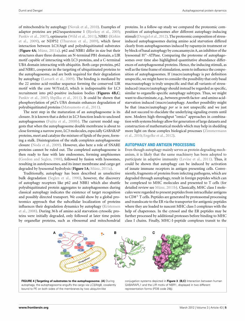
of mitochondria by autophagy (Novak et al., 2010). Examples ofadaptor proteins are p62/sequestosome 1 (Bjorkoy et al., 2005;Pankiv et al., 2007), optineurin (Wild et al., 2011), NBR1 (Kirkinet al., 2009), or NDP52 (Thurston et al., 2009), which favorinteraction between LC3/Atg8 and polyubiquitinated substrates(Figure 4A; Münz, 2011a). p62 and NBR1 differ in size but theirstructures share three domains: an N-terminal PB1 domain, a LIRmotif capable of interacting with LC3 proteins, and a C-terminalUBA domain interacting with ubiquitin. Both cargo proteins, p62and NBR1, cooperate in the targeting of ubiquitinated proteins tothe autophagosome, and are both required for their degradationby autophagy (Lamark et al., 2009). The binding is mediated bythe 22 amino acid-residue sequence forming the conserved LIRmotif with the core W/YxxL/I, which is indispensable for LC3recruitment into p62-positive inclusion bodies (Figures 4B,C;Pankiv et al., 2007; Shpilka et al., 2011). It could be shown thatphosphorylation of p62’s UBA domain enhances degradation ofpolyubiquitinated proteins (Matsumoto et al., 2011).
The next step in the maturation of an autophagosome is itsclosure. It is known that a defect in LC3 function leads to unclosedautophagosomes (Fujita et al., 2008b). The current model sug-gests that when the autophagosome double membrane is about toclose forming a narrow pore, LC3 molecules, especially GABARAPproteins, meet and catalyze the mixture of lipids of the pore, form-ing a stalk. Disintegration of the stalk completes autophagosomeclosure (Noda et al., 2009). However, also here a role of SNAREproteins cannot be ruled out. The completed autophagosome isthen ready to fuse with late endosomes, forming amphisomes(Gordon and Seglen, 1988), followed by fusion with lysosomes,resulting in autolysosomes, and its inner membrane and cargo getdegraded by lysosomal hydrolysis (Figure 1A; Münz, 2011a).
Traditionally, autophagy has been described as unselectivebulk degradation (Seglen et al., 1990), however, the discoveryof autophagy receptors like p62 and NBR1 which also shuttlepolyubiquitinated protein aggregates to autophagosomes duringclassical autophagy indicates the existence of target recognitionand possibly directed transport. We could show by a global pro-teomics approach that the subcellular localization of proteinsinfluences their degradation dynamics by autophagy (Kristensenet al., 2008). During 36 h of amino acid starvation cytosolic pro-teins were initially degraded, only followed at later time pointsby organellar proteins, such as ribosomal and mitochondrial
proteins. In a follow-up study we compared the proteomic com-position of autophagosomes after different autophagy-inducingstimuli (Dengjel et al., 2012). The proteomic composition of stress-induced autophagosomes during amino acid starvation differedclearly from autophagosomes induced by rapamycin treatment orby block of basal autophagy by concanamycin A, an inhibitor of thelysosomal H+-ATPase. Comparing the proteome of autophago-somes over time also highlighted quantitative abundance differ-ences of autophagosomal proteins. Hence, the inducing stimuli, aswell as the time frame of stimulation, seem to influence the compo-sition of autophagosomes. If (macro)autophagy is per definitionunspecific, we might have to consider the possibility that only basalmacroautophagy is truly unspecific and that all variants of stress-induced (macro)autophagy should instead be regarded as specific,similar to organelle-specific autophagy subtypes. Thus, we mightwant to discriminate, e.g., between growth factor- and amino acid-starvation induced (macro)autophagy. Another possibility mightbe that (macro)autophagy per se is not unspecific and we justdid not succeed to elucidate the underlying signaling events untilnow. Modern high-throughput “omics” approaches in combina-tion with systems biology allow for generation of large datasets andconstruction of mathematical models which may help in sheddingmore light on these complex biological processes (Zimmermannet al., 2010; Engelke et al., 2012).
AUTOPHAGY AND ANTIGEN PROCESSINGEven though autophagy mainly serves as protein degrading mech-anism, it is likely that the same machinery has been adopted toparticipate in adaptive immunity (Levine et al., 2011). Thus, itcould be shown that autophagy can be induced by activationof innate immune receptors in antigen presenting cells. Conve-niently, fragments of proteins from infecting pathogens, which aredegraded through autophagy, result in foreign peptides which canbe complexed to MHC molecules and presented to T cells (fordetailed review see Münz, 2011b). Classically, MHC class I mole-cules were regarded to present peptides from intracellular antigensto CD8+ T cells. Peptides are generated by proteasomal processingand translocate to the ER via the transporter for antigenic peptideswhere they are loaded to nascent MHC class I complexes with thehelp of chaperones. In the cytosol and the ER peptides may befurther processed by additional proteases before binding to MHCclass I chains. Finally, MHC-I-peptide complexes transit to the
FIGURE 4 |Targeting of substrates to the autophagosome. (A) Duringautophagy, the autophagosome engulfs the cargo via LC3/Atg8, covalentlybound to PE on both sides of the membranes by two ubiquitin-like
conjugation systems depicted in Figure 3. (B,C) Interaction between humanGABARAPL1 and the LIR motiv of NBR1, displayed in two differentrepresentation forms (PDB code 2l8j).
cell surface where they can be recognized by CD8+ T cells. Analternative pathway, called cross-presentation, allows presentationof peptides from exogenous antigens on MHC class I molecules(Crotzer and Blum, 2010). Whereas the classical MHC class Ipresentation pathway seems not to be influenced by autophagy,it could be shown that autophagy modulates MHC class I pre-sentation during late stage herpes simplex virus (HSV) infection(English et al., 2009).
Under normal conditions, MHC class II molecules present pep-tides on antigen presenting cells to CD4+ T cells, but duringinfection or inflammation MHC-II expression can be induced innon-immune cells as well. MHC class II molecules were viewedto present mainly peptides from extracellular antigens. However,MHC-II peptide analyses revealed that also peptides from intra-cellular source proteins are presented on MHC class II molecules.MHC class II α and β chains are synthesized into the ER and thechaperone invariant chain (Ii) prevents the binding of antigenicpeptides to the class II binding groove. In acidic vesicular com-partments Ii is cleaved and antigenic peptides can bind to MHCclass II heterodimers with the help of chaperones. In contrast toMHC-I presentation, the role of autophagy in MHC-II presenta-tion is more clear. Thus, it could be shown by several groups thatmacroautophagy and chaperone-mediated autopahgy play majorroles in promoting presentation of peptides derived from cytoplas-mic and nuclear proteins on MHC-II (Nimmerjahn et al., 2003;Dengjel et al., 2005; Zhou et al., 2005). This is also true for the pre-sentation of some virus derived peptides, like from Epstein Barrvirus nuclear antigen 1 (Paludan et al., 2005) and from influenzavirus matrix protein (Schmid et al., 2007). Interestingly, for MHCclass I as well as for MHC class II presentation a dual dependencyon proteasome- and autophagy-activity could be observed high-lighting a crosstalk between the two degradation pathways (Dörfelet al., 2005; English et al., 2009). Along this line, we could show thatthe proteasome is one of the “favorite substrates” of autophago-somes and that proteasome activity is modulated by functionalautophagy (Dengjel et al., 2012). However, the exact molecularmechanisms underlying autophagy–proteasome crosstalk are stillnot fully unveiled and more work has to be done. E.g., it is not clearif proteasomes are active inside autophagosomes and if autophago-somes may thus be regarded as scaffolds bringing together theproteasome with its substrates.
INFLUENZA VIRUS VERSUS AUTOPHAGY. WHO TAKESCONTROL?Many pathogens compromise peptide presentation on MHC mol-ecules by blocking the induction of autophagy or the maturation ofautophagosomes. Moreover, it is known that some viruses induceautophagy but inhibit autophagosome–lysosome fusion (Dereticand Levine, 2009). Hence, several viruses inhibit autophagy atthe level of autophagosome initiation by antagonizing Beclin-1.Examples of them are the α-herpervirus (HSV-1; Orvedahl et al.,2007), and γ-herpesviruses, which include human pathogens suchas Epstein Barr virus, Kaposi’s sarcoma associated herpesvirus(KSHV) and murine γ-HV68 (Liang et al., 2008). In contrastto DNA viruses, RNA viruses, including HIV (Kyei et al., 2009;Blanchet et al., 2010), hepatitis C (Ait-Goughoulte et al., 2008;Dreux et al., 2009), and poliovirus (Dales et al., 1965; Jackson et al.,
2005), block autophagosome maturation and consequently degra-dation, possibly to benefit from vesicular organelles for their repli-cation (Rossman and Lamb, 2009; Münz, 2011a). Non-maturingautophagosomes offer a propitious environment for virus replica-tion, due to the fact that high concentrations of viral proteins canbe accumulated while being unnoticed by the adaptive immunesystem (Rossman and Lamb, 2009). Importantly, viruses inhibit-ing autophagosome maturation target Beclin-1 as well, as it is alsoinvolved in the maturation process as binding partner of UVRAG.
Although it is well accepted that influenza virus infectionaffects autophagy, controversy still remains in many aspects ofthe underlying mechanisms and functional strategies employedby the virus to succeed in its infective purpose. Various indepen-dent studies suggest that an increasingly sophisticated connectionexists between autophagy, apoptosis and viral replication. More-over, interconnection between these three processes appears tobe cell-line dependent, further complicating interpretation of thegathered data on the effects of influenza virus infection.
Autophagy is induced by reactive oxygen species (Huang et al.,2011) which are also produced after influenza infection (Vlahoset al., 2012) highlighting a potential point of crosstalk. Oxidiz-ing molecules are suggested to modulate ATG4 activity leading toLC3-II accumulation (Scherz-Shouval et al., 2007). Influenza virushas been also proposed to up-regulate the expression of ATG7,ATG5, and ATG12 (Dai et al., 2012). Hence, all of these actionslead to autophagosome accumulation and may be tracked backto influenza virus infection. Significantly, it has been suggestedthat autophagy is involved in virus-dependent cytokine induc-tion, which is thought to be the main cause of death of infectedpatients (Law et al., 2010).
A closer look at the association between influenza virus infec-tion and autophagy raises many questions. It is well documentedthat influenza virus inhibits autophagy at the stage of autophago-some fusion with lysosomes, and thus leads to an accumulationof autophagosomes in human lung carcinoma-derived cells (Gan-nagé et al., 2009). On the other hand, it was shown that differentstrains of influenza virus induce functional autophagy, as detectedby degradation of the autophagy receptor p62 in infected pri-mary human blood macrophages (Law et al., 2010). A third reportstated as well that influenza virus infection does induce func-tional autophagy in several different cell lines, with no detectableblock in the pathway, as concluded from both GFP-LC3 and p62degradation measurements (Comber et al., 2011). In an attemptto conciliate all apparently contradicting results, the authors ofthe latter work suggested that discrepancies may be due to thecell types or the influenza virus strains used for the experiments(Comber et al., 2011).
Another polemic aspect is the functional association betweenautophagy and viral replication. The purpose of compromising akey homeostatic pathway of the cell by influenza virus is still underdebate. It was observed that influenza virus infection decreasescell survival by inducing apoptosis and inhibiting autophagy. Theinduction of apoptosis was suggested to circumvent an anti-viralimmune response (Gannagé et al., 2009). However, autophagy hadapparently a negligible influence on viral yields, given that loss ofthe degradation process did not affect virus replication. There-fore it was concluded that viral replication does not require the
Frontiers in Immunology | Antigen Presenting Cell Biology March 2012 | Volume 3 | Article 43 | 6
autophagosome environment to take its course (Gannagé et al.,2009). Hence, it has been suggested that influenza virus seemsto remain in the cytoplasm and nucleus for its replication (Ross-man and Lamb, 2009). But why does influenza virus compromiseautophagy? In contrast to the mentioned study, another investi-gation showed that inhibition of autophagy reduces replication ofinfluenza virus (Zhou et al., 2009). On top, it was suggested thatinfluenza virus induces autophagy only when apoptosis is firstinhibited (McLean et al., 2009). This evidence was proposed tobe the reason for the apparently opposing results attained before(Rossman and Lamb, 2009).
On the molecular level, there is solid evidence that binding ofinfluenza virus M2 protein to Beclin-1 compromises autophagy atthe step of lysosome fusion to autophagosomes (Gannagé et al.,2009). Beclin-1 contains a conserved BH3 domain (Obersteinet al., 2007). Such domains were first discovered in the context ofapoptosis, but then could also be related to regulation of autophagy(Sinha and Levine, 2008). Figures 5A,B show the amphipathicBH3 helix of Beclin-1 interacting with a conserved hydrophobicgroove of Bcl-XL and M11, respectively. Bcl-XL belongs to the Bcl-2 family of proteins, known to regulate apoptotic and autophagicprocesses in the cell (Sinha and Levine, 2008). M11 is a Bcl-2homolog present in the human pathogen γ-herpesvirus 68, able toregulate autophagy through interaction with Beclin-1 (Sinha et al.,2008).
It could be shown that influenza virus M2 integral mem-brane protein is necessary and sufficient to block autophagosome–lysosome fusion (Gannagé et al., 2009). Transient expression ofinfluenza A virus M2 protein reproduced the same phenotypeas viral infection, i.e., autophagosomes accumulation due to a
FIGURE 5 | Crystallographic structure of the BH3 helix of Beclin-1,
shown in yellow, complexed to (A) Bcl-XL (PDB code 2p1l) and (B) M11
(PDB code 3bl2). (C) Crystallographic structure of M2 transmembraneprotein from influenza virus (PDB code 2kih); residues 23–49 including thetransmembrane domain are shown in green and the cytoplasmic helices(residues 50–60) in blue. (D) Superimposition of Beclin-1 complexed toBcl-XL and influenza virus M2 protein. The picture was prepared using theprogram VMD (Humphrey et al., 1996), and MultiProt Server was employedfor protein alignment (Shatsky et al., 2004).
block in autophagosome maturation and not to an increase inautophagy. Silencing M2 expression during influenza A virusinfection, or infecting cells with a M2 knockout influenza Avirus, reverted the phenotype of classical infection, allowingautophagosome–lysosome fusion (Gannagé et al., 2009, 2010). M2is a proton-selective ion channel responsible for acidification ofthe viral core once the virus reaches endosomes, causing disso-ciation of the viral particles and release of the genome into thecytoplasm (Wang et al., 2011). Surprisingly, it has been observedthat autophagosome maturation is not inhibited by the M2 ionchannel activity itself and that the first 60 residues of the proteinare sufficient to inhibit autophagy by binding to Beclin-1 (Gan-nagé et al., 2009, 2010). The precise mechanism of inhibition hasnot been clearly determined, but it has been proposed that M2is likely to interact with Beclin-1 through either the ectodomain(residues 1-24) or the cytoplasmic amphipatic helix (residues 46–62), but not through the transmembrane domain which should beshielded from access (Rossman and Lamb, 2009).
As shown in Figure 5, the secondary structures of both Bcl-XLand M11 contain mostly α-helices, as do the first 70 amino acid-residues of influenza virus M2 protein, which are needed to inhibitBeclin-1. If crystallographic structures of the complex Beclin-1–Bcl-XL and M2 are aligned, one of the transmembrane α-helicesof M2 superimposes with one of the α-helices of Bcl-XL whichinteracts with Beclin-1 (Figures 5C,D). In our opinion, it cannotbe excluded that this ion channel transmembrane domain of M2binds to Beclin-1. This may happen before the M2 homotetramercomplex is fully assembled in the membrane indicating a second,non-membrane-bound, function/role of this protein and possiblyexplaining the observation that the M2 ion channel activity seemsnot to be involved in its functions in autophagy regulation. Tofully understand the modulations of autophagy by influenza virusand to outline the underlying molecular mechanisms more workhas to be done, e.g., specifically addressing protein dynamics andprotein–protein interactions under various conditions.
CONCLUSIONAutophagy is a highly complex process and only the identificationof autophagy-related genes in the genetically tractable organismyeast and the fact that the process is conserved in humans haveallowed the elucidation of underlying molecular mechanisms lead-ing to the generation of autophagosomes in mammalian cells.Although we have gained a tremendous amount of knowledge inthe last decade there are still many unanswered questions, espe-cially related to human diseases. Viruses employ autophagy fortheir own goods and the studying of autophagy modulation byviral infection and viral proteins will allow a deeper insight intounderlying molecular mechanisms shedding more light onto thisbasal cell biological process. We are confident that the newly gen-erated knowledge will not only allow the design of new anti-viraltherapies but will also help in targeting autophagy in other diseasesettings.
Regarding macroautophagy, a lot of work has to be done tofully understand target selection. Can a cell actually allow a com-pletely unspecific bulk degradation process to happen? Large-scale“omics” approaches should help in generating enough data tocomprehensively tackle this problem on a global scale.
ACKNOWLEDGMENTSThe authors are grateful to Victoria Küttner for carefully readingthe manuscript. The research leading to these results has receivedfunding from the Excellence Initiative of the German Federal andState Governments through FRIAS and BIOSS, from the DeutscheForschungsgemeinschaft (GZ DE1757/2-1), and from the Federal
Ministry of Education and Research through GerontoSys II –NephAge (031 5896 A). We thank all Protein Dynamics groupmembers, as well as M. Boerries, H. Busch, A. Schlosser, and col-leagues from the ZBSA, for helpful discussions and support. Weapologize to colleagues whose work was not covered in this reviewdue to space limitations.
REFERENCESAbeliovich, H., Dunn,W. A., Kim, J., and
Klionsky, D. J. (2000). Dissectionof autophagosome biogenesis intodistinct nucleation and expansionsteps. J. Cell Biol. 151, 1025–1034.
Ait-Goughoulte, M., Kanda, T., Meyer,K., Ryerse, J. S., Ray, R. B., andRay, R. (2008). Hepatitis C virusgenotype 1a growth and induc-tion of autophagy. J. Virol. 82,2241–2249.
Axe, E. L., Walker, S. A., Manifava, M.,Chandra, P., Roderick, H. L., Haber-mann, A., Griffiths, G., and Ktistakis,N. T. (2008). Autophagosome for-mation from membrane compart-ments enriched in phosphatidyli-nositol 3-phosphate and dynami-cally connected to the endoplas-mic reticulum. J. Cell Biol. 182,685–701.
Bjorkoy, G., Lamark, T., Brech, A., Out-zen, H., Perander, M., Overvatn,A., Stenmark, H., and Johansen, T.(2005). p62/SQSTM1 forms proteinaggregates degraded by autophagyand has a protective effect onhuntingtin-induced cell death. J. CellBiol. 171, 603–614.
Blanchet, F. P., Moris, A., Nikolic, D. S.,Lehmann, M., Cardinaud, S., Stalder,R., Garcia, E., Dinkins, C., Leuba,F., Wu, L., Schwartz, O., Deretic,V., and Piguet, V. (2010). Humanimmunodeficiency virus-1 inhibi-tion of immunoamphisomes in den-dritic cells impairs early innate andadaptive immune responses. Immu-nity 32, 654–669.
Burman, C., and Ktistakis, N. T. (2010).Regulation of autophagy by phos-phatidylinositol 3-phosphate. FEBSLett. 584, 1302–1312.
Chan, E. Y. (2009). mTORC1 phospho-rylates the ULK1-mAtg13-FIP200autophagy regulatory complex. Sci.Signal. 2, pe51.
Chen, Y., and Klionsky, D. J. (2011).The regulation of autophagy – unan-swered questions. J. Cell Sci. 124,161–170.
Comber, J. D., Robinson, T. M., Sicil-iano, N. A., Snook, A. E., andEisenlohr, L. C. (2011). Func-tional macroautophagy induction byinfluenza A virus without a contri-bution to major histocompatibilitycomplex class II-restricted presenta-tion. J. Virol. 85, 6453–6463.
Crotzer, V. L., and Blum, J. S. (2010).Autophagy and adaptive immunity.Immunology 131, 9–17.
Dai, J., Wang, G., Li, W., Zhang,L., Yang, J., Zhao, X., Chen,X., Xu, Y., and Li, K. (2012).High-throughput screening foranti-influenza a virus drugs andstudy of the mechanism of procyani-din on influenza a virus inducedautophagy. J. Biomol. Screen.doi:10.1177/1087057111435236
Dales, S., Eggers, H. J., Tamm, I., andPalade, G. E. (1965). Electron micro-scopic study of the formation ofpoliovirus. Virology 26, 379–389.
Dengjel, J., Hoyer-Hansen, M., Nielsen,M. O., Eisenberg, T., Harder, L.M., Schandorff, S., Farkas, T.,Kirkegaard, T., Becker, A. C.,Schroeder, S., Vanselow, K., Lund-berg, E., Nielsen, M. M., Kristensen,A. R., Akimov, V., Bunkenborg, J.,Madeo, F., Jäättelä, M., and Ander-sen, J. S. (2012). Identification ofautophagosome-associated proteinsand regulators by quantitativeproteomic analysis and geneticscreens. Mol. Cell. Proteomics.doi:10.1074/mcp.M111.014035
Dengjel, J., Schoor, O., Fischer, R., Reich,M., Kraus, M., Müller, M., Kreym-borg, K., Altenberend, F., Branden-burg, J., Kalbacher, H., Brock, R.,Driessen, C., Rammensee, H. G., andStevanovic, S. (2005). Autophagypromotes MHC class II presentationof peptides from intracellular sourceproteins. Proc. Natl. Acad. Sci. U.S.A.102, 7922–7927.
Deretic, V., and Levine, B. (2009).Autophagy, immunity, and micro-bial adaptations. Cell Host Microbe5, 527–549.
Deshaies, R. J., and Joazeiro, C. A.(2009). RING domain E3 ubiqui-tin ligases. Annu. Rev. Biochem. 78,399–434.
Dice, J. F. (2007). Chaperone-mediatedautophagy. Autophagy 3, 295–299.
Dörfel, D., Appel, S., Grünebach, F.,Weck, M. M., Müller, M. R., Heine,A., and Brossart, P. (2005). Pro-cessing and presentation of HLAclass I and II epitopes by dendriticcells after transfection with in vitro-transcribed MUC1 RNA. Blood 105,3199–3205.
Dreux, M., Gastaminza, P., Wieland, S.F., and Chisari, F. V. (2009). The
autophagy machinery is required toinitiate hepatitis C virus replication.Proc. Natl. Acad. Sci. U.S.A. 106,14046–14051.
Engelke, R., Becker, A. C., andDengjel, J. (2012). The degrada-tive inventory of the cell: pro-teomic insights. Antioxid Redox Sig-nal. doi:10.1089/ars.2011.4393
English, L., Chemali, M., Duron, J., Ron-deau, C., Laplante, A., Gingras, D.,Alexander, D., Leib, D., Norbury, C.,Lippé, R., and Desjardins, M. (2009).Autophagy enhances the presenta-tion of endogenous viral antigenson MHC class I molecules duringHSV-1 infection. Nat. Immunol. 10,480–487.
Fujita, N., Itoh, T., Omori, H., Fukuda,M., Noda, T., and Yoshimori, T.(2008a). The Atg16L complex spec-ifies the site of LC3 lipidation formembrane biogenesis in autophagy.Mol. Biol. Cell 19, 2092–2100.
Fujita, N., Hayashi-Nishino, M., Fuku-moto, H., Omori, H., Yamamoto, A.,Noda, T., and Yoshimori, T. (2008b).An Atg4B mutant hampers the lipi-dation of LC3 paralogues and causesdefects in autophagosome closure.Mol. Biol. Cell 19, 4651–4659.
Gannagé, M., Dormann, D., Albrecht,R., Dengjel, J., Torossi, T., Rämer, P.C., Lee, M., Strowig, T., Arrey, F.,Conenello, G., Pypaert, M., Ander-sen, J., García-Sastre, A., and Münz,C. (2009). Matrix protein 2 ofinfluenza A virus blocks autophago-some fusion with lysosomes. CellHost Microbe 6, 367–380.
Gannagé, M., Rämer, P. C., and Münz,C. (2010). Targeting Beclin 1 forviral subversion of macroautophagy.Autophagy 6, 166–167.
Glatter, T., Schittenhelm, R. B., Rinner,O., Roguska, K., Wepf, A., Jünger,M. A., Köhler, K., Jevtov, I., Choi,H., Schmidt, A., Nesvizhskii, A. I.,Stocker, H., Hafen, E., Aebersold,R., and Gstaiger, M. (2011). Mod-ularity and hormone sensitivity ofthe Drosophila melanogaster insulinreceptor/target of rapamycin inter-action proteome. Mol. Syst. Biol. 7,547–547.
Gordon, P. B., and Seglen, P. O.(1988). Prelysosomal convergenceof autophagic and endocytic path-ways. Biochem. Biophys. Res. Com-mun. 151, 40–47.
Grabbe, C., Husnjak, K., and Dikic, I.(2011). The spatial and temporalorganization of ubiquitin networks.Nat. Rev. Mol. Cell Biol. 12, 295–307.
Hailey, D. W, Rambold, A. S., Satpute-Krishnan, P., Mitra, K., Sougrat,R., Kim, P. K., and Lippincott-Schwartz, J. (2010). Mitochondriasupply membranes for autophago-some biogenesis during starvation.Cell 141, 656–667.
Hanada, T., Noda, N. N., Satomi, Y.,Ichimura, Y., Fujioka, Y., Takao, T.,Inagaki, F., and Ohsumi, Y. (2007).The Atg12-Atg5 conjugate has anovel E3-like activity for protein lip-idation in autophagy. J. Biol. Chem.282, 37298–37302.
Hayashi-Nishino, M., Fujita, N., Noda,T., Yamaguchi, A., Yoshimori, T., andYamamoto, A. (2009). A subdomainof the endoplasmic reticulum formsa cradle for autophagosome forma-tion. Nat. Cell Biol. 11, 1433–1437.
Hayashi-Nishino, M., Fujita, N., Noda,T., Yamaguchi, A., Yoshimori, T.,and Yamamoto, A. (2010). Elec-tron tomography reveals the endo-plasmic reticulum as a membranesource for autophagosome forma-tion. Autophagy 6, 301–303.
Hicke, L., Schubert, H. L., and Hill, C. P.(2005). Ubiquitin-binding domains.Nat. Rev. Mol. Cell Biol. 6, 610–621.
Hochstrasser, M. (2009). Origin andfunction of ubiquitin-like proteins.Nature 458, 422–429.
Huang, J., Lam, G. Y., and Brumell,J. H. (2011). Autophagy sig-naling through reactive oxygenspecies. Antioxid Redox Signal. 14,2215–2231.
Humphrey, W., Dalke, A., and Schulten,K. (1996). VMD: visual moleculardynamics. J. Mol. Graph. 14, 33–38.
Inoue, Y., and Klionsky, D. J. (2010).Regulation of macroautophagy inSaccharomyces cerevisiae. Semin. CellDev. Biol. 21, 664–670.
Jackson, W. T., Giddings, T. H., Taylor,M. P., Mulinyawe, S., Rabinovitch,M., Kopito, R. R., and Kirkegaard,K. (2005). Subversion of cellu-lar autophagosomal machinery byRNA viruses. PLoS Biol. 3, 861–871.doi:10.1371/journal.pbio.0030156
Kim, J., and Guan, K. L. (2011).Amino acid signaling in TOR acti-vation. Annu. Rev. Biochem. 80,1001–1032.
Frontiers in Immunology | Antigen Presenting Cell Biology March 2012 | Volume 3 | Article 43 | 8
Kirkin,V., Lamark, T., Sou,Y. S., Bjorkoy,G., Nunn, J. L., Bruun, J. A., Shvets,E., McEwan, D. G., Clausen, T. H.,Wild, P., Bilusic, I., Theurillat, J.P., Øvervatn, A., Ishii, T., Elazar,Z., Komatsu, M., Dikic, I., andJohansen, T. (2009). A role for NBR1in autophagosomal degradation ofubiquitinated substrates. Mol. Cell33, 505–516.
Klionsky, D. J., Cuervo, A. M., andSeglen, P. O. (2007). Methods formonitoring autophagy from yeast tohuman. Autophagy 3, 181–206.
Krauss, M., and Haucke, V. (2007).Phosphoinositide-metabolizingenzymes at the interface betweenmembrane traffic and cell signalling.EMBO Rep. 8, 241–246.
Kristensen, A. R., Schandorff, S., Hoyer-Hansen, M., Nielsen, M. O., Jäät-telä, M., Dengjel, J., and Andersen, J.S. (2008). Ordered organelle degra-dation during starvation-inducedautophagy. Mol. Cell. Proteomics 7,2419–2428.
Kyei, G. B., Dinkins, C., Davis, A.S., Roberts, E., Singh, S. B., Dong,C., Wu, L., Kominami, E., Ueno,T., Yamamoto, A., Federico, M.,Panganiban, A., Vergne, I., andDeretic, V. (2009). Autophagy path-way intersects with HIV-1 biosyn-thesis and regulates viral yieldsin macrophages. J. Cell Biol. 186,255–268.
Lamark, T., Kirkin, V., Dikic, I., andJohansen, T. (2009). NBR1 andp62 as cargo receptors for selectiveautophagy of ubiquitinated targets.Cell Cycle 8, 1986–1990.
Law, A. H., Lee, D. C., Yuen, K. Y., Peiris,M., and Lau, A. S. (2010). Cellularresponse to influenza virus infec-tion: a potential role for autophagyin CXCL10 and interferon-alphainduction. Cell. Mol. Immunol. 7,263–270.
Leevers, S. J., Vanhaesebroeck, B.,and Waterfield, M. D. (1999). Sig-nalling through phosphoinositide 3-kinases: the lipids take centre stage.Curr. Opin. Cell Biol. 11, 219–225.
Levine, B., Mizushima, N., and Virgin,H. W. (2011). Autophagy in immu-nity and inflammation. Nature 469,323–335.
Liang, C., E, X., and Jung, J. U. (2008).Downregulation of autophagyby herpesvirus Bcl-2 homologs.Autophagy 4, 268–272.
Matsumoto, G., Wada, K., Okuno,M., Kurosawa, M., and Nukina, N.(2011). Serine 403 phosphoryla-tion of p62/SQSTM1 regulates selec-tive autophagic clearance of ubiq-uitinated proteins. Mol. Cell 44,279–289.
McLean, J. E., Datan, E., Matassov,D., and Zakeri, Z. F. (2009).Lack of Bax prevents influenza Avirus-induced apoptosis and causesdiminished viral replication. J. Virol.83, 8233–8246.
Mizushima, N. (2010). The role of theAtg1/ULK1 complex in autophagyregulation. Curr. Opin. Cell Biol. 22,132–139.
Mizushima, N., Yoshimori, T., andOhsumi, Y. (2011). The role of atgproteins in autophagosome forma-tion. Annu. Rev. Cell Dev. Biol. 27,107–132.
Münz, C. (2011a). Beclin-1 targetingfor viral immune escape. Viruses 3,1166–1178.
Münz, C. (2011b). Antigen process-ing by macroautophagy for MHCpresentation. Front. Immunol. 2:1–7.doi:10.3389/fimmu.2011.00042
Nair, U., Jotwani, A., Geng, J., Gammoh,N., Richerson, D., Yen, W. L., Grif-fith, J., Nag, S., Wang, K., Moss, T.,Baba, M., McNew, J. A., Jiang, X.,Reggiori, F., Melia, T. J., and Klion-sky, D. J. (2011). SNARE proteins arerequired for macroautophagy. Cell146, 290–302.
Nakatogawa, H., Ichimura, Y., andOhsumi,Y. (2007). Atg8,a ubiquitin-like protein required for autophago-some formation, mediates mem-brane tethering and hemifusion. Cell130, 165–178.
Nimmerjahn, F., Milosevic, S.,Behrends, U., Jaffee, E. M., Par-doll, D. M., Bornkamm, G. W., andMautner, J. (2003). Major histocom-patibility complex class II-restrictedpresentation of a cytosolic antigenby autophagy. Eur. J. Immunol. 33,1250–1259.
Noda, T., Fujita, N., and Yoshimori, T.(2009). The late stages of autophagy:how does the end begin? Cell DeathDiffer. 16, 984–990.
Noda, T., Matsunaga, K., Taguchi-Atarashi, N., and Yoshimori, T.(2010). Regulation of membranebiogenesis in autophagy via PI3Pdynamics. Semin. Cell Dev. Biol. 21,671–676.
Novak, I., Kirkin, V., McEwan, D.G., Zhang, J., Wild, P., Rozenknop,A., Rogov, V., Löhr, F., Popovic,D., Occhipinti, A., Reichert, A. S.,Terzic, J., Dötsch, V., Ney, P. A.,and Dikic, I. (2010). Nix is a selec-tive autophagy receptor for mito-chondrial clearance. EMBO Rep. 11,45–51.
Oberstein, A., Jeffrey, P. D., and Shi, Y.(2007). Crystal structure of the Bcl-XL-Beclin 1 peptide complex: Beclin1 is a novel BH3-only protein. J. Biol.Chem. 282, 13123–13132.
Orvedahl, A., Alexander, D., Tallóczy,Z., Sun, Q., Wei, Y., Zhang, W.,Burns, D., Leib, D. A., and Levine,B. (2007). HSV-1 ICP34.5 con-fers neurovirulence by targeting theBeclin 1 autophagy protein. Cell HostMicrobe 1, 23–35.
Paludan, C., Schmid, D., Landthaler, M.,Vockerodt, M., Kube, D., Tuschl, T.,and Münz, C. (2005). EndogenousMHC class II processing of a viralnuclear antigen after autophagy. Sci-ence 307, 593–596.
Pankiv, S., Clausen, T. H., Lamark, T.,Brech, A., Bruun, J. A., Outzen,H., Øvervatn, A., Bjorkoy, G., andJohansen, T. (2007). p62/SQSTM1binds directly to Atg8/LC3 to facil-itate degradation of ubiquitinatedprotein aggregates by autophagy. J.Biol. Chem. 282, 24131–24145.
Polson, H. E., de Lartigue, J., Rigden, D.J., Reedijk, M., Urbé, S., Clague, M.J., and Tooze, S. A. (2010). Mam-malian Atg18 (WIPI2) localizes toomegasome-anchored phagophoresand positively regulates LC3 lipida-tion. Autophagy 6, 506–522.
Ravikumar, B., Moreau, K., Jahreiss,L., Puri, C., and Rubinsztein, D.C. (2010). Plasma membrane con-tributes to the formation of pre-autophagosomal structures. Nat.Cell Biol. 12, 747–757.
Rossman, J. S., and Lamb, R. A.(2009). Autophagy, apoptosis, andthe influenza virus M2 protein. CellHost Microbe 6, 299–300.
Scherz-Shouval, R., Shvets, E., Fass, E.,Shorer, H., Gil, L., and Elazar, Z.(2007). Reactive oxygen species areessential for autophagy and specif-ically regulate the activity of Atg4.EMBO J. 26, 1749–1760.
Schmid, D., Pypaert, M., and Münz, C.(2007). Antigen-loading compart-ments for major histocompatibilitycomplex class II molecules con-tinuously receive input fromautophagosomes. Immunity 26,79–92.
Seglen, P. O., Gordon, P. B., and Holen,I. (1990). Non-selective autophagy.Semin. Cell Biol. 1, 441–448.
Shatsky, M., Nussinov, R., and Wolfson,H. J. (2004). A method for simulta-neous alignment of multiple proteinstructures. Proteins 56, 143–156.
Shpilka, T., Weidberg, H., Pietrokovski,S., and Elazar, Z. (2011). Atg8:an autophagy-related ubiquitin-likeprotein family. Genome Biol. 12,226–226.
Sinha, S., Colbert, C. L., Becker, N.,Wei, Y., and Levine, B. (2008).Molecular basis of the regulationof Beclin 1-dependent autophagyby the gamma-herpesvirus 68
Bcl-2 homolog M11. Autophagy 4,989–997.
Sinha, S., and Levine, B. (2008).The autophagy effector Beclin 1: anovel BH3-only protein. Oncogene27(Suppl. 1), 137–148.
Thurston, T. L., Ryzhakov, G., Bloor,S., von Muhlinen, N., and Randow,F. (2009). The TBK1 adaptor andautophagy receptor NDP52 restrictsthe proliferation of ubiquitin-coated bacteria. Nat. Immunol. 10,1215–1221.
Tsukada, M., and Ohsumi, Y. (1993).Isolation and characterization ofautophagy-defective mutants of Sac-charomyces cerevisiae. FEBS Lett. 333,169–174.
Vergne, I., Roberts, E., Elmaoued, R. A.,Tosch, V., Delgado, M. A., Proikas-Cezanne, T., Laporte, J., and Deretic,V. (2009). Control of autophagyinitiation by phosphoinositide 3-phosphatase Jumpy. EMBO J. 28,2244–2258.
Vlahos, R., Stambas, J., and Selemidis,S. (2012). Suppressing productionof reactive oxygen species (ROS) forinfluenza A virus therapy. TrendsPharmacol. Sci. 33, 3–8.
Wang, J., Qiu, J. X., Soto, C., andDeGrado, W. F. (2011). Structuraland dynamic mechanisms for thefunction and inhibition of the M2proton channel from influenza Avirus. Curr. Opin. Struct. Biol. 21,68–80.
Weidberg, H., Shvets, E., and Elazar, Z.(2011a). Biogenesis and cargo selec-tivity of autophagosomes. Annu.Rev. Biochem. 80, 125–156.
Weidberg, H., Shpilka, T., Shvets, E.,Abada, A., Shimron, F., and Elazar,Z. (2011b). LC3 and GATE-16 Ntermini mediate membrane fusionprocesses required for autophago-some biogenesis. Dev. Cell 20,444–454.
Wild, P., Farhan, H., McEwan, D. G.,Wagner, S., Rogov, V. V., Brady, N. R.,Richter, B., Korac, J., Waidmann, O.,Choudhary, C., Dötsch, V., Bumann,D., and Dikic, I. (2011). Phospho-rylation of the autophagy recep-tor optineurin restricts Salmonellagrowth. Science 333, 228–233.
Ylä-Anttila, P., Vihinen, H., Jokitalo,E., and Eskelinen, E. L. (2009).3D tomography reveals connectionsbetween the phagophore and endo-plasmic reticulum. Autophagy 5,1180–1185.
Zhou, D., Li, P., Lin, Y., Lott, J. M.,Hislop, A. D., Canaday, D. H.,Brutkiewicz, R. R., and Blum, J.S. (2005). Lamp-2a facilitates MHCclass II presentation of cytoplasmicantigens. Immunity 22, 571–581.
Zhou, Z., Jiang, X., Liu, D., Fan, Z.,Hu, X., Yan, J., Wang, M., and Gao,G. F. (2009). Autophagy is involvedin influenza A virus replication.Autophagy 5, 321–328.
Zimmermann, A. C., Zarei, M., Eise-lein, S., and Dengjel, J. (2010). Quan-titative proteomics for the analysisof spatio-temporal protein dynam-ics during autophagy. Autophagy 6,1009–1016.
Zoncu, R., Efeyan, A., and Sabatini, D.M. (2011). mTOR: from growth sig-nal integration to cancer, diabetesand ageing. Nat. Rev. Mol. Cell Biol.12, 21–35.
Conflict of Interest Statement: Theauthors declare that the research wasconducted in the absence of any com-mercial or financial relationships that
could be construed as a potential con-flict of interest.
Received: 09 December 2011; paper pend-ing published: 11 January 2012; accepted:23 February 2012; published online: 14March 2012.Citation: Dumit VI and Dengjel J (2012)Autophagosomal protein dynamics andinfluenza virus infection. Front. Immun.3:43. doi: 10.3389/fimmu.2012.00043