AD_________________ Award Number: W81XWH-04-1-0014 TITLE: Characterization of a Novel Intracellular Receptor for Phorbol Esters and Diacylglycerol in Prostate Cancer PRINCIPAL INVESTIGATOR: Jose Olivia Martinez, Ph.D. CONTRACTING ORGANIZATION: The University of Pennsylvania Philadelphia, PA 19104-6205 REPORT DATE: February 2006 TYPE OF REPORT: Annual Summary PREPARED FOR: U.S. Army Medical Research and Materiel Command Fort Detrick, Maryland 21702-5012 DISTRIBUTION STATEMENT: Approved for Public Release; Distribution Unlimited The views, opinions and/or findings contained in this report are those of the author(s) and should not be construed as an official Department of the Army position, policy or decision unless so designated by other documentation.
Transcript
AD_________________ Award Number: W81XWH-04-1-0014 TITLE: Characterization of a Novel Intracellular Receptor for Phorbol Esters and Diacylglycerol in Prostate Cancer PRINCIPAL INVESTIGATOR: Jose Olivia Martinez, Ph.D. CONTRACTING ORGANIZATION: The University of Pennsylvania Philadelphia, PA 19104-6205 REPORT DATE: February 2006 TYPE OF REPORT: Annual Summary PREPARED FOR: U.S. Army Medical Research and Materiel Command Fort Detrick, Maryland 21702-5012 DISTRIBUTION STATEMENT: Approved for Public Release; Distribution Unlimited The views, opinions and/or findings contained in this report are those of the author(s) and should not be construed as an official Department of the Army position, policy or decision unless so designated by other documentation.
REPORT DOCUMENTATION PAGE Form Approved
OMB No. 0704-0188 Public reporting burden for this collection of information is estimated to average 1 hour per response, including the time for reviewing instructions, searching existing data sources, gathering and maintaining the data needed, and completing and reviewing this collection of information. Send comments regarding this burden estimate or any other aspect of this collection of information, including suggestions for reducing this burden to Department of Defense, Washington Headquarters Services, Directorate for Information Operations and Reports (0704-0188), 1215 Jefferson Davis Highway, Suite 1204, Arlington, VA 22202-4302. Respondents should be aware that notwithstanding any other provision of law, no person shall be subject to any penalty for failing to comply with a collection of information if it does not display a currently valid OMB control number. PLEASE DO NOT RETURN YOUR FORM TO THE ABOVE ADDRESS. 1. REPORT DATE (DD-MM-YYYY)01-02-2006
2. REPORT TYPEAnnual Summary
3. DATES COVERED (From - To)1 Feb 2004 – 31 Jan 2006
4. TITLE AND SUBTITLE Characterization of a Novel Intracellular Receptor for Phorbol Esters and
7. PERFORMING ORGANIZATION NAME(S) AND ADDRESS(ES)
8. PERFORMING ORGANIZATION REPORT NUMBER
The University of Pennsylvania Philadelphia, PA 19104-6205
9. SPONSORING / MONITORING AGENCY NAME(S) AND ADDRESS(ES) 10. SPONSOR/MONITOR’S ACRONYM(S)U.S. Army Medical Research and Materiel Command Fort Detrick, Maryland 21702-5012 11. SPONSOR/MONITOR’S REPORT NUMBER(S)
12. DISTRIBUTION / AVAILABILITY STATEMENT Approved for Public Release; Distribution Unlimited
13. SUPPLEMENTARY NOTES 14. ABSTRACT The small GTP-binding protein Rac controls essential functions, including actin cytoskeleton reorganization, cell proliferation, cell cycle progression, adhesion, migration and invasion. The relationship of Rac to prostate carcinogenesis has not been extensively studied. However upstream activators of Rac have been described to be hyperactivated in prostate cancer, and it is well known that growth factors are very important in the control of prostate cancer proliferation and progression, as well as in the maintenance of growth during androgen independency. Chimaerins, through their Rac-GAP activity, accelerate the hydrolysis of GTP from Rac, leading to its inactivation. To date four chimaerin isoforms have been isolated and reported: α1, α2-, β1- and β2-Chimaerin. While α1- and β1-chimaerin are restricted to brain and testis, respectively, α2- and β2-chimaerin are widely expressed. No experimental information has been reported about the possible role of chimaerins in prostate cancer. Likewise, there are no information available about the expression of different chimaerin in prostate cancer cell lines. Our work hypothesis is that by inhibiting Rac function in prostate cancer cells, chimaerins will impair proliferation and reduce the invasive properties of prostate cancer cells.
Standard Form 298 (Rev. 8-98)Prescribed by ANSI Std. Z39.18
3
Table of Contents Cover……………………………………………………………………………………1 SF 298………………………………………………………………...…………………2
Introduction…………………………………………………………….…………..…4 Body…………………………………………………………………………………….4 Key Research Accomplishments………………………………………….………12 Reportable Outcomes……………………………………………………………….13 Conclusions…………………………………………………………………………..13 References…………………………………………………………………………….13 Appendices……………………………………………………………………………14
INTRODUCTION
The small GTP-binding protein Rac controls essential functions, including actin cytoskeleton reorganization, cell proliferation, cell cycle progression, adhesion, migration and invasion. Whereas the relationship of Rac to prostate carcinogenesis has not been extensively studied, several papers have described that upstream activators of Rac are hyperactivated in prostate cancer, and it is well known that growth factors are very important in the control of prostate cancer proliferation and progression, as well as in the maintenance of growth during androgen independency. Rac belongs to the Rho family of small GTP-binding proteins, and cycles between an “on” (GTP-bound) and an “off” (GDP-bound) state, steps that involve guanine nucleotide exchange factors (GEFs) and GTPase activating proteins (GAPs), which accelerate GTP hydrolysis, respectively (1-3)
Chimaerins, through their Rac-GAP activity, accelerate the hydrolysis of GTP from Rac1, leading to its inactivation. To date four chimaerin isoforms have been isolated: α1, α2-, β1- and β2-chimaerin. While α1- and β1-chimaerin are restricted to brain and testis, respectively, α2- and β2-chimaerin are widely expressed (4,5). However, there are no reported information or experimental data describing chimaerin activity and/or expression in prostate cancer cells. Since chimaerins inhibit Rac function, it is predictable that they will have profound effects on Rac mediated signaling, and therefore impact on the proliferative and invasive capacity of prostate cancer cells. BODY 1- Characterization of chimaerin isozymes in prostate cancer cells
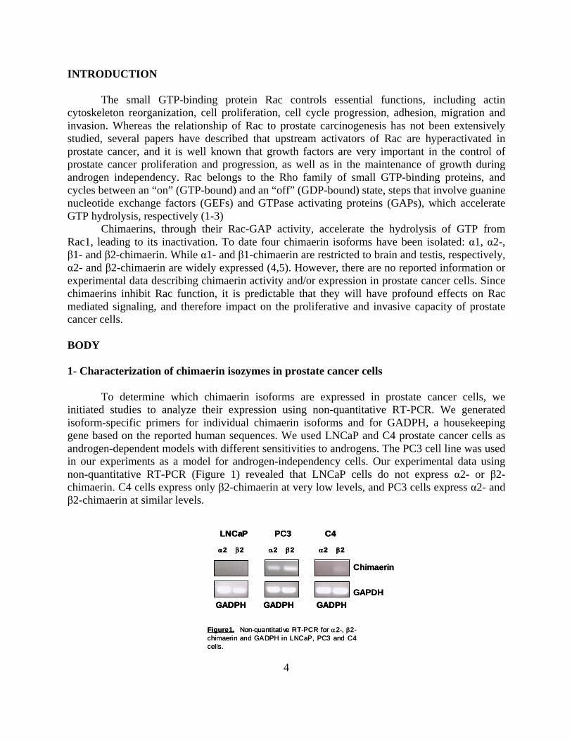
To determine which chimaerin isoforms are expressed in prostate cancer cells, we initiated studies to analyze their expression using non-quantitative RT-PCR. We generated isoform-specific primers for individual chimaerin isoforms and for GADPH, a housekeeping gene based on the reported human sequences. We used LNCaP and C4 prostate cancer cells as androgen-dependent models with different sensitivities to androgens. The PC3 cell line was used in our experiments as a model for androgen-independency cells. Our experimental data using non-quantitative RT-PCR (Figure 1) revealed that LNCaP cells do not express α2- or β2-chimaerin. C4 cells express only β2-chimaerin at very low levels, and PC3 cells express α2- and β2-chimaerin at similar levels.
α2 β2
GADPH
α2 β2 α2 β2
GADPH GADPH
LNCaP PC3 C4
Figure1. Non-quantitative RT-PCR for α2-, β2-chimaerin and GADPH in LNCaP, PC3 and C4 cells.
Chimaerin
GAPDH
α2 β2
GADPH
α2 β2 α2 β2
GADPH GADPH
LNCaP PC3 C4
α2 β2
GADPH
α2 β2 α2 β2
GADPH GADPH
LNCaP PC3 C4
Figure1. Non-quantitative RT-PCR for α2-, β2-chimaerin and GADPH in LNCaP, PC3 and C4 cells.
Chimaerin
GAPDH
4
In our laboratory, we have developed an anti-β2-chimaerin antibody (rat monoclonal) wich unfortunately was not highly specific, as it cross-reacts with α2-chimaerin (Figure 2A). Therefore we designed a peptide based on a β2-chimaerin specific sequence as well as a peptide for a α2-chimaerin specific sequence. These peptides were inoculated into rabbits in order to generate polyclonal antibodies. Different bleedings were obtained and subsequently tested by Western blot. Determination of chimaerin expression by Western blot with specific antibodies for each isoform was unsuccessful because unfortunately both antibodies showed cross-reaction (Figure 2B and C). It is not clear why this happened, as specific peptides were used. As the generation of specific antibodies is very important for this work as well as for the work of other members in our laboratory, we are currently generating new antibodies based on other peptide sequences.
HA-α2-chim
aerin
HA-β2-ch
imaerin
Endogenous
chim
aerin
Anti α2-Chimaerin
Actin
CP70Neuro
2A
Actin
CP70Neuro
2A
Anti β2-Chimaerin
Chimaerin
Actin
A
B C
Figure 2. Analysis of chimaerin isoform-specific antibodies by Western blot. (A and C) Control cell lysates from the human ovarian cancer cell line CP70, that express α2- but not β2-chimaerin and murine Neuro 2A cell line, that expresses both isoforms. (B) Cell lysates from the human ovarian cancer cell line HeLa infected with α2- and β2-chimaerin AdV.
2- Cloning of α2 chimaerin
As a cDNA for the α2-chimaerin isoform was not available in our laboratory, and this tool would be needed not only for functional experiments but also for the characterization of the antibody, we decided to clone this cDNA using RT-PCR. We first generated cDNA from fresh PC3 mRNA, wich was used for RT-PCR using specific primers. Restriction sites were included in order to facilitate subcloning into various expression vectors. We have been successful in cloning this cDNA and in subcloning it into TOPO®- vector (Invitrogen). The identity of the cDNA was then confirmed by sequencing. Results revealed 100% identity with the sequence reported in NCBI databases, suggesting that no mutations have not been introduced in the PCR reaction.
5
6
3- Generation of chimaerin adenoviruses
The generation of recombinant adenoviruses to study the role of specific chimaerins represents an essential step to assess their cellular functions, as we have developed conditions in our laboratory to achieve nearly ~100% of the prostate cancer cells expressing recombinant proteins using adenoviral techniques. We used the AdEasy system (Stratagene, La Jolla, CA) to generate chimaerin adenoviruses. The multicloning site of this vector was modified to facilitate easy subcloning, and a N-terminal HA-tag has been included for easy detection. Previously, in our laboratory we have successfully generated adenoviruses for wild type β2-chimaerin (β2-chim-AdV) and the β2-chimaerin GAP domain (β-GAP-AdV).
I have been actively involved in the generation of other adenoviruses that would be needed in further experiments, as follows: 3a- β2 chimaerin AdVs for GAP inactive mutants: β2-chim-∆EIE-AdV and β2-chim-298RRR-AdV. AdVs for phorbol ester/DAG unresponsive chimaerins: β2-chim-C246A-AdV. AdVs for SH2 domain : β2-chim-∆SH2-AdV.
The β2-chimaerin crystal structure of β2-chimaerin has been recently reported, as part of a collaborative effort between our laboratory and thelaboratory of Dr. Jim Hurley at the NIH (6). In this study it was found that several β2-chimaerin mutants present high sensitivity to phorbol ester-induced translocation as well as enhanced RacGAP activity in vivo. Due to this important finding we decided to incorporate these mutants to our study, as they will greatly contribute to our functional studies. Thus, I began the generation of adenoviruses encoding for two hyperactive mutants: β2-chimaerin Q32A and β2-chimaerin -I130A 3b- α 2 chimaerin
Using the AdEasy system I generated an adenovirus for wild type α2-chimaerin (α2-chim-AdV) and we are currently generating hiperactive mutants: α2-chimaerin Q24A and α2-chimaerin I122A, and the inactive mutant α2-chimaerin P216A. Determination of Rac-GTP levels in prostate cancer cell lines
In order to determine whether Rac hyperactivation is observed in prostate cancer cells, Rac-GTP levels were determined in the presence of two serum concentrations (2% and 10%). This is important to detect constitutive Rac hyperactivation under limiting conditions of stimulation by growth factors. Rac-GTP levels were determined using a Rac-GTP pull down assay. This is a non-radioactive methodology based on the specific binding of Rac-GTP (but not Rac-GDP) to PBD (the PAK1 p21Rac binding domain) coupled to GST using glutathione-Sepharose 4B beads. After the pull-down samples were analyzed by Western blot with an anti-Rac1 antibody, and expression is normalized to total Rac levels in the corresponding cell extracts. Our experimental data (Figure 3) showed that Rac-GTP levels in LNCaP and C4 cells are similar in both, low and high serum,
whereas the androgen independent PC3 cells showed higher levels of Rac-GTP in 10% serum compared to cells growing in 2% serum. This data suggest that the androgen-dependent cell lines LNCaP and C4 probably have constitutive Rac activation in the absence of growth factors or mitogenic stimuli.
Rac-GTP
Total Rac
2FBS(%) 10 2 10 2 10
LNCaP C4 PC3
Figure 3. LNCaP and C4 androgen-dependent prostate cancer cells shows high levels of activate Rac at low serum concentrations. Human prostate cancer cell lines LNCaP, C4 and PC3 were grown in the presence of low serum (2%) or normal serum (10%). Active Rac (Rac-GTP) were determined using a pull-down assay.
5- Determination of Rac-GTP levels upon chimaerin AdV infection
LNCaP cells do not express α2- or β2-chimaerin, and C4 cells express only β2-chimaerin at very low levels, therefore we decided to use these model to assess if wether chimaerin overexpression by adenoviral delivery reduces Rac-GTP levels. LNCaP and C4 cells were infected with HA-tagged α2- and β2-chimaerin AdVs at different MOIs (25, 50 and 100 pfu/cell) and cultured in the presence of 2% or 10%l serum. Rac-GTP levels were determined 48 h after infection. We used a LacZ AdV as a negative control at 100 MOI. Experimental data (Figure 4) show that β2-chimaerin overexpression reduces Rac-GTP levels in LNCaP and C4 cells both in 2% and 10% serum, whereas α2-chimaerin only reduces Rac-GTP levels in the presence of 2% serum. This data suggest that β2-chimaerin may reduce the levels of hyperactive Rac both in the absence or presence of mitogenic factors, whereas α2-chimaerin only in conditions of low serum.. Further analysis will be performed to determine the mechanisms that explain the differences in sensitivity to either chimaerin isoform.
7
8
6- Evaluation of MAPK signaling
Activation of Rac plays an important role in the control of the different MAPK signaling cascades. Since α2- and β2-chimaerin reduces Rac-GTP levels in LNCaP and C4 cells, we decided to study the effect of chimaerin overexpression on the activation of the various MAPK signaling pathways (ERK, JNK and p38 MAPK). LNCaP and C4 cells were infected with HA-tagged α2- and β2-chimaerin AdVs at different MOI (25, 50 and 100 pfu/cell) and cultured in the presence of medium with either 2% and 10% serum. Cell lysates were prepared 48 h after infection and subjected to Western blot analysis. Levels of activated ERK, JNK and p38 MAPK and the corresponding total levels were determined specific commercial antibodies. Experimental data shows that overexpression of β2-chimaerin (Figure 5, A, B) reduces the levels of activated ERK at MOI=50 pfu/cell both in the presence of 2% and 10% serum, but on the other hand, it induces a robust activation of p38 both in LNCaP and C4 cells. α2-chimaerin (Figure 5, C, D) reduces activated ERK levels only at high MOI (100 pfu/cell) and it reduces the levels of activated p38. No effect of chimaerin AdVs was found on JNK (data not shown). These data suggest a diferential regulation of MAPK cascades by chimaerin isoforms, an interesting finding that may translate into a differential regulation of genes and functional responses by either Rac-GAP. We are currently exploring the mechanisms involved in this differential effect of chimaerins isoforms.
25 50 100
Lac-Z
100
β2-chim
2%FBS
AdvMOI (pfu/cell) 25 50 100
Lac-Z
100
β2-chim
HA- β2-chimaerin
Rac-GTP
10%FBS
Rac
25 50 100
Lac-Z
100
α2-chim
2%FBS
AdvMOI (pfu/cell) 25 50 100
Lac-Z
100
α2-chim
10%FBS
HA-α2-chimaerin
Rac-GTP
Rac
LNCaP
25 50 100
Lac-Z
100
β2-chim
2%FBS
AdvMOI (pfu/cell) 25 50 100
Lac-Z
100
β2-chim
HA- β2-chimaerin
Rac-GTP
10%FBS
Rac
25 50 100
Lac-Z
100
α2-chim
2%FBS
AdvMOI (pfu/cell) 25 50 100
Lac-Z
100
α2-chim
10%FBS
HA-α2-chimaerin
Rac-GTP
Rac
LNCaP
25 50 100
Lac-Z
100
β2-chim
2%FBS
AdvMOI (pfu/cell) 25 50 100
Lac-Z
100
β2-chim
HA- β2-chimaerin
Rac-GTP
10%FBS
Rac
25 50 100
Lac-Z
100
α2-chim
2%FBS
AdvMOI (pfu/cell) 25 50 100
Lac-Z
100
α2-chim
10%FBS
HA- α2-chimaerin
Rac-GTP
Rac
C4
25 50 100
Lac-Z
100
β2-chim
C42%FBS
AdvMOI (pfu/cell) 25 50 100
Lac-Z
100
β2-chim
HA- β2-chimaerin
Rac-GTP
10%FBS
Rac
25 50 100
Lac-Z
100
α2-chim
2%FBS
AdvMOI (pfu/cell) 25 50 100
Lac-Z
100
α2-chim
10%FBS
HA- α2-chimaerin
Rac-GTP
Rac
25 50 100
Lac-Z
100
β2-chim
2%FBS
AdvMOI (pfu/cell) 25 50 100
Lac-Z
100
β2-chim
HA- β2-chimaerin
Rac-GTP
10%FBS
Rac
25 50 100
Lac-Z
100
α2-chim
2%FBS
AdvMOI (pfu/cell) 25 50 100
Lac-Z
100
α2-chim
10%FBS
HA- α2-chimaerin
Rac-GTP
Rac
Figure 4. Chimaerin overexpression reduces Rac-GTP levels in LNCaP and C4 androgen-dependent prostate cancer cells. Human prostate cancer cell lines LNCaP and C4 were infected for 16 h with α2-, β2-chimaerin or LacZ AdV at different multiplicities of infections (MOIs) and grown in the presence of either 2% or 10% serum. Active Rac (Rac-GTP) was determined 48 h later using a pull-down assay.
9
25 50 100
Lac-Z
100
β2-chim
2%FBS
Adv
7- Studies on prostate cancer cell proliferation
Rac is required for proliferation and cell cycle progression (9,10,11). Our original hypothesis was that Rac inhibition by chimaerins impairs mitogenic responses, particularly in models of Rac hyperactivation. The fact that LNCaP and C4 cells shown Rac hyperactivation and that overexpression of α2- and β2-chimaerins reduces Rac activity and ERK activation was indeed strongly suggestive that chimaerins must impair cell proliferation. To addres this issue LNCaP and C4 cells were infected with HA-tagged α2- and β2-chimaerin AdVs at different MOIs (1, 5 and 50 pfu/cell) and cultured in the presence of 10% serum. Proliferation was assessed at 72 h after infection using an MTT assay. Experimental results (Figure 6) shows that only β2-chimaerin reduces cell proliferation (~ 25% reduction relative to non-infected cells). A control using LacZ AdV did not have any significant effect.
MOI (pfu/cell) 25 50 100
Lac-Z
100
β2-chim
10%FBS
HA- β2-chimaerin
p-ERK
ERK
p-p38
p38
LNCaPA
25 50 100
Lac-Z
100
β2-chim
2%FBS
AdvOI (pfu/cell) 25 50 100
Lac-Z
100M
β2-chim
10%FBS
HA- β2-chimaerin
p-ERK
ERK
p-p38
p38
LNCaP
25 50 100
Lac-Z
100
β2-chim
2%FBS
AdvOI (pfu/cell) 25 50 100
Lac-Z
100M
β2-chim
10%FBS
HA- β2-chimaerin
p-ERK
ERK
p-p38
p38
25 50 100
Lac-Z
100
β2-chim
2%FBS
AdvOI (pfu/cell) 25 50 100
Lac-Z
100M
β2-chim
10%FBS
HA- β2-chimaerin
p-ERK
ERK
p-p38
p38
LNCaPA
25 50 100
Lac-Z
100
β2-chim
2%FBS
AdvMOI (pfu/cell) 25 50 100
Lac-Z
100
B
25 50 100
Lac-Z
100
β2-chim
10%FBS
HA- β2-chimaerin
p-ERK
ERK
p-p38
p38
C4
β2-chim
2%FBS
AdvMOI (pfu/cell) 25 50 100
Lac-Z
100
β2-chim
10%FBS
HA- β2-chimaerin
p-ERK
ERK
p-p38
p38
C4
25 50 100
Lac-Z
100
β2-chim
2%FBS
AdvMOI (pfu/cell) 25 50 100
Lac-Z
100
B
25 50 100
Lac-Z
100
C4
β2-chim
10%FBS
HA- β2-chimaerin
p-ERK
ERK
p-p38
p38
α2-chim
2%FBS
AdvOI (pfu/cell) 25 50 100
Lac-Z
100M
α2-chim
10%FBS
HA-α2-chimaerin
p-ERK
ERK
p-p38
p38
LNCaPC
25 50 100
Lac-Z
100
α2-chim
2%FBS
AdvOI (pfu/cell) 25 50 100
Lac-Z
100M
α2-chim
10%FBS
HA-α2-chimaerin
p-ERK
ERK
p-p38
p38
LNCaP
25 50 100
Lac-Z
100
α2-chim
2%FBS
AdvOI (pfu/cell) 25 50 100
Lac-Z
100M
α2-chim
10%FBS
HA-α2-chimaerin
p-ERK
ERK
p-p38
p38
LNCaPC
25 50 100
Lac-Z
100
α2-chim
2%FBS
AdvMOI (pfu/cell) 25 50 100
Lac-Z
100
D
25 50 100
Lac-Z
100
D C4C4C4
α2-chim
10%FBS
HA-α2-chimaerin
p-ERK
ERK
p-p38
p38
α2-chim
2%FBS
AdvMOI (pfu/cell) 25 50 100
Lac-Z
100
α2-chim
10%FBS
HA-α2-chimaerin
p-ERK
ERK
p-p38
p38
25 50 100
Lac-Z
100
α2-chim
2%FBS
AdvMOI (pfu/cell) 25 50 100
Lac-Z
100
α2-chim
10%FBS
HA-α2-chimaerin
p-ERK
ERK
p-p38
p38
Figure 5. Effect of chimaerin isoforms on ERK and p38 phosphorylation. Human LNCaP and C4 prostate cancer cell lines were infected for 16 h with α2-, β2-chimaerin or LacZ AdV at different MOIs and grown in the presence of either 2% or 10% serum. P-ERK, ERK, p-p38 and p38 levels were determined 48 h later.
10
5 501100 5 501
α2-chimβ2-chimLac-Z
AdvMOI (pfu/cell)
% i
ncre
ase
of c
ontr
olLNCaP
0
20
40
60
80
100
120
A
8- Cloning of a putative β2-chimaerin promoter
Transcription in eukaryotic cells is regulated at multiple levels. It has been determined that the methylation status of CpG islands in genes affects DNA-protein interactions. Most of the CpG islands in the promoter remain unmethylated, but when methylation occurs the gene is silenced. Thus, DNA methylation acts as a major epigenetic modification to maintain stable gene silencing. Aberrant DNA methylation impacts on gene transcription, and this event has been linked to cancer formation (7,8). Since several prostate cancer cell lines have low chimaerin expression, we sought to investigate whether the effect was at the promoter level. To date, there is no information on the β2 chimaerin promoter. We therefore decided to pursue this studies. Analysis of the gene using established programs reveal a region in the β2-chimaerin locus with promoter characteristics. Interestingly this region contains a putative CpG island. A 1 kb genomic DNA fragment 5´ upstream from the β2-chimaerin start codon was cloned by PCR into TOPO®- vector (Invitrogen) using specific primers.
Using PCR we have amplified and subcloned different fragments (see list above) of the 1 kb genomic segment into pRTK-Luc a plasmid that encodes for a luciferase gene under the control of a TK promoter. The constructs generated were as follows: pR-81-Luc, pR-181-Luc, pR-281-Luc, pR-381-Luc, pR-481-Luc, pR-581-Luc, pR-1000-Luc (the number represent the position of the first nucleotide upstream of the ATG). pR-BASIC (luciferase with no promoter) was used as a negative control and pRTK-Luc was used as a positive control.
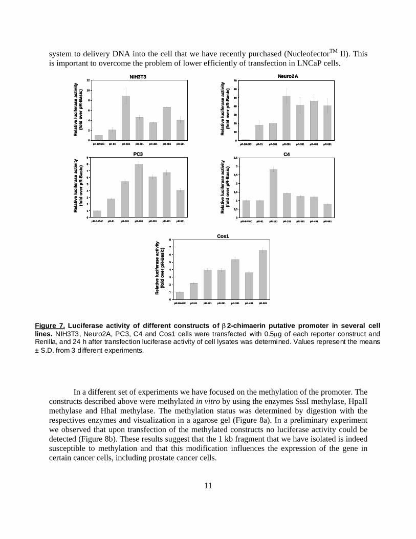
These luciferase reporters were co-tranfected together with a Renilla expression vector (for normalization) into several cell lines. Experimental data revealed that all constructs induce luciferase expression upon transfection (Figure 7). We are currently analyzing in a comparative manner the various constructs upon transfection into LNCaP prostate cancer cells by using a new
5 501100 5 501
α2-chimβ2-chimLac-Z
AdvMOI (pfu/cell)
% i
ncre
ase
of c
ontr
olLNCaP
0
20
40
60
80
100
120
5 501100 5 501
α2-chimβ2-chimLac-Z
AdvMOI (pfu/cell)
% i
ncre
ase
of c
ontr
olLNCaP
A
0
20
40
60
80
100
120
5 501100 5 501
α2-chimβ2-chimLac-Z
AdvMOI (pfu/cell)
% i
ncre
ase
of c
ontr
ol
0
20
40
60
80
100
120
C4B
5 501100 5 501
B
α2-chimβ2-chimLac-Z
AdvMOI (pfu/cell)
% i
ncre
ase
of c
ontr
ol
0
20
40
60
80
100
120
C4
5 501100 5 501
α2-chimβ2-chimLac-Z
AdvMOI (pfu/cell)
% i
ncre
ase
of c
ontr
ol
0
20
40
60
80
100
120
5 501100 5 501
α2-chimβ2-chimLac-Z
AdvMOI (pfu/cell)
% i
ncre
ase
of c
ontr
ol
5 501100 5 501
α2-chimβ2-chimLac-Z
AdvMOI (pfu/cell)
% i
ncre
ase
of c
ontr
ol
0
20
40
60
80
100
120
C4
Figure 6. β2-chimaerin overexpression inhibits LNCaP and C4 cell proliferation. Human LNCaP and C4 prostate cancer cell lines were infected for 16 h with α2-, β2-chimaerin or LacZ AdV at different MOIs. MTT assay was performed 72 h later.
system to delivery DNA into the cell that we have recently purchased (NucleofectorTM II). This is important to overcome the problem of lower efficiently of transfection in LNCaP cells.
Figure 7. Luciferase activity of different constructs of β2-chimaerin putative promoter in several cell lines. NIH3T3, Neuro2A, PC3, C4 and Cos1 cells were transfected with 0.5μg of each reporter construct and Renilla, and 24 h after transfection luciferase activity of cell lysates was determined. Values represent the means ± S.D. from 3 different experiments.
In a different set of experiments we have focused on the methylation of the promoter. The
constructs described above were methylated in vitro by using the enzymes SssI methylase, HpaII methylase and HhaI methylase. The methylation status was determined by digestion with the respectives enzymes and visualization in a agarose gel (Figure 8a). In a preliminary experiment we observed that upon transfection of the methylated constructs no luciferase activity could be detected (Figure 8b). These results suggest that the 1 kb fragment that we have isolated is indeed susceptible to methylation and that this modification influences the expression of the gene in certain cancer cells, including prostate cancer cells.
11
1 2 43
pRB-BASIC
pRB-281
0
0,5
1
1,5
2
2,5
3
3,5
4
4,5
pR-BASIC pR-281 pR-BASIC pR-281
Rela
tive
luci
fera
se a
ctiv
ity(fo
ld o
ver p
R-B
asic
)
Mock methylated HpaII methylated
A. B.
1 2 43
pRB-BASIC
pRB-281
1 2 43
pRB-BASIC
pRB-281
0
0,5
1
1,5
2
2,5
3
3,5
4
4,5
pR-BASIC pR-281 pR-BASIC pR-281
Rela
tive
luci
fera
se a
ctiv
ity(fo
ld o
ver p
R-B
asic
)
Mock methylated HpaII methylated
0
0,5
1
1,5
2
2,5
3
3,5
4
4,5
pR-BASIC pR-281 pR-BASIC pR-281
Rela
tive
luci
fera
se a
ctiv
ity(fo
ld o
ver p
R-B
asic
)
Mock methylated HpaII methylated
A. B. Figure 8. Effect of in vitro methylation on β2-chimaerin putative promoter activity. A, pR-BASIC
and pRB-281 were methylated in vitro with HpaII methylase. The extent of methylation was assessed by comparing digestion patterns of unmethylated (lanes 2-4) and methylated (lanes 1-3) constructs with HpaII. B, methylated and unmethylated pR-BASIC and pR-281 constructs were transfected into Cos1 cells and assayed for luciferase activity. Values represent the means ± S.D. from 3 different experiments.
Future directions. The next goals will be: 1. To assess wether chimaerins regulates ERK and p38 signaling pathways in prostate cancer cells. 2. To determine how chimaerins affect to cytoskeleton reorganization, migration and invasion in prostate cancer cells (Aim 3 from proposal). We will uso the NucleofectorTM II, a new DNA delivery system, to transfect GFP-chimaerin in LNCaP and C4 cells. The adenoviruses for different chimaerin isoforms and mutants will be used in addition in these experiments. These experiments were part of Specific Aim 3 in the original proposal, but we could not complete them in time. It is expected that these experiments will be done in the next few months. 3. To continue the characterization of the putative β2-chimaerin promoter and to determine whether methylation is a critical factor in β2 chimaerin expression. KEY RESEARCH ACCOMPLISHEMENTS
1. We characterize the expression of chimaerin isoforms in prostate cancer cell lines. 2. We generated adenoviruses or adenoviral constructs that are essential for functional
studies in prostate cancer cells. 3. We determined that Rac is hyperactive in some prostate cancer cell lines and that this
hyperactivation could be reverted by chimaerin overexpression.
12
13
4. We determined that ERK activation levels could be reduced by chimaerin overexpression.
5. We determined that α2- and β2-chimaerin exerts opposites effects on p38 activation. 6. We cloned a 1 kb fragment that corresponds to the β2-chimaerin promoter. 7. We generated reporter constructs for different regions of the β2-chimaerin promoter,
which will be used in luciferase assays. 8. We initiated studies aimed at determining whether the β2-chimaerin promoter is
regulated by methylation.
REPORTABLE OUTCOMES A manuscript is currently in preparation. CONCLUSIONS
In the last year we completed experiments aimed at characterizing the regulation and function of β2-chimaerin and the related α2-chimaerin isoforms in prostate cancer cells. These proteins regulate the function of the small GTP-binding protein Rac, which plays essential roles in mitogenesis, transformation, and the metastatic cascade. We have obtained data regarding the expression of chimaerin isozymes in prostate cancer cell lines. We found that expression is very low in some prostate cancer cells, which is in agreement with data in other types of cancers, such breast cancer cells or gliomas. In addition, these prostate cancer cell lines presents high levels of activated Rac a situation that can be reversed by overexpression of chimaerins, wich also leads to reduce ERK activation and proliferation rate. The only divergence that we found between α2- and β2-chimaerin effects in prostate cancer cells is the different behavior on p38 phosphorylation. This is indeed the first evidence that probably α2- and β2-chimaerin may be having differential effect. The mechanisms have yet to be characterized.
The efficiency of transfection in LNCaP and C4 cells with usual DNA delivery systems is very poor (~5%). Since transfection in this cell lines is not efficient data about spreading and cytoskeleton reorganization were not consistent. Because that, at the present time we are using a new cDNA delivery system, NucleofectorTM II with provide a 80-90% of transfection efficiency. The development of the different adenoviruses will also help in this endeavour.
Understanding the events that control the expression of the β2-chimaerin gene is very important because the gene seems to be down-regulated in a variety of cancers. It would be important in the future to compare the expression of chimaerin in normal vs. cancer specimens. The cloning of a putative promoter and its characterization will further enhance the understanding of the regulation of this molecule.
REFERENCES 1. Mackay, D.J., and Hall, A. Rho GTPases. J. Biol. Chem. 273: 20685-20688 (1998). 2. Bar-Sagi, D., and Hall, A. Ras and Rho GTPases: a family reunion. Cell 103: 227-238 (2000).
14
3. Ridley, A.J. Rho family proteins: coordinating cell responses. Trends Cell Biol. 11: 471-477 (2001). 4. Caloca, M.J., Wang, H.B., Delemos, A., Wang, S., and Kazanietz, M.G. Phorbol esters and related analogs regulate the subcellular localization of β2-chimaerin, a non-PKC. phorbol ester receptor. J.Biol. Chem. 276: 18303-18312 (2001). 5. Wang, H., and Kazanietz, M.G. Chimaerins, novel .non-PKC. phorbol ester receptors, associate withTmp21-I (p23). Evidence for a novel anchoring mechanism involving the chimaerin C1 domain. J. Biol.Chem. 277: 4541-4550 (2002). 6. Canagarajah B, Leskow FC, Ho JY, Mischak H, Saidi LF, Kazanietz MG, Hurley JH. Structural mechanism for lipid activation of the Rac-specific GAP, beta2-chimaerin. Cell. 2004 119:407-418. 7. Jones PA: Epigenetics in carcinogenesis and cancer prevention. Ann N Y Acad Sci 2003, 983:213-219. 8. Feninberg A: Cancer epigenetics takes center stage. Proc Natl Acad Sci USA 2001, 98(2):392-394.) 9. Olson, M.F., Ashworth, A., and Hall, A. An essential role for Rho, Rac, and Cdc42 GTPases in cell cycle progression though G1. Science 269: 1270-1272 (1995). 10. Joneson, T., and Bar-Sagi, D. A Rac1 effector site controlling mitogenesis through superoxide production. J. Biol. Chem. 273: 17991-17994 (1998). 11. Bouzahzah, J.D., Fu, M., Albanese, C., D.Amico, M., Steer, J., Klein, J.U., Lee, R.J., Segall, J.E., Westwick, J.K., Der, C.J., and Pestell, R.G. Integration of Rac-dependent regulation of cyclin D1 transcription through a nuclear factor-kappaB-dependent pathway. J. Biol. Chem. 274: 25245-25249 (1999). APPENDICES None. ABBREVIATIONS AdV: adenovirus GAPDH: Glyceraldehyde-3-phosphate dehydrogenase DAG: diacylglycerol GAP: GTPase activating protein Kb: Kilobase MOI: Multiplicity of infection NCBI: National Center for Biotechnology Information PKC: protein kinase C PMA: Phorbol 12-myristate 13-acetate PCR: Polymerase Chain Reaction Rac-GAP: Rac-GTPase activating protein TK: Thymidine Kinase