Award Number: W81XWH-12-1-0245 TITLE: Evaluation of Multimodal Imaging Biomarkers of Prostate Cancer PRINCIPAL INVESTIGATOR: Christopher Chad Quarles CONTRACTING ORGANIZATION: Vanderbilt University Medical Center Nashville, TN 37232-2675 REPORT DATE: November 2016 TYPE OF REPORT: Final PREPARED FOR: U.S. Army Medical Research and Materiel Command Fort Detrick, Maryland 21702-5012 DISTRIBUTION STATEMENT: Approved for Public Release; Distribution Unlimited The views, opinions and/or findings contained in this report are those of the author(s) and should not be construed as an official Department of the Army position, policy or decision unless so designated by other documentation.
Transcript
Award Number: W81XWH-12-1-0245
TITLE: Evaluation of Multimodal Imaging Biomarkers of Prostate Cancer
PRINCIPAL INVESTIGATOR: Christopher Chad Quarles
CONTRACTING ORGANIZATION: Vanderbilt University Medical Center Nashville, TN 37232-2675
REPORT DATE: November 2016
TYPE OF REPORT: Final
PREPARED FOR: U.S. Army Medical Research and Materiel Command Fort Detrick, Maryland 21702-5012
DISTRIBUTION STATEMENT: Approved for Public Release; Distribution Unlimited
The views, opinions and/or findings contained in this report are those of the author(s) and should not be construed as an official Department of the Army position, policy or decision unless so designated by other documentation.
REPORT DOCUMENTATION PAGE Form Approved
OMB No. 0704-0188 Public reporting burden for this collection of information is estimated to average 1 hour per response, including the time for reviewing instructions, searching existing data sources, gathering and maintaining the data needed, and completing and reviewing this collection of information. Send comments regarding this burden estimate or any other aspect of this collection of information, including suggestions for reducing this burden to Department of Defense, Washington Headquarters Services, Directorate for Information Operations and Reports (0704-0188), 1215 Jefferson Davis Highway, Suite 1204, Arlington, VA 22202-4302. Respondents should be aware that notwithstanding any other provision of law, no person shall be subject to any penalty for failing to comply with a collection of information if it does not display a currently valid OMB control number. PLEASE DO NOT RETURN YOUR FORM TO THE ABOVE ADDRESS. 1. REPORT DATENovember 2016
2. REPORT TYPEFinal
3. DATES COVERED9/1/12 - 8/31/16
4. TITLE AND SUBTITLEEvaluation of Multimodal Imaging Biomarkers of Prostate Cancer
5a. CONTRACT NUMBER
5b. GRANT NUMBER W81XWH-12-1-02455c. PROGRAM ELEMENT NUMBER
6. AUTHOR(S)Christopher Chad Quarles Betty Diamond
7. PERFORMING ORGANIZATION NAME(S) AND ADDRESS(ES)
AND ADDRESS(ES)
8. PERFORMING ORGANIZATION REPORTNUMBER
Vanderbilt University Medical Center1161 21st Avenue South Nashville, TN 37203 and Meherry Medical College: 1005 Dr D.B. Todd Blvd, Nashville, TN 37208 9. SPONSORING / MONITORING AGENCY NAME(S) AND ADDRESS(ES) 10. SPONSOR/MONITOR’S ACRONYM(S)U.S. Army Medical Research and Materiel Command Fort Detrick, Maryland 21702-5012
11. SPONSOR/MONITOR’S REPORTNUMBER(S)
12. DISTRIBUTION / AVAILABILITY STATEMENTApproved for Public Release; Distribution Unlimited
13. SUPPLEMENTARY NOTES
14. ABSTRACT The goals of the proposed studies are to develop, optimize and use imaging methods to non-invasively assessthe temporal relationship prostate cancer growth, androgen receptor (AR) levels, hypoxia, and translocator protein (TSPO) levels. As described in the statement of work, the first year of this award focused on implementation and optimization of the imaging methods in the Pten / p53 double null mutant mouse model. Towards that end, we have systematically optimized and characterized protocols for imaging tumor morphology using MRI, AR levels using [18F]-fluoro-5alpha-dihydrotestosterone (FDHT), hypoxia using, 18F-fluoromisonidazole, and TSPO using an in-house tracer called VUIIS-1008. The major finding of this work is the validation of TSPO as a highly sensitive and specific marker of prostate cancer with favorable imaging characteristics (e.g. low bladder uptake) that enable robust detection of small prostate cancers. In contrast, high background and variable uptake of FDHT and FMISO confounded the reliable evaluation of AR and hypoxia in the Pten/p53 mouse model. The observed high uptake of TSPO justifies its further evaluation in both preclinical and clinical studies as a novel biomarker for prostate cancer detection and treatment response.
In its advanced stages, prostate cancer (PCa) becomes clinically difficult to restrain due to failure of therapy and the development of castration resistant prostate cancer (CRPC). Thus, there is a compelling need to investigate the mechanisms leading to CRPC in order to develop more effective treatment strategies. The most common approach to biologically assess disease progression in mouse models of PCa is through pathological examination, which requires the sacrifice of mice at multiple arbitrary time points and, consequently, is unsuitable for the temporal characterization of physiological, cellular and molecular events leading to CRPC growth in a given animal. In recent years, however, there have been dramatic increases in the range and quality of information available from non-invasive imaging methods so that many potentially valuable imaging metrics are now available to quantitatively measure tumor growth, assess tumor status, and predict treatment response. To this end, this study aimed to evaluate emerging, clinically-viable imaging metrics in an appropriate PCa animal model to serially assess tumor progression. In particular, we proposed to non-invasively assess the relationship between growth, androgen receptor levels, hypoxia, and translocator protein expression using Magnetic Resonance Imaging (MRI) and Positron Emission Tomography (PET). Such studies could provide the scientific basis for the acceleration of these emerging imaging methods into clinical care and could have a direct impact on prostate cancer detection, staging and treatment monitoring. The proposed studies were carried out in the genetically engineered Pten/p53 conditional mouse model (Ptenpc-/-; Trp53pc-/- double-null mutants). Our preliminary studies revealed that prostate tumors in these mutant mice are initially sensitive to castration, as evidenced by tumor regression, but this is followed by tumor recurrence that is ultimately lethal. The regression response to castration and subsequent CRPC growth in these mice clinically recapitulate the disease progression observed in human prostate cancer undergoing androgen-ablation therapy. Therefore, this authentic mouse model provides a valuable and unique tool with which to validate multiparametric imaging of cellular and molecular imaging parameters.
2
2. KEYWORDS: castration resistant prostate cancer, MRI, PET, multiparametric imaging 3. ACCOMPLISHMENTS: What were the major goals of the project? Goal 1: Synthesize 18F-FDHT for imaging androgen receptors in Pten/p53 mice Goal 2: Optimize and characterize MRI and PET (using radiotracers for hypoxia (18F-FMISO), Translocator protein expression (18F-VUIIS1008) and AR (15F-FDHT)) prostate cancer imaging methods for application to Pten/p53 mice Goal 3: Acquire serial PET and MRI anatomic data to determine which marker best correlates with progression and correlate imaging with histologic data
What was accomplished under these goals? Goal 1: Synthesize 18F-FDHT for imaging androgen receptors in Pten/p53 mice Conditions for radiosynthesis of 18b-[18F]fluorodihydrotestosterone ([18F]FDHT) were developed using a combination of an automated reaction module (GE TRACERlab FXF-
N) and manual manipulations. Preparation (Scheme 1) began with standard nucleophilic radiofluorination (step 1) of protected precursor 16a-[[(trifluoromethyl)sulfonyl]oxy]-3,3-(ethylenedioxy)androstan-17-one (Memorial Sloan-Kettering Cancer Center) within the reaction module. After completion of radiofluorination, the material was then transferred out of the module and reduced via lithium aluminum hydride (LiAlH4) at -78 °C (step 2). This was followed by reaction quenching and then deprotection using standard acid hydrolysis at 55 °C (step 3) to reveal the final product. The final product was then purified by semi-preparative HPLC (Hitachi High Technologies) and formulated in a solution containing no greater than 10% ethanol in saline. Quality control was performed by analytical HPLC (Hitachi High Technologies) with inline UV and radiation detectors enabling comparison of the radioactive product to the reference standard ([19F]FDHT) (Figure 1). Results showed a perfect overlay with a typical radiochemical purity greater than 98%.
Figure 1: Overlay of the radioactive HPLC trace (Blue) and reference standard UV trace (Red). Retention time difference is due to the time delay between the UV detector and the radiation detector.
Goal 2: Optimize MRI and PET (using radiotracers for hypoxia (18F-FMISO), Translocator protein expression (18F-VUIIS1008) and AR (15F-FDHT)) prostate cancer imaging methods
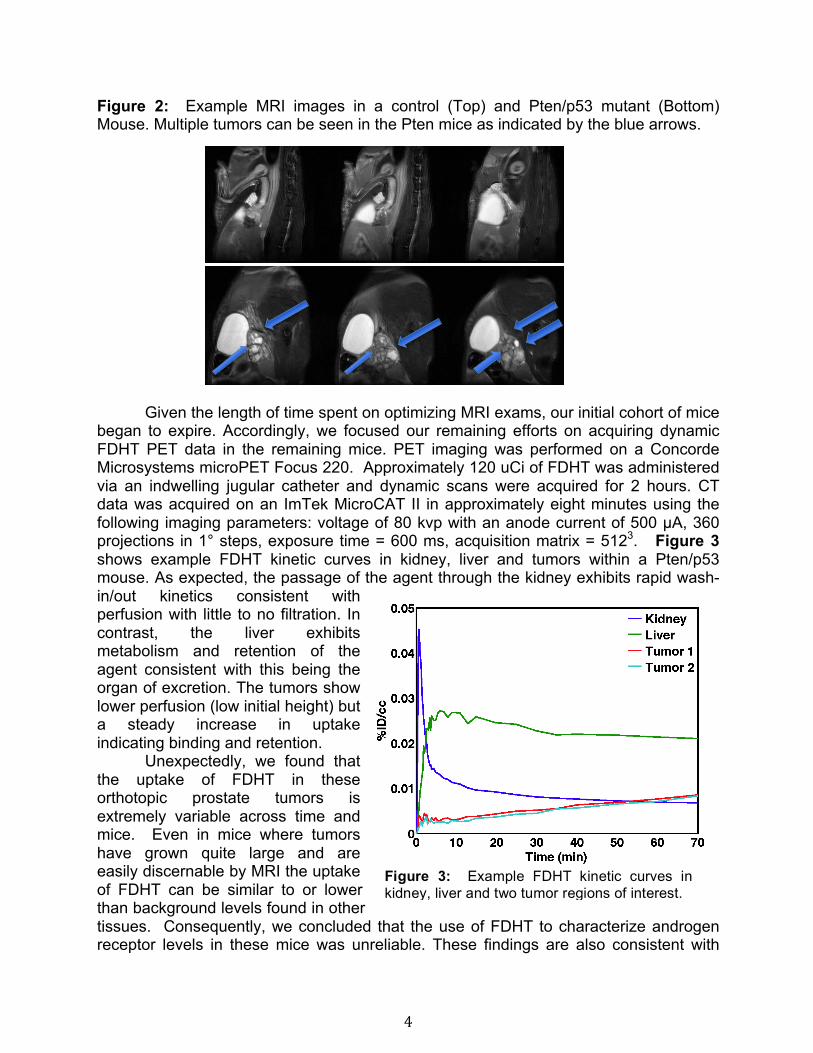
In the Pten/p53 conditional mice we were successfully able to use MRI to identify the prostate and developing tumors. We did find a mix success rate of tumor growth in the pten/p53 mice acquired from Meharry. We were able to track the growth of tumors over the course of two to three months. The primary goal of each of these scans was to optimize the acquisition protocols needed to: i) minimize motion artifacts, ii) reliably detect the prostate and any associated tumors, and iii) enhance contrast within tumors. Robust image quality and tumor detection was best achieved with a 2D T2-weighted high-resolution fast spin echo scan with the following imaging parameters (Varian 7T MRI system, TR = 2s, TE = 40ms, echo train length = 8, echo spacing = 10ms, matrix = 256 x 256, FOV = 25.6 x 25.6 mm, 20 slices, 1 mm slice thickness, number of excitations = 20, scan duration ~ 21 min). We also have found that placing the animal on its back (rather than the usual stomach position) enabled us to avoid the use of respiratory gating in the scans, thereby reducing scan durations. It also reduced scan-to-scan variability in the location and appearance of the tumors. This feature also made the acquisition of the DCE-MRI data much more reliable as it removes any confounding issues that could occur due to variable breathing rates. Figure 2 shows example images of a Pten/p53 mouse tumor. Across the small cohort of mice in this study, the number of tumors, their size, appearance and location was heterogeneous.
4
Figure 2: Example MRI images in a control (Top) and Pten/p53 mutant (Bottom) Mouse. Multiple tumors can be seen in the Pten mice as indicated by the blue arrows.
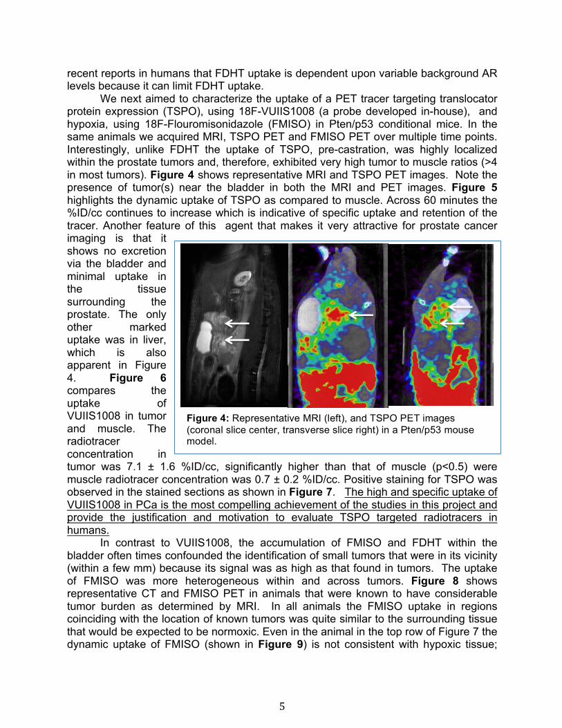
Given the length of time spent on optimizing MRI exams, our initial cohort of mice began to expire. Accordingly, we focused our remaining efforts on acquiring dynamic FDHT PET data in the remaining mice. PET imaging was performed on a Concorde Microsystems microPET Focus 220. Approximately 120 uCi of FDHT was administered via an indwelling jugular catheter and dynamic scans were acquired for 2 hours. CT data was acquired on an ImTek MicroCAT II in approximately eight minutes using the following imaging parameters: voltage of 80 kvp with an anode current of 500 μA, 360 projections in 1° steps, exposure time = 600 ms, acquisition matrix = 5123. Figure 3 shows example FDHT kinetic curves in kidney, liver and tumors within a Pten/p53 mouse. As expected, the passage of the agent through the kidney exhibits rapid wash-in/out kinetics consistent with perfusion with little to no filtration. In contrast, the liver exhibits metabolism and retention of the agent consistent with this being the organ of excretion. The tumors show lower perfusion (low initial height) but a steady increase in uptake indicating binding and retention.
Unexpectedly, we found that the uptake of FDHT in these orthotopic prostate tumors is extremely variable across time and mice. Even in mice where tumors have grown quite large and are easily discernable by MRI the uptake of FDHT can be similar to or lower than background levels found in other tissues. Consequently, we concluded that the use of FDHT to characterize androgen receptor levels in these mice was unreliable. These findings are also consistent with
Figure 3: Example FDHT kinetic curves in kidney, liver and two tumor regions of interest.
5
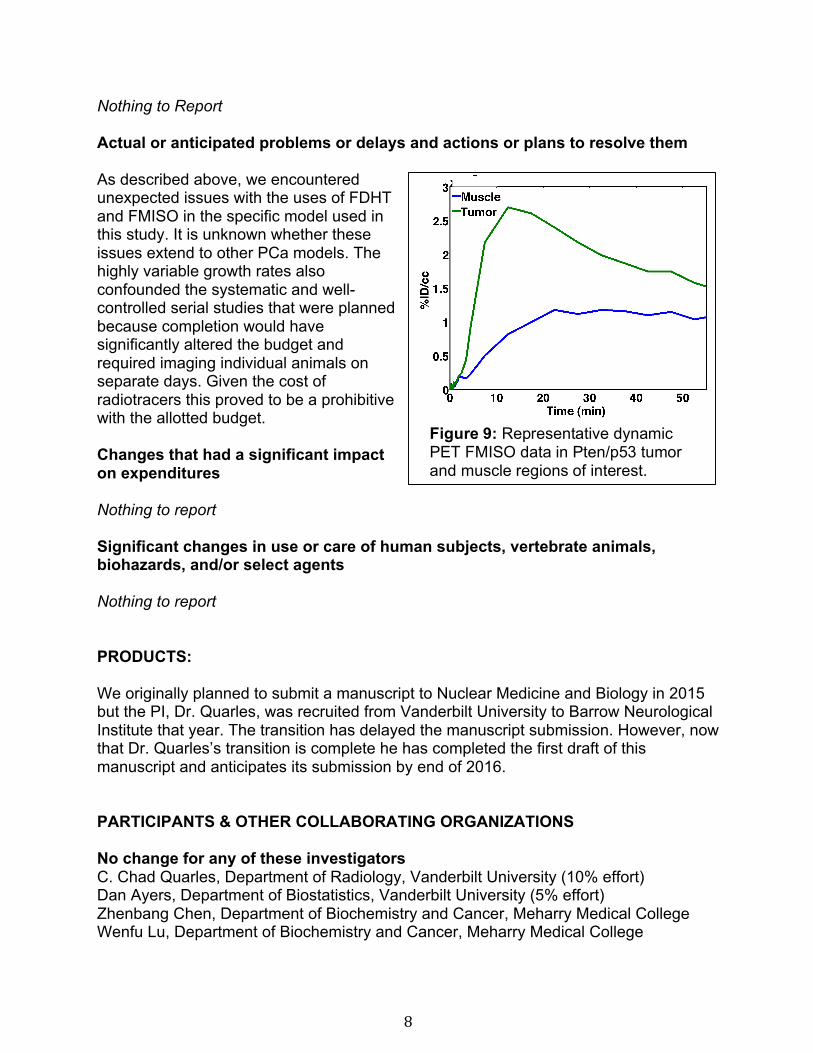
recent reports in humans that FDHT uptake is dependent upon variable background AR levels because it can limit FDHT uptake. We next aimed to characterize the uptake of a PET tracer targeting translocator protein expression (TSPO), using 18F-VUIIS1008 (a probe developed in-house), and hypoxia, using 18F-Flouromisonidazole (FMISO) in Pten/p53 conditional mice. In the same animals we acquired MRI, TSPO PET and FMISO PET over multiple time points. Interestingly, unlike FDHT the uptake of TSPO, pre-castration, was highly localized within the prostate tumors and, therefore, exhibited very high tumor to muscle ratios (>4 in most tumors). Figure 4 shows representative MRI and TSPO PET images. Note the presence of tumor(s) near the bladder in both the MRI and PET images. Figure 5 highlights the dynamic uptake of TSPO as compared to muscle. Across 60 minutes the %ID/cc continues to increase which is indicative of specific uptake and retention of the tracer. Another feature of this agent that makes it very attractive for prostate cancer imaging is that it shows no excretion via the bladder and minimal uptake in the tissue surrounding the prostate. The only other marked uptake was in liver, which is also apparent in Figure 4. Figure 6 compares the uptake of VUIIS1008 in tumor and muscle. The radiotracer concentration in tumor was 7.1 ± 1.6 %ID/cc, significantly higher than that of muscle (p<0.5) were muscle radiotracer concentration was 0.7 ± 0.2 %ID/cc. Positive staining for TSPO was observed in the stained sections as shown in Figure 7. The high and specific uptake of VUIIS1008 in PCa is the most compelling achievement of the studies in this project and provide the justification and motivation to evaluate TSPO targeted radiotracers in humans.
In contrast to VUIIS1008, the accumulation of FMISO and FDHT within the bladder often times confounded the identification of small tumors that were in its vicinity (within a few mm) because its signal was as high as that found in tumors. The uptake of FMISO was more heterogeneous within and across tumors. Figure 8 shows representative CT and FMISO PET in animals that were known to have considerable tumor burden as determined by MRI. In all animals the FMISO uptake in regions coinciding with the location of known tumors was quite similar to the surrounding tissue that would be expected to be normoxic. Even in the animal in the top row of Figure 7 the dynamic uptake of FMISO (shown in Figure 9) is not consistent with hypoxic tissue;
Figure 4: Representative MRI (left), and TSPO PET images (coronal slice center, transverse slice right) in a Pten/p53 mouse model.
6
exhibiting rapid and strong influx of tracer followed up efflux that is similar to surrounding tissue. In our other tumor models (e.g. breast, glioma), the FMISO dynamics in hypoxic reg ons after its initial delivery slowly continues to increase indicating its reduction and cellular retention. Accordingly, it is unlikely that FMISO is serving as a useful marker of hypoxia in this prostate tumor model. Goal 3: Acquire serial, multi-parametric PET and MRI anatomic data to determine during PCa progression and correlate imaging with histologic data While we were able to serially evaluate tumor size changes using MRI and TSPO levels using VUIIS-1008 PET the inconsistency of FDHT and FMISO hindered the completion of his goal. The primary hypothesis was that multi-parametric imaging, which is sensitive to a range of PCa biological features, would provide a better prediction of cancer progression and response to castration. This goal was further confounded by the variable growth observed in the Pten/p53 animals, with a fair proportion of animals exhibiting no tumors a nd others with highly inconsistent growth rates.
What opportunities for training and professional development has the project provided? Nothing to Report
Figure 5: Average time-activity curves (TACs) of mice (n = 9) injected with 18F-VUIIS1008 and imaged in a microPET for 60 min. The error bars are the standard deviation in curves.
Tumor
Muscle
0
5
10
15
18F-
VUIIS
1008
con
cent
ratio
n (%
ID/c
c)
Figure 6: Tumor versus muscle uptake of 18F-VUIIS1008 in mice bearing prostrate tumors.
7
How were the results disseminated to communities of interest? Nothing to Report What do you plan to do during the next reporting period to accomplish the goals? Nothing to Report 4. IMPACT: What was the impact on the development of the principal discipline(s) of the project? The results of this study will guide future preclinical
imaging efforts that are seeking to gain new insights into prostate cancer biology. The project also identified a potentially novel and highly sensitive imaging method that could ultimately be useful in diagnosis and tracking prostate cancer in patients. What was the impact on other disciplines? The finding that the TSPO imaging marker, VUIIS-1008, further demonstrates the potential of this biological target in cancer and justifies its evaluation in other cancer types and in humans. What was the impact on technology tr ansfer? Nothing to Report
What was the impact on society beyond science and technology? Nothing to Report." CHANGES/PROBLEMS: Changes in approach and reasons for change
Figure 8: Representative FMISO PET images in two PTEN/p53 mouse with apparent tumor uptake (top) and no tumor specific uptake (bottom). Both animals had substantial tumor burden as determined by MRI.
Figure 7: Confirmation of TSPO expression (brown) in a PTEN/p53 mouse model exhibiting high 18F-VUIIS1008 uptake.
8
Nothing to Report Actual or anticipated problems or delays and actions or plans to resolve them
As described above, we encountered unexpected issues with the uses of FDHT and FMISO in the specific model used in this study. It is unknown whether these issues extend to other PCa models. The highly variable growth rates also confounded the systematic and well-controlled serial studies that were planned because completion would have significantly altered the budget and required imaging individual animals on separate days. Given the cost of radiotracers this proved to be a prohibitive with the allotted budget.
Changes that had a significant impact on expenditures Nothing to report Significant changes in use or care of human subjects, vertebrate animals, biohazards, and/or select agents Nothing to report PRODUCTS: We originally planned to submit a manuscript to Nuclear Medicine and Biology in 2015 but the PI, Dr. Quarles, was recruited from Vanderbilt University to Barrow Neurological Institute that year. The transition has delayed the manuscript submission. However, now that Dr. Quarles’s transition is complete he has completed the first draft of this manuscript and anticipates its submission by end of 2016. PARTICIPANTS & OTHER COLLABORATING ORGANIZATIONS No change for any of these investigators C. Chad Quarles, Department of Radiology, Vanderbilt University (10% effort) Dan Ayers, Department of Biostatistics, Vanderbilt University (5% effort) Zhenbang Chen, Department of Biochemistry and Cancer, Meharry Medical College Wenfu Lu, Department of Biochemistry and Cancer, Meharry Medical College
Figure 9: Representative dynamic PET FMISO data in Pten/p53 tumor and muscle regions of interest.
9
Has there been a change in the active other support of the PD/PI(s) or senior/key personnel since the last reporting period? Nothing to Report What other organizations were involved as partners? Organization Name: Meharry Medical College Partner's contribution to the project: Dr. Zhenbang and Wenfu Lu maintained the Pten/p53 mouse colony that was used in these studies