Brommer et al. BMC Veterinary Research 2014, 10:272http://www.biomedcentral.com/1746-6148/10/272
RESEARCH ARTICLE Open Access
Axial osteitis of the proximal sesamoid bones anddesmitis of the intersesamoidean ligament in thehindlimb of Friesian horses: review of 12 cases(2002-2012) and post-mortem analysis of thebone-ligament interfaceHarold Brommer1*, Margreet Voermans2,3, Stefanie Veraa4, Antoon JM van den Belt4, Annette van der Toorn5,Margreet Ploeg6, Andrea Gröne6 and Willem Back1,7
Abstract
Background: Axial osteitis of the proximal sesamoid bones and desmitis of the intersesamoidean ligament hasbeen described in Friesian horses as well as in other breeds. The objectives of this study were to review the outcomeof clinical cases of this disease in Friesian horses and analyse the pathology of the bone-ligament interface. Case recordsof Friesian horses diagnosed with axial osteitis of the proximal sesamoid bones and desmitis of the intersesamoideanligament in the period 2002-2012 were retrospectively evaluated. Post-mortem examination was performed on horsesthat were euthanized (n = 3) and included macroscopic necropsy (n = 3), high-field (9.4 Tesla) magnetic resonanceimaging (n = 1) and histopathology (n = 2).
Results: Twelve horses were included, aged 6.8 ± 2.7 years. The hindlimb was involved in all cases. Lameness wasacute in onset and severe, with a mean duration of 1.9 ± 1.0 months. Three horses were euthanized after diagnosis; 9horses underwent treatment. Two horses (22%) became sound for light riding purposes, 2 horses (22%) becamepasture sound (comfortable at pasture, but not suitable for riding), 5 horses (56%) remained lame. In addition tobone resorption at the proximo-axial margin of the proximal sesamoid bones, magnetic resonance imaging andhistopathology showed osteoporosis of the peripheral compact bone and spongious bone of the proximal sesamoidbones and chronic inflammation of the intersesamoidean ligament.
Conclusions: Axial osteitis of the proximal sesamoid bones and desmitis of the intersesamoidean ligament in thehindlimb of Friesian horses carries a poor prognosis. Pathological characterization (inflammation, proximo-axialbone resorption and remodelling of the peripheral compact bone and spongious bone of the proximal sesamoidbones) may help in unravelling the aetiology of this disease.
* Correspondence: [email protected] of Equine Sciences, Faculty of Veterinary Medicine, UtrechtUniversity, P.O. Box 80.163, 3584 CM Utrecht, The NetherlandsFull list of author information is available at the end of the article
Brommer et al. BMC Veterinary Research 2014, 10:272 Page 2 of 11http://www.biomedcentral.com/1746-6148/10/272
BackgroundAxial osteitis of the proximal sesamoid bones (PSBs)with desmitis of the intersesamoidean ligament (ISL)has been documented in several reports during the lasttwo decades [1-8]. The clinical and diagnostic imagingfeatures have been evaluated recently [7]. The disease ischaracterized by focal areas of bone lysis at the axialmargin of the PSBs in combination with fraying and/ordetachment of the ISL from the PSBs. The disorder isnot new. A possible relationship between osteolyticchanges of the PSBs and changes in the fibrillar struc-ture of the ISL had already been hypothesized 80 yearsago [9]. Causes of ISL desmitis that have been consideredinclude primary disruption of the ISL [2,3], traumaticallyinduced inflammation with secondary disruption of theligament [2,3,7], disruption of the ISL secondary tosepsis of the metacarpophalangeal (MCPJ) or metatar-sophalangeal joint (MTPJ) or digital flexor tendon sheath(DFTS) [2,5,8], fungal osteomyelitis of the PSBs [6], andischemia-induced lysis of bone and secondary disrup-tion of the ISL as a consequence of disturbance of theblood supply [1,2].The architecture of the (micro-)vasculature of the
PSBs had been reported to be of clinical relevance inthose pathologies of the PSBs in which bone lysis is apredominant feature [10,11]. The vascular pattern ofthe PSBs and ISL is not essentially different betweenthe medial and lateral PSBs and between fore- andhindlimbs: the arteries course through the bone inabaxial-to-axial, proximal-to-distal, and palmar-to-dorsaldirections [10,11]. The vascularization of the ISL origi-nates from a proximal branch of the sesamoid arterythat arborizes into smaller branches in the ISL [10].Traumatic disruption of the vessels or formation ofvascular thrombosis may lead to ischemia-induced lysisof bone at the axial aspect of the PSBs at the level of theinterface with the ISL [1,2].The high number of Friesian horses (39%) in the study
population of Vanderperren et al. [7] may suggest a rela-tively high susceptibility of the Friesian horse for devel-opment of axial osteitis of the PSBs with desmitis of theISL. This study focuses entirely on this breed. Wherediagnostic imaging (radiography (Rx), ultrasonography (US)and contrast enhanced computed tomography (CT))was the central theme of the paper of Vanderperrenet al. [7], the aim of the present study was firstly toreview the outcome of Friesian horses diagnosed withand treated for axial osteitis of the PSBs with desmitisof the ISL, and secondly to describe the pathology ofthe bone-ligament interface. For the latter, low-field(0.27 Tesla (T)) and high-field (9.4 T) magnetic reson-ance (MR) imaging and histopathology were applied toa limited number of horses with this disease that be-came available for scientific research.
MethodsCase selectionCase records of Friesian horses admitted to the Departmentof Equine Sciences of Utrecht University (The Netherlands)between 2002-2012 (n = 7) and to the Equine VeterinaryHospital Bodegraven (The Netherlands) between 2009-2010 (n = 5) that were diagnosed with axial osteitis ofthe PSBs and desmitis of the ISL in the hindlimb werereviewed. Information obtained from these recordsincluded: age and gender of the horses, affected limb,duration and severity of lameness (graded according tothe American Association of Equine Practitioners classifi-cation [12]), results of diagnostic tests and diagnosticimaging, treatments, and outcome (Table 1). Informationof horses that were euthanized and subsequently subjectedto post-mortem examination was also included.
Animal care and ethics committeeThe evaluated horses were all patients that were referred tothe clinic and were examined and treated with informedconsent of the owner. According to Dutch law there was noneed for an Animal Care and Ethics Committee approval.
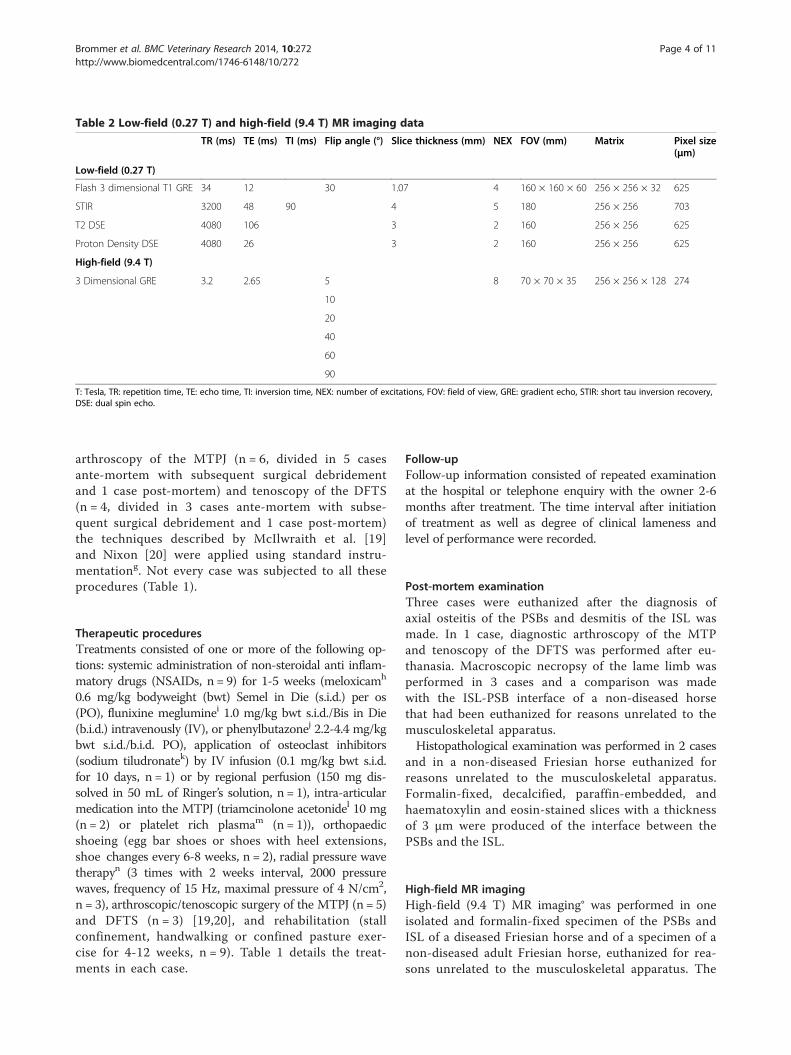
Diagnostic proceduresDiagnostic procedures consisted of a clinical orthopaedicexamination (n = 12) including diagnostic analgesia(n = 11) [13], clinical laboratory evaluation and bac-teriological analysis of synovial fluid (n = 2) and variousimaging modalities. Clinical examination was performedby diplomates of the European College of VeterinarySurgeons (ECVS, n = 4) or by a resident (n = 1) whowas supervised by ECVS diplomates. The radiographicinvestigation of the MTPJ (n = 12) consisted of laterome-dial, dorsal 5-10° proximal-plantarodistal oblique, dorsal45° lateral-plantaromedial oblique, dorsal 45° medial-plantarolateral oblique, and high contrast dorsal 5-10°proximal-plantarodistal oblique views [14] using standardequipmenta. Loss of radiopacity, which is consistent withloss of bone, was described in a subjective way in terms of‘minor’, ‘moderate’ or ‘severe’. Decrease of radiopacity atthe proximo-axial aspect was compared to the disto-axialaspect of the PSBs and was assessed in terms of ‘more’ or‘equal’. USb of the plantar aspect of the MTPJ (n = 11) wasperformed in the transverse and longitudinal planes with a12 MHz linear transducer [15]. In the cases examined byCTc (n = 2), contiguous slices of 3 mm thickness were ac-quired at settings of 120 kV and 220 mA with a rotationspeed of 1 s [16]. For intra-arterial contrast enhanced CTof the MTPJ (n = 2) similar settings were used. Contrastmediumd (350 mg I/mL) was infused into the dorsal meta-tarsal artery using a remotely controllable infusion pumpe
at a continuous rate of 2 mL/s [17]. Low-field (0.27 T) MRimagingf (n = 2) was performed using techniques describedby Werpy [18]. Table 2 details the MR imaging data. For
Table 1 Cases (n = 12) diagnosed with axial osteitis of the PSBs and desmitis of the ISL
Case Age(years)
Gender Affected limb, durationand severity of lameness
T: Tesla, TR: repetition time, TE: echo time, TI: inversion time, NEX: number of excitations, FOV: field of view, GRE: gradient echo, STIR: short tau inversion recovery,DSE: dual spin echo.
Brommer et al. BMC Veterinary Research 2014, 10:272 Page 4 of 11http://www.biomedcentral.com/1746-6148/10/272
arthroscopy of the MTPJ (n = 6, divided in 5 casesante-mortem with subsequent surgical debridementand 1 case post-mortem) and tenoscopy of the DFTS(n = 4, divided in 3 cases ante-mortem with subse-quent surgical debridement and 1 case post-mortem)the techniques described by McIlwraith et al. [19]and Nixon [20] were applied using standard instru-mentationg. Not every case was subjected to all theseprocedures (Table 1).
Therapeutic proceduresTreatments consisted of one or more of the following op-tions: systemic administration of non-steroidal anti inflam-matory drugs (NSAIDs, n = 9) for 1-5 weeks (meloxicamh
0.6 mg/kg bodyweight (bwt) Semel in Die (s.i.d.) per os(PO), flunixine megluminei 1.0 mg/kg bwt s.i.d./Bis in Die(b.i.d.) intravenously (IV), or phenylbutazonej 2.2-4.4 mg/kgbwt s.i.d./b.i.d. PO), application of osteoclast inhibitors(sodium tiludronatek) by IV infusion (0.1 mg/kg bwt s.i.d.for 10 days, n = 1) or by regional perfusion (150 mg dis-solved in 50 mL of Ringer’s solution, n = 1), intra-articularmedication into the MTPJ (triamcinolone acetonidel 10 mg(n = 2) or platelet rich plasmam (n = 1)), orthopaedicshoeing (egg bar shoes or shoes with heel extensions,shoe changes every 6-8 weeks, n = 2), radial pressure wavetherapyn (3 times with 2 weeks interval, 2000 pressurewaves, frequency of 15 Hz, maximal pressure of 4 N/cm2,n = 3), arthroscopic/tenoscopic surgery of the MTPJ (n = 5)and DFTS (n = 3) [19,20], and rehabilitation (stallconfinement, handwalking or confined pasture exer-cise for 4-12 weeks, n = 9). Table 1 details the treat-ments in each case.
Follow-upFollow-up information consisted of repeated examinationat the hospital or telephone enquiry with the owner 2-6months after treatment. The time interval after initiationof treatment as well as degree of clinical lameness andlevel of performance were recorded.
Post-mortem examinationThree cases were euthanized after the diagnosis ofaxial osteitis of the PSBs and desmitis of the ISL wasmade. In 1 case, diagnostic arthroscopy of the MTPand tenoscopy of the DFTS was performed after eu-thanasia. Macroscopic necropsy of the lame limb wasperformed in 3 cases and a comparison was madewith the ISL-PSB interface of a non-diseased horsethat had been euthanized for reasons unrelated to themusculoskeletal apparatus.Histopathological examination was performed in 2 cases
and in a non-diseased Friesian horse euthanized forreasons unrelated to the musculoskeletal apparatus.Formalin-fixed, decalcified, paraffin-embedded, andhaematoxylin and eosin-stained slices with a thicknessof 3 μm were produced of the interface between thePSBs and the ISL.
High-field MR imagingHigh-field (9.4 T) MR imaging° was performed in oneisolated and formalin-fixed specimen of the PSBs andISL of a diseased Friesian horse and of a specimen of anon-diseased adult Friesian horse, euthanized for rea-sons unrelated to the musculoskeletal apparatus. The
Brommer et al. BMC Veterinary Research 2014, 10:272 Page 5 of 11http://www.biomedcentral.com/1746-6148/10/272
specimens were placed in a quadrature volume coil;Table 2 further details the MR imaging data. Data werestored in neuroimaging informatics technology initiative(NIfTI) format and were read with image analysis softwarep.
ResultsCase detailsTwelve cases of axial osteitis of the PSBs with desmitis ofthe ISL were included in the study (Table 1). The meanage was 6.8 ± 2.7 years (range 3-13 years). All horses hada history of intermittent severe hindlimb lameness of anacute onset with a mean duration at the time of ortho-paedic examination of 1.9 ± 1.0 months (range 1 week - 4months). No treatments prior to referral other thanadministration of NSAIDs and a period of box-rest hadbeen applied by the owners or referring veterinarianswith no or minor improvement of the lameness.
Clinical findingsAt the time of examination, the horses displayed a lame-ness of 3-4/5. The right hindlimb and left hindlimb wereaffected in an equal number of 6 cases. All horsesresented normal loading of the fetlock and there wasreduced extension of the fetlock at walk, especially onthe circle when the affected limb was inside. All horseshad effusion of the MTPJ (the plantar pouch being moreobviously distended than the dorsal pouch) and a marginalto moderate effusion of the DFTS. Pain could not beevoked by palpation or by passive movement of the distallimb. Distal limb flexion increased the lameness. Plantarnerve blocks (n = 11) performed at the base of the PSBsdid not result in any substantial improvement of the lame-ness. However, there was 60% to 80% improvement afterlow 6 point nerve blocks (perineural anaesthesia of themedial and lateral plantar nerves, medial and lateral plan-tar metatarsal nerves and medial and lateral dorsal meta-tarsal nerves at a level just proximal to the MTPJ, n = 11).After intra-articular anaesthesia of the MTPJ (using 8-10mL of local anaesthetic solution, n = 7), a mean improve-ment of 40-60% was noted after 10 minutes. Intrathecalanaesthesia of the DFTS (using 6-8 mL of local anaestheticsolution, n = 4) also resulted in a mean improvement of40-60% 10 minutes after injection. The synovial fluidwas yellow and clear at inspection, viscosity appeared tobe slightly reduced, mean polymorphonucleated cellcount was 4.0 ± 1.0 × 10⁹ cells/L and mean total proteinconcentration was 1.2 ± 0.2 g/dL (n = 2). Culture of thesynovial fluid for bacterial or fungal organisms wasnegative in both of the tested samples (n = 2).
Findings on diagnostic imagingCharacteristic radiographic changes could be discerned onthe (high contrast) dorsal 5-10° proximal-plantarodistaloblique views of the MTPJ. Loss of radiopacity of variable
extent from minor to severe was present at the proximo-axial aspects of the PSBs, symmetrically medially versuslaterally. Loss of radiopacity was more extensive at theproximo-axial and less extensive at the disto-axial aspectsof the PSBs. The margins of the areas where loss ofradiopacity was noted, were not well defined.US showed diffuse and irregular hypoechogenicity of
the ISL on transverse US images. The interface betweenthe ISL and the axial compact bone of the PSBs wasirregular in all cases in which US was performed (n = 11).In 3 cases small hyperechogenic particles were detected atthe interface between compact bone and ligament at theproximo-axial aspect of the PSBs, which was most likelyconsistent with avulsion fragments.On pre-contrast CT images (n = 2) ill-defined osteo-
lytic areas at the proximal aspect of the axial border ofthe PSBs were detected. The area of bone lysis was moreor less symmetrical medially and laterally with respectto size and location. Intra-arterial contrast CT imaging(n = 2) revealed a focal deposit of contrast medium atthe level of the proximal part of the ISL, which wasconsistent with either abnormal blood vessel permeability/disruption or neovascularization related with tissue repair.On low-field MR imaging (n = 2), the proximal part of
the ISL had an iso-intense signal (compared to surround-ing tissues) on T1-weighted gradient echo (GRE) imagesand an irregular hyperintense signal on proton density,T2-weighted and short tau inversion recovery (STIR) im-ages. These findings may be consistent with inflammationand fibrous tissue (scar) formation in the ligament andadjacent region. On all sequences, a slightly ill-definedincrease in signal intensity was also present at theproximo-axial region of the PSBs, the region that showsloss of radiopacity on radiographs and that normally con-sists of compact bone. At these sites, the compact bonedid not have a regular margin and there was no definiteinterface with adjacent spongious bone. Adjacent spon-gious bone of the PSBs had a diffuse hypointense signal in-tensity on T1-weighted GRE images and a slightly diffusehyperintense signal on proton density, T2-weighted andSTIR images; taken all together these findings are suggest-ive for loss of compact bone at the proximo-axial regionand edema whether or not accompanied by necrosis in theadjacent spongious bone of the PSBs (Figure 1).During arthroscopy of the MTPJ and tenoscopy of the
DFTS, mild hypertrophy of the synovial membrane wasvisible. In the MTPJ, discolouration and fraying of theISL was found in 3 cases. Complete tearing of the ISLwith penetration into the DFTS was present in 2 cases.In the other case, the plantar surface of the ISL was notaffected. At the level of insertion of the ISL onto the PSBs,the articular cartilage was soft at manual probing. Debride-ment of malacic bone using hooked curettes and motor-ized equipmentg was performed until healthy subchondral
Figure 1 Low-field (0.27 T) MR imaging of the MTPJ. A) T1 weighted GRE transverse image at the level of the PSBs and ISL. B) STIR transverseimage at the same level. C) T1 weighted GRE dorsal image. The proximal part of the ISL shows an iso-intense signal compared to surroundingtissues on T1 weighted GRE images (A, C, white line marked area) and an irregular hyperintense signal on STIR images (B, white line marked area).A slightly ill-defined increase in signal intensity was also present at the proximo-axial aspect of the compact bone of the PSBs and extendingslightly into the spongiosa on all sequences. The margins of the compact bone are irregular. Note the diffuse hypointense signal of the PSBson the T1-weighted images (A, C). These findings may be consistent with inflammation and fibrous tissue (scar) formation in the ligament and adjacentregion, loss of compact bone at the proximo-axial aspect and edema in the adjacent spongious bone of the PSBs.
Brommer et al. BMC Veterinary Research 2014, 10:272 Page 6 of 11http://www.biomedcentral.com/1746-6148/10/272
bone was encountered. Torn fibres of the ISL were alsodebrided. Debridement was performed by either a MTPJapproach in cases in which the dorsal surface of the ISLwas affected but not the plantar surface, or by a com-bined MTPJ and DFTS approach in the cases in whichcomplete tearing of the ISL into the DFTS was present.We had no cases with incomplete tearing of the plantarsurface of the ISL in combination with an intact dorsalsurface. There were no changes in the superficial digitalflexor tendon, deep digital flexor tendon, proximal anddistal manica flexoria, or the proximal annular ligamentin any of the cases.
Follow-upNine horses were subjected to treatment (Table 1).Two horses (22%) became sound for light riding pur-poses 3-4 months after treatment. Two horses (22%)were pasture sound 3-6 months after treatment butwere not used for riding. Five horses (56%) remainedintermittently or persistently lame. One of these de-veloped axial osteitis of the PSBs with desmitis of theISL in the contralateral hindlimb after 2 months andwas subsequently euthanized. One other horse waseuthanized 6 months after treatment due to persistentlameness.
Post-mortem examinationThree horses were euthanized on request of the ownerafter the diagnosis of axial osteitis of the PSBs withdesmitis of the ISL and were subjected to post-mortemexamination. Macroscopic inspection showed a disco-loured, irregular and frayed ISL. Focal loss of bone waspresent at the proximo-axial aspects of the PSBs. The
attachment of the ISL to the bone was irregular withfocal detachment of the ISL (Figure 2).At histopathology, severe multifocal to coalescing
areas of inflammation of the ISL, characterized byabundance of fibroblasts, lymphocytes, plasma cellsand a small amount of necrotic debris were visible inthe specimens of the diseased horse. The transitionfrom ligament to the compact bone was very irregular.In addition, compared to the non-diseased horse, in thespecimens of the diseased horse there was a decrease ofavailable surface at which ligament tissue could mergeto the compact bone due to loss of bone at the bone-ligament interface. Remaining adjacent bone showedincreased osteoclastic bone resorption when comparingthe diseased horse to the non-diseased horse. Bonemarrow was hypercellular due to an invasion of lym-phocytes and plasma cells. These histological abnor-malities were found along the entire line of attachmentof the ISL to the PSBs and were most severe in themedial PSB (Figure 3).
High-field MR imagingCompared to the non-diseased horse, a clear increasein signal intensity could be seen in the compact boneat the proximo-axial margin of the PSBs on the T1-weighted images of the diseased horse (Figure 4). Theinterface with the spongious bone at the proximo-axialregion is irregular and ill-defined. Moreover, a diffusehypointense signal without difference in signal intensitybetween the central spongious bone and the peripheralcompact bone was present on T1-weighted GRE imagesof the diseased horse. The signal intensity was more orless homogeneously distributed across the spongiosa and
Figure 2 Macroscopic view of the PSBs and the ISL of a diseased and a non-diseased Friesian horse. Note the focal loss of bone at theproximo-axial aspects of the PSBs (open arrows). Concurrent partial rupture of the ISL (white line marked area), starting at the proximal level ofthe bone and coursing to distal with locally a detachment from the bone (black arrows). The rupture did not enter the plantar surface so therewas no penetration into the DFTS in this case.
Brommer et al. BMC Veterinary Research 2014, 10:272 Page 7 of 11http://www.biomedcentral.com/1746-6148/10/272
compacta. In the non-diseased horse, signal intensityof the PSBs was heterogeneous with the central spon-gious bone having more intense signals compared tothe peripheral compact bone. Evaluating the absoluteT1 values (T1 map) of the diseased horse, especiallythe proximal (apical) part and the peripheral compactbone showed local increases of T1 values compared tothe non-diseased horse, leading to a heterogeneouslydistributed T1 pattern across the PSBs of the diseasedhorse. Moreover, the ISL ligament of the diseased horseshowed higher T1 values than the non-diseased horseon the T1 map. The T1 map of the non-diseased horseshowed a more homogeneous pattern across spongiosaand compacta of the PSBs with hardly any signal. Protondensity images of the diseased horse had a clear increasein signal intensity of the compact bone at the proximo-axial margin of the PSBs compared to the images of thenon-diseased horse. The remainder cortical and spongiousbone of the PSBs showed a homogeneous distribution ofsignal intensity in the diseased horse. In the non-diseasedhorse there was a more heterogeneous signal intensity pat-tern across the PSBs, the signal of the central spongiosawas slightly more intense compared to the peripheral
Figure 3 Histopathology of the interface of the PSBs and the ISL. Tranhaematoxylin and eosin staining. Note the multifocal to coalescing inflammatand plasma cells in the diseased horse. The transition from ligament tothe non-diseased horse, there is a decrease of surface area where ligamebone shows increased osteoclastic bone resorption (osteoclasts markedlymphocytes and plasma cells.
compacta. Taking the findings of all images together, theresults are consistent with loss of compact bone at theproximo-axial margin of the PSBs, osteoporosis of the per-ipheral compact bone and spongious bone of the PSBsand inflammation and fibrous (scar) tissue formation ofthe ISL in the diseased horse.
DiscussionThe clinical profile and pathologic findings of thesecases of axial osteitis of the PSBs with desmitis of theISL in Friesian horses showed a consistent pattern. Thedisease was typically diagnosed in young to middle-agedFriesian horses and characterized by acute hindlimblameness localised in the MTPJ. Findings on Rx, US and(contrast enhanced) CT matched closely with those pre-viously reported [1,2,6-8]. The overall prognosis in thiscase series was poor with return to light riding purposesas the best achieved result. The majority of the horsesremained lame and, depending on severity of the lame-ness, became pasture sound or had to be euthanized.It should be realized that retrospective evaluation of data
carries limitations, especially when it is used to answerquestions about outcome in terms of relative performance
sverse sections of a diseased and a non-diseased Friesian horse,ion of the ISL which is characterized by abundant fibroblasts, lymphocytesbone (arrow) is very irregular in the diseased horse and compared tont tissue merges to bone in the diseased horse. Remaining adjacentwith asterisks). Bone marrow was hypercellular due to invasion of
Figure 4 (See legend on next page.)
Brommer et al. BMC Veterinary Research 2014, 10:272 Page 8 of 11http://www.biomedcentral.com/1746-6148/10/272
(See figure on previous page.)Figure 4 High-field MR imaging (9.4 T, flip angle 40°) of the PSBs and the ISL. Post-mortem analysis of a specimen of a Friesian horse withaxial osteitis of the PSBs and desmitis of the ISL (left images) and a non-diseased Friesian horse (right images). Top: T1 weighted GRE dorsal sequences.Middle: T1 mapping. Bottom: proton density sequences. On the T1 weighted images, a clear increase in signal intensity could be seen in the compactbone at the proximo-axial area of the PSBs in the diseased horse (white line marked area). Compared to the non-diseased horse, signal intensity of thespongiosa (asterisks) was reduced, signal intensity of the compacta was increased in the T1 weighted GRE (open arrows) and proton density(black arrows) images leading to homogeneous signal intensity across the PSBs in the diseased animal. In the non-diseased horse, heterogeneoussignal intensity was present in T1 weighted and proton density images with the spongiosa having more intense signaling (asterisks) and peripheralcompacta having less intense signaling (open arrows). Compared to the non-diseased horse, on the T1 map of the diseased horse, especially the apicalpart and the peripheral compact bone showed an increase in T1 values (black and white arrows), the ISL ligament also showed an increase in T1 values(white line marked area). The non-diseased horse showed a homogeneous pattern of the PSBs with hardly any signal on the T1 map. Integration ofthe findings on all images could be interpreted as loss of compact bone at the proximo-axial margin of the PSBs, osteoporosis of the peripheralcompact bone and spongious bone of the PSBs, and inflammation and fibrous (scar) tissue formation of the ISL in the diseased horse.
Brommer et al. BMC Veterinary Research 2014, 10:272 Page 9 of 11http://www.biomedcentral.com/1746-6148/10/272
after treatment. Further, the highly heterogenic characterof the treatment regimens to which the horses weresubjected precludes drawing of any evidence-based con-clusions regarding treatment efficacy, which is a weaknessof this study. Treatments of the horses in the study ofDabareiner et al. [2] were more uniform and they reportedbetter results in a case series involving 4 Quarter Horses,2 Polo Ponies, 1 Thoroughbred and 1 Appaloosa. In thatstudy five MTPJs and three MCPJs were treated with acombination of surgical intervention (arthroscopic/teno-scopic debridement) and medical treatment and an overallresult of 63% return to previous level of performance wasreported [2]. Although the horses in that study differ fromFriesian horses in several aspects such as conformationand equestrian use, the results may suggest that aggressivetreatment in the form of arthroscopic/tenoscopic debride-ment of bone and inflamed ligament tissue may optimizethe conditions for healing. In our opinion, if the plantarsurface of the ISL (i.c. at the site of the DFTS) lookedunaffected, care should be taken not to transect the ISL asthis may lead to instability of the MTPJ.Another factor that may influence outcome is the dur-
ation of lameness before treatment. In our case series, themean lameness duration before referral was 1.9 months. Inthe case series of Wisner et al. [8], who also reported a pooroutcome, lameness duration was still longer with a mean of5.6 months. In the study of Dabareiner et al. [2], whichreported better results, mean lameness duration was only3.1 weeks. This may indicate that, as with many disorders,the sooner treatment is started, the better the prognosis.Some of the horses described by Dabareiner et al. [2]
had an infective component which was a negativeprognostic indicator in that study and in other studiesreported a few years later [1,6]. Infection should, however,be regarded as a separate condition. In our cases, therewere no histories or signs of infection.The rationale for the use of corticosteroids in some of
our cases was its anti-inflammatory effect and reductionof joint/sheath effusion which may lead to reductionof the lameness. However, corticosteroids also haveanti-anabolic action which is an undesired side-effect
in damaged tissue. It is therefore necessary to weighup carefully how corticosteroids should be used in thisdisease.In one case axial osteitis of the PSBs with desmitis
of the ISL had been developed in the contralateralhindlimb after 2 months of rehabilitation. We usuallyadvise owners to radiograph the limbs bilaterally atinitial examination. In this case, the owner refused toradiograph the contra-lateral MTPJ for financial rea-sons. In every case and at every time, the cost-benefitratio should be considered. From a prognostic pointof view this case showed the importance for bilateralimaging and this is a valid argumentation of acquisitionof at least a dorsoplantar radiograph of the contralaterallimb.The cost-benefit ratio should also be discussed in
every clinical case when considering advanced diagnosticimaging such as (contrast enhanced) CT of MR imagingversus surgical arthroscopy/tenoscopy. Advanced diag-nostic imaging techniques as (contrast enhanced) CTand MR imaging as used in this paper have provided infurther documentation of this disease, but we believethat for routine clinical practice these techniques havevery little additional value over Rx and US for differenti-ation between different treatment options or for definingthe prognosis. As we believe that surgical debridement islikely to give the best outcome, we would advise theowner to invest his/her money for surgery rather thanfor advanced diagnostic imaging in cases in which theowner opts for treatment.It has been speculated that excessive or abnormal
forces within the MTPJ, provoked by severe overexten-sion of the joint in combination with hindlimb rotationin particular, will heavily load the plantar supportingstructures, especially the PSBs and the ISL [2]. Friesianhorses might be predisposed for axial osteitis of the PSBswith desmitis of the ISL. Tendon mechanical propertiesin this breed have been shown to be on average morecompliant than in ponies, resulting in hyperextensionof the MTPJ [21]. Fetlock angles were not measuredin this study because any increase in hyperextension due
Brommer et al. BMC Veterinary Research 2014, 10:272 Page 10 of 11http://www.biomedcentral.com/1746-6148/10/272
to different material properties would potentially bemasked by decreased limb loading, related to the gradeof lameness.In discussions on the aetiopathogenesis of this dis-
order, it is debatable whether ligament pathology comesfirst with bone pathology as a secondary event, or theother way round. It is also a matter of debate whethertrauma is the primary initiating factor that evokes acascade of secondary events consisting of inflammation,weakening/rupture of the ISL, disturbance of the bloodsupply of the bone-ligament interface and bone resorption.Another possibility is that disturbance of the blood supplydue to vessel rupture or thrombosis formation is primaryand bone lysis with inflammation of the ISL secondary. Theinformation in the current paper does not permit to drawconclusions on these hypothesised pathophysiological se-quences. In this sense this study was limited as macro-scopic necropsy and histopathology were restricted tothe lame limb. Concurrent post-mortem examination ofthe contralateral non-lame limb might possibly haveresulted in additional information that may have assistedin unravelling the aetiopathogenesis of this disease.The nature of the bone and the type of injury may play
a role in the type of response that develops after tearingand avulsion of tendoligamentous insertions [22]. Newbone formation as well as osseous cyst-like lesions havebeen reported in cases of desmopathy of collateral liga-ments of the distal interphalangeal joint [23]. In axialosteitis of the PSBs with desmitis of the ISL bone lysiswas the predominant feature of bone pathology. Bonelysis was seen at the axial aspect of the PSBs and morelysis was seen at the proximo-axial margin compared tothe disto-axial aspect of the PSBs. The same distributionpattern of bone lysis was reported by Vanderperren et al.[7]. Dabareiner et al. [2], Sedrish et al. [5] and Shermanet al. [6] found relatively more bone lysis in the midaxialregion of the PSBs, whereas Barr et al. [1] observedmore bone lysis in the disto-axial region of the PSBs.Given the vascularization pattern of the PSBs and ISL
[10,11], an explanation for the typical bone resorption atthe proximo-axial location could be the different densityof afferent arterioles per unit of bone volume. Trumbleet al. [11] observed a relative ‘absence of vessels’ in theapical region of the PSB in contrast to the rich vascularityin the remainder of the bone. Trauma to blood vessels orthrombosis formation will lead to changes in perfusionand disturbances in oxygen and nutrient supply at theapical region of the PSB. Intra-arterial contrast CT showedan increased density of vasculature in the region of theISL, comparable to the findings described by Vanderper-ren et al. [7]. This indicates that the vascular pattern hadchanged in diseased horses, supporting the hypothesis thatchanges in the blood supply may play a role, either pri-mary or secondary, in the development of this disease.
In addition to loss of bone at the proximo-axial marginof the PSBs, high-field MR imaging has learnt us that theremainder part of the PSBs also responds in this disease.High field MR imaging showed changes that could beinterpreted as bone remodelling (development of osteo-porosis) which was evident in the compact bone at theperipheral margins and the spongious bone of the PSBs. Itremains elusive whether these phenomena are hallmarksof the primary disease process or represent disuseosteopenia which is a physiological adaptive response asa consequence of Wolff ’s law [24] caused by the factthat the horse had been lame during a period of time.
ConclusionsAxial osteitis of the PSBs and desmitis of the ISL shouldbe considered in the differential diagnosis of severehindlimb fetlock lameness in Friesian horses. Affectedhorses typically showed clinical signs of acute onset. Theresults of currently used treatments were disappointing.High-field MR imaging and histopathology providedevidence of interrelated changes of bone lysis at the axialaspect of the PSBs, inflammation with subsequent fibroustissue (scar) formation of the adjacent ISL and additionalremodelling (development of osteoporosis) of the compactbone at the periphery and the spongious bone of the PSBs.Further studies are needed to unravel the exact aetio-pathogenesis of this disorder.
EndnotesaPhilips Healthcare, Eindhoven, The Netherlands or Agfa
Health Care Nederland B.V, Rijswijk, The Netherlands.bPhilips Healthcare, Eindhoven, The Netherlands or
Aloka®, Biomedic Nederland B.V., Almere, The Netherlands.cPhilips Healthcare, Eindhoven, The Netherlands.dXenetix®, Guerbet B.V., Gorinchem, The NetherlandseMark V plus®, Medrad Co., Warrendale, USA.fSiemens Healthcare Solutions Diagnostics, Breda, The
Netherlands.mRecover®, Biomet Biologics Inc., Warsaw Indiana.nDolorClast®, Enraf-Nonius N.V., Aartselaar, Belgium.oHorizontal bore MRI®, Varian Inc., Palo Alto, USA.pImageJ®, National Institute of Heath (NIH), Maryland,
USA.
Brommer et al. BMC Veterinary Research 2014, 10:272 Page 11 of 11http://www.biomedcentral.com/1746-6148/10/272
Abbreviationsb.i.d: Bis in Die (Latin, twice a day); bwt: bodyweight; CT: Computedtomography; DFTS: Digital flexor tendon sheath; DSE: Dual spin echo;ECVS: European College of Veterinary Surgeons; FOV: Field of view;GRE: Gradient echo; ISL: Intersesamoidean ligament; IV: Intravenously;MR: Magnetic resonance; MCPJ: Metacarpophalangeal joint;MTPJ: Metatarsophalangeal joint; NEX: Number of excitations;NIfTI: Neuroimaging informatics technology initiative; NSAIDs: Non steroidalanti inflammatory drugs; PO: Per os; PSBs: Proximal sesamoid bones;Rx: Radiography; s.i.d: Semel in Die (Latin, once a day); T: Tesla; TE: Echo time;TI: Inversion time; TR: Repetition time; US: Ultrasonography; STIR: Short tauinversion recovery.
Competing interestsThe authors declare that they have no competing interests. None of theauthors has a financial or personal relationship with other people ororganizations that could inappropriately have influenced or biased thecontent of the paper.
Authors’ contributionsMV, WB and HB initiated the study, HB and MV processed and analyzed theclinical and laboratory data. SV, AJMVDB and AVDT were responsible for thediagnostic imaging, MP an AG were responsible for the pathologicalexaminations. HB drafted the manuscript, all authors have participated in theinterpretation of the data and in revising the manuscript. All authorsapproved the final manuscript.
AcknowledgementsProfessor P.R. van Weeren, Professor A. Barneveld and Professor G. Voorhoutare acknowledged for their suggestions and critical reading of the manuscript.
Author details1Department of Equine Sciences, Faculty of Veterinary Medicine, UtrechtUniversity, P.O. Box 80.163, 3584 CM Utrecht, The Netherlands. 2EquineVeterinary Hospital Bodegraven, Bodegraven, The Netherlands. 3Equi-Tech,Puenta Piedra, Bogota, Columbia. 4Division of Diagnostic Imaging, Faculty ofVeterinary Medicine, Utrecht University, Utrecht, The Netherlands.5Biomedical MR Imaging and Spectroscopy Group, Image Sciences Institute,University Medical Center, Utrecht, The Netherlands. 6Department ofPathobiology, Faculty of Veterinary Medicine, Utrecht University, Utrecht, TheNetherlands. 7Department of Surgery and Anaesthesiology, Faculty ofVeterinary Medicine, Ghent University, Merelbeke, Belgium.
Received: 18 November 2013 Accepted: 8 November 2014
References1. Barr ED, Clegg PD, Senior JM, Singer ER: Destructive lesions of the
proximal sesamoid bones as a complication of the dorsal metatarsalartery catheterization in three horses. Equine Vet J 2005, 34:159–166.
2. Dabareiner RM, Watkins JP, Carter GK, Honnas CM, Eastman T: Osteitis ofthe axial border of the proximal sesamoid bones in horses: eight cases(1993-1999). J Am Vet Med Ass 2001, 219:82–86.
3. Hauri S, Finsler S, Walliser U: Clinical findings and treatment of asepticnecrosis of the proximal sesamoid bones in an Icelandic pony.Pferdeheilk 2009, 25:54–60.
4. Lawrence CP, Fraser BSL: Septic osteitis of the axial border of the proximalsesamoid bones in two foals. Equine Vet Educ 2012, 25:63–66.
5. Sedrish S, Burba D, Williams J: Radiographic diagnosis of axial sesamoidosteomyelitis in a horse. Vet Radiol Ultrasound 1996, 37:417–418.
6. Sherman KM, Myhre GD, Heymann EI: Fungal osteomyelitis of the axialborder of the proximal sesamoid bones in a horse. J Am Vet Med Ass2006, 229:1607–1611.
7. Vanderperren K, Bergman HJ, Spoormakers TJP, Pille F, Duchateau L,Puchalski SM, Saunders JH: Clinical, radiographic, ultrasonographic andcomputed tomographic features of nonseptic osteitis of the axial borderof the proximal sesamoid bones. Equine Vet J 2014, 46:463–467.
8. Wisner ER, O’Brien TR, Pool RR: Osteomyelitis of the axial border of theproximal sesamoid bones in seven horses. Equine Vet J 1991, 23:383–389.
9. Berge E: Űber die locale Malazie der Sesambeine am Fesselgelenk desPferdes. Berl Tierärztl Wschr 1933, 49:629–645.
10. Németh F: Vascularization of normal and pathological proximal sesamoidbones in the horse. Tijdschr Diergeneesk 1972, 17:1117–1126.
11. Trumble TN, Arnoczky SP, Stick JA, Stickle RL: Clinical relevance of themicrovasculature of the equine proximal sesamoid bone. Am J Vet Res1995, 6:720–724.
12. Anonymous: Guide for veterinary service and judging of EquestrianEvents. In American Association of Equine Practitioners. 4th edition.Lexington, Kentucky: 1996:19.
13. Bassage LH, Ross MW: Diagnostic analgesia. In Diagnosis and Managementof Lameness in the Horse. 2nd edition. Edited by Ross MW, Dyson SJ. St.Louis: Saunders; 2011:100–135.
14. Butler JA, Colles CM, Dyson SJ, Kold SE, Poulos PW: Foot, pastern and fetlock.In Clinical Radiology of the Horse. 3rd edition. Edited by Butler JA, Colles CM,Dyson SJ, Kold SE, Poulos PW. Oxford: Wiley-Blackwell; 2008:53–187.
15. Reef VB: Ultrasound of the locomotor apparatus. In Equine DiagnosticUltrasound. Edited by Reef VB. Philadelphia: Saunders; 1998:39–186.
16. Puchalski SM: Computed tomography. In Diagnosis and Management ofLameness in the Horse. 2nd edition. Edited by Ross MW, Dyson SJ. St. Louis:Saunders; 2011:234–239.
17. Puchalski SM, Galuppo LD, Hornof WJ, Wisner ER: Intraarterialcontrast- enhanced computed tomography of the equine distalextremity. Vet Radiol Ultrasound 2007, 48:21–29.
18. Werpy N: Low-field MRI in horses: practicalities and image acquisition.In Equine MRI. Edited by Murray RC. Oxford: Blackwell publishing;2011:75–99.
19. McIlwraith CW, Nixon AJ, Wright IM, Boening KJ: Diagnostic and surgicalarthroscopy of the metacarpophalangeal and metatarsophalangealjoints. In Diagnostic and Surgical Arthroscopy in the Horse. 3rd edition. Editedby McIlwraith CW, Nixon AJ, Wright IM, Boening KJ. London: Elsevier;2005:129–196.
20. Nixon AJ: Endoscopy of the digital flexor tendon sheath in horses.Vet Surg 1990, 4:266–271.
21. Gussekloo SW, Lankester J, Kersten W, Back W: Effect of differences intendon properties on functionality of the passive stay apparatus inhorses. Am J Vet Res 2011, 4:474–483.
22. Rooney JR, Robertson JL: Foreleg. In Equine Pathology. Edited by Rooney JR,Robertson JL. Iowa: State University Press; 1996:172–173.
23. Dakin SG, Dyson SJ, Murray RC, Tranquille C: Osseous abnormalitiesassociated with collateral desmopathy of the distal interphalangeal joint:Part 1. Equine Vet J 2009, 41:786–793.
24. Wolff J: Das Gesetz der Transformation der Knochen. Reprint 2010. Berlin:ProBusiness Verlag; 1892.
doi:10.1186/s12917-014-0272-xCite this article as: Brommer et al.: Axial osteitis of the proximal sesamoidbones and desmitis of the intersesamoidean ligament in the hindlimb ofFriesian horses: review of 12 cases (2002-2012) and post-mortem analysisof the bone-ligament interface. BMC Veterinary Research 2014 10:272.
Submit your next manuscript to BioMed Centraland take full advantage of:
• Convenient online submission
• Thorough peer review
• No space constraints or color figure charges
• Immediate publication on acceptance
• Inclusion in PubMed, CAS, Scopus and Google Scholar
• Research which is freely available for redistribution
Submit your manuscript at www.biomedcentral.com/submit


![Untitled-1 []...months months months months](https://static.documents.pub/doc/80x56/60cc6a9b3d3a423bd0058c49/-untitled-1-months-months-months-months.jpg)


![Veterans Benefits: Compensation & · PDF file[VETERANS BENEFITS: COMPENSATION & PENSION] ... Osteitis deformans (Paget’s disease). Osteomalacia. Palsy, bulbar. Paralysis agitans.](https://static.documents.pub/doc/80x56/5a7353817f8b9aa2538e908e/veterans-benefits-compensation-pensioncymcdncomsites-veterans-benefits.jpg)


