54
Axilla Dr. Sara Soleimani Asl Department of Anatomy, HUMS
| Date post: | 26-Dec-2015 |
| Category: |
Documents |
| Upload: | eustace-greene |
| View: | 236 times |
| Download: | 1 times |

AxillaDr. Sara Soleimani Asl
Department of Anatomy, HUMS

Figure 7.20 Clavicle.
Downloaded from: StudentConsult (on 13 November 2008 04:30 PM)
© 2005 Elsevier
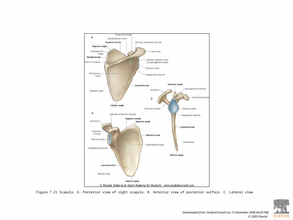
Figure 7.21 Scapula. A. Posterior view of right scapula. B. Anterior view of posterior surface. C. Lateral view.
Downloaded from: StudentConsult (on 13 November 2008 04:30 PM)
© 2005 Elsevier
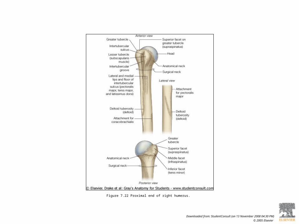
Figure 7.22 Proximal end of right humerus.
Downloaded from: StudentConsult (on 13 November 2008 04:30 PM)
© 2005 Elsevier
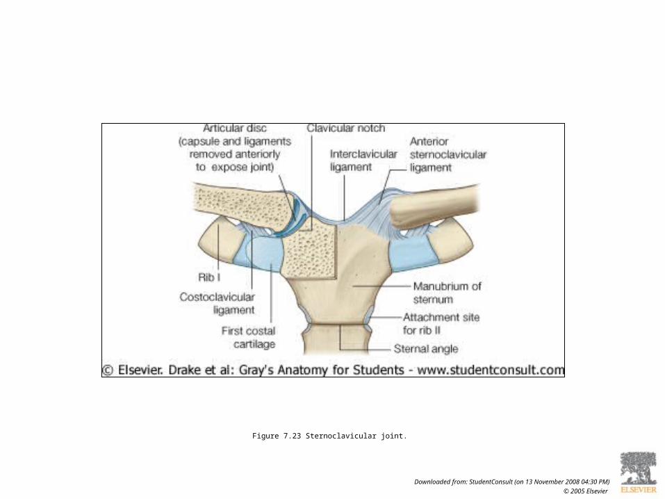
Figure 7.23 Sternoclavicular joint.
Downloaded from: StudentConsult (on 13 November 2008 04:30 PM)
© 2005 Elsevier

Figure 7.24 Right acromioclavicular joint.
Downloaded from: StudentConsult (on 13 November 2008 04:30 PM)
© 2005 Elsevier

Figure 7.25 Glenohumeral joint. A. Articular surfaces of right glenohumeral joint. B. Radiograph of a normal glenohumeral joint.
Downloaded from: StudentConsult (on 13 November 2008 04:30 PM)
© 2005 Elsevier
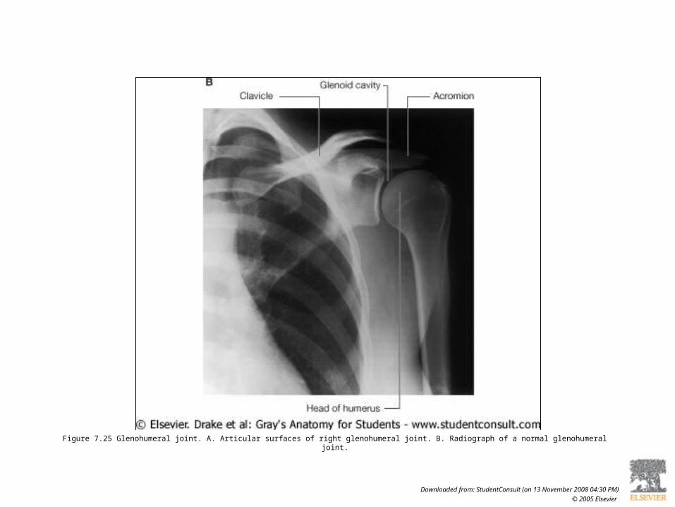
Figure 7.25 Glenohumeral joint. A. Articular surfaces of right glenohumeral joint. B. Radiograph of a normal glenohumeral joint.
Downloaded from: StudentConsult (on 13 November 2008 04:30 PM)
© 2005 Elsevier

Figure 7.26 Synovial membrane and joint capsule of right glenohumeral joint.
Downloaded from: StudentConsult (on 13 November 2008 04:30 PM)
© 2005 Elsevier

Figure 7.27 Capsule of right glenohumeral joint.
Downloaded from: StudentConsult (on 13 November 2008 04:30 PM)
© 2005 Elsevier

Figure 7.28 Lateral view of right glenohumeral joint and surrounding muscles with proximal end of humerus removed.
Downloaded from: StudentConsult (on 13 November 2008 04:30 PM)
© 2005 Elsevier

Figure 7.29 Magnetic resonance image (T1-weighted) of a normal glenohumeral joint in the sagittal plane. Ant, anterior; Post., posterior.
Downloaded from: StudentConsult (on 13 November 2008 04:30 PM)
© 2005 Elsevier

Figure 7.31 Radiograph showing an anterior dislocation of the left glenohumeral joint.
Downloaded from: StudentConsult (on 13 November 2008 04:30 PM)
© 2005 Elsevier
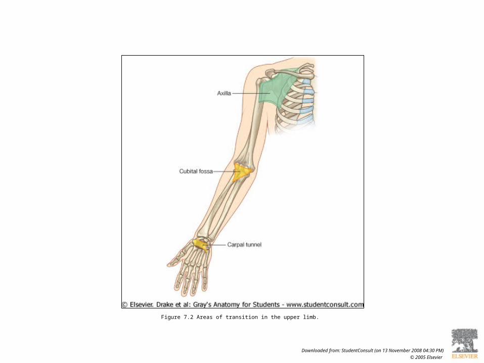
Figure 7.2 Areas of transition in the upper limb.
Downloaded from: StudentConsult (on 13 November 2008 04:30 PM)
© 2005 Elsevier

Figure 7.1 Upper limb. A. Anterior view of the upper limb. B. Superior view of the shoulder.
Downloaded from: StudentConsult (on 13 November 2008 04:30 PM)
© 2005 Elsevier

Figure 7.39 Axilla. A. Walls and transition between neck and arm. Axilla. B. Boundaries. C. Continuity with the arm.
Downloaded from: StudentConsult (on 13 November 2008 04:31 PM)
© 2005 Elsevier

Downloaded from: StudentConsult (on 13 November 2008 04:31 PM)
© 2005 Elsevier
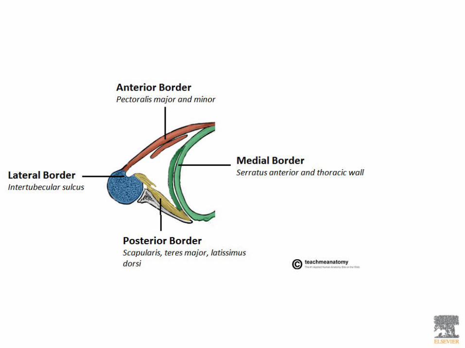

Figure 7.11 Relationship of the upper limb to the neck.
Downloaded from: StudentConsult (on 13 November 2008 04:30 PM)
© 2005 Elsevier
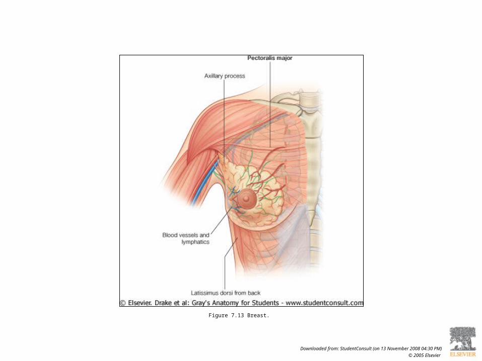
Figure 7.13 Breast.
Downloaded from: StudentConsult (on 13 November 2008 04:30 PM)
© 2005 Elsevier

Figure 7.40 Pectoralis major muscle.
Downloaded from: StudentConsult (on 13 November 2008 04:31 PM)
© 2005 Elsevier
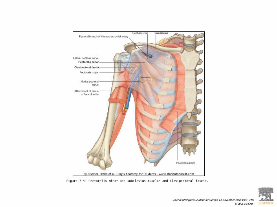
Figure 7.41 Pectoralis minor and subclavius muscles and clavipectoral fascia.
Downloaded from: StudentConsult (on 13 November 2008 04:31 PM)
© 2005 Elsevier

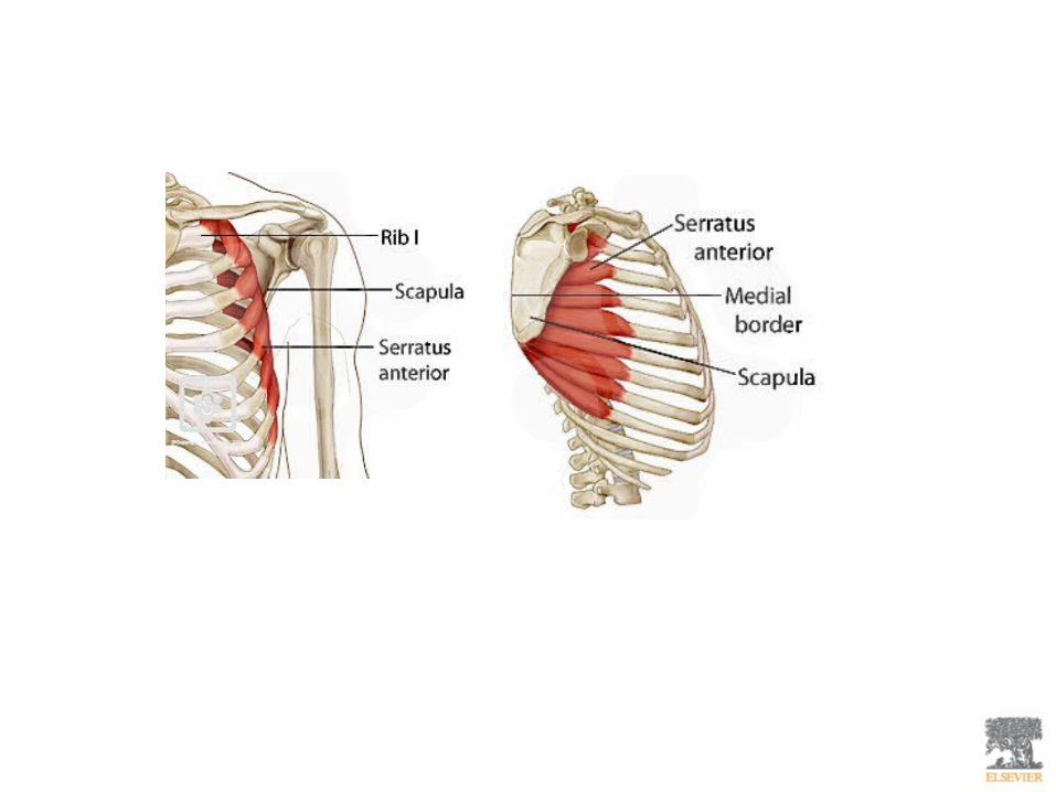

Figure 7.42 Medial wall of the axilla.
Downloaded from: StudentConsult (on 13 November 2008 04:31 PM)
© 2005 Elsevier
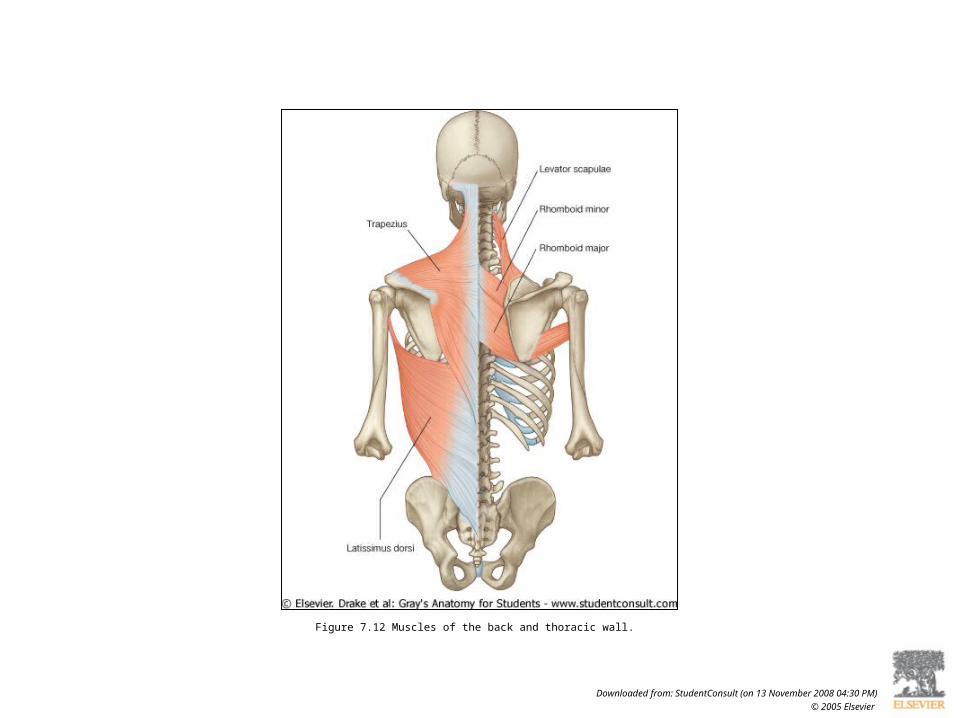
Figure 7.12 Muscles of the back and thoracic wall.
Downloaded from: StudentConsult (on 13 November 2008 04:30 PM)
© 2005 Elsevier

Figure 7.43 Lateral wall of the axilla.
Downloaded from: StudentConsult (on 13 November 2008 04:31 PM)
© 2005 Elsevier
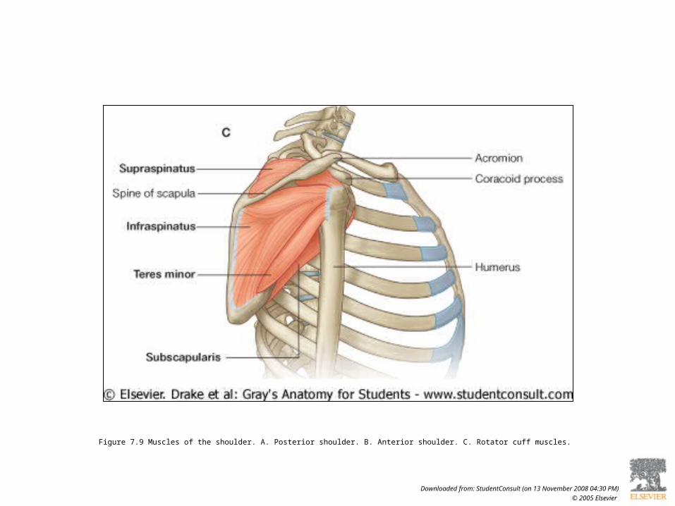
Figure 7.9 Muscles of the shoulder. A. Posterior shoulder. B. Anterior shoulder. C. Rotator cuff muscles.
Downloaded from: StudentConsult (on 13 November 2008 04:30 PM)
© 2005 Elsevier

Figure 7.36 Right posterior scapular region.
Downloaded from: StudentConsult (on 13 November 2008 04:30 PM)
© 2005 Elsevier

Figure 7.44 Posterior wall of the axilla.
Downloaded from: StudentConsult (on 13 November 2008 04:31 PM)
© 2005 Elsevier

Figure 7.37 Arteries and nerves associated with gateways in the posterior scapular region.
Downloaded from: StudentConsult (on 13 November 2008 04:31 PM)
© 2005 Elsevier

Figure 7.34 Lateral view of trapezius and deltoid muscles.
Downloaded from: StudentConsult (on 13 November 2008 04:30 PM)
© 2005 Elsevier

Figure 7.35 Attachment and neurovascular supply of the trapezius and deltoid muscles.
Downloaded from: StudentConsult (on 13 November 2008 04:30 PM)
© 2005 Elsevier

Figure 7.45 Magnetic resonance image of the glenohumeral joint in the transverse or horizontal plane. Ant., anterior; Post., posterior.
Downloaded from: StudentConsult (on 13 November 2008 04:31 PM)
© 2005 Elsevier

Figure 7.51 Brachial plexus. A. Major components in the neck and axilla. Brachial plexus. B. Schematic showing parts of the brachial plexus.
Downloaded from: StudentConsult (on 13 November 2008 04:31 PM)
© 2005 Elsevier

Figure 7.14 Innervation of the upper limb.
Downloaded from: StudentConsult (on 13 November 2008 04:30 PM)
© 2005 Elsevier

Figure 7.51 Brachial plexus. A. Major components in the neck and axilla. Brachial plexus. B. Schematic showing parts of the brachial plexus.
Downloaded from: StudentConsult (on 13 November 2008 04:31 PM)
© 2005 Elsevier

Figure 7.52 Brachial plexus. A. Schematic showing branches of the brachial plexus. B. Relationships to the axillary artery.
Downloaded from: StudentConsult (on 13 November 2008 04:31 PM)
© 2005 Elsevier

Figure 7.15 Dermatomes and myotomes in the upper limb. A. Dermatomes. B. Movements produced by myotomes.
Downloaded from: StudentConsult (on 13 November 2008 04:30 PM)
© 2005 Elsevier

Figure 7.15 Dermatomes and myotomes in the upper limb. A. Dermatomes. B. Movements produced by myotomes.
Downloaded from: StudentConsult (on 13 November 2008 04:30 PM)
© 2005 Elsevier
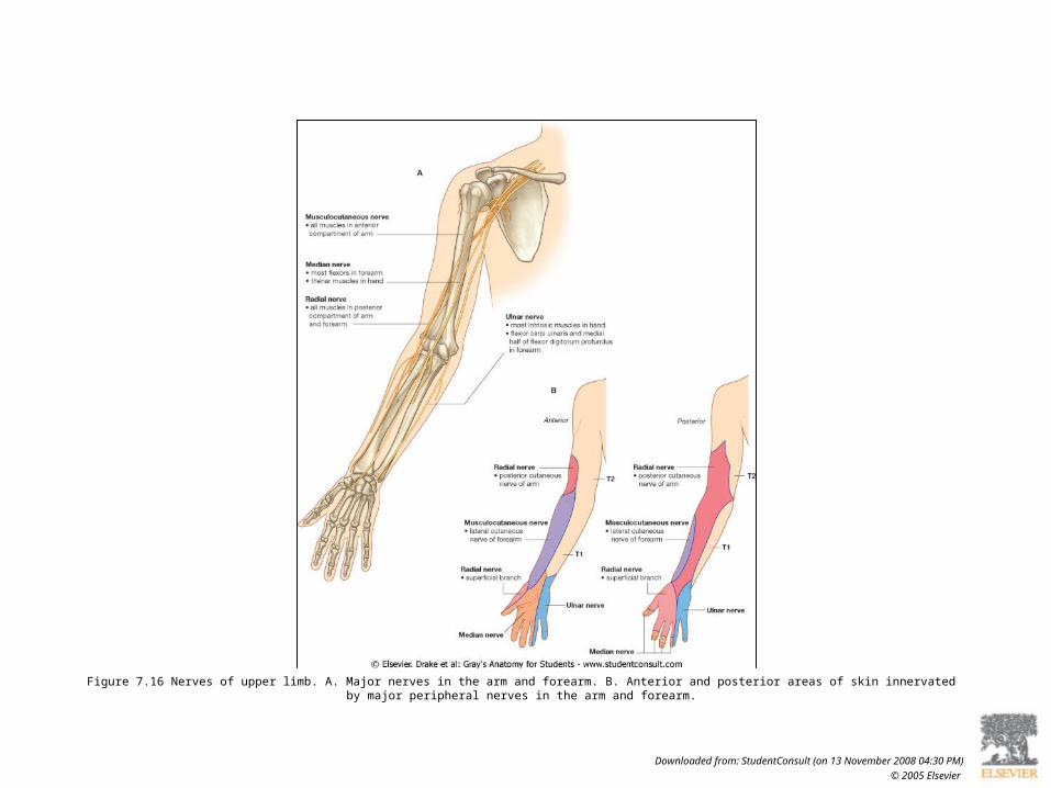
Figure 7.16 Nerves of upper limb. A. Major nerves in the arm and forearm. B. Anterior and posterior areas of skin innervated by major peripheral nerves in the arm and forearm.
Downloaded from: StudentConsult (on 13 November 2008 04:30 PM)
© 2005 Elsevier

Figure 7.17 Nerves related to the humerus.
Downloaded from: StudentConsult (on 13 November 2008 04:30 PM)
© 2005 Elsevier
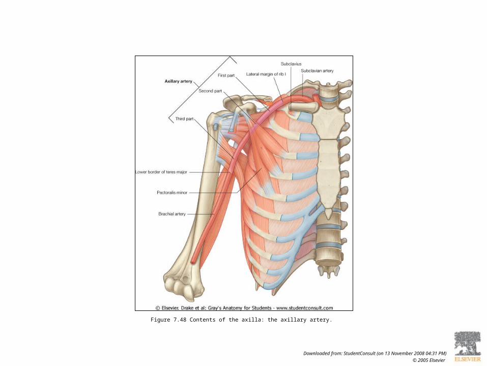
Figure 7.48 Contents of the axilla: the axillary artery.
Downloaded from: StudentConsult (on 13 November 2008 04:31 PM)
© 2005 Elsevier
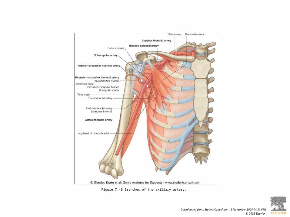
Figure 7.49 Branches of the axillary artery.
Downloaded from: StudentConsult (on 13 November 2008 04:31 PM)
© 2005 Elsevier
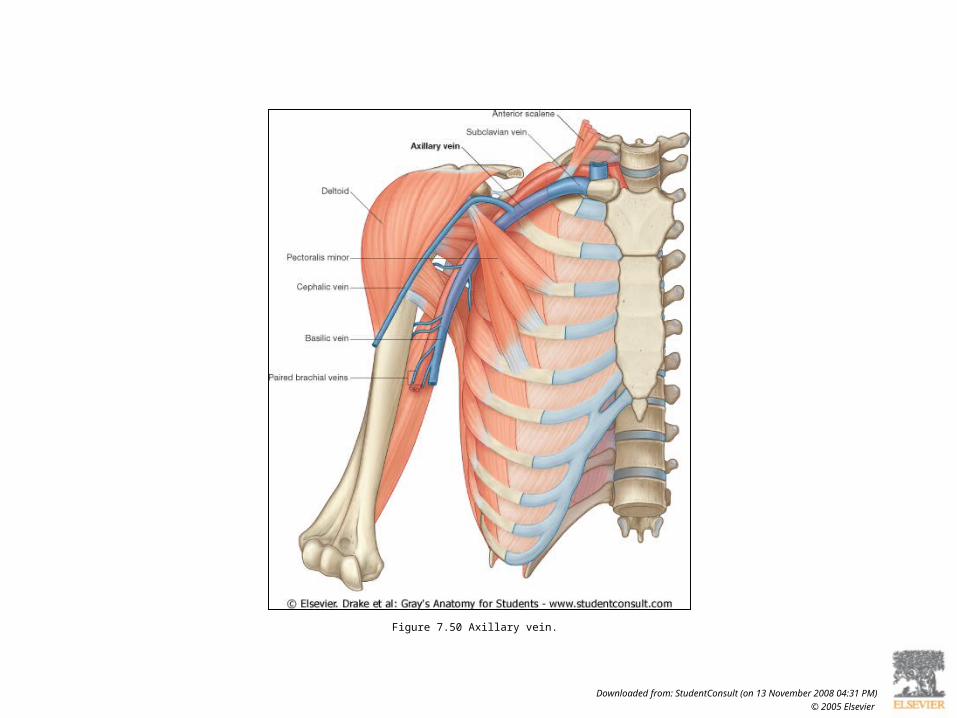
Figure 7.50 Axillary vein.
Downloaded from: StudentConsult (on 13 November 2008 04:31 PM)
© 2005 Elsevier


Figure 7.38 Arterial anastomoses around the shoulder.
Downloaded from: StudentConsult (on 13 November 2008 04:31 PM)
© 2005 Elsevier

Figure 7.18 Veins in the superficial fascia of upper limb. The area of the cubital fossa is shown in yellow.
Downloaded from: StudentConsult (on 13 November 2008 04:30 PM)
© 2005 Elsevier

Figure 7.53 Branches of the roots and trunks of the brachial plexus.
Downloaded from: StudentConsult (on 13 November 2008 04:31 PM)
© 2005 Elsevier
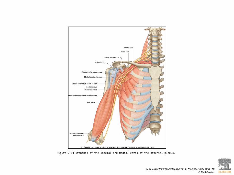
Figure 7.54 Branches of the lateral and medial cords of the brachial plexus.
Downloaded from: StudentConsult (on 13 November 2008 04:31 PM)
© 2005 Elsevier

Figure 7.55 Branches of the posterior cord of the brachial plexus.
Downloaded from: StudentConsult (on 13 November 2008 04:31 PM)
© 2005 Elsevier

Figure 7.56 Lymph nodes and vessels in the axilla.
Downloaded from: StudentConsult (on 13 November 2008 04:31 PM)
© 2005 Elsevier


