Comprehensive Summaries of Uppsala Dissertations from the Faculty of Medicine 1116 _____________________________ _____________________________ Studies on Prediction of Axillary Lymph Node Status in Invasive Breast Cancer BY JOHAN AHLGREN ACTA UNIVERSITATIS UPSALIENSIS UPPSALA 2002
Transcript
Comprehensive Summaries of Uppsala Dissertationsfrom the Faculty of Medicine 1116
Studies on Prediction ofAxillary Lymph Node Status in
Invasive Breast Cancer
BY
JOHAN AHLGREN
ACTA UNIVERSITATIS UPSALIENSISUPPSALA 2002
2
Dissertation for the Degree of Doctor of Philosophy (Faculty of Medicine) in Oncology presented at UppsalaUniversity in 2002
ABSTRACT
Ahlgren, J. 2002. Studies on prediction of axillary lymph node status in invasive breast
cancer. Acta Universitatis Upsaliensis. Comprehensive Summaries of Uppsala Dissertations
from the Faculty of Medicine 1116. 63pp. Uppsala. ISBN 91-554-5221-3.
Breast cancer is the most common malignancy among females in Sweden. Axillarylymph-node dissection is a standard procedure in the management of breast cancer,aiming at obtaining prognostic information for adjuvant therapy decisions. Axillarydissection entails considerable morbidity. The aims of this study were to establish moreselective surgical approaches and to investigate angiogenesis, a potential predictor forlymph-node metastases and prognosis.
Clinical nodal status, tumour size and S-phase were associated with nodal metastasesin cohort of 1145 women. The proportion of nodal metastases was 13% in the subgroupwith the lowest risk.
In a study from two registries, 675 and 1035 breast cancers ≤ 10 mm diagnosed byscreening mammography had nodal metastases in 6,5% and 7%, respectively. Clinicallydetected cancers had a risk of 16% and 14%, respectively.
In a study on 415 women, a 5-node biopsy of the axilla had a sensitivity of 97,3%and a false negative rate of 2,7% in comparison with axillary dissection.
Six sections from 21 breast cancers were analysed for microvessel density (MVD).The inter-section variation contributed more to the total variance than inter-tumourvariation, 45,0% and 37,3%, respectively.
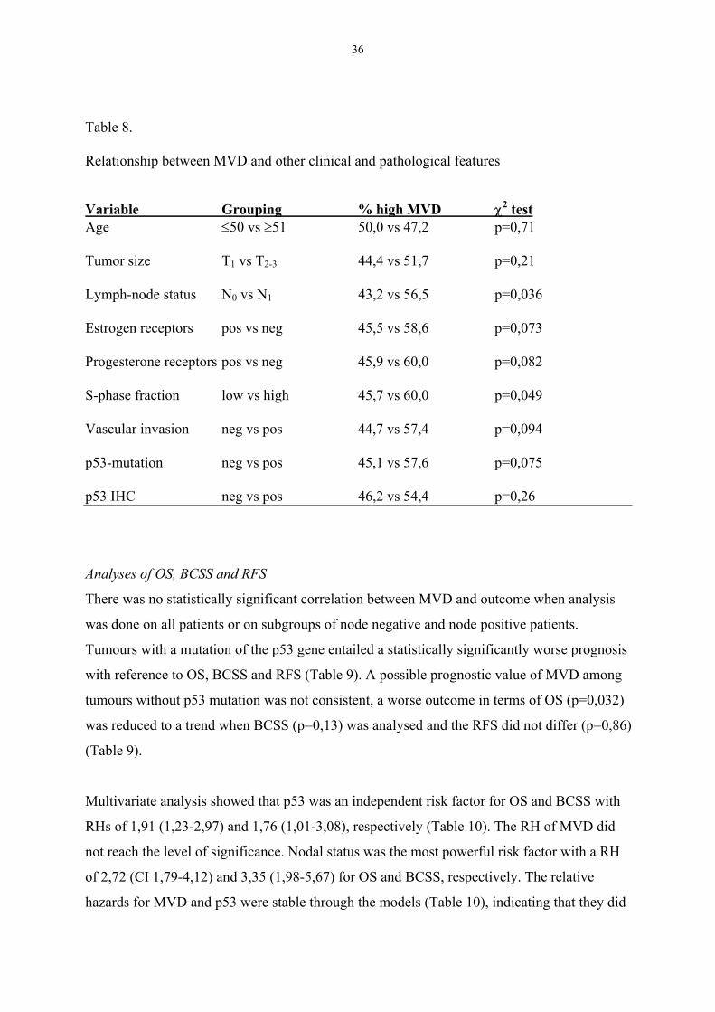
In a cohort of 315 women, breast cancers with high MVD more frequently had p53mutations (27,1%) compared with cases with low MVD (18,4%). This difference wasnot statistically significant (p=0,075). p53 mutations were associated with a worseoutcome, whereas MVD was not.
In conclusion, women with screening detected ≤10 mm breast cancers have a lowrisk of lymph node metastases and some may not need axillary dissection in the future.The 5-node biopsy could be an alternative to axillary dissection. MVD is associatedwith methodological weaknesses and routine use is not recommended.
pN- denotes node negative and pN+ node positive patients. 1Estimated in 856 cases.2Estimated in 730 cases.
Paper II
From the South-East Swedish Region 2325 tumours ≤15 mm were included. The proportion
of lymph-node metastases was 11% among tumours ≤10 mm and 24% for tumours 11-15 mm.
The lowest risk of having lymph-node metastases was 7% and this was found among
screening detected tumours ≤10 mm. The corresponding figure for clinically detected tumours
≤10 mm was 14%.
26
The figure for screening detected tumours ≤10 mm from the Uppsala-Örebro Region was
6,5% (30/464), whereas the proportion of lymph node positivity among patients with
clinically detected breast cancers ≤10 mm was 14% (34/211).
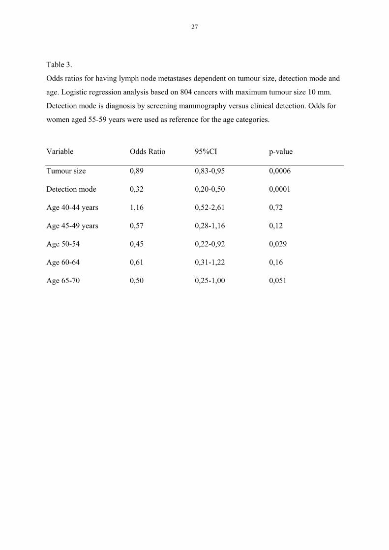
Results from extended analysis made in December 2001
It is reasonable to believe that the subgroup of clinically detected tumours has a greater mean
tumour size compared with tumours detected by screening mammography. Moreover, since
the screening detected subgroup consists of women aged 40-70, the clinically detected
subgroup will have a different age distribution. In December 2001 we performed a
multivariate analysis on the relation between positive lymph node status and detection mode
adjusted for tumour size and age using data from the registry at the Regional Oncologic
Centre of Uppsala/Örebro. Data from 803 tumours with a maximum diameter of 10 mm
diagnosed from September 1992 to December 1996 was selected. Detection mode, tumour
size and age were entered in a logistic regression model with lymph node status as response
variable (Table 3). The results from this analysis show that the mode of detection is the
strongest predictor of lymph node metastases, clinically detected tumours being more likely to
have positive lymph nodes. Both tumour size and age are also independent risk factors for
nodal metastases, whereas decreasing tumour size as expected shows a correlation with
decreasing risk for metastases, age have a non-linear relationship. Age 50-54 years was the
only age group with an odds ratio that was statistically significant.
27
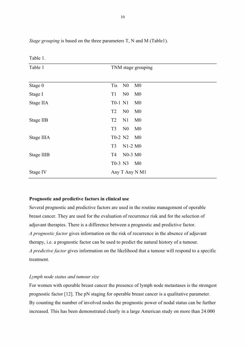
Table 3.
Odds ratios for having lymph node metastases dependent on tumour size, detection mode and
age. Logistic regression analysis based on 804 cancers with maximum tumour size 10 mm.
Detection mode is diagnosis by screening mammography versus clinical detection. Odds for
women aged 55-59 years were used as reference for the age categories.
Variable Odds Ratio 95%CI p-value
Tumour size 0,89 0,83-0,95 0,0006
Detection mode 0,32 0,20-0,50 0,0001
Age 40-44 years 1,16 0,52-2,61 0,72
Age 45-49 years 0,57 0,28-1,16 0,12
Age 50-54 0,45 0,22-0,92 0,029
Age 60-64 0,61 0,31-1,22 0,16
Age 65-70 0,50 0,25-1,00 0,051
28
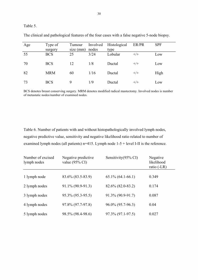
Paper III
Th number of women with positive lymph node status was 149/415 (36%). The number
of lymph node negative axillae was 266/415. The number of lymph node positive axillae
that were missed in the 5-node biopsy was 4/149, thus, according to the 5-node biopsy
270 were classified as lymph node negative which gives a false negative rate of 1,5%.
The distribution in different subgroups of the 4 erroneously classified cases is given in
Table 4. No risk factor for erroneous classification can be observed due to the low
number of such cases. The clinical and pathological features of the four cases with a
false negative 5-node biopsy are given in Table 5.
The 5-node biopsy had a sensitivity of 97,3% (CI 97,1-97,5) and a negative predictive
value of 98,5 (CI 98,4-98,6). Among women with detection by screening (n=204) the
sensitivity was 95,8% (CI 95,7-96,0) and the negative predictive value was 98,7 (CI
98,7-98,8). The corresponding estimates for clinically detected cases (n=197) were
97,9% (CI 97,9-98,0) and 98,0 (CI 98,0-98,1). The –LR was 0,027 for all women, for
those with screening and clinically detected tumours the –LR was 0,042 and 0,021
respectively.
As expected, the sensitivity of the 5-node biopsy increased for each lymph node
examined but the difference with reference to negative predictive value and sensitivity
was only marginally increased when the performance of 4 nodes was compared with 5
nodes (Table 6).
29
Table 4.Clinical and pathological characteristics of study population in relation to lymph-nodepositivity and numbers of cases with false negative 5-node biopsy
Variable Grouping Node positive Number of node pos cases % Numbers % missed in 5 node biopsyAll patients 100 149/415 36 4(n=415)
Age <=50 24 41/9742 0(n=415) >50 76 108/318 34 4
Menopause, pre 27 46/109 42 0(n=412) post 73 101/303 33 4
S-phase (high vs low) - - 1.2 (0.67-2.2)MVD denotes microvessel density. Mut denotes mutation T size is tumour size. N+ is axillarylymph-node positive. High S-phase: Diploid tumours >7%; non-diploid tumours >12%. CIdenotes confidence interval.
40
General DiscussionPaper I-III
Clinical palpation as the only means of deciding whether a patient must undergo axillary
surgery or not is inadequate. Fisher and co-workers [136] reported a false negative rate of 39%
and a false positive rate of 27%. The corresponding figures in our study (I) were 31% and
19%. These somewhat lower figures are probably partly due to a lower prevalence of nodal
metastases in a screened population. Clinical node negativity can not be considered sufficient
for allowing omission of axillary surgery. However, the current praxis that all women with
preoperatively palpable lymph nodes are recommended axillary operation is justifiable, given
a proportion of positive nodes of 52% for cN1a, and 81% for cN1b (Paper I).
Larger tumour size was strongly correlated with the presence of lymph node metastases in our
study (Paper I). This correlation has been reported several times before [33, 34]. However,
tumour size alone cannot be considered as a sufficient means to delineate a low risk group
since the proportion with lymph node metastases was 13% among women with cN0 status and
tumour size ≤10 mm (Paper I). This risk is too high to allow omission of axillary surgery.
The third factor that correlated with lymph node metastases was SPF (Paper I). The
correlation was limited to tumours ≤20mm. Stål and co-writers [42] reviewed 16 articles
regarding the relation between SPF and nodal status, 13 did not show a statistically significant
correlation whereas 3 did. The majority of cases in our study (70%) had a tumour size of 20
mm or less, most likely because of screening, and this could explain why most other
investigators did not find a correlation. Data from 605 women was entered into a logistic
regression analysis of correlation between high SPF, adjusted for tumour size among cN0
tumours (Paper I). The frequency of pN1 in the ≤10mm subgroup was 14% and 21% in the
low SPF and high SPF groups respectively. The clinical consequence of this finding is that
one should not consider omitting axillary surgery in women with high SPF tumours. On the
other hand, low SPF is not useful as a low risk criterion for a tentative subgroup in which
axillary surgery could be omitted. The reason for this is that the correlation between high SPF
and pN1 is not strong enough and that small tumours with high SPF are rare.
41
Several studies with a similar design as in our study (Paper I) have been published [137-151].
The main findings regarding the most common risk factors of 15 such studies are summarised
in Table 11. From Table 11 one can conclude that tumour size and peritumoural vascular
invasion are the histopathological factors that are most consistently associated with nodal
metastases with positive correlation in 12/15 [137-151] and 9/10 studies respectively.
Table 11.
Reference Numberof cases
Inclusioncriteria
% pN1 T size(larger)
Youngage
Highgrade
Clinicaldetection/nonpalpable
Vascularinvasion
Chadha 139 263 pT1; cN0 27 + NS NSN ND +
Ravdin 148 59631 Completedata on riskfactors
39 + + ND ND ND
Barth 138 918 pT1 23 + NS +N + +
Fein 141 1598 pT1-2;≥10N
28 + NS NST + +
Mustafa 146 1641 pT<=10mm 16 NS + +N ND ND
Cutuli 140 893 cT<=30mm 25 NS NS +T +4 ND
Velanovich 151 851 - ND + ND NSN ND ND
Olivotto 147 4312 <90 years 32 + + +N+T +3 +
Gann 143 14993 ≥6N;<79 years
36 + + +T ND ND
Gajdos 142 850 pT1 25 + + NST ND +
Gonzales-Vela 144 102 ≥10N 53 + ND NST ND +
Shoup 150 204 pT1 25 +2 NS +2, N ND +2
Anan 137 1003 pT1 25 NS + ND NS +
Rivadeneira 149 919 pT;<=10mm
18 + + +T ND NS
Guarnieri 145 547 pT1; ≥5N 29 + NS NST ND +1An additional 6001 cases in a validatin set. 2 Univariate analysis only. 3 Palpable tumour and/or palpable lymphnodes. 4 T0 vs. T1 vs. T2. N Nuclear grade. T Tumour grade. + denotes statistically significant correlation withlymph node status. NS = not statistically significant. ND = No data reported. N =lymph nodes.
As one can see from Table 11 the size of the cohorts varies largely and the selection of cases
is also quite different between different studies. The prevalence of lymph-node metastases
ranges from 16-53%. Two studies included only cancers with size ≤ 10mm [146, 149], while
stage I-III cancers were included in another [147]. The fifteen studies [137-151] all tried to
define a group with low risk for lymph-node metastases. The risk for having lymph-node
metastases in these subgroups ranged from 0-17% (data not shown in table).
42
Since increasing tumour size is the most consistent risk factor for lymph node metastasis, our
second study (Paper II) was focused on women with a tumour size of 10 mm or less. The use
of detection mode (screening mammography vs. clinical) was based on findings in a study by
Arnesson and co-workers [152] who reported that 9% of 229 screening detected cancers were
lymph node positive compared to 20% of 89 clinically detected tumours. Our study (Paper II),
based on data from two large breast cancer registries, showed that detection by screening
mammography is associated with a considerable lower risk of positive lymph nodes when
compared with clinically detected tumours. This finding was later confirmed with a
multivariate analysis (Table 3), in which detection mode was adjusted for age and tumour
size. This is in accordance with a study reported by Fein and co-writers [141], who also
identified mammographical detection as a predictor of low risk for nodal metastases. The
correlation between mode of detection and node positivity was retained in multivariate
analysis [141]. The proportion of lymph node metastases was 15,8% among 487 women with
mammographically detected tumours whereas 32,7% had lymph node metastases if the
tumour was detected by physical examination. Fein and colleagues reported a 0% risk of
lymph node metastases among mammographically detected tumours ≤ 5 mm and a 5-10% risk
for mammographically detected tumours with histopathologic size 6-10 mm and age > 40.
Other groups [138, 147] have used palpable versus non-palpable breast tumour as a potential
predictor for nodal metastases. Both these studies showed that palpability is an independent
risk factor for axillary metastases. A French study [140] reported that clinical tumour size (T0
vs. T1 vs. T2) was independently correlated with pN status.
Thus, the reason for mammographical detection being a predictor for node negativity seems to
be something more than a mere matter of tumour size. This phenomenon is probably partly
due to the tendency for mammography to detect biologically less aggressive cancers. Duffy
and co-workers found that interval cancers and cancers among non-attenders had a
significantly higher proportion of grade 3 compared with incident cases detected with
mammography [153]. Hakama and co-writers [154] found that the proportion of diploid
tumours was higher among cancers detected in incident screens compared with interval cases
and cancers among non-attenders.
43
Based on the findings in our study (Paper II), we proposed a prospective Swedish multicentric
cohort-study. After careful ethical and medical considerations, the study started accrual of
patients in 1997. In this study axillary dissection is omitted in screening detected tumours with
size ≤10 mm and grade I-II according to Elston and Ellis and/or low SPF. Women with
multifocality or a prior history of cancer are excluded. The primary end point is axillary
recurrence and a total of 1500 women are to be recruited. The accrual is estimated to stop
early 2002. No results are yet available from this study.
The five node biopsy
Although there is hope that it will be possible to forgo axillary surgery in a low risk
group, this subset comprises only a minority of newly diagnosed breast cancers. Thus, in
order to improve the management of the axilla for the vast majority of women with
breast cancer other strategies are needed. One alternative is lymph-node sampling of the
axilla, a method that is less extensive than axillary dissection. The Scottish trial on
sampling versus axillary clearance showed a sensitivity of 100% of a four-node biopsy
from the axilla [118]. However, the estimation of sensitivity, which is a key parameter,
was based on only 67 women. Moreover, the Scottish trial included patients with on
average larger tumours than currently are seen in areas with well functioning screening.
In contrast, our study of the 5-node biopsy (Paper III) included a large number of women
(n 415) and half of them had screening detected cancers. The false negative rate in our
study was 2,7% (Paper III), this estimate is encouraging since several studies of the
sentinel node procedure have shown false negative rates of 6,7-11,4% [122-124]. The
negative predictive value in our series was 98,5%, the corresponding estimate reported
from the sentinel node procedure is 93-96%. The reproducibility of our study can be
questioned, since one surgeon made all operations. However, for a specialised team a
training period with 30-50 operations with the same technique as in this trial would be
easily accomplished. The recommendations for the sentinel node procedure are similar.
The only direct comparison between a sampling procedure and the sentinel node technique
that has been published [155] included 200 women. Ten out of 60 node positive patients were
not detected with the sentinel node procedure. The corresponding figure for 4-node sampling
was 1/60. The value of this study has been questioned [156] because of the use of suboptimal
44
amounts of radiocolloid and the lack of an established reference method for comparison, i.e.
no axillary dissection was performed.
An interesting aspect is that serial sectioning and/or immunohistochemical staining of the
sentinel node(-s) often are employed [122, 124], whereas only routine pathological
examination was performed in our study (Paper III). In a recent review [157] it is estimated
that the use of IHC and/or serial sectioning produces a conversion to node positivity in 9-33%
of tumours initially classified as node negative due to detection of micrometastases. It is
reasonable to believe that the use of more sensitive pathological methods is an important part
of the sentinel node procedure, it is even tempting to hypothesise that the sensitivity could be
higher with the sentinel node procedure in comparison with level I-II dissection. A possible
argument against this is that micrometastatic sentinel nodes sometimes are accompanied with
macrometastases in non-sentinel nodes [158]. Serial sectioning and IHC are feasible when
used in conjunction with the sentinel node procedure but have been considered too unpractical
to be used in routine handling of specimens from level I-II dissections. The 5-node biopsy
represents something in between. One could speculate that the performance of the 5-node
biopsy could be improved by using these refined histopathological techniques. Although there
seems to be a worse outcome for women with lymph node micrometastases compared with
those without [157], the long-term prognostic value of IHC-positive sentinel nodes is still
unknown.
In contrast to most studies on the sentinel node procedure we reported the –LR. This measure
of a test’s performance is not affected by differences in prevalence between populations and it
can be used to calculate the post-test probability if the pre-test probability is known. In this
context the pre-test probability is the prevalence of lymph node metastases in a population
with breast cancer. An LR of 1 means that the post-test probability is exactly the same as the
pre-test probability. Likelihood ratios greater than 10 or less than 0,1 generate large and often
conclusive changes from pre-test to post-test probability [159]. The –LR of 0,027 for the 5-
node biopsy thus supports the view that this procedure can be a valuable diagnostic tool.
Although axillary surgery per se not seems to affect survival [109] the risk of withholding
adjuvant treatment for the women that were falsely defined as node negative must be
considered. This problem has decreased during recent years since most breast cancer patients
45
nowadays will be given adjuvant tamoxifen and even chemotherapy regardless of lymph-node
status [160]. In our series 3 out of 4 women with lymph node metastases not detected in the 5-
node biopsy would have been treated with tamoxifen according to current guidelines. The
fourth woman would not have been offered adjuvant systemic treatment if her nodal status had
been based on the 5-node biopsy only. This potential risk of under-treatment must be balanced
with the benefits of a less traumatic surgical procedure in the rest of the patients.
Even though we do not know the true incidence of arm symptoms after a 5-node biopsy, the
Scottish group has reported less morbidity of 4-node sampling compared with axillary
clearance of level I-III [120, 161]. Moreover, several studies indicate that increasing number
of removed nodes causes increased incidence of arm morbidity [14, 116, 117]. It is also most
likely that the sentinel node procedure causes less arm morbidity than a 5-node biopsy.
Although the sentinel node procedure probably is superior, in terms of less associated
morbidity, the 5-node biopsy seems safe to use as an alternative to level I-II dissection in
women not suitable for a sentinel node procedure. Ideally, the 5-node biopsy should be
compared with the sentinel node procedure in a randomised study.
Paper IV-V
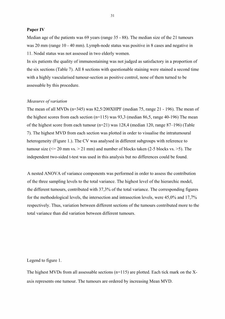
Estimation of angiogenesis, methodological considerations
A large number of studies have reported that prognostic information can be obtained from
estimation of MVD [18, 81-85, 87-92], but several studies question the use of MVD as a
prognostic factor [93-99]. The reason for these conflicting results is probably methodological
problems, maybe related to the immunohistochemical technique, inter observer variability
and/or intra tumoural heterogeneity. Our methodological study (Paper IV) focuses on the
latter. Present methodological recommendations [162, 163] advocate determination of MVD
in the areas of highest vascular density (hotspots). The underlying hypothesis for the hotspot
method is that systemic dissemination of cancer cells is more likely in the areas of highest
vessel density [18]. Since identification of hotspots is of great importance for the method, the
chance of identifying the “hottest-spots” would most likely be influenced by the potential
variation of vascular density between different parts of a tumour.
46
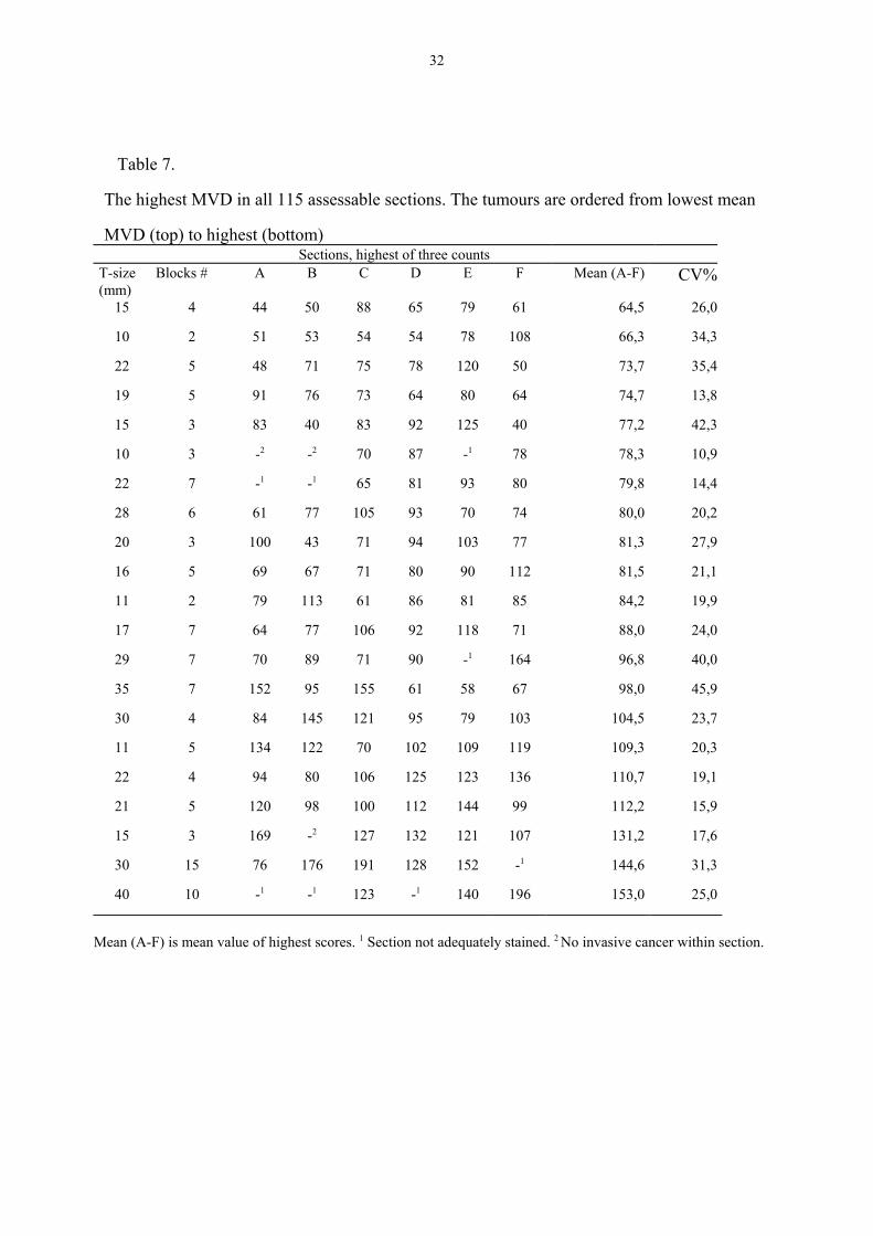
Our prospective study, which employed a predefined and systematic handling of all tumour
material, demonstrates that there is a marked intra tumoural variation of MVD (Paper IV). In
the nested ANOVA analysis the variation between different sections contributed more to the
total variance than the variation between different tumours, 37,3% and 45,0% of the total
variance, respectively. This means that there is a risk that a MVD score is more influenced by
the sampling procedure and less by the true angiogenic capacity of the tumour. The influence
of intra tumoural heterogeneity on the results has been addressed in previous studies but, as
can be noticed below, there has been no standardised way of designing or reporting studies on
heterogeneity. In a Dutch study [164], a design similar to ours was used. They analysed all
available blocks (2 - 4) from 10 breast cancers. Their data showed that the variation between
blocks contributed more to the total variation than the variation between tumours, which is
identical to what we found. The CVs for different blocks from the same tumour in the Dutch
study ranged from 5,7 - 54,9%. Although the sampling technique was not identical in our
study, the corresponding range of CVs in our material, based on twice as many cases, is quite
similar (9,4 - 44,1%) (Paper IV). The Dutch group concluded that sections from multiple
blocks should be analysed to identify the “hottest-spot” for each tumour, a handling, which in
routine use would be unpractical. Bosari and co-workers [82] retrospectively examined all
available blocks (on average three) from 120 breast cancers. In 14% of the cases the difference
between the average and the maximum score was more than 20%. However, it is unclear if
this moderate variation predominantly was the result of intra- or intersection heterogeneity.
The variation within each section constituted 17,7% of the total variance in our study. The
difference between average score and maximum score exceeded 20% in 19% of our 115
sections (Paper IV). The intra section variation was much more pronounced in a study
reported by Axelsson and colleagues [95] with a greater than 20% difference in 49% of 220
breast cancers. They could not find any prognostic value of MVD and concluded that this was
mainly due to variability from field to field within the same section. Van Hoef and co-writers
[94] retrospectively analysed MVD in 93 breast cancers, without finding any independent
prognostic information. In 41 of the tumours, MVD was assessed in two sections from two
different blocks. This comparison showed a concordance with reference to MVD categories of
71 - 78%, which could introduce a substantial error in the method according to the authors.
47
Some studies have claimed that intra tumoural heterogeneity is of less importance [165, 166].
The study by Martin and co-workers [165] comprised 20 breast cancers and included an
analysis of three different blocks from each tumour. Eighty-five percent of the patients were
correctly assigned to the high MVD and low MVD groups regardless if one or three sections
were taken into account, they drew the conclusion that the MVD measured in one section is
representative of whole tumour vascularity. The Danish study [166] mainly dealt with the
reproducibility of different counting procedures but they also concluded that intra tumoural
variation does not substantially affect the results. The relevance of this conclusion must be
questioned since they selected 40 archival cancers with the object of obtaining different
degrees of MVD. This could lead to overestimation of the inter tumoural variation in relation
to the intra tumoural variation.
It has been argued that the probability of finding the hotspots can be increased by counting the
ten apparent highest fields [84]. In another study, counting 4 hotspots gave better prognostic
information than 10 [91]. The suggestion to count several hotspots seems to be supported by
our findings. The problem with this approach according to our data, is that if one counts a
large number of apparent hotspots it is likely that one will end up with a high score in most
tumours. In our study (Paper IV), all tumours had at least one score above the median MVD if
all scores from each tumour were taken into account.
We used an antibody to CD31 in both our studies on angiogenesis (Paper IV-V). Anti-CD31
has been recommended by the international consensus on the methodology and criteria for
quantification of angiogenesis as being the most useful antibody, followed by anti-CD34 and
anti-FVIII-Rag [163]. One advantage with anti-CD31 is that it does not react with lympho-
endothelial cells [163]. Martin and co-authors found anti-CD31 less reliable with up to 13%
failed stainings [84] compared with 2% for anti-CD34 and 1% for anti-FVIIIRag. In our
studies, 6% (Paper IV) and 0% (Paper V) were judged as inadequate.
Interobserver variability is not a negligible problem, variable reproducibility between different
observers has been reported (0,45< r <0,99)[166]. By following the consensus
recommendations [163], and having two observers that simultaneously assessed the slides
(Paper IV), we tried to reduce the impact of interobserver variability. In the other study (Paper
V) we had only one observer but a Chalkley eyepiece graticule was used for the assessment of
48
MVD. Hansen and co-workers [166] have shown that the interobserver variability is lower for
the Chalkley method compared with classical counting of microvessels. The Chalkley method
is probably more robust since no decisions have to be made on whether adjacent stained
structures are separate countable microvessels or not. Furthermore, the Chalkley graticule
method makes the procedure less time consuming [135].
To conclude the methodological discussion, it seems quite clear that intratumoural
heterogeneity can influence the results and that this represents a major problem for the
method. Intratumoural variation seems to be one main reason for the conflicting data on the
usefulness of MVD as a prognostic factor in breast cancer. Interobserver variability and
immunohistochemical technique also are likely contributors to diverging results.
The relation between angiogenesis and p53
Although there are methodological problems associated with MVD, we chose to continue our
studies on MVD (paper V) due to the following reasons: Firstly, the important role of
angiogenesis in the malignant progression of solid tumours is undisputed. Secondly, mutations
of the tumor-suppressor gene p53 has been demonstrated to regulate not only apoptosis and
proliferation but also angiogenesis [78, 79], which gives a molecular rationale for a
relationship between MVD and mutations of p53. Thirdly, the opportunity to analyse the
potential relationship between MVD and cDNA determined p53 mutations for exons 2-11,
which has not been investigated before, to the best f our knowledge. Fourthly, the opportunity
to study this relationship on a large population based cohort of women with complete and
long-term follow-up.
The main finding of our study is that high MVD, assessed by the Graticule counting method,
seems to correlate with mutations in the p53 tumour suppressor gene. The proportion of p53
mutation was 27,1% among women whose tumour had a high MVD, the corresponding figure
for those with a low MVD was 18,4%. The correlation between high MVD and p53 mutation
reached only borderline statistical significance (χ2-test, p=0,075). However, we found that
there was a statistically significant correlation between high MVD and p53 mutations (χ2-test,
p=0,037) when tumours (n10) with a mutation in the evolutionary conserved regions 2 or 5
were excluded. In fact, 6/10 tumours with mutation in region 2 or 5 had a low MVD. We do
not know if this is a reflection of a more complicated relationship between angiogenesis and
49
p53 status or if it is merely a play of chance due to the relatively low number of women with a
p53 mutated tumour. Therefore, this retrospective subanalysis must be interpreted cautiously.
Our study could not show any correlation between MVD and p53 analysis by IHC and this
finding is in agreement with several previously published studies [102, 167-170]. Thus,
studies on angiogenesis and p53 IHC fail to support the thrombospondin-1 based mechanism.
The reason for this could be that immunohistochemistry is inferior to sequenced based
determination of p53 status [60, 61, 171]. The false negative and false positive rate with p53
IHC was 33% and 30%, respectively, compared with sequenced based p53 status [60].
MVD and lymph-node status
Among standard prognostic factors, high MVD was associated with positive lymph-node
status (p=0,036) and high S-phase (p=0,049). The proportion of lymph-node positive women
with high MVD was 56,5%, i.e., more than 40% of lymph-node positive women had primary
tumour with a low MVD. Thus, MVD is unlikely to be of practical value for predicting the
axillary lymph-node status. In a simple predictive model consisting of only a few criteria, that
ideally should be easy to adopt in the routine management of breast cancer, there is a need of
powerful predictive factors like tumour size and detection mode (Paper I-II). Moreover, the
methodological aspects of MVD are problematic as have been outlined above.
MVD and p53 in relation to prognosis
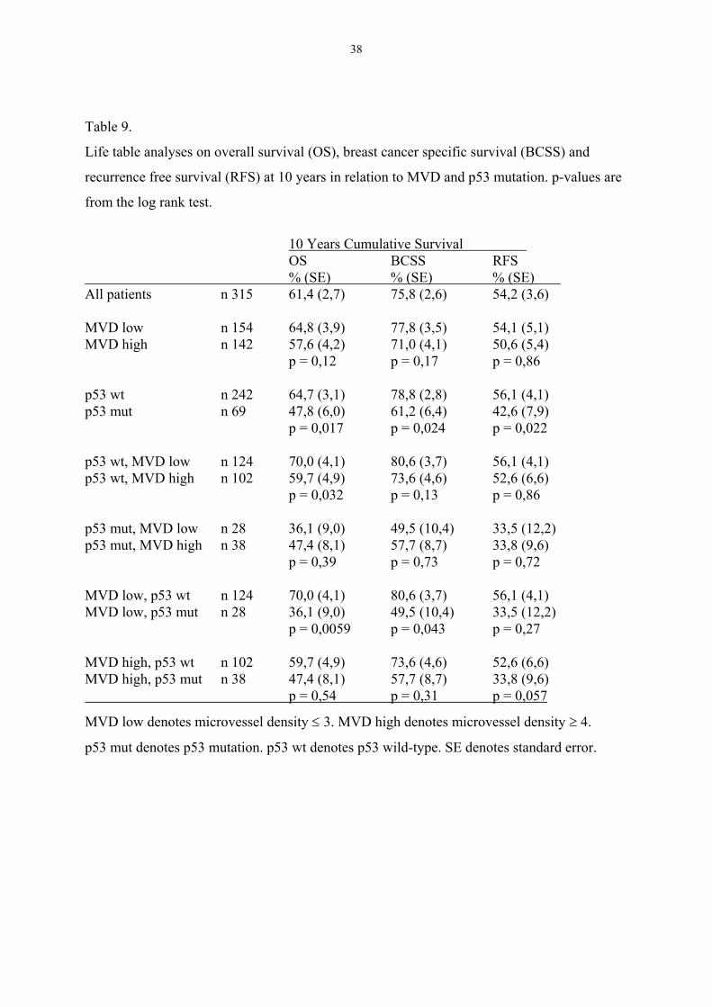
No prognostic information could be obtained from MVD status in our study. Women whose
tumours had a mutation of the p53 gene had a statistically significantly worse prognosis with
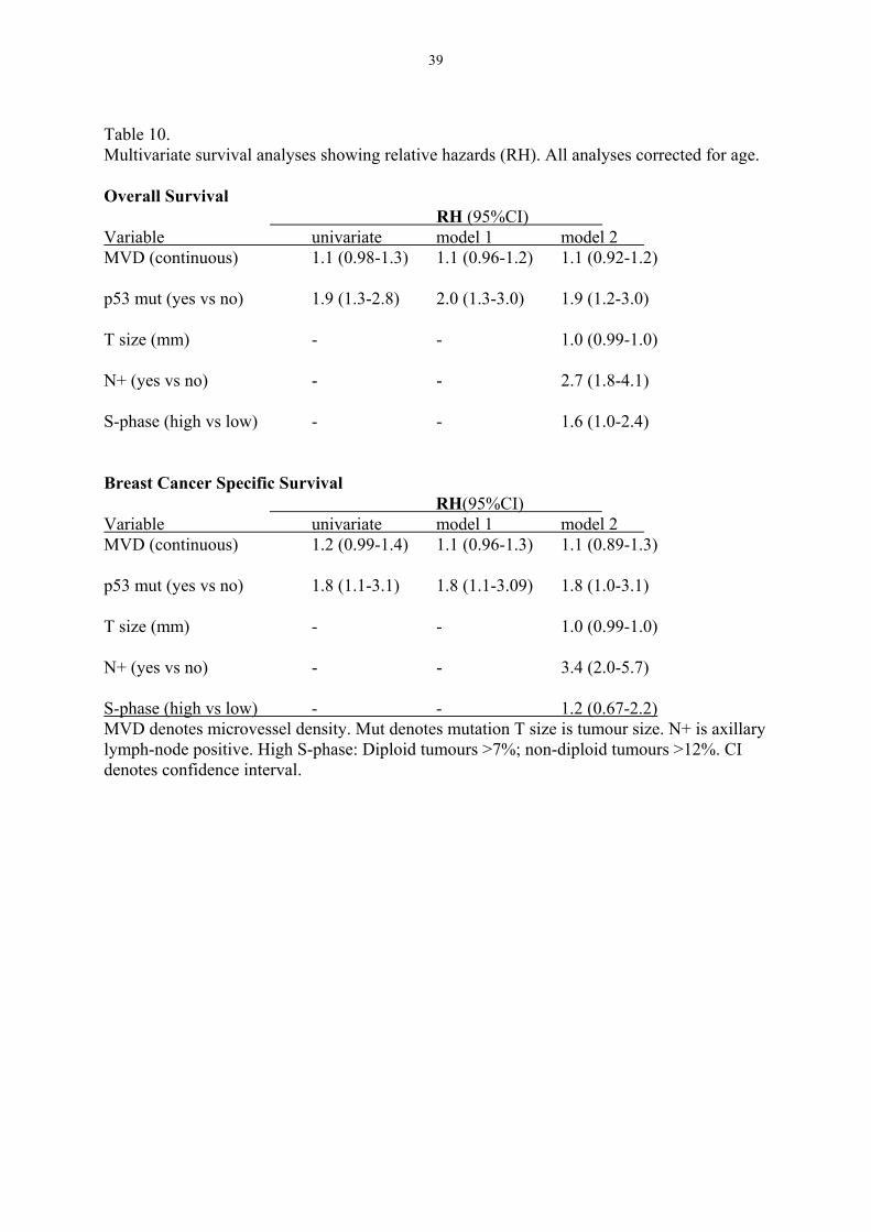
reference to OS, BCSS and RFS according to univariate analysis and this effect was retained
in multivariate modelling. Women with tumours with low MVD and mutation seemed to do
worse with reference to OS and BCCS compared with women with high MVD and p53
mutation. Although this difference was not statistically significant, it is a bit surprising since
the difference was in the wrong direction compared with what one would expect. When we
explored the possibility of a more complicated relationship between MVD status and p53
mutation in terms of prognosis and introduced an interaction term in the multivariate
modelling, our findings were not convincing for an interaction. Although the number of
women in these subgroups were limited and the data must be interpreted with caution, we
50
found the pattern interesting enough to be worth studying further when we accumulate more
events.
The lack of prognostic power of MVD in our study is not surprising. It has been mentioned
before that several studies have reported negative findings [93-99]. In order to avoid
methodological pitfalls as much as possible we followed the recommendations that has been
published [163] with the exception that the readings of the scores were done by one person.
We also used the Chalkley eye-piece graticule counting method which is time saving [135]
and possibly superior from a methodological standpoint [166]. One limitation of our study
was the use of relatively small tumour sections. This could lead to underestimation of MVD,
especially since the intratumoural heterogeneity of this parameter is pronounced.
Nevertheless, the lack of prognostic information from MVD in this rather large cohort of
women with complete and long-term follow-up is not supportive for the use of MVD as a
prognostic factor in breast cancer.
51
General conclusions
• Positive clinical lymph node status, increasing tumour size and high S-phase fraction are
all associated with increased risk of axillary lymph-node metastases in breast cancer.
However, even if these factors are used in combination, they can not define a subgroup of
women with a sufficiently low risk of having lymph node metastases to allow omission of
axillary surgery.
• Breast cancers diagnosed with screening mammography have a lower risk of having
axillary lymph node metastases than clinically detected tumours. This association is
independent of the woman’s age and the size of the tumour. Diagnosis by screening
mammography and tumour size ≤ 10 mm are criteria, combined with other low risk
features that may be useful for characterisation o a subgroup of women in which axillary
surgery may be omitted as a part of a prospective study.
• Five-node biopsy of the axilla has a high sensitivity and a low false negative rate
compared with axillary dissection of level I-II in women with breast cancer. The 5-node
biopsy is an alternative to axillary dissection and compares very favourably with results
reported in the literature from the sentinel node biopsy procedure.
• Angiogenesis estimated with MVD shows a marked variation between different sections
from the same tumour. Intra tumoural hetereogeneity in breast cancer makes the use of
MVD questionable.
• There seems to be a relationship between angiogenesis (MVD) and cDNA sequenced
determined p53 mutations for exons 2-11 in primary breast cancer. Tumours with high
MVD more often had p53 mutations. p53 status had independent prognostic value in a
multivariate analysis whereas MVD had not.
52
Acknowledgements
I want to express my most sincere gratitude to all that helped me with this thesis, especially to:
Jonas Bergh, my supervisor, for all his never ending support, his excellent knowledge onscience and oncology and spiritual intellect, for friendship and for giving me food and shelterin his house when my home town Gävle was buried in snow. Without You this thesis wouldnot have been accomplished.
Lars Holmberg, my co-supervisor, his profound knowledge, for good discussions and forgenerous help, especially with statistical problems.
Gunnar Westman, for being my mentor and co-supervisor during the years in Örebro, forsharing some of the basic ideas to this thesis, for encouraging me to start with clinicalresearch, for friendship and good laughs.
Göran Liljegren, for good and generous collaboration since my early years in Örebro, forhaving a sincere interest in bandy.
Olle Ståhl, for co-authorship and being my supervisor during the start of my PhD studies inÖrebro.
all other co-authors: Lars-Gunnar Arnesson, Björn Risberg, Anders Lindgren, SigridKlaar, Thomas Lindahl and Kenneth Villman, for giving me of your time and knowledge.
all clinicians reporting data on women with breast cancer to the registries in Uppsala andLinköping and to the staff at ROC in Uppsala, especially Marit Holmqvist.
all my colleagues at the Department of Oncology in Gävle, for encouragement and forstanding extra work load while I have worked with this thesis.
all my colleagues at the departments of Oncology in Uppsala and Örebro, for goodcomradeship and a creative and stimulating atmosphere.
the institution of Oncology, Radiology and Clinical Immunology, section of Oncology,Professor Ingela Turesson for giving me the opportunity and support to do my PhD thesis,Didde Westerström for all help.
Torpgänget, the club for men of men, Hans, Ulf, Tomas, Per and Magnus, for good work inthe timber forests.
Karin, Anna, Nils for love and trust.
I would also express my thankfulness to the following institutions that have supported thisresearch financially: Örebro Medical Council Resarch Committee, the Swedish CancerSociety, Stiftelsen Onkologiska klinikens i Uppsala Forskningsfond, King Gustav the Vth
Jubilee Fund, the Stockholm Cancer Society and the Research Fund of Dalarna.
53
References
1. Socialstyrelsen. Cancer incidence in Sweden 1999. http://www.sos.se/sos/stat.htm 2001.2. EBCTCG. Effects of radiotherapy and surgery in early breast cancer: an overview of the
randomized trials. N Engl J Med 1995, 333, 1444-1455.3. Peto, R. Oral presentation on behalf of the EBCTCG. European Breast Cancer Conference
Brussels, 2000.4. EBCTCG. Polychemotherapy for early breast cancer: an overview of the randomised trials.
Lancet 1998, 352, 930-942.5. EBCTCG. Tamoxifen for early breast cancer: an overview of the randomised trials. Lancet
1998, 351, 1451-1467.6. Slamon, D, Clark G, Wong S, Levin W, Ullrich A, McGuire W. Human breast cancer:
correlation of relapse and survival with amplification of the HER-2/neu oncogene. Science1987, 235, 177-182.
7. Bergh, J, Norberg T, Sjögren S, Lindgren A, Holmberg L. Complete sequencing of the p53gene provides prognostic information in breast cancer patients, particularly in relation toadjuvant systemic therapy and radiotherapy. Nature Med 1995, 1, 1029-1034.
8. Perou, CM, Sorlie T, Eisen MB, van de Rijn M, Jeffrey SS, Rees CA, Pollack JR, Ross DT,Johnsen H, Akslen LA, Fluge O, Pergamenschikov A, Williams C, Zhu SX, Lonning PE,Borresen-Dale AL, Brown PO, Botstein D. Molecular portraits of human breast tumours.Nature 2000, 406, 747-52.
9. Sorlie, T, Perou CM, Tibshirani R, Aas T, Geisler S, Johnsen H, Hastie T, Eisen MB, vande Rijn M, Jeffrey SS, Thorsen T, Quist H, Matese JC, Brown PO, Botstein D, EysteinLonning P, Borresen-Dale AL. Gene expression patterns of breast carcinomas distinguishtumor subclasses with clinical implications. Proc Natl Acad Sci U S A 2001, 98, 10869-74.
10. Miki, Y, Swensen J, Shattuck-Eidens D, Futreal PA, Harshman K, Tavtigian S, Liu Q,Cochran C, Bennett LM, Ding W, et al. A strong candidate for the breast and ovariancancer susceptibility gene BRCA1. Science 1994, 266, 66-71.
11. Wooster, R, Bignell G, Lancaster J, Swift S, Seal S, Mangion J, Collins N, Gregory S,Gumbs C, Micklem G. Identification of the breast cancer susceptibility gene BRCA2.Nature 1995, 378, 789-92.
12. Clark, GM. Prognostic and predictive factors. In Harris J, Lippman M, Morrow M,Hellman S, eds. Diseases of the breast. Philadephia, Lippincott-Raven Publishers, 1996,461-485.
13. Ivens, D, Hoe AL, Podd TJ, Hamilton CR, Taylor I, Royle GT. Assessment of morbidityfrom complete axillary dissection. Br J Cancer 1992, 66, 136-8.
14. Liljegren, G, Holmberg L. Arm morbidity after sector resection and axillary dissectionwith or without postoperative radiotherapy in breast cancer stage I. Results from arandomised trial. Uppsala-Orebro Breast Cancer Study Group. Eur J Cancer 1997, 33, 193-9.
15. Tabár, L, Fagerberg CJG, Gad A, Baldetorp L, Holmberg LH, Gröntoft O, Ljungquist U,Lundström B, J.C. M. Reduction in Mortality from Breast Cancer After Mass Screeningwith Mammography: Randomized Trial from the Breast Cancer Screening Working Groupof the Swedish National Board of Health and Welfare. Lancet 1985, i, 829-832.
16. Folkman, J. Angiogenesis and breast cancer. J Clin Oncol 1994, 12, 441-443.17. Weidner, N, Semple J, Welch W, Folkman J. Tumor angiogenesis and metastasis--
correlation in invasive breast carcinoma. New Engl J Med 1991, 324, 1-8.
54
18. Horak, E, Leek R, Klenk N, LeJeune S, Smith K, Stuart N, Greenall M, Stepniewska K,Harris A. Angiogenesis, assessed by platelet/endothelial cell adhesion moleculeantibodies, as indicator of node metastases and survival in breast cancer. Lancet 1992,340, 1120-1124.
19. Socialstyrelsen. Causes of death 1999. http://www.sos.se/sos/stat.htm 2001.20. Peto, R, Boreham J, Clarke M, Davies C, Beral V. UK and USA breast cancer deaths
down 25% in year 2000 at ages 20-69 years. Lancet 2000, 355, 1822.21. Winer, E, Morrow M, Osborne C, Harris J. Malignant tumors of the breast. In DeVita VT,
Hellman S, Rosenberg S, eds. Cancer Principles & Practice of Oncology. Philadelphia,Lippincott Williams & Wilkins, 2001, 1652-1717.
22. John, EM, Kelsey JL. Radiation and other environmental exposures and breast cancer.Epidemiol Rev 1993, 15, 157-62.
23. Howe, GR, Friedenreich CM, Jain M, Miller AB. A cohort study of fat intake and risk ofbreast cancer. J Natl Cancer Inst 1991, 83, 336-40.
24. van den Brandt, PA, van't Veer P, Goldbohm RA, Dorant E, Volovics A, Hermus RJ,Sturmans F. A prospective cohort study on dietary fat and the risk of postmenopausalbreast cancer. Cancer Res 1993, 53, 75-82.
25. Willett, WC, Hunter DJ, Stampfer MJ, Colditz G, Manson JE, Spiegelman D, Rosner B,Hennekens CH, Speizer FE. Dietary fat and fiber in relation to risk of breast cancer. An 8-year follow-up. Jama 1992, 268, 2037-44.
26. Vatten, LJ, Kvinnsland S. Prospective study of height, body mass index and risk of breastcancer. Acta Oncol 1992, 31, 195-200.
27. Longnecker, MP, Berlin JA, Orza MJ, Chalmers TC. A meta-analysis of alcoholconsumption in relation to risk of breast cancer. Jama 1988, 260, 652-6.
28. Weber, BL, Garber JE. Familial breast cancer. In Harris J, Lippman M, Morrow M,Hellman S, J.R., eds. Diseases of the breast. Philadelphia, Lippincott-Raven Publishers,1996, 168-185.
29. Regionalt Onkologiskt Centrum, Uppsala-Örebro. Bröstcancer registret. Data on file 2001.30. Kerlikowske, K, Grady D, Rubin SM, Sandrock C, Ernster VL. Efficacy of screening
mammography. A meta-analysis. Jama 1995, 273, 149-54.31. Larsson, LG, Andersson I, Bjurstam N, Fagerberg G, Frisell J, Tabar L, Nystrom L.
Updated overview of the Swedish Randomized Trials on Breast Cancer Screening withMammography: age group 40-49 at randomization. J Natl Cancer Inst Monogr 1997, 22,57-61.
32. UICC. Illustrated guide to the TNM/pTNM classification of malignant tumours. In SpiesslB, Beahrs O, Hermanek P, Hutter R, Scheibe O, Sobin L, Wagner G, eds. TNM Atlas.Berlin, Heidelberg, Springer-Verlag, 1990, 173-183.
33. Carter, C, Allen C, Henson D. Relation of tumor size, lymph node status, and survival in24,740 breast cancer cases. Cancer 1989, 63, 181-187.
34. Axelsson, C, Mouridsen H, Zedeler K. Axillary Dissection of Level I and II Lymph Nodesis Important in Breast Cancer Classification. Eur J Cancer 1992, 28A, 1415-1418.
35. Westman, G, Ahlgren J, Liljegren G, Risberg B. Förbättra utbytet av axillingrepp.Läkartidningen 1995, 92, 2477-2483.
36. Tubiana, M, Koscielny S. Natural History of Human Breast Cancer: Recent Data andClinical Implications. Breast Cancer Res Treat 1991, 18, 125-140.
37. Tabár, L, Fagerberg G, Day N, Duffy S, Kitchin R. Breast Cancer Treatment and NaturalHistory: New Insights From Results of Screening. Lancet 1992, 339, 412-414.
38. Bloom, H, Richardson W. Histological grading and prognosis in breast cancer. Br JCancer 1957, 11, 357-377.
55
39. Elston, C, Ellis I. Pathological prognostic factors in breast cancer. I. The value ofhistological grade in breast cancer: experience from a large study with long-term follow-up. Histopathology 1991, 19, 403-410.
40. Sundquist, M, Thorstenson S, Brudin L, Nordenskjold B. Applying the NottinghamPrognostic Index to a Swedish breast cancer population. South East Swedish BreastCancer Study Group. Breast Cancer Res Treat 1999, 53, 1-8.
41. Boiesen, P, Bendahl PO, Anagnostaki L, Domanski H, Holm E, Idvall I, Johansson S,Ljungberg O, Ringberg A, Ostberg G, Ferno M. Histologic grading in breast cancer--reproducibility between seven pathologic departments. South Sweden Breast CancerGroup. Acta Oncol 2000, 39, 41-5.
42. Stål, O, Hatschek T, Carstensen J, Nordenskjöld B. DNA analysis in the management ofbreast cancer. Diagn Oncol 1991, 1, 140-154.
43. Wenger, CR, Clark GM. S-phase fraction and breast cancer--a decade of experience.Breast Cancer Res Treat 1998, 51, 255-65.
44. Knight, W, Livingston R, Gregory E. Estrogen Receptor as an Independent PrognosticFactor for Early Recurrence in Breast Cancer. Cancer Res 1977, 37, 4669-4671.
45. Osborne, CK. Steroid hormone receptors in breast cancer management. Breast Cancer ResTreat 1998, 51, 227-38.
46. McGuire, WL. Steroid receptors in human breast cancer. Cancer Res 1978, 38, 4289-91.47. Kinsel, LB, Szabo E, Greene GL, Konrath J, Leight GS, McCarty KS, Jr.
Immunocytochemical analysis of estrogen receptors as a predictor of prognosis in breastcancer patients: comparison with quantitative biochemical methods. Cancer Res 1989,49, 1052-6.
48. Pertschuk, LP, Kim DS, Nayer K, Feldman JG, Eisenberg KB, Carter AC, Rong ZT,Thelmo WL, Fleisher J, Greene GL. Immunocytochemical estrogen and progestinreceptor assays in breast cancer with monoclonal antibodies. Histopathologic,demographic, and biochemical correlations and relationship to endocrine response andsurvival. Cancer 1990, 66, 1663-70.
49. Blomqvist, C, von Boguslawski K, Stenman UH, Maenpaa H, von Smitten K, Nordling S.Long-term prognostic impact of immunohistochemical estrogen receptor determinationscompared with biochemical receptor determination in primary breast cancer. Acta Oncol1997, 36, 530-2.
50. Coussens, L, Yang-Feng T, Liao Y-C, Chen E, Gray A, McGrath J, Seeburg P, LibermannT, Schlessinger J, Francke U, Levinson A, Ullrich A. Tyrosine kinase receptor withextensive homology to EGF receptor shares chromosomal Location with neu oncogene.Science 1985, 230, 1132-1139.
51. Rajkumar, T, Gullick W. The type I growth factor receptors in human breast cancer.Breast Cancer Res Treat 1994, 29, 3-9.
52. Sjögren, S, Inganäs M, Lindgren A, Holmberg L, Bergh J. The prognostic and predictivevalue of c-erbB-2 overexpression in primary breast cancer, alone and in combination withother prognostic markers. J Clin Oncol 1998, 16, 462-469.
53. Prost, S, Le M, Douc-Rasy S, Ahomadegbe J, Spielmann M, Guerin M, Riou G.Association of c-erbB2-gene amplification with poor prognosis in non-inflammatorybreast carcinomas but not in carcinomas of the inflammatory type. Int J Cancer 1994, 58,763-768.
54. Isola, J, Visakorpi T, Holli K, Kallioniemi OP. Association of overexpression of tumorsuppressor protein p53 with rapid cell proliferation and poor prognosis in node-negativebreast cancer patients. J Natl Cancer Inst 1992, 84, 1109-14.
56
55. Piccart, M, Lohrisch C, Di Leo A, Larsimont D. The Predictive Value of HER2 in BreastCancer. Oncology 2001, 61 Suppl S2, 73-82.
56. Slamon, DJ, Leyland-Jones B, Shak S, Fuchs H, Paton V, Bajamonde A, Fleming T,Eiermann W, Wolter J, Pegram M, Baselga J, Norton L. Use of chemotherapy plus amonoclonal antibody against HER2 for metastatic breast cancer that overexpressesHER2. N Engl J Med 2001, 344, 783-92.
57. Levine, A, Momand J, Finlay C. The p53 tumor suppressor gene. Nature 1991, 351, 453-456.
58. Lane, DP. Cancer. p53, guardian of the genome. Nature 1992, 358, 15-6.59. Davidoff, A, Kerns B, Pence J, Marks J, Iglehart J. p53 alterations in all stages of breast
cancer. J Surg Oncol 1991, 48, 260-267.60. Sjögren, S, Inganäs M, Norberg T, Lindgren A, Nordgren H, Holmberg L, Bergh J. The
p53 gene in breast cancer: Prognostic value of complementary DNA sequencing versusimmunohistochemistry. J Natl Cancer Inst 1996, 88, 173-182.
61. Falette, N, Paperin MP, Treilleux I, Gratadour AC, Peloux N, Mignotte H, Tooke N,Lofman E, Inganas M, Bremond A, Ozturk M, Puisieux A. Prognostic value of P53 genemutations in a large series of node-negative breast cancer patients. Cancer Res 1998, 58,1451-5.
62. Kandioler-Eckersberger, D, Ludwig C, Rudas M, Kappel S, Janschek E, Wenzel C,Schlagbauer-Wadl H, Mittlbock M, Gnant M, Steger G, Jakesz R. TP53 mutation andp53 overexpression for prediction of response to neoadjuvant treatment in breast cancerpatients. Clin Cancer Res 2000, 6, 50-6.
63. Berns, EM, Foekens JA, Vossen R, Look MP, Devilee P, Henzen-Logmans SC, vanStaveren IL, van Putten WL, Inganas M, Meijer-van Gelder ME, Cornelisse C, ClaassenCJ, Portengen H, Bakker B, Klijn JG. Complete sequencing of TP53 predicts poorresponse to systemic therapy of advanced breast cancer. Cancer Res 2000, 60, 2155-62.
64. Folkman, J. Tumor angiogenesis: therapeutic implications. N Engl J Med 1971, 285,1182-6.
65. Folkman, J. What is the evidence that tumors are angiogenesis dependent? J Natl CancerInst 1990, 82, 4-6.
66. Liotta, LA, Kleinerman J, Saidel GM. Quantitative relationships of intravascular tumorcells, tumor vessels, and pulmonary metastases following tumor implantation. Cancer Res 1974, 34, 997-1004.
67. McCulloch, P, Choy A, Martin L. Association between tumour angiogenesis and tumourcell shedding into effluent venous blood during breast cancer surgery. Lancet 1995, 346,1334-5.
68. Rak, J, Filmus J, Kerbel RS. Reciprocal paracrine interactions between tumour cells andendothelial cells: the 'angiogenesis progression' hypothesis. Eur J Cancer 1996, 32A,2438-50.
69. Sandstrom, M, Johansson M, Sandstrom J, Bergenheim AT, Henriksson R. Expression ofthe proteolytic factors, tPA and uPA, PAI-1 and VEGF during malignant gliomaprogression. Int J Dev Neurosci 1999, 17, 473-81.
70. Senger, DR, Van de Water L, Brown LF, Nagy JA, Yeo KT, Yeo TK, Berse B, JackmanRW, Dvorak AM, Dvorak HF. Vascular permeability factor (VPF, VEGF) in tumorbiology. Cancer Metastasis Rev 1993, 12, 303-24.
71. Obermair, A, Kucera E, Mayerhofer K, Speiser P, Seifert M, Czerwenka K, Kaider A,Leodolter S, Kainz C, Zeillinger R. Vascular endothelial growth factor (VEGF) in humanbreast cancer: correlation with disease-free survival. Int J Cancer 1997, 74, 455-8.
57
72. Gasparini, G, Toi M, Gion M, Verderio P, Dittadi R, Hanatani M, Matsubara I, Vinante O,Bonoldi E, Boracchi P, Gatti C, Suzuki H, Tominaga T. Prognostic significance ofvascular endothelial growth factor protein in node-negative breast carcinoma. J NatlCancer Inst 1997, 89, 139-47.
73. Linderholm, B, Tavelin B, Grankvist K, Henriksson R. Vascular endothelial growth factoris of high prognostic value in node-negative breast carcinoma. J Clin Oncol 1998, 16,3121-3128.
74. Mukhopadhyay, D, Tsiokas L, Sukhatme VP. Wild-type p53 and v-Src exert opposinginfluences on human vascular endothelial growth factor gene expression. Cancer Res1995, 55, 6161-5.
75. Linderholm, B, Lindh B, Tavelin B, Grankvist K, Henriksson R. p53 and vascular-endothelial-growth-factor (VEGF) expression predicts outcome in 833 patients withprimary breast carcinoma. Int J Cancer 2000, 89, 51-62.
76. Linderholm, BK, Lindahl T, Holmberg L, Klaar S, Lennerstrand J, Henriksson R, Bergh J.The expression of vascular endothelial growth factor correlates with mutant p53 and poorprognosis in human breast cancer. Cancer Res 2001, 61, 2256-60.
77. Weinstat-Saslow, DL, Zabrenetzky VS, VanHoutte K, Frazier WA, Roberts DD, Steeg PS.Transfection of thrombospondin 1 complementary DNA into a human breast carcinomacell line reduces primary tumor growth, metastatic potential, and angiogenesis. CancerRes 1994, 54, 6504-11.
78. Dameron, KM, Volpert OV, Tainsky MA, Bouck N. Control of angiogenesis in fibroblastsby p53 regulation of thrombospondin-1. Science 1994, 265, 1582-4.
79. Grant, SW, Kyshtoobayeva AS, Kurosaki T, Jakowatz J, Fruehauf JP. Mutant p53correlates with reduced expression of thrombospondin-1, increased angiogenesis, andmetastatic progression in melanoma. Cancer Detect Prev 1998, 22, 185-94.
80. Brem, S, Cotran R, Folkman J. Tumor angiogenesis: a quantitative method for histologicgrading. J Natl Cancer Inst 1972, 48, 347-56.
81. Weidner, N, Folkman J, Pozza F, Bevilacqua P, Allred E, Moore D, Meli S, Gasparini G.Tumor angiogenesis: a new significant and independent prognostic indicator in early-stage breast carcinoma. J Natl Cancer Inst 1992, 84, 1875-1887.
82. Bosari, S, Lee A, DeLellis R, Wiley B, Heatley G, Silverman M. Microvessel quantitationand prognosis in invasive breast carcinoma. Hum Pathol 1992, 23, 755-761.
83. Toi, M, Kashitani J, Tominaga T. Tumor angiogenesis is an independent prognosticindicator in primary breast carcinoma. Int J Cancer 1993, 55, 371-374.
84. Martin, L, Green B, Renshaw C, Lowe D, Rudland P, Leinster S, Winstanley J. Examiningthe technique of angiogenesis assessment in invasive breast cancer. Br J Cancer 1997, 76, 1046-1054.
85. Hansen, S, Grabau DA, Sorensen FB, Bak M, Vach W, Rose C. The prognostic value ofangiogenesis by Chalkley counting in a confirmatory study design on 836 breast cancerpatients. Clin Cancer Res 2000, 6, 139-146.
86. Hansen, S, Grabau DA, Sorensen FB, Bak M, Vach W, Rose C. Vascular grading ofangiogenesis: prognostic significance in breast cancer. Br J Cancer 2000, 82, 339-47.
87. Gasparini, G, Weidner N, Bevilacqua P, Maluta S, Dalla Palma P, Caffo O, BarbareschiM, Boracchi P, Marubini E, Pozza F. Tumor microvessel density, p53 expression, tumorsize, and peritumoral lymphatic vessel invasion are relevant prognostic markers in node-negative breast carcinoma. J Clin Oncol 1994, 12, 454-466.
88. Fox, S, Leek R, Smith K, Hollyer J, Greenall M, Harris A. Tumor angiogenesis in node-negative breast carcinomas--relationship with epidermal growth factor receptor, estrogenreceptor, and survival. Breast Cancer Res Treat 1994, 29, 109-116.
58
89. Heimann, R, Ferguson D, Powers C, Recant W, Weichselbaum R, Hellman S.Angiogenesis as a predictor of long-term survival for patients with node-negative breastcancer. J Natl Cancer Inst 1996, 88, 1764-1769.
90. Gasparini, G, Toi M, Verderio P, Ranieri G, Dante S, Bonoldi E, Boracchi P, Fanelli M,Tominaga T. Prognostic significance of p53, angiogenesis, and other conventionalfeatures in operable breast cancer: subanalysis in node-positive and node-negativepatients. Int J Oncol 1998, 12, 1117-1125.
91. de Jong, JS, van Diest PJ, Baak JP. Hot spot microvessel density and the mitotic activityindex are strong additional prognostic indicators in invasive breast cancer.Histopathology 2000, 36, 306-12.
92. Kato, T, Kameoka S, Kimura T, Nishikawa T, Kasajima T. Angiogenesis and blood vesselinvasion as prognostic indicators for node-negative breast cancer. Breast Cancer ResTreat 2001, 65, 203-15.
93. Hall, N, Fish D, Hunt N, Goldin R, Guillou P, Monson J. Is the relationship betweenangiogenesis and metastasis in breast cancer real? Surg Oncol 1992, 1, 223-229.
94. Van Hoef, M, Knox W, Dhesi S, Howell A, Schor A. Assessment of tumour vascularity asa prognostic factor in lymph node negative invasive breast cancer. Eur J Cancer 1993,29A, 1141-1145.
95. Axelsson, K, Ljung B, Moore 2nd D, Thor A, Chew K, Edgerton S, Smith H, Mayall B.Tumor angiogenesis as a prognostic assay for invasive ductal breast carcinoma. J NatlCancer Inst 1995, 87, 997-1008.
96. Costello, P, McCann A, Carney D, Dervan P. Prognostic significance of microvesseldensity in lymph node negative breast carcinoma. Hum Pathol 1995, 26, 1181-1184.
97. Siitonen, S, Haapasalo H, Rantala I, Helin H, Isola J. Comparison of differentimmunohistochemical methods in the assessment of angiogenesis: lack of prognosticvalue in a group of 77 selected node-negative breast carcinomas. Mod Pathol 1995, 8,845-752.
98. Goulding, H, Abdul Rashid NF, Robertson JF, Bell JA, Elston CW, Blamey RW, Ellis IO.Assessment of angiogenesis in breast carcinoma: an important factor in prognosis? [seecomments]. Hum Pathol 1995, 26, 1196-200.
99. Morphopoulos, G, Pearson M, Ryder WD, Howell A, Harris M. Tumour angiogenesis as aprognostic marker in infiltrating lobular carcinoma of the breast [see comments]. J Pathol1996, 180, 44-9.
100. Miliaras, D, Kamas A, Kalekou H. Angiogenesis in invasive breast carcinomas: is itassociated with parameters of prognostic significance? Histopathol 1995, 26, 165-169.
101. Fox, SB, Leek RD, Bliss J, Mansi JL, Gusterson B, Gatter KC, Harris AL. Association oftumor angiogenesis with bone marrow micrometastases in breast cancer patients. J NatlCancer Inst 1997, 89, 1044-9.
102. Jacquemier, JD, Penault-Llorca FM, Bertucci F, Sun ZZ, Houvenaeghel GF, Geneix JA,Puig BD, Bardou VJ, Hassoun JA, Birnbaum D, Viens PJ. Angiogenesis as a prognosticmarker in breast carcinoma with conventional adjuvant chemotherapy: a multiparametricand immunohistochemical analysis. J Pathol 1998, 184, 130-5.
103. Gasparini, G, Bonoldi E, Viale G, Verderio P, Boracchi P, Panizzoni GA, Radaelli U, DiBacco A, Guglielmi RB, Bevilacqua P. Prognostic and predictive value of tumourangiogenesis in ovarian carcinomas. Int J Cancer 1996, 69, 205-11.
104. Gasparini, G, Fox SB, Verderio P, Bonoldi E, Bevilacqua P, Boracchi P, Dante S,Marubini E, Harris AL. Determination of angiogenesis adds information to estrogenreceptor status in predicting the efficacy of adjuvant tamoxifen in node-positive breastcancer patients. Clin Cancer Res 1996, 2, 1191-8.
59
105. Deplanque, G, Harris AL. Anti-angiogenic agents: clinical trial design and therapies indevelopment. Eur J Cancer 2000, 36, 1713-24.
106. Westphal, JR, Ruiter DJ, De Waal RM. Anti-angiogenic treatment of human cancer:pitfalls and promises. Int J Cancer 2000, 86, 870-3.
107. Aspegren, K, Holmberg L, Adami HO. Standardization of the surgical technique inbreast-conserving treatment of mammary cancer. Br J Surg 1988, 75, 807-10.
108. Halsted, W. The results of operation for the cure of cancer of the breast performed at TheJohns Hopkins Hospital from June 1889 to January 1894. Johns Hopkins Hosp Rep 1894,4, 297-350.
109. Fisher, B, Redmond C, Fisher ER, Bauer M, Wolmark N, Wickerham D, Deutsch M,Montague E, Margolese R, Foster R. Ten Year Results of a Randomized Clinical TrialComparing Radical Mastectomy and Total Mastectomy With or Without Radiation. NEngl J Med 1985, 312, 674-681.
110. Veronesi, U, Marubini E, Mariani L, Valagussa P, Zucali R. The dissection of internalmammary nodes does not improve the survival of breast cancer patients. 30-year resultsof a randomised trial. Eur J Cancer 1999, 35, 1320-5.
111. Harris, J, Morrow M. Treatment of early-stage breast cancer. In Harris J, Lippman M,Morrow M, Hellman S, eds. Diseases of the breast. Philadelphia, Lippincott-RavenPublishers, 1996, 487-547.
112. Recht, A, Pierce SM, Abner A, Vicini F, Osteen RT, Love SM, Silver B, Harris JR.Regional Nodal Failure after Conservative Surgery and Radiotherapy for Early-Stagebreast Carcinoma. J Clin Oncol 1991, 9, 988-996.
113. Rosen, PP, Lesser ML, Kinne DW, Beattie EJ. Discontinuous or "skip" metastases inbreast carcinoma. Analysis of 1228 axillary dissections. Ann Surg 1983, 197, 276-83.
114. Veronesi, U, Luini A, Galimberti V, Marchini S, Sacchini V, Rilke F. Extent ofMetastatic Axillary Involvement in 1446 Cases of Breast Cancer. Eur J Surg Oncol 1990,16, 127-133.
115. Petrek, J, Lerner R. Lymphedema. In Harris J, Lippman M, Morrow M, Hellman S, eds.Diseases of the breast. Philadelphia, Lippincott-Raven Publishers, 1996, 896-903.
116. Larson, D, Weinstein M, Goldberg I. Edema of the Arm as a Function of the Extent ofAxillary Surgery in patients With Stage I-II Carcinoma of the Breast treated With PrimaryRadiotherapy. Int J Radiat Oncol Biol Phys 1986, 12, 1575-1582.
117. Borup Christensen, S, Lundgren E. Sequelae of Axillary Dissection vs Axillary SamplingWith or Without Irradiation for Breast Cancer. Acta Chir Scand 1989, 155, 515-520.
118. Steele, R, Forrest A, Gibson T, Stewart H, Chetty U. The Efficacy of Lower AxillarySampling in Obtaining Lymph Node Status in Breast cancer: A Controlled RandomizedTrial. Br J Surg 1985, 72, 368-369.
119. Forrest, AP, Everington D, McDonald CC, Steele RJ, Chetty U, Stewart HJ. TheEdinburgh randomized trial of axillary sampling or clearance after mastectomy. Br J Surg1995, 82, 1504-8.
120. Chetty, U, Jack W, Prescott RJ, Tyler C, Rodger A. Management of the axilla in operablebreast cancer treated by breast conservation: a randomized clinical trial. Edinburgh BreastUnit. Br J Surg 2000, 87, 163-9.
121. Kissin, MW, Thompson EM, Price AB, Slavin G, Kark AE. The inadequacy of axillarysampling in breast cancer. Lancet 1982, 1, 1210-2.
122. Veronesi, U, Paganelli G, Viale G, Galimberti V, Luini A, Zurrida S, Robertson C,Sacchini V, Veronesi P, Orvieto E, De Cicco C, Intra M, Tosi G, Scarpa D. Sentinellymph node biopsy and axillary dissection in breast cancer: results in a large series. J NatlCancer Inst 1999, 91, 368-73.
60
123. Krag, D, Weaver D, Ashikaga T, Moffat F, Klimberg VS, Shriver C, Feldman S,Kusminsky R, Gadd M, Kuhn J, Harlow S, Beitsch P. The sentinel node in breast cancer--a multicenter validation study. N Engl J Med 1998, 339, 941-6.
124. Frisell, J, Bergqvist L, Liljegren G, Thorn M, Damm S, Rydman H, Danielsson R.Sentinel node in breast cancer--a Swedish pilot study of 75 patients. Eur J Surg 2001,167, 179-83.
125. EBCTCG. Favourable and unfavourable effects on long-term survival of radiotherapy forearly breast cancer: an overview of the randomised trials. Early Breast Cancer Trialists'Collaborative Group. Lancet 2000, 355, 1757-70.
126. Overgaard, M, Hansen PS, Overgaard J, Rose C, Andersson M, Bach F, Kjaer M,Gadeberg CC, Mouridsen HT, Jensen MB, Zedeler K. Postoperative radiotherapy in high-risk premenopausal women with breast cancer who receive adjuvant chemotherapy.Danish Breast Cancer Cooperative Group 82b Trial. N Engl J Med 1997, 337, 949-55.
127. Overgaard, M, Jensen MB, Overgaard J, Hansen PS, Rose C, Andersson M, Kamby C,Kjaer M, Gadeberg CC, Rasmussen BB, Blichert-Toft M, Mouridsen HT. Postoperativeradiotherapy in high-risk postmenopausal breast-cancer patients given adjuvanttamoxifen: Danish Breast Cancer Cooperative Group DBCG 82c randomised trial. Lancet1999, 353, 1641-8.
128. Ragaz, J, Jackson S, Le N, Plenderleith I, Spinelli J, Basco V, Wilson K, Knowling M,Coppin C, Paradis M, Coldman A, Olivotto I. Adjuvant radiotherapy and chemotherapyin node-positive premenopausal women with breast cancer. N Engl J Med 1997, 337,956-962.
129. Dewar, JA, Sarrazin D, Benhamou E. Management of the Axilla in ConservativelyTreated Breast Cancer: 592 Patients treated at Institut Gustave-Roussy. Int J RadiatOncol Biol Phys 1987, 13, 475-481.
130. Meek, AG. Breast radiotherapy and lymphedema. Cancer 1998, 83, 2788-97.131. Cold, S, Jensen N, Brincker H, Rose C. The influence of chemotherapy on survival after
recurrence in breast cancer - A population-based study of patients treated in the 1950s,1960s and 1970s. Eur J Cancer 1993, 29A, 1146-1152.
132. Fossati, R, Confalonieri C, Torri V, Ghislandi E, Penna A, Pistotti V, Tinazzi A, LiberatiA. Cytotoxic and hormonal treatment for metastatic breast cancer: a systematic review ofpublished randomized trials involving 31,510 women. J Clin Oncol 1998, 16, 3439-60.
133. Nabholtz, J, Thuerlimann B, Beswoda W, Melnychuk D, Deschenes L, Douma J,Vandenberg T, Rapoport B, Rosso R, Trillet-Lenoir V, Drbal J, Aapro M, Alakl M,Murawsky M, Riva A. Taxotere improves survival over mitomycin C vinblastine inpatients with metastatic breast cancer who have failed on anthracycline containingregimen: final results of a phase III randomized trial. In Perry M, ed. Thirty-FourthAnnual Meeting 17. Los Angeles, CA, Am Soc Clin Oncol, 1998, 101a, abstract 390.
134. Jassem, J, Pienkowski T, Pluzanska A, Jelic S, Gorbunova V, Mrsic-Krmpotic Z, BerzinsJ, Nagykalnai T, Wigler N, Renard J, Munier S, Weil C. Doxorubicin and paclitaxelversus fluorouracil, doxorubicin, and cyclophosphamide as first-line therapy for womenwith metastatic breast cancer: final results of a randomized phase III multicenter trial. JClin Oncol 2001, 19, 1707-15.
135. Fox, SB, Leek RD, Weekes MP, Whitehouse RM, Gatter KC, Harris AL. Quantitationand prognostic value of breast cancer angiogenesis: comparison of microvessel density,Chalkley count, and computer image analysis. J Pathol 1995, 177, 275-83.
136. Fisher, B, Wolmark N, Bauer M, Redmond C, Gebhardt M. The Accuracy of ClinicalNodal Staging and of Limited Axillary Dissection as a Determinant of Histological NodalStatus in Carcinoma of the Breast. Surg Gyn Obst 1981, 152, 765-772.
61
137. Anan, K, Mitsuyama S, Tamae K, Nishihara K, Iwashita T, Abe Y, Ihara T, ToyoshimaS. Axillary lymph node metastases in patients with small carcinomas of the breast: isaccurate prediction possible? Eur J Surg 2000, 166, 610-5.
138. Barth, A, Craig PH, Silverstein MJ. Predictors of axillary lymph node metastases inpatients with T1 breast carcinoma. Cancer 1997, 79, 1918-22.
139. Chadha, M, Chabon AB, Friedmann P, Vikram B. Predictors of axillary lymph nodemetastases in patients with T1 breast cancer. A multivariate analysis. Cancer 1994, 73,350-3.
140. Cutuli, B, Velten M, Rodier JF, Janser JC, Quetin P, Jaeck D, Renaud R, Duperoux G.[Evaluating the risk of axillary lymph node involvement in inferior breast cancermeasuring 3 centimeters. Analysis of a predictive model based on 893 cases]. Chirurgie1998, 123, 175-81; discussion 181-2.
141. Fein, DA, Fowble BL, Hanlon AL, Hooks MA, Hoffman JP, Sigurdson ER, Jardines LA,Eisenberg BL. Identification of women with T1-T2 breast cancer at low risk of positiveaxillary nodes. J Surg Oncol 1997, 65, 34-9.
142. Gajdos, C, Tartter PI, Bleiweiss IJ. Lymphatic invasion, tumor size, and age areindependent predictors of axillary lymph node metastases in women with T1 breastcancers. Ann Surg 1999, 230, 692-6.
143. Gann, PH, Colilla SA, Gapstur SM, Winchester DJ, Winchester DP. Factors associatedwith axillary lymph node metastasis from breast carcinoma: descriptive and predictiveanalyses. Cancer 1999, 86, 1511-9.
144. Gonzalez-Vela, MC, Garijo MF, Fernandez FA, Buelta L, Val-Bernal JF. Predictors ofaxillary lymph node metastases in patients with invasive breast carcinoma by acombination of classical and biological prognostic factors. Pathol Res Pract 1999, 195,611-8.
145. Guarnieri, A, Neri A, Correale PP, Lottini M, Testa M, Mariani F, Tucci E, Megha T,Cintorino M, Carli A. Prediction of lymph node status by analysis of prognostic factorsand possible indications for elective axillary dissection in T1 breast cancers. Eur J Surg2001, 167, 255-9.
146. Mustafa, IA, Cole B, Wanebo HJ, Bland KI, Chang HR. The impact of histopathology onnodal metastases in minimal breast cancer. Arch Surg 1997, 132, 384-90; discussion 390-1.
147. Olivotto, IA, Jackson JS, Mates D, Andersen S, Davidson W, Bryce CJ, Ragaz J.Prediction of axillary lymph node involvement of women with invasive breast carcinoma:a multivariate analysis. Cancer 1998, 83, 948-55.
148. Ravdin, PM, De Laurentiis M, Vendely T, Clark GM. Prediction of axillary lymph nodestatus in breast cancer patients by use of prognostic indicators. J Natl Cancer Inst 1994,86, 1771-5.
149. Rivadeneira, DE, Simmons RM, Christos PJ, Hanna K, Daly JM, Osborne MP.Predictive factors associated with axillary lymph node metastases in T1a and T1b breastcarcinomas: analysis in more than 900 patients. J Am Coll Surg 2000, 191, 1-6;discussion 6-8.
150. Shoup, M, Malinzak L, Weisenberger J, Aranha GV. Predictors of axillary lymph nodemetastasis in T1 breast carcinoma. Am Surg 1999, 65, 748-52; discussion 752-3.
151. Velanovich, V, Szymanski W. Lymph node metastasis in breast cancer: commonprognostic markers lack predictive value. Ann Surg Oncol 1998, 5, 613-9.
152. Arnesson, LG, Smeds S, Fagerberg G. Recurrence-free survival in patients with smallbreast cancer. An analysis of cancers 10 mm or less detected clinically and by screening.Eur J Surg 1994, 160, 271-6.
62
153. Duffy, SW, Tabar L, Fagerberg G, Gad A, Gröntoft O, South MC, Day NE. BreastScreening, Prognostic Factors and Survival - Results From the Swedish Two CountyStudy. Br J Cancer 1991, 64, 1133-1138.
154. Hakama, M, Holli K, Isola J, Kallioniemi OP, Karkkainen A, Visakorpi T, Pukkala E,Saarenmaa I, Geiger U, Ikkala J, et al. Aggressiveness of screen-detected breast cancers.Lancet 1995, 345, 221-4.
155. Macmillan, RD, Barbera D, Hadjiminas DJ, Rampaul RS, Lee AH, Pinder SE, Ellis IO,Blamey RW, Geraghty JG. Sentinel node biopsy for breast cancer may have little to offerfour-node-samplers. results of a prospective comparison study. Eur J Cancer 2001, 37,1076-80.
156. Hansen, NM, Giuliano AE. Why remove four by chance when one will suffice? Eur JCancer 2001, 37, 1067-9.
157. Dowlatshahi, K, Fan M, Snider HC, Habib FA. Lymph node micrometastases frombreast carcinoma: reviewing the dilemma. Cancer 1997, 80, 1188-97.
158. Chua, B, Ung O, Taylor R, Bilous M, Salisbury E, Boyages J. Treatment implications ofa positive sentinel lymph node biopsy for patients with early-stage breast carcinoma.Cancer 2001, 92, 1769-1774.
159. Jaeschke, R, Guyatt GH, Sackett DL. Users' guides to the medical literature. III. How touse an article about a diagnostic test. B. What are the results and will they help me incaring for my patients? The Evidence-Based Medicine Working Group. Jama 1994, 271,703-7.
160. Goldhirsch, A, Glick JH, Gelber RD, Coates AS, Senn HJ. Meeting highlights:International Consensus Panel on the Treatment of Primary Breast Cancer. SeventhInternational Conference on Adjuvant Therapy of Primary Breast Cancer. J Clin Oncol2001, 19, 3817-27.
161. Aitken, RJ, Gaze MN, Rodger A, Chetty U, Forrest AP. Arm morbidity within a trial ofmastectomy and either nodal sample with selective radiotherapy or axillary clearance. BrJ Surg 1989, 76, 568-71.
162. Gasparini, G, Harris A. Clinical importance of the determination of tumor angiogenesisin breast carcinoma: much more than a new prognostic tool. J Clin Oncol 1995, 13, 765-782.
163. Vermeulen, P, Gasparini G, Fox S, Toi M, Martin L, McCulloch P, Pezzella F, Viale G,Weidner N, Harris A, Dirix L. Quantification of angiogenesis in solid human tumours: aninternational consensus on the methodology and criteria oevaluation. Eur J Cancer 1996Dec;32A(14):2474-84 1996, 32A, 2474-2484.
164. de Jong, J, van Diest P, Baak J. Methods in laboratory investigation. Heterogeneity andreproducibility of microvessel counts in breast cancer. Lab Invest 1995, 73, 922-926.
165. Martin, L, Holcombe C, Green B, Leinster SJ, Winstanley J. Is a histological sectionrepresentative of whole tumour vascularity in breast cancer? Br J Cancer 1997, 76, 40-3.
166. Hansen, S, Grabau DA, Rose C, Bak M, Sorensen FB. Angiogenesis in breast cancer: acomparative study of the observer variability of methods for determining microvesseldensity. Lab Invest 1998, 78, 1563-73.
167. Marinho, A, Soares R, Ferro J, Lacerda M, Schmitt FC. Angiogenesis in breast cancer isrelated to age but not to other prognostic parameters. Pathol Res Pract 1997, 193, 267-73.
168. Bevilacqua, P, Barbareschi M, Verderio P, Boracchi P, Caffo O, Dalla Palma P, Meli S,Weidner N, Gasparini G. Prognostic value of intratumoral microvessel density, a measureof tumor angiogenesis, in node-negative breast carcinoma--results of a multiparametricstudy. Breast Cancer Res Treat 1995, 36, 205-17.
63
169. Tas, F, Yavuz E, Aydiner A, Saip P, Disci R, Iplikci A, Topuz E. Angiogenesis and p53protein expression in breast cancer: prognostic roles and interrelationships. Am J ClinOncol 2000, 23, 546-53.
170. Kato, T, Kimura T, Ishii N, Fujii A, Yamamoto K, Kameoka S, Nishikawa T, KasajimaT. The methodology of quantitation of microvessel density and prognostic value ofneovascularization associated with long-term survival in Japanese patients with breastcancer. Breast Cancer Res Treat 1999, 53, 19-31.
171. Borresen, AL. Subgroups of p53 mutations may predict the clinical behaviour of cancersin the breast and colon and contribute to therapy response. In Klijn JGM, ed. Prognosticand predictive value of p53 1. Amsterdam, Elsevier Science, 1997, 23-33.
Studies on Prediction of Axillary Lymph Node Status in Invasive Breast Cancer
Errata:
Page 28, first paragraph: ”…false negative rate of 1,5%…” is wrong”…false negative rate of 2,7%…” is corrrect