ORIGINAL RESEARCH published: 22 September 2015 doi: 10.3389/fmicb.2015.01008 Edited by: Mysore V. Tejesvi, University of Oulu, Finland Reviewed by: Raffaella Balestrini, Consiglio Nazionale delle Ricerche, Italy Oswaldo Valdes-Lopez, National Autonomous University of Mexico, Mexico *Correspondence: Anna C. Frank, Life and Environmental Sciences, School of Natural Sciences, University of California, Merced, 5200 North Lake Road, Merced, CA 95343, USA [email protected]Specialty section: This article was submitted to Plant Biotic Interactions, a section of the journal Frontiers in Microbiology Received: 19 June 2015 Accepted: 07 September 2015 Published: 22 September 2015 Citation: Carrell AA and Frank AC (2015) Bacterial endophyte communities in the foliage of coast redwood and giant sequoia. Front. Microbiol. 6:1008. doi: 10.3389/fmicb.2015.01008 Bacterial endophyte communities in the foliage of coast redwood and giant sequoia Alyssa A. Carrell 1,2,3 and Anna C. Frank 1,4 * 1 Life and Environmental Sciences, School of Natural Sciences, University of California, Merced, Merced, CA, USA, 2 Department of Biology, Duke University, Durham, NC, USA, 3 Environmental Sciences Division, Oak Ridge National Laboratory, Oak Ridge, TN, USA, 4 Sierra Nevada Research Institute, University of California, Merced, Merced, CA, USA The endophytic bacterial microbiome, with an emerging role in plant nutrient acquisition and stress tolerance, is much less studied in natural plant populations than in agricultural crops. In a previous study, we found consistent associations between trees in the pine family and acetic acid bacteria (AAB) occurring at high relative abundance inside their needles. Our objective here was to determine if that pattern may be general to conifers, or alternatively, is more likely restricted to pines or conifers growing in nutrient limited and exposed environments. We used 16S rRNA pyrosequencing to characterize the foliar endophyte communities of two conifers in the Cupressaceae family: Two coast redwood (CR; Sequoia sempervirens) populations and one giant sequoia (GS; Sequoiadendron giganteum) population were sampled. Similar to the pines, the endophyte communities of the giant trees were dominated by Proteobacteria, Firmicutes, Acidobacteria, and Actinobacteria. However, although some major operational taxonomic units (OTUs) occurred at a high relative abundance of 10–40% in multiple samples, no specific group of bacteria dominated the endophyte community to the extent previously observed in high-elevation pines. Several of the dominating bacterial groups in the CR and GS foliage (e.g., Bacillus, Burkholderia, Actinomycetes) are known for disease- and pest suppression, raising the possibility that the endophytic microbiome protects the giant trees against biotic stress. Many of the most common and abundant OTUs in our dataset were most similar to 16S rRNA sequences from bacteria found in lichens or arctic plants. For example, an OTU belonging to the uncultured Rhizobiales LAR1 lineage, which is commonly associated with lichens, was observed at high relative abundance in many of the CR samples. The taxa shared between the giant trees, arctic plants, and lichens may be part of a broadly defined endophyte microbiome common to temperate, boreal, and tundra ecosystems. Keywords: bacterial endophytes, 16S rRNA, foliage, microbiome, giant sequoia, redwood, Sequoia sempervirens, Sequoiadendron giganteum Introduction The plant microbiome is essential to plant health (Turner et al., 2013; Berg, 2014; Peñuelas and Terradas, 2014), but the role of microbes colonizing most wild plants still remains unknown. For example, while a number of studies have examined the fungal endophyte communities inside the leaves of forest trees (Ganley et al., 2004; Arnold et al., 2007; Oono et al., 2014; Qadri et al., 2014), Frontiers in Microbiology | www.frontiersin.org 1 September 2015 | Volume 6 | Article 1008
Transcript
ORIGINAL RESEARCHpublished: 22 September 2015
doi: 10.3389/fmicb.2015.01008
Edited by:Mysore V. Tejesvi,
University of Oulu, Finland
Reviewed by:Raffaella Balestrini,
Consiglio Nazionale delle Ricerche,Italy
Oswaldo Valdes-Lopez,National Autonomous University
of Mexico, Mexico
*Correspondence:Anna C. Frank,
Life and Environmental Sciences,School of Natural Sciences, University
of California, Merced, 5200 NorthLake Road, Merced, CA 95343, USA
Plant Biotic Interactions,a section of the journal
Frontiers in Microbiology
Received: 19 June 2015Accepted: 07 September 2015Published: 22 September 2015
Citation:Carrell AA and Frank AC (2015)
Bacterial endophyte communitiesin the foliage of coast redwood
and giant sequoia.Front. Microbiol. 6:1008.
doi: 10.3389/fmicb.2015.01008
Bacterial endophyte communities inthe foliage of coast redwood andgiant sequoiaAlyssa A. Carrell1,2,3 and Anna C. Frank1,4*
1 Life and Environmental Sciences, School of Natural Sciences, University of California, Merced, Merced, CA, USA,2 Department of Biology, Duke University, Durham, NC, USA, 3 Environmental Sciences Division, Oak Ridge NationalLaboratory, Oak Ridge, TN, USA, 4 Sierra Nevada Research Institute, University of California, Merced, Merced, CA, USA
The endophytic bacterial microbiome, with an emerging role in plant nutrient acquisitionand stress tolerance, is much less studied in natural plant populations than in agriculturalcrops. In a previous study, we found consistent associations between trees in the pinefamily and acetic acid bacteria (AAB) occurring at high relative abundance inside theirneedles. Our objective here was to determine if that pattern may be general to conifers,or alternatively, is more likely restricted to pines or conifers growing in nutrient limited andexposed environments. We used 16S rRNA pyrosequencing to characterize the foliarendophyte communities of two conifers in the Cupressaceae family: Two coast redwood(CR; Sequoia sempervirens) populations and one giant sequoia (GS; Sequoiadendrongiganteum) population were sampled. Similar to the pines, the endophyte communitiesof the giant trees were dominated by Proteobacteria, Firmicutes, Acidobacteria, andActinobacteria. However, although some major operational taxonomic units (OTUs)occurred at a high relative abundance of 10–40% in multiple samples, no specific groupof bacteria dominated the endophyte community to the extent previously observedin high-elevation pines. Several of the dominating bacterial groups in the CR and GSfoliage (e.g., Bacillus, Burkholderia, Actinomycetes) are known for disease- and pestsuppression, raising the possibility that the endophytic microbiome protects the gianttrees against biotic stress. Many of the most common and abundant OTUs in ourdataset were most similar to 16S rRNA sequences from bacteria found in lichensor arctic plants. For example, an OTU belonging to the uncultured Rhizobiales LAR1lineage, which is commonly associated with lichens, was observed at high relativeabundance in many of the CR samples. The taxa shared between the giant trees, arcticplants, and lichens may be part of a broadly defined endophyte microbiome commonto temperate, boreal, and tundra ecosystems.
The plant microbiome is essential to plant health (Turner et al., 2013; Berg, 2014; Peñuelas andTerradas, 2014), but the role of microbes colonizing most wild plants still remains unknown. Forexample, while a number of studies have examined the fungal endophyte communities inside theleaves of forest trees (Ganley et al., 2004; Arnold et al., 2007; Oono et al., 2014; Qadri et al., 2014),
Frontiers in Microbiology | www.frontiersin.org 1 September 2015 | Volume 6 | Article 1008
Carrell and Frank Endophytes of redwood and giant sequoia
less is known about the role and diversity of their bacterialcounterparts. The motivation for studying endophyticmicrobiomes comes mainly from studies of agricultural crops:Over the last two decades or so, a number of studies—mostof them focused on bacterial isolates—have demonstrated thatendophytes can benefit plants and crop yield through enhancednutrient uptake, disease suppression, increased abiotic stresstolerance, and direct stimulation of plant growth, all from withinthe plant tissues (Rosenblueth and Martinez-Romero, 2006;Hardoim et al., 2008; Reinhold-Hurek and Hurek, 2011). Inaddition, a few studies on natural plant populations suggest thatthe bacterial endophytes associated with wild plants can affectplant traits, for example by fixing nitrogen (N), altering soilgeochemical cycles to enable plant persistence, and producingcompounds that are antagonistic against fungal pests (Adamset al., 2008; Anand et al., 2013; Rout et al., 2013; Knoth et al.,2014).
A better appreciation of how wild plants interact with theirnative microbiomes may be critical for understanding andpredicting how terrestrial ecosystems will respond to currentand projected global change (Rodriguez et al., 2004; Porras-Alfaro and Bayman, 2011; Berg, 2014). The coniferous forests inthe Northern Hemisphere are potential major carbon (C) sinks,and their response to warming, elevated CO2, and increaseddisease pressure will influence the amount of C they canstore. Many of the traits that influence this response can bemicrobially mediated, including defense, N-fixation, and abioticstress tolerance (Friesen et al., 2011).
Community 16S rRNA sequencing can yield some insightinto the relationship between a plant host and its associatedmicrobiome, as well as detect endophyte community memberswith potential functional importance. Recent work on model-, agricultural-, and biofuel plants (e.g., Arabidopsis, Oryza, Zea,and Populus) suggests that bacterial endophyte communities aregenerally influenced by a combination of host species identity,host genotype, season, and environment, with substantialvariation in taxonomic composition across plant individuals orspecies (Gottel et al., 2011; Bulgarelli et al., 2012; Lundberget al., 2012; Shakya et al., 2013; Schlaeppi et al., 2014; Aleklettet al., 2015; Edwards et al., 2015; Müller et al., 2015; Shenand Fulthorpe, 2015). There are exceptions to this pattern, forexample in Sphagnum mosses, where Burkholderia sp. dominateacross individual plants as well as plant species, likely dueto their vertical transmission (Bragina et al., 2013). Similarly,our recent study of limber pine (Pinus flexilis) and Engelmannspruce (Picea engelmannii) growing at high elevation (3000–3400 m), showed that their foliar endophyte microbiomes wereconsistently dominated by a few operational taxonomic units(OTUs) in the Acetobacteraceae, or acetic acid bacteria (AAB),a family of Alphaproteobacteria commonly associated with N2-fixation (Fuentes-Ramirez et al., 2001; Kersters et al., 2006; Duttaand Gachhui, 2007).
In order to determine whether the pattern we observedin the high elevation conifers—recurring dominance by a fewendophytic taxa—is unique to trees in the pine family, and/orthe extreme subalpine environment, or alternatively, is commonto conifer species across habitats, we here explore the foliar
bacterial endophytic communities of coast redwood (CR) andgiant sequoia (GS).
Coast redwood and giant sequoia are the tallest and largestliving tree species on Earth, respectively. The oldest known GSindividuals are about 3,500 years old, and CR individuals have lifespans that can extend 2000 years. Both are the only extant speciesin their respective genera, with extremely restricted distributions;CR occurs exclusively in the cloud-inundated humid areas alongthe coast of central and northern California; GS occurs inscattered groves along a narrow belt along the western SierraNevada, California, at elevations that generally range from 1400to 2000 m. While fungal endophytes of CR have received someattention, to our knowledge, no studies of bacterial endophytesin CR or GS exist. The investigation of fungal endophytes inCR was pioneered by Carroll and Carroll (1978), who isolatedfour different endophyte species. A follow-up study that alsoexamined spatial patterns in fungal endophyte communitiesfound a higher diversity of fungal species (Espinosa-Garciaand Langenheim, 1990). The most extensive study to datedocumented 16 different endophyte species, and found that thefungal endophyte community was stable among host individualsand along a north to south distribution of CR, with dominance ofPleuroplacoema sp. (Rollinger and Langenheim, 1993).
Here, we used 16S rRNA pyrosequencing to characterize thetaxonomic composition of bacteria in surface-sterilized foliageof two populations of CR (one in Northern California and onein Central California), and one population of GS. At each site,we sampled three individuals. To contrast inter- and intra treevariation in the endophytic community, we took samples fromthe lower, middle, and upper canopy of each tree.
Materials and Methods
Sample Collection and SterilizationWe collected CR needles from a Northern California site(Samuel P. Taylor State Park, Lagunitas) in November 2011and a Central California site (Big Creek UC Natural Reserve,in Big Sur) in October 2011. We collected GS needles fromtrees growing at Freeman Creek Grove in Sequoia NationalMonument, Porterville, CA, USA in August 2011. To assessthe difference in endophytic communities across individuals,locations, and species, we collected needles from three individualstrees in each of the three locations; GS trees A, B, and C fromFreeman Creek Grove, CR trees D, E, and F from Big Creek,and trees G, I, and H from Samuel P. Taylor SP). To investigateintra-tree variation in the endophytic community, we sampledneedles from three canopy heights (lower, middle, and upper)from each tree. For all downstream processing and analysis,we treated the resulting 27 samples individually (i.e., we didnot pool them). For each sample, we removed approximately10 g of needles with a sterile razor blade, placed them in aziplock bag, and transported them to the University of California,Merced at 4◦C. We sterilized the needles via submersion inethanol for 1 min, 30% hydrogen peroxide for three minutes,followed by three rinses with sterile de-ionized water, and storedthem at −20◦C. We confirmed surface sterility of foliage by
Frontiers in Microbiology | www.frontiersin.org 2 September 2015 | Volume 6 | Article 1008
Carrell and Frank Endophytes of redwood and giant sequoia
negative PCR amplification (but not sequencing) of the finalrinse.
DNA ExtractionWe pulverized the needles to a fine powder using liquid nitrogenin a sterile mortar. We extracted DNA from 0.6 g of the pulverizedtissue in a 2 ml screw cap tube containing 800 µl of CTABsolution (1 ml CTAB buffer, 0.04 g of polyvinylpyrrolidone, 5 µlof 2-mercaptoethanol), incubated in a dry bath at 60◦C for 2 h,and then homogenized with 0.3 g of 0.11 mm sterile glass beadswith a bead beater for 3 min. We removed proteins by addingan equal amount of chloroform and centrifuged the sample for10 min at 16 rcf. We placed the aqueous top phase in a sterile2 ml snap cap tube with 1/10 volume of cold 3 M sodium acetateand 1/2 volume cold isopropanol and placed it in a –20◦C freezerovernight to precipitate the nucleic acids. We then centrifugedthe sample for 30 min at 16 rcf, decanted the supernatant, added700µl of 70% ethanol, and centrifuged the sample for 10min.Weresuspended the air-dried pellet with 30 µl of DNA resuspensionfluid (1.0 M Tris-HCL, 0.1 M EDTA) and stored it at –20◦C.
OTU GenerationWe analyzed and processed the sequences using the QIIMEpackage (Caporaso et al., 2010b). We quality filtered thesequences (minimum quality score of 25, minimum length of200 bp, and no ambiguity in primer sequence) and assignedthem to their corresponding sample by the barcode sequences.We removed sample EU (CR tree E, upper canopy) due to aninsufficient number of sequences (59 sequences). One GS sample(tree A, middle canopy), was dominated by Staphylococcusepidermis, a common member of the skin microbiota, at high
relative abundance (40%), and we discarded it due to likelycontamination. We clustered the remaining sequences intoOTUs using UCLUST, with a minimum coverage of 99% anda minimum similarity of 97%. A representative sequence waschosen for each OTU by selecting the longest sequence that hadthe highest number of hits to other sequences of that particularOTU. We detected chimeric sequences with ChimeraSlayer andremoved them before taxonomic analysis (Edgar et al., 2011).We aligned representative sequences using PyNAST (Caporasoet al., 2010a) against the Greengenes core set (DeSantis et al.,2006). We made taxonomic assignments for the representativesequences using the Ribosomal Database Project (RDP) classifier(Wang et al., 2007) with greengenes representative set ofsequences as reference. We removed sequences classified as“Chloroplast” (0.5%), “Mitochondria” (10%), or “Unassigned”from the alignment. We generated heatmaps using in-houseperl/perl Tk scripts. We identified core OTUs using the scriptcompute_core_microbiome.py in QIIME.
Community AnalysisTo evaluate communities at an equal sequencing depth, werarified all samples to the lowest number of sequences occurringin a sample (594). We inferred an approximate maximum-likelihood phylogeny with FastTree (Price et al., 2009).We constructed unweighted and weighted UniFrac distancematrices from the phylogenetic tree to analyze dissimilarityof sample communities (Lozupone and Knight, 2005). Toanalyze the strength and statistical significance of samplegroupings, we used Anosim and PERMANOVA as implementedin QIIME. We used the Kruskal–Wallis test as implementedin QIIME to determine whether differences in the relativeabundances of individual bacterial taxa across sample types weresignificant.
Phylogenetic TreeTo build a phylogenetic tree of the Alphaproteobacteria inour dataset, we created a dataset that contained only OTUscorresponding to Alphaproteobacteria present more than 50times in our samples (34 OTUs total). First, we used this datasetas a query for BLAST searches against the NCBI 16S rRNA andGenBank non-redundant (nr) databases to identify the five topmatching isolates or uncultured taxa that matched each OTUat or above 96% identity. We added matching sequences to ourdataset, and aligned the sequences using infernal (Nawrocki et al.,2009). We removed highly variable regions and gap-only sitesfrom the alignment using the filer_alignment.py script in theQIIME package and trimmed it to the ∼300 nucleotides coveredby our 16S rRNA pyrosequences. We used RAxML (Stamatakiset al., 2005) to infer a maximum likelihood tree with 1000bootstrap replicates, and plotted it using the Interactive Tree ofLife tool (Letunic and Bork, 2011).
Results
A total of 26 out of our 27 samples were successfully amplifiedand sequenced, and the negative PCR control was blank. One CR
Frontiers in Microbiology | www.frontiersin.org 3 September 2015 | Volume 6 | Article 1008
Carrell and Frank Endophytes of redwood and giant sequoia
sample (from the Central CA location) had only 59 sequences,and was discarded from further analysis. We also removed onesample due to likely contamination (see Materials and Methods),giving us eight sequenced samples from GS, eight sequencedsamples from the Central CA CR population, and nine sequencedsamples from the Northern CA CR population (25 sequencedsamples total). The samples yielded an average of 1741 sequencesafter plant DNA was removed. Rarefaction plots did not saturate,indicating that we under-sampled the bacterial communitiesat the 97% OTU level (data not shown). The sequence datahave been submitted to the GenBank databases under projectaccession number SRP045230.
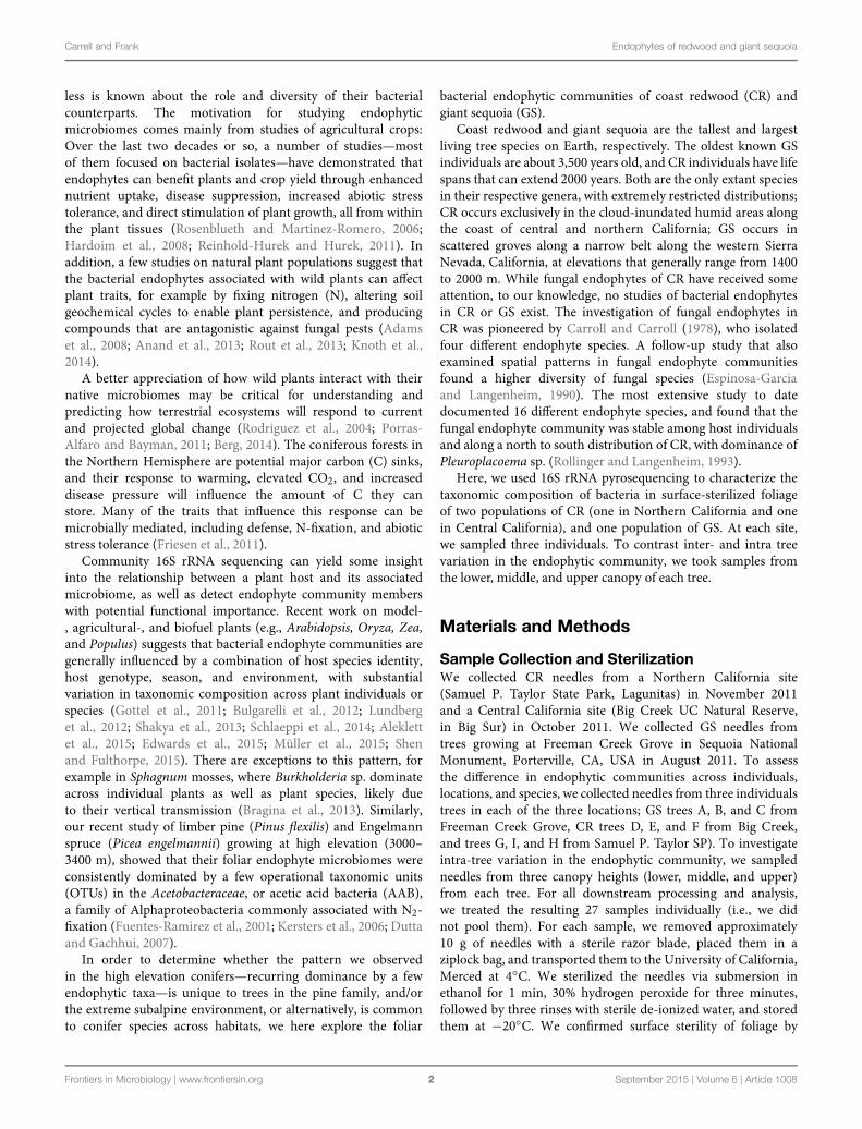
Across all samples, the most abundant phyla in all sampleswere Proteobacteria and Firmicutes, followed by Acidobacteria,Actinobacteria, TM7, and Bacteroidetes. The relative abundanceof bacterial phyla varied among samples, but Proteobacteriaor Firmicutes dominated most samples (Figure 1). Firmicuteswere significantly more abundant in GS (35% of the sequenceson average) than in CR (13% of the sequences on average;P < 0.05), and significantly different across locations (35,22, and 6% of sequences from Freeman Creek, Central CA,and Northern CA, respectively; P < 0.005). Among theProteobacteria, Alphaproteobacteria was the most prominentclass, followed by Betaproteobacteria. Among the Firmicutes,Bacilli dominated.
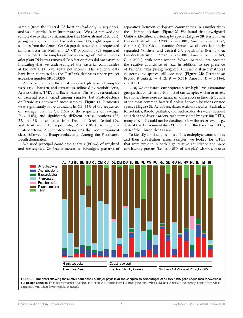
We used principal coordinate analysis (PCoA) of weightedand unweighted UniFrac distances to investigate patterns of
separation between endophyte communities in samples fromthe different locations (Figure 2). We found that unweightedUniFrac identified clustering by species (Figure 2B: Permanova:Pseudo-F statistic = 3.2009, P = 0.001; Anosim: R = 0.4557,P < 0.001). The CR communities formed two clusters that largelyseparated Northern and Central CA populations (Permanova:Pseudo-F statistic = 2.7375, P < 0.001; Anosim: R = 0.5349,P < 0.001), with some overlap. When we took into accountthe relative abundance of taxa in addition to the presenceof bacterial taxa (using weighted UniFrac distance matrices)clustering by species still occurred (Figure 2B: Permanova:Pseudo-F statistic = 6.12, P = 0.001; Anosim: R = 0.5464,P < 0.001).
Next, we examined our sequences for high-level taxonomicgroups that consistently dominated our samples within or acrosslocations. There were no significant differences in the distributionof the most common bacterial orders between locations or treespecies (Figure 3). Acidobacteriales, Actinomycetales, Bacillales,Rhizobiales, Rhodospirillales, and Burkholderiales were the mostabundant and diverse orders, each represented by over 100OTUs,many of which could not be classified below the order level (e.g.,43% of the Actinomycetales OTUs, 35% of the Bacillales OTUs,70% of the Rhizobiales OTUs).
To identify dominant members of the endophyte communitiesand their distribution across samples, we looked for OTUsthat were present in both high relative abundance and wereconsistently present (i.e., in >85% of samples) within a species
FIGURE 1 | Bar chart showing the relative abundance of major phyla in all the samples as percentages of all 16S rRNA gene sequences recovered inour foliage samples. Each bar represents a sample, and letters A–I indicate individual trees (nine total), while L, M, and U indicate the canopy location from whichthe sample was taken (lower, middle, or upper).
Frontiers in Microbiology | www.frontiersin.org 4 September 2015 | Volume 6 | Article 1008
Carrell and Frank Endophytes of redwood and giant sequoia
FIGURE 2 | Principal coordinate analysis (PCoA) of the (A) unweightedand (B) weighted UniFrac distance matrices. Points that are closertogether on the ordination have communities that are more similar. Each pointcorresponds to a sample, and shapes correspond to host tree populations.Coast redwood (CR) samples are shown in pink, and giant sequoia (GS)samples are shown in gray.
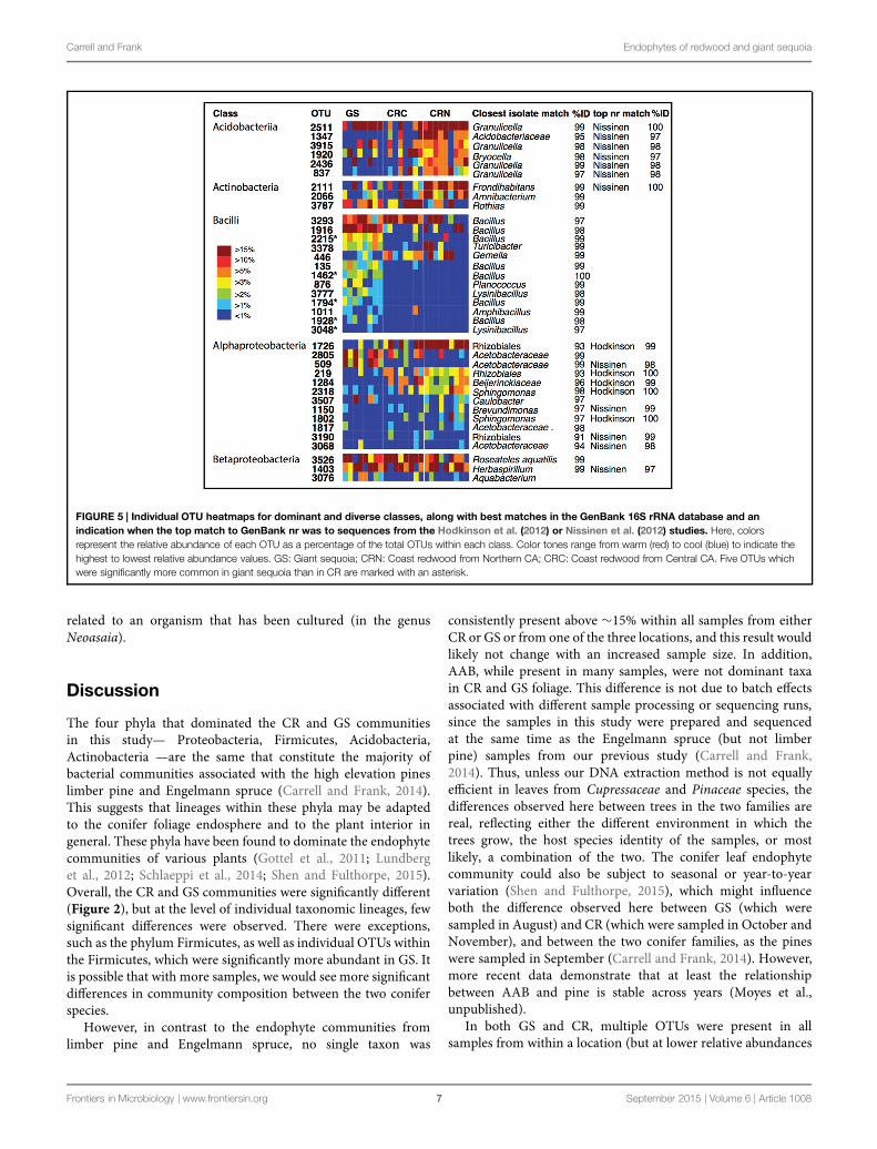
or population (hereafter referred to as core OTUs). Figure 4shows the overall 20 most common OTUs in our dataset,along with their status as core OTUs across all samples orwithin a population. In addition, to capture the dominanceand variation of OTUs within major groups (Figure 4), butthat were not necessarily among the 20 top OTUs in theentire dataset, we did this separately for the OTU-rich classesAcidobacteria, Actinobacteriia, Bacilli, Alphaproteobacteria, andBetaproteobacteria (Figure 4). The results are shown in Figure 5,where the relative abundance of each OTU is calculated as
the percentages of total OTUs within each class. In both cases(Figures 4 and 5), the resulting OTU sequences were usedto query the NCBI 16S rRNA and nr databases for matchesto isolates or uncultured sequences described in peer-reviewedmanuscripts.
In contrast to the endophyte communities from high elevationpines in our previous study, where single AAB OTUs madeup at least 15% in Engelmann spruce and 19% in limber pine(Carrell and Frank, 2014), no single taxon was consistentlypresent with a relative abundance above 15% across the samplesfrom within a site. The most common OTUs belonged to generapreviously identified as endophytes, e.g., Bacillus,Herbaspirillum,Pseudomonas, and Acetobacteraceae (Elbeltagy et al., 2001;Cocking et al., 2006; Bacon and Hinton, 2011; Bordiec et al.,2011; Figures 4 and 5). Many of our dominant OTUs in theclasses Acidobacteriia and Alphaproteobacteria (but not Bacillus)were most similar to sequences from one of two particularstudies; a study of lichen-associated bacteria (Hodkinson et al.,2012), and a study of endophytes of the cold-tolerant arcticplants Alpine sorrel (Oxyria digyna), pincushion plant (Diapensialapponica), and highland rush (Juncus trifidus) (Nissinen et al.,2012; Figures 4 and 5).
The most common OTU in our dataset (1726, Figure 4) waspresent in 85% of all CR samples, and in 100% of samples fromthe Northern CA population, where it was found in high relativeabundance (6–34%) in all but one sample. This OTU is notclosely related to any known isolate, but shares 99% identity touncultured clones in the Lichen-Associated Rhizobiales-1 (LAR1)lineage. Taxa in this lineage are prevalent and recurring in thelichen microbiome (Hodkinson and Lutzoni, 2009; Hodkinsonet al., 2012). While this OTU was not completely absent fromour GS samples, it was only present in a few samples, andonly at low relative abundance (Figure 4). In GS, severaltop OTUs were present across all samples (i.e., OTU 3293,3526, 2805, 348; Figure 5). OTU 3293, which belongs to thegenus Bacillus, was also present in all CR samples from theCentral CA population; in a few of the samples at high relativeabundance (20-40%; Figure 4). AAB, the family that was foundto recur at high relative abundance in the subalpine conifers(Carrell and Frank, 2014), did not consistently dominate thefoliar endophyte community of CR and GS, although taxabelonging to this group were present in many samples. Forexample, OTUs 2805 and 509 (Figure 4) belong to this group.OTU 2805 was found in the majority of samples from bothspecies, while OTU 509 was absent from most of the CRsamples (Figure 4). Also notable, half of the sequences fromone of the GS samples fell within an OTU with 99% identityto database sequences from to the insect symbiont Sodalisglossinidus.
We looked for dominant OTUs that were significantly morecommon in a particular location and/or species, but found nosignificant differences in the distribution of the 20most dominantOTUs between locations or tree species (Kruskal–Wallis). Onlyfive Bacillus OTUs that were not among the most dominantoverall, but which were dominant within the class Bacilli, weresignificantly more common in GS than in CR (indicated inFigure 5).
Frontiers in Microbiology | www.frontiersin.org 5 September 2015 | Volume 6 | Article 1008
Carrell and Frank Endophytes of redwood and giant sequoia
FIGURE 3 | Heatmap showing the 20 most dominant orders in our dataset and their average relative abundances as percentages of all 16S rRNAgene sequences recovered in our foliage samples, along with the total number of operational taxonomic units (OTUs) in each order. The number ofOTUs in each order that could not be classified below the order level is shown within parenthesis. Color tones range from white to dark gray to indicate the highestto lowest relative abundance values.
FIGURE 4 | Heatmap showing the 20 most dominant OTUs in our dataset, along with best matches in the GenBank 16S rRNA database, an indicationif the top GenBank nr match was a sequence from the Hodkinson et al. (2012) or Nissinen et al. (2012) studies, and their status as core OTUs acrossall samples (ALL), GS samples, CR samples, Coast redwood from Northern CA (CRN) or Coast redwood from Central CA (CRC). Within parenthesis, thepercentage of samples above which the OTU is present. Color tones range from warm (red) to cool (blue) to indicate the highest to lowest relative abundance values.
To gain better taxonomic resolution for dominantAlphaproteobacterial OTUs (such as those belonging toLAR1 and AAB discussed above), we constructed a maximumlikelihood phylogenetic tree from the Alphaproteobacterialsequences occurring more than 50 times in our samples,along with similar sequences in GenBank (≥96% identity).The phylogeny is shown in Figure 6. All Rhodospirillalessequences fell within the family Acetobacteraceae but couldnot be classified below the family level. Many were similar tosequences from Nissinen et al.’s (2012) study on arctic plants.Similarly, Rhizobiales sequences fell in uncultured lineages with
the majority putatively in the LAR1 lineage commonly associatedwith lichens (Hodkinson et al., 2012). This includes some of themost common OTUs in our dataset (e.g., 1726 and 1284), whichfell within clades together with LAR1 sequences. The sequencesclassified as belonging to the order Sphingomonadales also hadmatches to sequences from the study on arctic plants (Nissinenet al., 2012). While several of the Sphingomonadales OTUs wereclosely related (≥97% identity) to isolated bacteria (in the genusSphingomonas), only one OTU in the Rhizobiales was closelyrelated to known isolates (in the genus Methylobacterium).Similarly, only one OTU in the Rhodospirillales was closely
Frontiers in Microbiology | www.frontiersin.org 6 September 2015 | Volume 6 | Article 1008
Carrell and Frank Endophytes of redwood and giant sequoia
FIGURE 5 | Individual OTU heatmaps for dominant and diverse classes, along with best matches in the GenBank 16S rRNA database and anindication when the top match to GenBank nr was to sequences from the Hodkinson et al. (2012) or Nissinen et al. (2012) studies. Here, colorsrepresent the relative abundance of each OTU as a percentage of the total OTUs within each class. Color tones range from warm (red) to cool (blue) to indicate thehighest to lowest relative abundance values. GS: Giant sequoia; CRN: Coast redwood from Northern CA; CRC: Coast redwood from Central CA. Five OTUs whichwere significantly more common in giant sequoia than in CR are marked with an asterisk.
related to an organism that has been cultured (in the genusNeoasaia).
Discussion
The four phyla that dominated the CR and GS communitiesin this study— Proteobacteria, Firmicutes, Acidobacteria,Actinobacteria —are the same that constitute the majority ofbacterial communities associated with the high elevation pineslimber pine and Engelmann spruce (Carrell and Frank, 2014).This suggests that lineages within these phyla may be adaptedto the conifer foliage endosphere and to the plant interior ingeneral. These phyla have been found to dominate the endophytecommunities of various plants (Gottel et al., 2011; Lundberget al., 2012; Schlaeppi et al., 2014; Shen and Fulthorpe, 2015).Overall, the CR and GS communities were significantly different(Figure 2), but at the level of individual taxonomic lineages, fewsignificant differences were observed. There were exceptions,such as the phylum Firmicutes, as well as individual OTUs withinthe Firmicutes, which were significantly more abundant in GS. Itis possible that with more samples, we would see more significantdifferences in community composition between the two coniferspecies.
However, in contrast to the endophyte communities fromlimber pine and Engelmann spruce, no single taxon was
consistently present above ∼15% within all samples from eitherCR or GS or from one of the three locations, and this result wouldlikely not change with an increased sample size. In addition,AAB, while present in many samples, were not dominant taxain CR and GS foliage. This difference is not due to batch effectsassociated with different sample processing or sequencing runs,since the samples in this study were prepared and sequencedat the same time as the Engelmann spruce (but not limberpine) samples from our previous study (Carrell and Frank,2014). Thus, unless our DNA extraction method is not equallyefficient in leaves from Cupressaceae and Pinaceae species, thedifferences observed here between trees in the two families arereal, reflecting either the different environment in which thetrees grow, the host species identity of the samples, or mostlikely, a combination of the two. The conifer leaf endophytecommunity could also be subject to seasonal or year-to-yearvariation (Shen and Fulthorpe, 2015), which might influenceboth the difference observed here between GS (which weresampled in August) and CR (which were sampled in October andNovember), and between the two conifer families, as the pineswere sampled in September (Carrell and Frank, 2014). However,more recent data demonstrate that at least the relationshipbetween AAB and pine is stable across years (Moyes et al.,unpublished).
In both GS and CR, multiple OTUs were present in allsamples from within a location (but at lower relative abundances
Frontiers in Microbiology | www.frontiersin.org 7 September 2015 | Volume 6 | Article 1008
Carrell and Frank Endophytes of redwood and giant sequoia
FIGURE 6 | Maximum likelihood tree inferred using the Alphaproteobacterial sequences in our dataset that occur above 50 times total. Nodes withbootstrap support at or above 80 are indicated with a gray circle. Taxa named ‘OTU’ and with terminal branches shown in solid lines are OTUs from our dataset.Other taxa are indicated by their GenBank accession number, and in the case of isolates of known species, by species name. Taxa from the Hodkinson et al. (2012)study of lichen-associated bacteria are marked ‘Hodkinson’ and appear in blue, and taxa from the Nissinen et al. (2012) study on endophytes of arctic plants aremarked ‘Nissinen’ and appear in red. A red arrow indicates that the OTU is among the 20 most abundant in the dataset (Figure 4).
than observed in the pines in our previous study). Such coreOTUs may represent bacteria that are selected by the host,adapted to the environment inside the foliage, or present inhigh abundance in the source community (e.g., leaf surface,dust, or soil). Most notably, in CR foliage, an OTU belongingto the uncultured LAR1 lineage, which previously has onlybeen described associated with the lichen symbiosis (Hodkinsonet al., 2012), was present in all samples from the NorthernCA population, and in most samples from the Central CApopulation. Our phylogenetic analysis of Alphaproteobacterialsequences, while limited by the length of the alignment (∼300 nt),
suggests that our CR samples contain a wide a diversity oftaxa belonging to LAR1 and/or other uncultured lineages in theRhizobiales (Figure 1).
Interestingly, many of the dominant OTUs in the classesAcidobacteriia, Alphaproteobacteria, and Betaproteobacteriawere most similar to uncultured endophytes of arctic plants(Nissinen et al., 2012), and several Alphaproteobacterial OTUs—in addition to those belonging to the LAR1 lineage—weremost similar to uncultured bacteria associated with the lichensymbiosis (Hodkinson et al., 2012; Figures 5 and 6). Nissinenet al. (2012) demonstrated that many of their isolates from arctic
Frontiers in Microbiology | www.frontiersin.org 8 September 2015 | Volume 6 | Article 1008
Carrell and Frank Endophytes of redwood and giant sequoia
plants were cold-tolerant. Endophytic mediation of planttolerance to low-temperature stress has been reported ingrapevine (Theocharis et al., 2012), however cold-tolerantendophytes do not necessarily provide cold-tolerance to the hostplant.
Some possible functions of the CR and GS endophytemicrobiome are protection against host biotic and abioticstress, and N2 fixation. Several of the major and diversebacterial groups in the CR and GS foliage (e.g., Bacillus,Burkholderia, Actinomycetes) are among those known to providedefense to plant hosts though, e.g., antimicrobial and antifungalactivity (Mendes et al., 2011; Raaijmakers and Mazzola, 2012).Taxa belonging to the class Bacilli were present in all threepopulations, but were especially prominent in GS; severalOTUs from this class were significantly more common in GSthan in CR (Figure 5). Bacteria in the genus Bacillus showantagonistic activity to a wide range of potential phytopathogens,stimulate plant host defense, and are consequently exploited forbiological control of plant diseases (Ongena and Jacques, 2008;Raaijmakers and Mazzola, 2012). For example, a Bacillus pumilusendophyte isolated from phloem of healthy lodgepole pine (Pinuscontorta) is antagonistic against the fungal symbionts of thebark beetle (Dendroctonus ponderosae) (Adams et al., 2008).Likewise, Actinomycetes are well-known for their wide diversityof secondary metabolite production, many of which includeantibiotic compounds (Tiwari and Gupta, 2012), includingstrains isolated from plants (Qin et al., 2011). Actinomycetes havebeen found to dominate the culturable antifungal population inthe roots of Douglas fir (Pseudotsuga menziesii) (Axelrood et al.,1996). The Burkholderiaceae and Pseudomonadacae also harborgenera and species with activity against plant pathogenic fungi(Postma et al., 2010; Kwak et al., 2012; Suárez-Moreno et al.,2012).
The presence of these bacterial lineages in the foliage alongwith the lack of reported outbreaks of pests or diseases onCR and giant foliage is an incentive to further study theirfoliar bacterial microbiomes. For example, while redwoodforests are one of the ecosystems most threatened by theoomycete sudden oak death agent Phytophthora ramorum,infection of CR is much lower than in co-occurring speciessuch as tanoak (Lithocarpus densiflorus) and California–laurel(Umbellularia californica) and results in substantially lesssporulation from infected needles (Davidson et al., 2008).Foliar endophytic fungi may contribute heterogeneity in defensechemicals that allows the giant trees to resist disease overcenturies to millennia; unlike the host tree, their short lifecycle should allow them to respond on ecological timescales toshort-cycle pathogens and pests (Carroll, 1988). The bacterialcommunity present within the foliage is another potentialsource of defense with high potential for spatial and temporalvariability.
We previously hypothesized that AAB endophytes fixatmospheric N2 inside the needles of high elevation pines(Carrell and Frank, 2014). While AAB bacterial were onlypresent at low relative abundance in CR and GS, we found thatLAR1, a potential N2 fixing lineage associated with lichen thalli
(Hodkinson and Lutzoni, 2009), was both consistently associatedwith CR (Figure 5), and represented by diverse taxa (Figure 6).Based on the phylogenetic affiliation of the nifH sequences fromlichen, it has been hypothesized that lichen-associated bacteriain the LAR1 lineage fix and contribute atmospheric N2 to thelichen symbiosis (Grube et al., 2009; Hodkinson and Lutzoni,2009). Endophytic N2-fixation may be a source of N2 to CRs,in addition to other suspected N sources such as fog (Ewinget al., 2009). Moreover, the presence of LAR1 taxa as endophytesin CR could reflect the high abundance of epiphytic lichens inthe CR canopy (Williams and Sillett, 2007), which may shareendophytic communities with their substrate tree. Redwoods,with their complex branch architecture and long lifespan, supportlarge communities of epiphytic ferns, shrubs, and even trees(Sillett and Pelt, 2007; Williams and Sillett, 2007), all potentialhosts of endophytic communities that could be shared withthe redwood. Given the phylogenetic affinity of many of ourdominant OTUswith endophytes from distant environments andhosts (i.e., arctic plants and lichens), the potential for endophytesharing among partners in the redwood canopy ecosystem isprobably high.
Conclusion
The GS and CR trees we sampled did not host specific recurringbacterial taxa to the extent observed in high elevation conifers(Carrell and Frank, 2014); major OTUs were present buttheir relative abundance was more variable among samples.Bacterial groups known to be involved in plant defense weremajor members of the CRs and GS microbiomes, suggesting apotential role in host defense. Further studies using culturingprotocols designed to maximize the recovery of specificbacteria such as Actinomycetes (Kaewkla and Franco, 2013)could be done to assess the antimicrobial and antifungalpotential of bacteria isolated from surface-sterilized CR and GSfoliage.
Author Contributions
AC and AF conceived and designed the sampling andexperiments. AC performed the DNA extraction, and PCRamplification. AC and AF analyzed the data and wrote the article.
Acknowledgments
The authors thank Anthony Ambrose, Rikke R. Naesborg,Cameron Williams, Wendy Baxter, Chris Wong, and ToddDawson at UC Berkeley for providing us with samples,and Jason Sexton, Lara Kueppers, Dana Carper, and MikeBeman at UC Merced, as well as the two reviewers forgiving constructive comments on the manuscript. This researchwas supported by a grant from the Save-the-RedwoodsLeague.
Frontiers in Microbiology | www.frontiersin.org 9 September 2015 | Volume 6 | Article 1008
Carrell and Frank Endophytes of redwood and giant sequoia
References
Adams, A. S., Six, D. L., Adams, S. M., and Holben, W. E. (2008). In vitrointeractions between yeasts and bacteria and the fungal symbionts of themountain pine beetle (Dendroctonus ponderosae). Microb. Ecol. 56, 460–466.doi: 10.1007/s00248-008-9364-0
Aleklett, K., Leff, J. W., Fierer, N., and Hart, M. (2015). Wild plant species growingclosely connected in a subalpine meadow host distinct root-associated bacterialcommunities. Peer J. 3:e804. doi: 10.7717/peerj.804
Anand, R., Grayston, S., and Chanway, C. (2013). N2-Fixation and seedling growthpromotion of lodgepole pine by endophytic Paenibacillus polymyxa. Microb.Ecol. 66, 369–374. doi: 10.1007/s00248-013-0196-1
Arnold, A. E., Henk, D. A., Eells, R. L., Lutzoni, F., and Vilgalys, R. (2007).Diversity and phylogenetic affinities of foliar fungal endophytes in loblolly pineinferred by culturing and environmental PCR. Mycologia 99, 185–206. doi:10.3852/mycologia.99.2.185
Axelrood, P. E., Clarke, A. M., Radley, R., and Zemcov, S. J. (1996). Douglas-firroot-associated microorganisms with inhibitory activity towards fungal plantpathogens and human bacterial pathogens. Can. J. Microbiol. 42, 690–700. doi:10.1139/m96-094
Bacon, C. W., and Hinton, D. M. (2011). In planta reduction of maize seedlingstalk lesions by the bacterial endophyte Bacillus mojavensis. Can. J. Microbiol.57, 485–492. doi: 10.1139/w11-031
Berg, G. (2014). The plant microbiome and its importance for plant and humanhealth. Front. Microbiol. 5:491. doi: 10.3389/fmicb.2014.00491
Bordiec, S., Paquis, S., Lacroix, H., Dhondt, S., Ait Barka, E., Kauffmann, S., et al.(2011). Comparative analysis of defence responses induced by the endophyticplant growth-promoting rhizobacteriumBurkholderia phytofirmans strain PsJNand the non-host bacterium Pseudomonas syringae pv. pisi in grapevine cellsuspensions. J. Exp. Bot. 62, 595–603. doi: 10.1093/jxb/erq291
Bragina, A., Cardinale, M., Berg, C., and Berg, G. (2013). Vertical transmissionexplains the specific Burkholderia pattern in Sphagnum mosses at multi-geographic scale. Front. Microbiol. 4:394. doi: 10.3389/fmicb.2013.00394
Bulgarelli, D., Rott, M., Schlaeppi, K., Ver Loren van Themaat, E.,Ahmadinejad, N., Assenza, F., et al. (2012). Revealing structure and assemblycues for Arabidopsis root-inhabiting bacterial microbiota. Nature 488, 91–95.doi: 10.1038/nature11336
Caporaso, J. G., Bittinger, K., Bushman, F. D., DeSantis, T. Z., Andersen, G. L., andKnight, R. (2010a). PyNAST: a flexible tool for aligning sequences to a templatealignment. Bioinformatics 26, 266–267. doi: 10.1093/bioinformatics/btp636
Caporaso, J. G., Kuczynski, J., Stombaugh, J., Bittinger, K., Bushman,F. D., Costello, E. K., et al. (2010b). QIIME allows analysis of high-throughput community sequencing data. Nat. Methods 7, 335–336. doi:10.1038/nmeth.f.303
Carrell, A. A., and Frank, A. C. (2014). Pinus flexilis and Picea engelmannii sharea simple and consistent needle endophyte microbiota with a potential role innitrogen fixation. Front. Microbiol. 5:333. doi: 10.3389/fmicb.2014.00333
Carroll, G. (1988). Fungal endophytes in stems and leaves: from latent pathogen tomutualistic symbiont. Ecology 69, 2–9. doi: 10.2307/1943154
Carroll, G. C., and Carroll, F. E. (1978). Studies on the incidence of coniferousneedle endophytes in the Pacific Northwest. Can. J. Bot. 56, 3034–3043. doi:10.1139/b78-367
Chelius, M. K., and Triplett, E. W. (2001). The diversity of archaea and bacteriain association with the roots of Zea mays L. Microb. Ecol. 41, 252–263. doi:10.1007/s002480000087
Cocking, E. C., Stone, J. T., and Davey, M. R. (2006). Intracellular colonization ofroots of arabidopsis and crop plants by Gluconacetobacter diazotrophicus. InVitro Cell. Dev. Biol. 42, 74–82. doi: 10.1079/IVP2005716
Davidson, J. M., Patterson, H. A., and Rizzo, D. M. (2008). Sources of inoculum forPhytophthora ramorum in a redwood forest. Phytopathology 98, 860–866. doi:10.1094/PHYTO-98-8-0860
DeSantis, T. Z., Hugenholtz, P., Larsen, N., Rojas, M., Brodie, E. L., Keller, K.,et al. (2006). Greengenes, a chimera-checked 16S rRNA gene database andworkbench compatible with ARB. Appl. Environ. Microbiol. 72, 5069–72. doi:10.1128/AEM.03006-05
Dutta, D., and Gachhui, R. (2007). Nitrogen-fixing and cellulose-producingGluconacetobacter kombuchae sp. nov., isolated from Kombucha tea. Int. J. Syst.Evol. Microbiol. 57, 353–7. doi: 10.1099/ijs.0.64638-0
Edgar, R. C., Haas, B. J., Clemente, J. C., Quince, C., and Knight, R. (2011).UCHIME improves sensitivity and speed of chimera detection. Bioinformatics27, 2194–2200. doi: 10.1093/bioinformatics/btr381
Edwards, J., Johnson, C., Santos-Medellín, C., Lurie, E., Podishetty, N. K.,Bhatnagar, S., et al. (2015). Structure, variation, and assembly of the root-associated microbiomes of rice. Proc. Natl. Acad. Sci. U.S.A. 112, E911–E920.doi: 10.1073/pnas.1414592112
Elbeltagy, A., Nishioka, K., Sato, T., Suzuki, H., Ye, B., Hamada, T., et al. (2001).Endophytic colonization and in planta nitrogen fixation by aHerbaspirillum sp.isolated from wild rice species. Appl. Environ. Microbiol. 67, 5285–5293. doi:10.1128/AEM.67.11.5285-5293.2001
Espinosa-Garcia, F. J., and Langenheim, J. H. (1990). The endophytic fungalcommunity in leaves of a coastal redwood population diversity and spatialpatterns. New Phytol. 116, 89–97. doi: 10.1111/j.1469-8137.1990.tb00513.x
Ewing, H. A., Weathers, K. C., Templer, P. H., Dawson, T. E., Firestone, M. K.,Elliott, A. M., et al. (2009). Fog water and ecosystem function: heterogeneity ina california redwood forest. Ecosystems 12, 417–433. doi: 10.1007/s10021-009-9232-x
Friesen, M. L., Porter, S. S., Stark, S. C., von Wettberg, E. J., Sachs, J. L., andMartinez-Romero, E. (2011). Microbially mediated plant functional traits.Annu. Rev. Ecol. Evol. Syst. 42, 23–46. doi: 10.1146/annurev-ecolsys-102710-145039
Fuentes-Ramirez, L. E., Bustillos-Cristales, R., Tapia-Hernandez, A., Jimenez-Salgado, T., Wang, E. T., Martinez-Romero, E., et al. (2001). Novelnitrogen-fixing acetic acid bacteria, Gluconacetobacter johannae sp. nov. andGluconacetobacter azotocaptans sp. nov., associated with coffee plants. Int. J.Syst. Evol. Microbiol. 51, 1305–1314
Ganley, R. J., Brunsfeld, S. J., and Newcombe, G. (2004). A community of unknown,endophytic fungi in western white pine. Proc. Natl. Acad. Sci. U.S.A. 101,10107–10112. doi: 10.1073/pnas.0401513101
Gottel, N. R., Castro, H. F., Kerley, M., Yang, Z., Pelletier, D. A., Podar, M., et al.(2011). Distinct microbial communities within the endosphere and rhizosphereof Populus deltoides roots across contrasting soil types.Appl. Environ. Microbiol.77, 5934–44. doi: 10.1128/AEM.05255-11
Grube, M., Cardinale, M., de Castro, J. V., Müller, H., and Berg, G. (2009). Species-specific structural and functional diversity of bacterial communities in lichensymbioses. ISME J. 3, 1105–1115. doi: 10.1038/ismej.2009.63
Hardoim, P. R., van Overbeek, L. S., and Elsas, J. D. (2008). Properties of bacterialendophytes and their proposed role in plant growth. Trends Microbiol. 16,463–471. doi: 10.1016/j.tim.2008.07.008
Hodkinson, B. P., Gottel, N. R., Schadt, C. W., and Lutzoni, F. (2012).Photoautotrophic symbiont and geography are major factors affecting highlystructured and diverse bacterial communities in the lichen microbiome.Environ. Microbiol. 14, 147–161. doi: 10.1111/j.1462-2920.2011.02560.x
Hodkinson, B. P., and Lutzoni, F. (2009). A microbiotic survey of lichen-associatedbacteria reveals a new lineage from the Rhizobiales. Symbiosis 49, 163–180. doi:10.1007/s13199-009-0049-3
Jiao, J. Y., Wang, H. X., Zeng, Y., and Shen, Y. M. (2006). Enrichment for microbesliving in association with plant tissues. J. Appl. Microbiol. 100, 830–837. doi:10.1111/j.1365-2672.2006.02830.x
Kaewkla, O., and Franco, C. M. (2013). Rational approaches to improving theisolation of endophytic actinobacteria from Australian native trees. Microb.Ecol. 65, 384–393. doi: 10.1007/s00248-012-0113-z
Kersters, K., Lisdiyanti, P., Komagata, K., and Swings, J. (2006). “The familyacetobacteraceae: the genera acetobacter, acidomonas, asaia, gluconacetobacter,gluconobacter, and kozakia,” in The prokaryotes, eds M. Dworkin, S. Falkow,E. Rosenberg, K. H. Schleifer, and E. Stackebrandt (New York, NY: Springer),163–200.
Knoth, J. L., Kim, S.-H., Ettl, G. J., and Doty, S. L. (2014). Biological nitrogenfixation and biomass accumulation within poplar clones as a result ofinoculations with diazotrophic endophyte consortia. New Phytol. 201, 599–609.doi: 10.1111/nph.12536
Kwak, M. -J., Song, J. Y., Kim, S.-Y., Jeong, H., Kang, S. G., Kim, B. K., et al.(2012). Complete genome sequence of the endophytic bacterium Burkholderiasp. strain KJ006. J. Bacteriol. 194, 4432–4433. doi: 10.1128/JB.00821-12
Letunic, I., and Bork, P. (2011). Interactive Tree Of Life v2: online annotation anddisplay of phylogenetic trees made easy. Nucleic Acids Res. 39, W475–W478.doi: 10.1093/nar/gkr201
Frontiers in Microbiology | www.frontiersin.org 10 September 2015 | Volume 6 | Article 1008
Carrell and Frank Endophytes of redwood and giant sequoia
Lozupone, C., and Knight, R. (2005). UniFrac: a new phylogenetic method forcomparing microbial communities. Appl. Environ. Microbiol. 71, 8228–8235.doi: 10.1128/AEM.71.12.8228-8235.2005
Lundberg, D. S., Lebeis, S. L., Paredes, S. H., Yourstone, S., Gehring, J., Malfatti, S.,et al. (2012). Defining the core Arabidopsis thaliana root microbiome. Nature488, 86–90. doi: 10.1038/nature11237
Mendes, R., Kruijt, M., de Bruijn, I., Dekkers, E., van der Voort, M., Schneider, J. H.,et al. (2011). Deciphering the rhizosphere microbiome for disease-suppressivebacteria. Science 332, 1097–1100. doi: 10.1126/science.1203980
Müller, H., Berg, C., Landa, B. B., Auerbach, A., Moissl-Eichinger, C., and Berg, G.(2015). Plant genotype-specific archaeal and bacterial endophytes but similarBacillus antagonists colonize Mediterranean olive trees. Front. Microbiol. 6:138.doi: 10.3389/fmicb.2015.00138
Nawrocki, E. P., Kolbe, D. L., and Eddy, S. R. (2009). Infernal 1.0: inference of RNAalignments. Bioinformatics 25, 1335–1337. doi: 10.1093/bioinformatics/btp157
Nissinen, R. M., Männistö, M. K., and van Elsas, J. D. (2012). Endophytic bacterialcommunities in three arctic plants from low arctic fell tundra are cold-adaptedand host-plant specific. FEMSMicrobiol. Ecol. 82, 510–522. doi: 10.1111/j.1574-6941.2012.01464.x
Ongena, M., and Jacques, P. (2008). Bacillus lipopeptides: versatile weaponsfor plant disease biocontrol. Trends Microbiol. 16, 115–125. doi:10.1016/j.tim.2007.12.009
Oono, R., Lutzoni, F., Arnold, A. E., Kaye, L., U’Ren, J. M., May, G., et al. (2014).Genetic variation in horizontally transmitted fungal endophytes of pine needlesreveals population structure in cryptic species. Am. J. Bot. 101, 1362–1374. doi:10.3732/ajb.1400141
Peñuelas, J., and Terradas, J. (2014). The foliar microbiome. Trends Plant Sci. 19,278–280. doi: 10.1016/j.tplants.2013.12.007
Porras-Alfaro, A., and Bayman, P. (2011). Hidden fungi, emergent properties:endophytes and microbiomes. Annu. Rev. Phytopathol. 49, 291–315. doi:10.1146/annurev-phyto-080508-081831
Postma, J., Scheper, R. W. A., and Schilder, M. T. (2010). Effect of successivecauliflower plantings and Rhizoctonia solani AG 2-1 inoculations on diseasesuppressiveness of a suppressive and a conducive soil. Soil Biol. Biochem. 42,804–812. doi: 10.1016/j.soilbio.2010.01.017
Price, M. N., Dehal, P. S., and Arkin, A. P. (2009). FastTree: computing largeminimum evolution trees with profiles instead of a distance matrix. Mol. Biol.Evol. 26, 1641–1650. doi: 10.1093/molbev/msp077
Qadri, M., Rajput, R., Abdin, M. Z., Vishwakarma, R. A., and Riyaz-Ul-Hassan, S.(2014). Diversity, molecular phylogeny, and bioactive potential of fungalendophytes associated with the himalayan blue pine (Pinus wallichiana).Microb. Ecol. 67, 877–887. doi: 10.1007/s00248-014-0379-4
Qin, S., Xing, K., Jiang, J.-H., Xu, L.-H., and Li, W.-J. (2011). Biodiversity, bioactivenatural products and biotechnological potential of plant-associated endophyticactinobacteria. Appl. Microbiol. Biotechnol. 89, 457–473. doi: 10.1007/s00253-010-2923-6
Raaijmakers, J. M., and Mazzola, M. (2012). Diversity and natural functions ofantibiotics produced by beneficial and plant pathogenic bacteria. Annu. Rev.Phytopathol. 50, 403–424. doi: 10.1146/annurev-phyto-081211-172908
Redford, A. J., Bowers, R. M., Knight, R., Linhart, Y., and Fierer, N. (2010). Theecology of the phyllosphere: geographic and phylogenetic variability in thedistribution of bacteria on tree leaves. Environ. Microbiol. 12, 2885–2893. doi:10.1111/j.1462-2920.2010.02258.x
Reinhold-Hurek, B., and Hurek, T. (2011). Living inside plants: bacterialendophytes. Curr. Opin. Plant Biol. 14, 435–443. doi: 10.1016/j.pbi.2011.04.004
Rodriguez, R. J., Redman, R. S., and Henson, J. M. (2004). the role of fungalsymbioses in the adaptation of plants to high stress environments.Mitig. Adapt.Strateg. Glob. Change 9, 1573–1596. doi: 10.1023/B:MITI.0000029922.31110.97
Rollinger, J. L., and Langenheim, J. H. (1993). Geographic survey of fungalendophyte community composition in leaves of coastal redwood.Mycologia 85,149. doi: 10.2307/3760450
Rosenblueth, M., and Martinez-Romero, E. (2006). Bacterial endophytes andtheir interactions with hosts. Mol. Plant Microbe Interact. 19, 827–837. doi:10.1094/MPMI-19-0827
Rout, M. E., Chrzanowski, T. H., Westlie, T. K., Deluca, T. H., Callaway, R. M., andHolben, W. E. (2013). Bacterial endophytes enhance competition by invasiveplants. Am. J. Bot. 100, 1726–1737. doi: 10.3732/ajb.1200577
Schlaeppi, K., Dombrowski, N., Oter, R. G., Ver Loren van Themaat, E.,and Schulze-Lefert, P. (2014). Quantitative divergence of the bacterial rootmicrobiota in Arabidopsis thaliana relatives. Proc. Natl. Acad. Sci. U.S.A. 111,585–592. doi: 10.1073/pnas.1321597111
Shakya, M., Gottel, N., Castro, H., Yang, Z. K., Gunter, L., LabbÃ, J., et al. (2013).A multifactor analysis of fungal and bacterial community structure in theroot microbiome of mature populus deltoides trees. PLoS ONE 8:e76382 doi:10.1371/journal.pone.0076382
Shen, S. Y., and Fulthorpe, R. (2015). Seasonal variation of bacterial endophytes inurban trees. Front. Microbiol. 6:427. doi: 10.3389/fmicb.2015.00427
Sillett, S. C., and Pelt, R. V. (2007). Trunk reiteration promotes epiphytes and waterstorage in an old-growth redwood forest canopy. Ecol. Monogr. 77, 335–359.doi: 10.1890/06-0994.1
Stamatakis, A., Ludwig, T., and Meier, H. (2005). RAxML-III: a fast program formaximum likelihood-based inference of large phylogenetic trees. Bioinformatics21, 456–463. doi: 10.1093/bioinformatics/bti191
Suárez-Moreno, Z. R., Caballero-Mellado, J., Coutinho, B. G., Mendonça-Previato, L., James, E. K., and Venturi, V. (2012). Common features ofenvironmental and potentially beneficial plant-associated burkholderia.Microb.Ecol. 63, 249–266. doi: 10.1007/s00248-011-9929-1
Theocharis, A., Bordiec, S., Fernandez, O., Paquis, S., Dhondt-Cordelier, S.,Baillieul, F., et al. (2012). Burkholderia phytofirmans PsJN primes Vitis viniferaL. and confers a better tolerance to low nonfreezing temperatures. Mol. PlantMicrobe Interact. 25, 241–249. doi: 10.1094/MPMI-05-11-0124
Tiwari, K., and Gupta, R. K. (2012). Rare actinomycetes: a potentialstorehouse for novel antibiotics. Crit. Rev. Biotechnol. 32, 108–132. doi:10.3109/07388551.2011.562482
Turner, T. R., James, E. K., and Poole, P. S. (2013). The plant microbiome. GenomeBiol. 14, 209. doi: 10.1186/gb-2013-14-6-209
Wang, Q., Garrity, G. M., Tiedje, J. M., and Cole, J. R. (2007). Naive Bayesianclassifier for rapid assignment of rRNA sequences into the new bacterialtaxonomy. Appl. Environ. Microbiol. 73, 5261–5267. doi: 10.1128/AEM.00062-07
Williams, C. B., and Sillett, S. C. (2007). Epiphyte communities on redwood(Sequoia sempervirens) in northwestern California. Bryologist 110, 420–452.doi: 10.1639/0007-2745(2007)11
Conflict of Interest Statement: The Guest Associate Editor Mysore Tejesvideclares that despite having hosted a Frontiers Research Topic with the authorAnna C. Frank, the review process was handled objectively. The authors declarethat the research was conducted in the absence of any commercial or financialrelationships that could be construed as a potential conflict of interest.