International Journal of Molecular Sciences Review Bacterial Responses to Glyoxal and Methylglyoxal: Reactive Electrophilic Species Changhan Lee † and Chankyu Park * Department of Biological Sciences, Korea Advanced Institute of Science and Technology, Yuseong-gu, Daejeon 305-701, Korea; [email protected]* Correspondence: [email protected]; Tel.: +82-42-350-2629; Fax: +82-42-350-2610 † Current address: Department of Microbiology, Tumor and Cell Biology, Karolinska Institute, 17177 Stockholm, Sweden. Academic Editor: Casper G. Schalkwijk Received: 15 December 2016; Accepted: 11 January 2017; Published: 17 January 2017 Abstract: Glyoxal (GO) and methylglyoxal (MG), belonging to α-oxoaldehydes, are produced by organisms from bacteria to humans by glucose oxidation, lipid peroxidation, and DNA oxidation. Since glyoxals contain two adjacent reactive carbonyl groups, they are referred to as reactive electrophilic species (RES), and are damaging to proteins and nucleotides. Therefore, glyoxals cause various diseases in humans, such as diabetes and neurodegenerative diseases, from which all living organisms need to be protected. Although the glyoxalase system has been known for some time, details on how glyoxals are sensed and detoxified in the cell have not been fully elucidated, and are only beginning to be uncovered. In this review, we will summarize the current knowledge on bacterial responses to glyoxal, and specifically focus on the glyoxal-associated regulators YqhC and NemR, as well as their detoxification mediated by glutathione (GSH)-dependent/independent glyoxalases and NAD(P)H-dependent reductases. Furthermore, we will address questions and future directions. Keywords: glyoxal; reactive electrophilic species (RES); glyoxalase 1. Introduction Reactive electrophilic species (RES) are compounds containing α,β-unsaturated carbonyl or other electrophilic groups (Figure 1A) [1]. Because of their reactivity, they interact with macromolecules and affect cellular redox, mediated by redox cofactors like glutathione (GSH) and NAD(P)H [2,3]. The results often involve cellular malfunction. α-Oxoaldehydes—glyoxal (GO) and methylglyoxal (MGO), containing two reactive carbonyl groups (Figure 1B)—are examples of RES. Although stress responses to reactive oxygen species (ROS) and nitrogen species (RNS) have been extensively studied, research on the response to RES have emerged only recently. GO and MGO were reported to be associated with aging and diseases such as diabetes, Alzheimer’s, and Parkinson’s diseases [4–6]. However, cellular responses to glyoxals are largely unknown. Although the major glyoxal-detoxification systems using glutathione are present in most species from bacteria to humans [7], a number of issues on sensing and detoxification of glyoxals still remains to be investigated. Recently, considerable efforts have been made to study cellular responses to glyoxals for bacteria as a model system in terms of their formation, cytotoxic targets, and detoxification. In this review, we will summarize recent findings on cellular responses to GO/MGO and its detoxification. Int. J. Mol. Sci. 2017, 18, 169; doi:10.3390/ijms18010169 www.mdpi.com/journal/ijms
Transcript
International Journal of
Molecular Sciences
Review
Bacterial Responses to Glyoxal and Methylglyoxal:Reactive Electrophilic Species
Changhan Lee † and Chankyu Park *
Department of Biological Sciences, Korea Advanced Institute of Science and Technology, Yuseong-gu,Daejeon 305-701, Korea; [email protected]* Correspondence: [email protected]; Tel.: +82-42-350-2629; Fax: +82-42-350-2610† Current address: Department of Microbiology, Tumor and Cell Biology, Karolinska Institute,
17177 Stockholm, Sweden.
Academic Editor: Casper G. SchalkwijkReceived: 15 December 2016; Accepted: 11 January 2017; Published: 17 January 2017
Abstract: Glyoxal (GO) and methylglyoxal (MG), belonging to α-oxoaldehydes, are produced byorganisms from bacteria to humans by glucose oxidation, lipid peroxidation, and DNA oxidation.Since glyoxals contain two adjacent reactive carbonyl groups, they are referred to as reactiveelectrophilic species (RES), and are damaging to proteins and nucleotides. Therefore, glyoxals causevarious diseases in humans, such as diabetes and neurodegenerative diseases, from which all livingorganisms need to be protected. Although the glyoxalase system has been known for some time,details on how glyoxals are sensed and detoxified in the cell have not been fully elucidated, and areonly beginning to be uncovered. In this review, we will summarize the current knowledge on bacterialresponses to glyoxal, and specifically focus on the glyoxal-associated regulators YqhC and NemR,as well as their detoxification mediated by glutathione (GSH)-dependent/independent glyoxalasesand NAD(P)H-dependent reductases. Furthermore, we will address questions and future directions.
Keywords: glyoxal; reactive electrophilic species (RES); glyoxalase
1. Introduction
Reactive electrophilic species (RES) are compounds containing α,β-unsaturated carbonyl or otherelectrophilic groups (Figure 1A) [1]. Because of their reactivity, they interact with macromoleculesand affect cellular redox, mediated by redox cofactors like glutathione (GSH) and NAD(P)H [2,3].The results often involve cellular malfunction. α-Oxoaldehydes—glyoxal (GO) and methylglyoxal(MGO), containing two reactive carbonyl groups (Figure 1B)—are examples of RES. Although stressresponses to reactive oxygen species (ROS) and nitrogen species (RNS) have been extensively studied,research on the response to RES have emerged only recently. GO and MGO were reported to be associatedwith aging and diseases such as diabetes, Alzheimer’s, and Parkinson’s diseases [4–6]. However, cellularresponses to glyoxals are largely unknown. Although the major glyoxal-detoxification systems usingglutathione are present in most species from bacteria to humans [7], a number of issues on sensingand detoxification of glyoxals still remains to be investigated. Recently, considerable efforts have beenmade to study cellular responses to glyoxals for bacteria as a model system in terms of their formation,cytotoxic targets, and detoxification. In this review, we will summarize recent findings on cellularresponses to GO/MGO and its detoxification.
Int. J. Mol. Sci. 2017, 18, 169; doi:10.3390/ijms18010169 www.mdpi.com/journal/ijms
Int. J. Mol. Sci. 2017, 18, 169 2 of 12Int. J. Mol. Sci. 2017, 18, 169 2 of 11
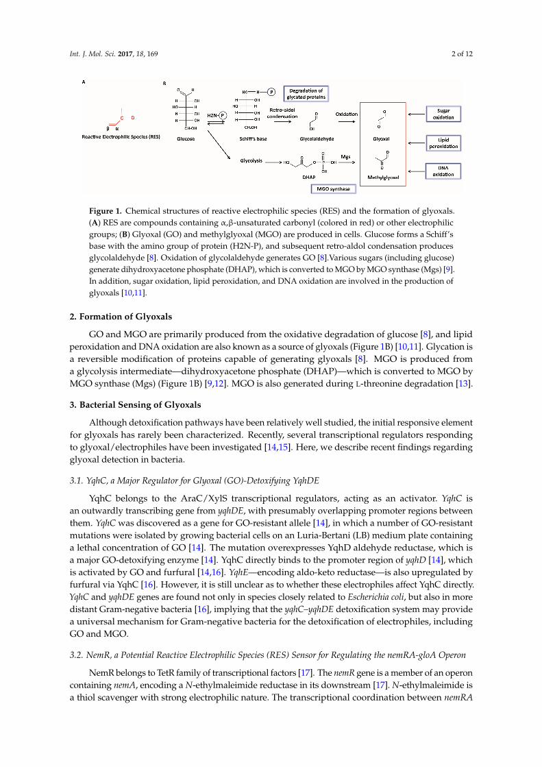
Figure 1. Chemical structures of reactive electrophilic species (RES) and the formation of glyoxals. (A) RES are compounds containing α,β-unsaturated carbonyl (colored in red) or other electrophilic groups; (B) Glyoxal (GO) and methylglyoxal (MGO) are produced in cells. Glucose forms a Schiff’s base with the amino group of protein (H2N-P), and subsequent retro-aldol condensation produces glycolaldehyde [8]. Oxidation of glycolaldehyde generates GO [8].Various sugars (including glucose) generate dihydroxyacetone phosphate (DHAP), which is converted to MGO by MGO synthase (Mgs) [9]. In addition, sugar oxidation, lipid peroxidation, and DNA oxidation are involved in the production of glyoxals [10,11].
2. Formation of Glyoxals
GO and MGO are primarily produced from the oxidative degradation of glucose [8], and lipid peroxidation and DNA oxidation are also known as a source of glyoxals (Figure 1B) [10,11]. Glycation is a reversible modification of proteins capable of generating glyoxals [8]. MGO is produced from a glycolysis intermediate—dihydroxyacetone phosphate (DHAP)—which is converted to MGO by MGO synthase (Mgs) (Figure 1B) [9,12]. MGO is also generated during L-threonine degradation [13].
3. Bacterial Sensing of Glyoxals
Although detoxification pathways have been relatively well studied, the initial responsive element for glyoxals has rarely been characterized. Recently, several transcriptional regulators responding to glyoxal/electrophiles have been investigated [14,15]. Here, we describe recent findings regarding glyoxal detection in bacteria.
3.1. YqhC, a Major Regulator for Glyoxal (GO)-Detoxifying YqhDE
YqhC belongs to the AraC/XylS transcriptional regulators, acting as an activator. YqhC is an outwardly transcribing gene from yqhDE, with presumably overlapping promoter regions between them. YqhC was discovered as a gene for GO-resistant allele [14], in which a number of GO-resistant mutations were isolated by growing bacterial cells on an Luria-Bertani (LB) medium plate containing a lethal concentration of GO [14]. The mutation overexpresses YqhD aldehyde reductase, which is a major GO-detoxifying enzyme [14]. YqhC directly binds to the promoter region of yqhD [14], which is activated by GO and furfural [14,16]. YqhE—encoding aldo-keto reductase—is also upregulated by furfural via YqhC [16]. However, it is still unclear as to whether these electrophiles affect YqhC directly. YqhC and yqhDE genes are found not only in species closely related to Escherichia coli, but also in more distant Gram-negative bacteria [16], implying that the yqhC–yqhDE detoxification system may provide a universal mechanism for Gram-negative bacteria for the detoxification of electrophiles, including GO and MGO.
3.2. NemR, a Potential Reactive Electrophilic Species (RES) Sensor for Regulating the nemRA-gloA Operon
NemR belongs to TetR family of transcriptional factors [17]. The nemR gene is a member of an operon containing nemA, encoding a N-ethylmaleimide reductase in its downstream [17]. N-ethylmaleimide is a thiol scavenger with strong electrophilic nature. The transcriptional coordination between nemRA and gloA genes was recently reported [15,18]. A mutation in the NemR-binding site of the nemRA operon was also isolated with a GO-resistant phenotype [15]. Since
Figure 1. Chemical structures of reactive electrophilic species (RES) and the formation of glyoxals.(A) RES are compounds containing α,β-unsaturated carbonyl (colored in red) or other electrophilicgroups; (B) Glyoxal (GO) and methylglyoxal (MGO) are produced in cells. Glucose forms a Schiff’sbase with the amino group of protein (H2N-P), and subsequent retro-aldol condensation producesglycolaldehyde [8]. Oxidation of glycolaldehyde generates GO [8].Various sugars (including glucose)generate dihydroxyacetone phosphate (DHAP), which is converted to MGO by MGO synthase (Mgs) [9].In addition, sugar oxidation, lipid peroxidation, and DNA oxidation are involved in the production ofglyoxals [10,11].
2. Formation of Glyoxals
GO and MGO are primarily produced from the oxidative degradation of glucose [8], and lipidperoxidation and DNA oxidation are also known as a source of glyoxals (Figure 1B) [10,11]. Glycation isa reversible modification of proteins capable of generating glyoxals [8]. MGO is produced froma glycolysis intermediate—dihydroxyacetone phosphate (DHAP)—which is converted to MGO byMGO synthase (Mgs) (Figure 1B) [9,12]. MGO is also generated during L-threonine degradation [13].
3. Bacterial Sensing of Glyoxals
Although detoxification pathways have been relatively well studied, the initial responsive elementfor glyoxals has rarely been characterized. Recently, several transcriptional regulators respondingto glyoxal/electrophiles have been investigated [14,15]. Here, we describe recent findings regardingglyoxal detection in bacteria.
3.1. YqhC, a Major Regulator for Glyoxal (GO)-Detoxifying YqhDE
YqhC belongs to the AraC/XylS transcriptional regulators, acting as an activator. YqhC isan outwardly transcribing gene from yqhDE, with presumably overlapping promoter regions betweenthem. YqhC was discovered as a gene for GO-resistant allele [14], in which a number of GO-resistantmutations were isolated by growing bacterial cells on an Luria-Bertani (LB) medium plate containinga lethal concentration of GO [14]. The mutation overexpresses YqhD aldehyde reductase, which isa major GO-detoxifying enzyme [14]. YqhC directly binds to the promoter region of yqhD [14], whichis activated by GO and furfural [14,16]. YqhE—encoding aldo-keto reductase—is also upregulated byfurfural via YqhC [16]. However, it is still unclear as to whether these electrophiles affect YqhC directly.YqhC and yqhDE genes are found not only in species closely related to Escherichia coli, but also in moredistant Gram-negative bacteria [16], implying that the yqhC–yqhDE detoxification system may providea universal mechanism for Gram-negative bacteria for the detoxification of electrophiles, includingGO and MGO.
3.2. NemR, a Potential Reactive Electrophilic Species (RES) Sensor for Regulating the nemRA-gloA Operon
NemR belongs to TetR family of transcriptional factors [17]. The nemR gene is a member of an operoncontaining nemA, encoding a N-ethylmaleimide reductase in its downstream [17]. N-ethylmaleimide isa thiol scavenger with strong electrophilic nature. The transcriptional coordination between nemRA
Int. J. Mol. Sci. 2017, 18, 169 3 of 12
and gloA genes was recently reported [15,18]. A mutation in the NemR-binding site of the nemRAoperon was also isolated with a GO-resistant phenotype [15]. Since the gloA gene is a member ofthe nemRA operon, a point mutation at the NemR-binding site results in over-expression of GloA,thereby conferring resistance to glyoxal. Consistently, a transcriptomic analysis revealed that thenemRA and gloA genes are co-expressed upon treatment with MGO [15,18]. NemA is a member ofthe old yellow enzymes with flavin mononucleotide (FMN) as a cofactor, reducing various aldehydecompounds, such as glyoxals and quinones [19,20]. Quinones are electrophilic, serving as electrondonors in the respiratory system. DNA binding affinity of NemR decreases upon the addition ofGO/MGO or quinones, but not with their reduction products (i.e., 1,2-ethandiol/1,2-propanediol andquinol) [15]. In addition, NemR serves as a sensor for reactive chlorine species (RCS), which are strongoxidants with electrophilic activity [21,22]. NemR appears to be a specialized sensor for RES, becausethe DNA-binding affinity and transcription level of the nemRA-gloA operon is not affected by ROS [15].On the other hand, the transcriptional regulator RutR binds to the upstream region of the nemRA-gloAoperon, although it does not play a role in the regulation of nemRA-gloA [15,17].
Cysteine residues play a crucial role in nemRA-gloA regulation. Among six cysteine residues(21, 98, 106, 116, 149, and 153) of E. coli NemR, two at 21 and 116 were demonstrated to be essential inresponding to electrophiles (including GO and MGO), in which formation of intermolecular disulfidebonds involving these residues were required both in vivo and in vitro [15]. RES and glyoxals oxidizecysteines, thereby forming disulfide bonds. This type of redox regulation is basically reversible, suchthat an oxidized NemR can be reactivated by a reducing agent. However, the cysteine at position 106that is conserved among NemR homologs has been identified as a critical residue for responding toRCS, such as hypochlorous acid [21]. Recently, the formation of a Cys106-Lys175 sulfenamide bondwas shown to serve as a thiol-based redox switch to modulate DNA-binding of NemR in responseto RCS [23]. It is of interest to note that different stress conditions involve modifications of differentcysteine residues to regulate the activity of NemR. In summary, NemR functions as an RES-specificregulator through various modifications (such as alkylation and oxidation) of cysteine residues.
3.3. CRP/cAMP-Mediated Detoxification Pathway for Glyoxal
CRP is a global transcriptional regulator for more than 180 genes, and requires cAMP [24]. The crpmutant was recently found to be associated with GO resistance [25], suggesting that cAMP and CRPare involved in responding to GO and its detoxification. Consistently, a mutation of cya gene encodingadenylate cyclase has been shown to confer resistance to GO/MGO [25]. From the study of genesregulated by cAMP/CRP (which are directly involved in the detoxification of GO/MGO), we were able toreveal new members of cAMP/CRP regulon: yqhC (transcriptional regulator), yqhD (NADPH-dependentaldehyde reductase), yafB (NADPH-dependent aldo-keto reductase), sodB (superoxide dismutase), andgloA (glyoxalase I). These genes are negatively regulated by CRP. In addition to cAMP/CRP, the gloAgene is also known to be regulated by NemR when it is expressed as part of the nemRA-gloA operon.It was shown that the level of nemRA transcript was unaffected by CRP. Therefore, it was inferredthat CRP only affects gloA by acting on its own promoter [15,25]. Conclusively, it was found thatgloA is regulated by dual promoters with two different transcriptional regulators. Since the cAMPlevel and glucose uptake are inversely correlated through the phosphotransferase system (PTS) [26],an expression of glyoxalase I might be necessary for the reduction of intracellular glyoxals producedfrom the oxidative degradation of glucose.
3.4. Fnr and NsrR—Regulators of YafB Aldo-Keto Reductase
Aldo-keto reductases (AKRs) play a crucial role in the detoxification of glyoxals by reducingGO and MGO to glycolaldehyde and acetol, respectively [27,28]. Among the several AKRs of E. coli,the YafB protein is a major one, based on the fact that the screening of GO-resistant mutant from thestrain lacking yqhD and gloA yielded a number of mutants overexpressing YafB through genomicrearrangements, such as multi-base deletions and recombination in the upstream region of the yafB
Int. J. Mol. Sci. 2017, 18, 169 4 of 12
gene [29]. As a result of the genomic rearrangements, transcriptional fusions of yafB to the upstreamrrn occurred. In addition, the negative regulatory sites for NsrR and Fnr were removed, resulting inenhancement of YafB expression [29]. NsrR is a nitrite-sensitive transcription repressor sensing nitritedue to its iron-sulfur [2Fe-2S] cluster essential for sensing redox change [30]. Fnr is a redox-responsivetranscriptional factor harboring [4Fe-4S] cluster to sense oxygen and nitric oxide [31]. Fnr is alsoinvolved in the activation and repression of genes in anaerobic and aerobic metabolism [31,32].YafB expression is increased upon GO treatment by de-repressing NsrR/Fnr [29]. However, thede-repression by NsrR/Fnr is directly influenced by GO or indirectly by redox imbalance caused byGO. Interestingly, among AKRs, transcripts of yghZ and yajO are reduced upon exposure to GO [28].This may indicate the presence of regulatory divergence in various stress conditions.
4. Detoxification of Glyoxals
Living organisms employ various means of detoxifying reactive GO and MGO. Bacterialdetoxifications are carried out primarily by the following two systems: glyoxalase and NAD(P)H-dependent detoxification enzyme (Figure 2). Glyoxalases are further classified based on their cofactorrequirement; GSH-dependent or -independent. Except for the well-known glyoxalase I/II, other enzymesassociated with GO/MGO are listed in Table 1 with their enzymatic characteristics.
Int. J. Mol. Sci. 2017, 18, 169 4 of 11
due to its iron-sulfur [2Fe-2S] cluster essential for sensing redox change [30]. Fnr is a redox-responsive transcriptional factor harboring [4Fe-4S] cluster to sense oxygen and nitric oxide [31]. Fnr is also involved in the activation and repression of genes in anaerobic and aerobic metabolism [31,32]. YafB expression is increased upon GO treatment by de-repressing NsrR/Fnr [29]. However, the de-repression by NsrR/Fnr is directly influenced by GO or indirectly by redox imbalance caused by GO. Interestingly, among AKRs, transcripts of yghZ and yajO are reduced upon exposure to GO [28]. This may indicate the presence of regulatory divergence in various stress conditions.
4. Detoxification of Glyoxals
Living organisms employ various means of detoxifying reactive GO and MGO. Bacterial detoxifications are carried out primarily by the following two systems: glyoxalase and NAD(P)H-dependent detoxification enzyme (Figure 2). Glyoxalases are further classified based on their cofactor requirement; GSH-dependent or -independent. Except for the well-known glyoxalase I/II, other enzymes associated with GO/MGO are listed in Table 1 with their enzymatic characteristics.
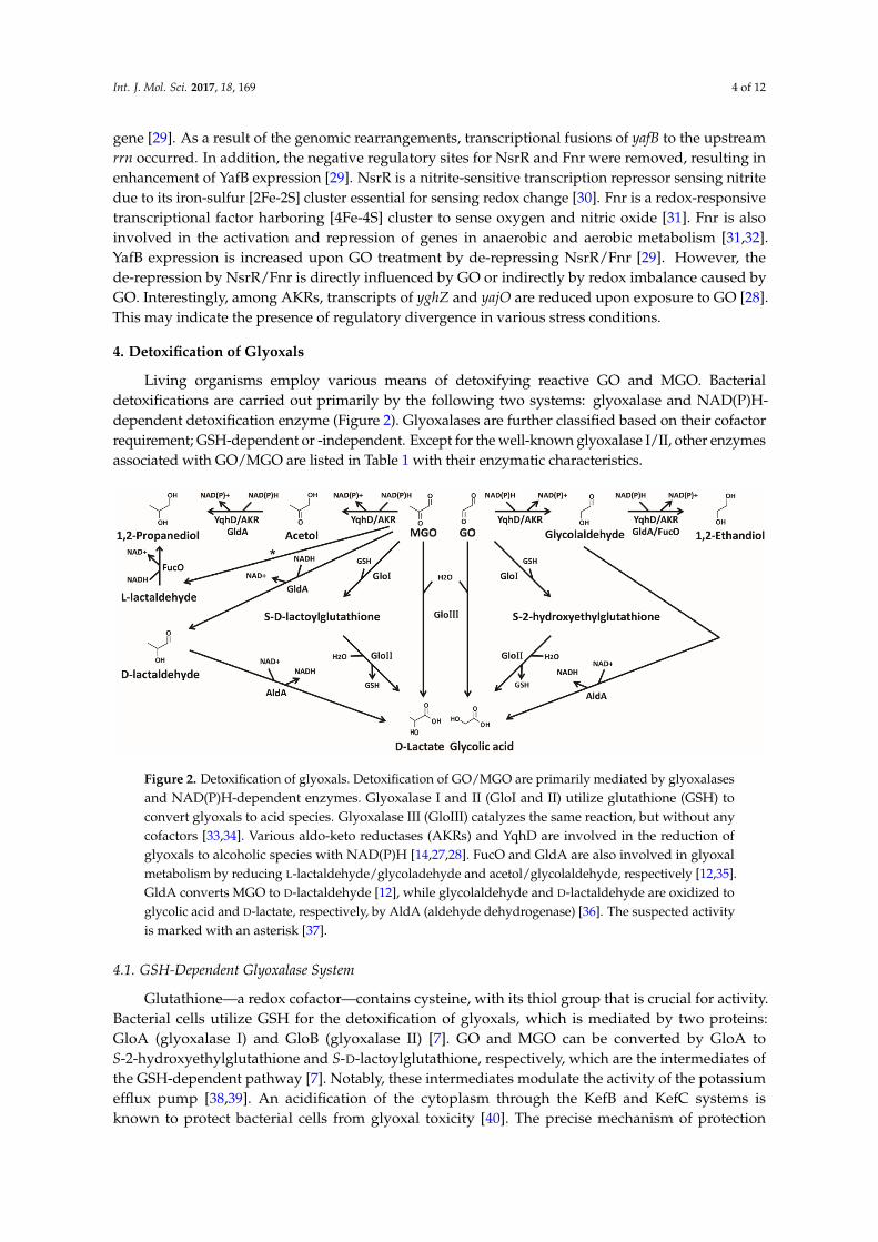
Figure 2. Detoxification of glyoxals. Detoxification of GO/MGO are primarily mediated by glyoxalases and NAD(P)H-dependent enzymes. Glyoxalase I and II (GloI and II) utilize glutathione (GSH) to convert glyoxals to acid species. Glyoxalase III (GloIII) catalyzes the same reaction, but without any cofactors [33,34]. Various aldo-keto reductases (AKRs) and YqhD are involved in the reduction of glyoxals to alcoholic species with NAD(P)H [14,27,28]. FucO and GldA are also involved in glyoxal metabolism by reducing L-lactaldehyde/glycoladehyde and acetol/glycolaldehyde, respectively [12,35]. GldA converts MGO to D-lactaldehyde [12], while glycolaldehyde and D-lactaldehyde are oxidized to glycolic acid and D-lactate, respectively, by AldA (aldehyde dehydrogenase) [36]. The suspected activity is marked with an asterisk [37].
4.1. GSH-Dependent Glyoxalase System
Glutathione—a redox cofactor—contains cysteine, with its thiol group that is crucial for activity. Bacterial cells utilize GSH for the detoxification of glyoxals, which is mediated by two proteins: GloA (glyoxalase I) and GloB (glyoxalase II) [7]. GO and MGO can be converted by GloA to S-2-hydroxyethylglutathione and S-D-lactoylglutathione, respectively, which are the intermediates of the GSH-dependent pathway [7]. Notably, these intermediates modulate the activity of the potassium efflux pump [38,39]. An acidification of the cytoplasm through the KefB and KefC systems is known to protect bacterial cells from glyoxal toxicity [40]. The precise mechanism of protection by cytoplasmic acidification is poorly understood, although it was suggested that this change is to minimize glyoxal-induced DNA damage [39]. GloB converts the above intermediates to glycolic and lactic acids. Therefore, an availability of GSH would be critical in the proper functioning of the GloAB system in the cell. Bacillus subtilis—a Gram-positive bacterium—has a similar glyoxalase system, but utilizes bacillithiol (BSH) instead of GSH [41]. While GSH is synthesized from L-cysteine, L-glutamic acid, and glycine, BSH consists of L-cysteinyl-D-glucosamine and malic acid.
Figure 2. Detoxification of glyoxals. Detoxification of GO/MGO are primarily mediated by glyoxalasesand NAD(P)H-dependent enzymes. Glyoxalase I and II (GloI and II) utilize glutathione (GSH) toconvert glyoxals to acid species. Glyoxalase III (GloIII) catalyzes the same reaction, but without anycofactors [33,34]. Various aldo-keto reductases (AKRs) and YqhD are involved in the reduction ofglyoxals to alcoholic species with NAD(P)H [14,27,28]. FucO and GldA are also involved in glyoxalmetabolism by reducing L-lactaldehyde/glycoladehyde and acetol/glycolaldehyde, respectively [12,35].GldA converts MGO to D-lactaldehyde [12], while glycolaldehyde and D-lactaldehyde are oxidized toglycolic acid and D-lactate, respectively, by AldA (aldehyde dehydrogenase) [36]. The suspected activityis marked with an asterisk [37].
4.1. GSH-Dependent Glyoxalase System
Glutathione—a redox cofactor—contains cysteine, with its thiol group that is crucial for activity.Bacterial cells utilize GSH for the detoxification of glyoxals, which is mediated by two proteins:GloA (glyoxalase I) and GloB (glyoxalase II) [7]. GO and MGO can be converted by GloA toS-2-hydroxyethylglutathione and S-D-lactoylglutathione, respectively, which are the intermediates ofthe GSH-dependent pathway [7]. Notably, these intermediates modulate the activity of the potassiumefflux pump [38,39]. An acidification of the cytoplasm through the KefB and KefC systems isknown to protect bacterial cells from glyoxal toxicity [40]. The precise mechanism of protection
Int. J. Mol. Sci. 2017, 18, 169 5 of 12
by cytoplasmic acidification is poorly understood, although it was suggested that this change isto minimize glyoxal-induced DNA damage [39]. GloB converts the above intermediates to glycolicand lactic acids. Therefore, an availability of GSH would be critical in the proper functioning of theGloAB system in the cell. Bacillus subtilis—a Gram-positive bacterium—has a similar glyoxalasesystem, but utilizes bacillithiol (BSH) instead of GSH [41]. While GSH is synthesized from L-cysteine,L-glutamic acid, and glycine, BSH consists of L-cysteinyl-D-glucosamine and malic acid.
4.2. GSH-Independent Glyoxalase System
From the previous report on the activity of GSH-independent glyoxalase [42], the gene encodingthe activity of glyoxalase III was identified [33,43]. This enzyme catalyzes the same reaction as thatof glyoxalase I/II enzymes without any cofactor. An E. coli homolog of glyoxalase III was firstdiscovered by the activity tracing of fractionated E. coli extract, leading to a characterization of thehchA (Hsp31) gene for GSH-independent glyoxalase III [33]. The Hsp31 protein is a member of theDJ-1 homologs that are present in all biological species [33]. In E. coli, there are four homologs: hchA,yajL, yhbO, and elbB. The Cys-185 and Glu-77 residues of Hsp31 and their corresponding positionsin other homologs are essential to catalysis [34]. Hsp31 and YhbO harbor an additional His residueat 186 [34]. YajL and ElbB have Phe and Ile residues, respectively, instead of His [34]. The DJ-1homologs share structural similarity of central β-strands surrounded by α-helices, forming a dimer oran oligomer (Figure 3) [34,44]. Analogous to atDJ-1d of Arabidopsis thaliana, YhbO likely forms a circularstructure of a hexamer being composed of three dimers [44–46]. Purified Hsp31, YajL, YhbO, and ElbBshow glyoxalase activity with different substrate specificity (Table 1) [33,34]. Over-expressions of thehomologs exhibit protection against exogenously added GO/MGO, and reduce the glyoxal-dependentaccumulation of advanced glycation end products (AGEs). Glycation is a nonenzymatic reactionbetween protein and sugar/glyoxal, presumably resulting in protein malfunction. Recent reportspropose that Hsp31 and human DJ-1 protein are deglycases likely associated with protein repair [47,48].Int. J. Mol. Sci. 2017, 18, 169 6 of 11
Figure 3. Structural and oligomeric diversities of DJ-1 homologs. Dimeric or hexameric structure of atDJ-1d (PDB code: 4OFW)/YhbO (PDB code: 1OI4), Hsp31 (PDB code: 1N57), ElbB (PDB code: 1OY1), and hDJ1 (PDB code: 1UCF, similar to E. coli YajL) are shown. DJ-1 homologs share α/β core domain. Formations of oligomeric structures are schematized with the letter “β” as a monomer and “a” as the catalytic site.
4.3. NAD(P)H-Dependent Detoxifying Enzymes
The electron donor NAD(P)H is a redox cofactor for various enzymes, including ones for glyoxal detoxification. YqhD is an NADPH-dependent aldehyde reductase converting GO/MGO to corresponding alcohols (Figure 2, Table 1) [14,49]. As described above, the existence of YqhD enzyme was revealed by characterizing GO-resistant mutations with constitutively-activating YqhC transcriptional regulator, resulting in YqhD overexpression [14]. YqhD turned out to be a major detoxification pathway for GO [14]. Although YqhD exhibits activity toward MGO, it does not play a major role in MGO detoxification. Instead, glyoxalase I contributes more in vivo [14,15]. AKRs are also NADPH-dependent enzymes, converting GO/MGO to less-reactive alcoholic species (Figure 2, Table 1) [27,28]. The E. coli genome encodes nine AKRs, among which five of them—YqhE, YafB, YghZ, YeaE, and YajO—show activity to GO/MGO [28]. The role of AKRs in glyoxal metabolism is likely to be multi-faceted in response to various physiological changes; i.e., intracellular concentrations of glyoxals, other metabolites, or growth states. Since the AKRs have different kinetic and expression profiles in relation to the intracellular concentration of glyoxal, they might have unique roles in the detoxification of glyoxal. YqhD and AKRs exhibit two-step reductions of glyoxals, converting GO/MGO to 1,2-ethandiol/1,2-propanediol via glycolaldehyde/acetol, respectively [14,27,28]. The first step is critical in decreasing the toxicity of glyoxal. FucO (1,2-propanediol oxidoreductase) is involved in the conversion of L-lactaldehyde and glycolaldehyde to 1,2-propanediol and 1,2-ethandiol, respectively [35,50]. GldA (glycerol dehydrogenase) is involved in NADH-dependent reductions of glycolaldehyde and acetol [12]. GldA also converts MGO to D-lactaldehyde, and AldA (aldehyde dehydrogenase) oxidizes glycolaldehyde and D-lactaldehyde to glycolic and D-lactic acids, respectively, using NAD+ as a cofactor [12,35,36] (Figure 2).
5. Toxicity of Glyoxals
Since GO and MGO are highly reactive (especially to nucleophilic macromolecules, including proteins and nucleotides), strong intracellular toxicity has been found. To protect from glyoxal
Figure 3. Structural and oligomeric diversities of DJ-1 homologs. Dimeric or hexameric structure ofatDJ-1d (PDB code: 4OFW)/YhbO (PDB code: 1OI4), Hsp31 (PDB code: 1N57), ElbB (PDB code: 1OY1),and hDJ1 (PDB code: 1UCF, similar to E. coli YajL) are shown. DJ-1 homologs share α/β core domain.Formations of oligomeric structures are schematized with the letter “β” as a monomer and “a” as thecatalytic site.
Int. J. Mol. Sci. 2017, 18, 169 6 of 12
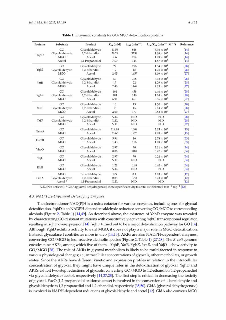
Table 1. Enzymatic constants for GO/MGO detoxification proteins.
Proteins Substrate Product Km (mM) kcat (min−1) kcat/Km (min−1·M−1) Reference
N.D. (Not detected); a GldA (glycerol dehydrogenase) shows specific activity to acetol as 4600 nmol·min−1·mg−1 [12].
4.3. NAD(P)H-Dependent Detoxifying Enzymes
The electron donor NAD(P)H is a redox cofactor for various enzymes, including ones for glyoxaldetoxification. YqhD is an NADPH-dependent aldehyde reductase converting GO/MGO to correspondingalcohols (Figure 2, Table 1) [14,49]. As described above, the existence of YqhD enzyme was revealedby characterizing GO-resistant mutations with constitutively-activating YqhC transcriptional regulator,resulting in YqhD overexpression [14]. YqhD turned out to be a major detoxification pathway for GO [14].Although YqhD exhibits activity toward MGO, it does not play a major role in MGO detoxification.Instead, glyoxalase I contributes more in vivo [14,15]. AKRs are also NADPH-dependent enzymes,converting GO/MGO to less-reactive alcoholic species (Figure 2, Table 1) [27,28]. The E. coli genomeencodes nine AKRs, among which five of them—YqhE, YafB, YghZ, YeaE, and YajO—show activity toGO/MGO [28]. The role of AKRs in glyoxal metabolism is likely to be multi-faceted in response tovarious physiological changes; i.e., intracellular concentrations of glyoxals, other metabolites, or growthstates. Since the AKRs have different kinetic and expression profiles in relation to the intracellularconcentration of glyoxal, they might have unique roles in the detoxification of glyoxal. YqhD andAKRs exhibit two-step reductions of glyoxals, converting GO/MGO to 1,2-ethandiol/1,2-propanediolvia glycolaldehyde/acetol, respectively [14,27,28]. The first step is critical in decreasing the toxicityof glyoxal. FucO (1,2-propanediol oxidoreductase) is involved in the conversion of L-lactaldehyde andglycolaldehyde to 1,2-propanediol and 1,2-ethandiol, respectively [35,50]. GldA (glycerol dehydrogenase)is involved in NADH-dependent reductions of glycolaldehyde and acetol [12]. GldA also converts MGO
Int. J. Mol. Sci. 2017, 18, 169 7 of 12
to D-lactaldehyde, and AldA (aldehyde dehydrogenase) oxidizes glycolaldehyde and D-lactaldehyde toglycolic and D-lactic acids, respectively, using NAD+ as a cofactor [12,35,36] (Figure 2).
5. Toxicity of Glyoxals
Since GO and MGO are highly reactive (especially to nucleophilic macromolecules, includingproteins and nucleotides), strong intracellular toxicity has been found. To protect from glyoxal toxicity,cellular redox cofactors such as GSH and NAD(P)H are required, without which cells are endangeredwith irreversible damages.
5.1. Protein Damage
Since the carbonyl groups of glyoxal are highly reactive, they interact with diverse moleculesincluding proteins and nucleotides. GO and MGO form a Schiff’s base with the amino group of aminoacids, and subsequent Amadori rearrangement produces AGEs [5]. The nucleophilic residues of proteins,such as lysine, cysteine, arginine, and histidine, are prone to react with GO/MGO. Carboxymethyl- andcarboxyethyl-lysines (CML/CEL) or products with other amino acids are formed from reactions withGO and MGO, respectively [51,52]. The spontaneous non-enzymatic glycation affects protein function.The glycation itself is basically reversible, although the reversal of AGE formation is still controversial.
5.2. Nucleotide Damage
Nucleotides are vulnerable targets of GO/MGO, forming adducts such as carboxymethyl- andcarboxyethyl guanosine (CMG/CEG) to increase mutation frequency [53,54]. However, the mechanismunderlying nucleotide modification caused by glyoxals is largely unknown. Recent study indicates thatgenes associated with DNA repair (recA, recC) and tRNA modification (truA, trmE, gidA) are involvedin GO sensitivity, suggesting that nucleotides are one of the main targets of GO [25]. The repair of DNAdamage and tRNA modification seem to be crucial in enduring GO assault. RecA catalyzes an exchangeof DNA strands in the recombinational repair, and also cleaves LexA repressor for an induction ofthe SOS response [55]. As a subunit of RecBCD exonuclease V, RecC is necessary for homologousrecombination in repairing double strand break [55]. Therefore, the isolation of recA and recC mutationsas GO-sensitive derivatives suggests that the strand break of DNA might occurs with GO, which islikely to be repaired by the recombinational repair system. The trmE, truA, and gidA genes are involvedin tRNA stability [56]. Thus, it is likely that unmodified tRNA is more sensitive to GO than modifiedtRNA. Alternatively, since the tRNA modification stabilizes the U·G pairing at the wobble positionand plays a role in decoding NNG codons [57], wobble pairing in translation may serve as a target forGO. The mutant strains related to DNA repair show comparable sensitivity to both GO and MGO,but mutations in tRNA modification exert more sensitivity to GO than to MGO [25]. Therefore, tRNAis more likely to interact with GO than MGO, implying different target specificity of glyoxal species.
5.3. Relation to Oxidative Stress
ROS have been known for some time and are reasonably well-characterized. Since the detoxificationof RES (including glyoxals) requires several redox cofactors (such as NAD(P)H and GSH), it mayaffect redox regulation associated with electron transport chain, which is a site for ROS production.In eukaryotes, a decrease in the activity of the respiratory complex III caused by MGO glycationresults in oxidative stress [58]. A removal of aldehyde is closely associated with oxidative stress—e.g., glycolaldehyde-dependent induction of SoxRS regulon [59]. Recent study of genetic screeningrevealed SodB (superoxide dismutase) as one of the GO-resistance genes under the regulation of CRP [25].On the other hand, a high level of oxidative stress increases the production of glyoxals, presumablyvia an oxidative degradation of sugar [8]. The other possibility would be an oxidation of aldehyde,which also likely generates glyoxal, especially under the condition lacking superoxide dismutase [60].Finally, the expression of glyoxalase exhibits protection from glyoxal as well as oxidative stress [61,62].Therefore, ROS and RES are somehow closely associated in vivo.
Int. J. Mol. Sci. 2017, 18, 169 8 of 12
6. Conclusions and Perspectives
The production of GO/MGO in vivo appears to be inevitable, since they are intrinsically associatedwith various physiological processes. Like ROS, RES are fairly reactive, thereby resulting in stress tothe cell. Due to their toxic effect, the cell has devised various enzyme systems to reduce their toxicity.Our current knowledge only focuses on individual detoxification pathways, without an organizedscheme to systematically detect their actions and respond appropriately. Ironically, bacteria havean MGO producing enzyme—MGO synthase [9]. This review attempted to delineate global processesresponding to glyoxal/RES (Figure 4). A systematic characterization of responsive and regulatoryplayers for glyoxals was only recently conducted in bacteria. It is expected that much of the futurenovelty lies in this aspect of RES response. Meanwhile, since the toxicity of RES is closely associatedwith various human diseases (particularly neurodegenerative diseases), a fundamental understandingof RES response obtained from the study of prokaryotes can be applied to higher eukaryotes for thedevelopment of novel therapy. Although we learned that ROS and RES are intimately associated,the detailed mechanisms of how they relate to each other are yet to be investigated. The molecularmechanisms involving the cAMP- and YqhC/NemR-mediated RES sensors also need to be unraveled,in addition to the effect of glyoxal on DNA damage and tRNA modification. Finally, the ubiquitouspresence of DJ-1 homologs (we propose the name “glyoxalin”, as in “peroxiredoxin”) and numerousAKRs present an issue of redundancy in single organism. Addressing these questions with regard totheir specified roles and expression patterns would be one of the future challenges.
Int. J. Mol. Sci. 2017, 18, 169 8 of 11
their toxicity. Our current knowledge only focuses on individual detoxification pathways, without an organized scheme to systematically detect their actions and respond appropriately. Ironically, bacteria have an MGO producing enzyme—MGO synthase [9]. This review attempted to delineate global processes responding to glyoxal/RES (Figure 4). A systematic characterization of responsive and regulatory players for glyoxals was only recently conducted in bacteria. It is expected that much of the future novelty lies in this aspect of RES response. Meanwhile, since the toxicity of RES is closely associated with various human diseases (particularly neurodegenerative diseases), a fundamental understanding of RES response obtained from the study of prokaryotes can be applied to higher eukaryotes for the development of novel therapy. Although we learned that ROS and RES are intimately associated, the detailed mechanisms of how they relate to each other are yet to be investigated. The molecular mechanisms involving the cAMP- and YqhC/NemR-mediated RES sensors also need to be unraveled, in addition to the effect of glyoxal on DNA damage and tRNA modification. Finally, the ubiquitous presence of DJ-1 homologs (we propose the name “glyoxalin”, as in “peroxiredoxin”) and numerous AKRs present an issue of redundancy in single organism. Addressing these questions with regard to their specified roles and expression patterns would be one of the future challenges.
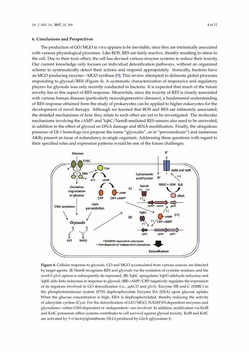
Figure 4. Cellular response to glyoxals. GO and MGO accumulated from various sources are detected by target agents. (I) NemR recognizes RES and glyoxals via the oxidation of cysteine residues, and the nemRA-gloA operon is subsequently de-repressed; (II) YqhC upregulates YqhD aldehyde reductase and YqhE aldo-keto reductase in response to glyoxal; (III) cAMP/CRP negatively regulates the expression of its regulons involved in GO detoxification (i.e., yqhCD and gloA). Enzyme IIB and C (EIIBC) in the phosphotransferase system (PTS) dephosphorylate Enzyme IIA (EIIA) upon glucose uptake. When the glucose concentration is high, EIIA is dephosphorylated, thereby reducing the activity of adenylate cyclase (Cya). For the detoxification of GO/MGO, NAD(P)H-dependent enzymes and glyoxalases—either GSH-dependent or -independent—are involved. In addition, acidification via KefB and KefC potassium efflux systems contributes to cell survival against glyoxal toxicity. KefB and KefC are activated by S-D-lactoylglutathione (SLG) produced by GloA (glyoxalase I).
Acknowledgments: The authors thank Dongwook Choi for Figure 3. This work was supported by a grant from the National Research Foundation of Korea (NRF) to Chankyu Park.
Author Contributions: Changhan Lee and Chankyu Park wrote the paper.
Conflicts of interest: The authors declare no conflict of interest.
Figure 4. Cellular response to glyoxals. GO and MGO accumulated from various sources are detectedby target agents. (I) NemR recognizes RES and glyoxals via the oxidation of cysteine residues, and thenemRA-gloA operon is subsequently de-repressed; (II) YqhC upregulates YqhD aldehyde reductase andYqhE aldo-keto reductase in response to glyoxal; (III) cAMP/CRP negatively regulates the expressionof its regulons involved in GO detoxification (i.e., yqhCD and gloA). Enzyme IIB and C (EIIBC) inthe phosphotransferase system (PTS) dephosphorylate Enzyme IIA (EIIA) upon glucose uptake.When the glucose concentration is high, EIIA is dephosphorylated, thereby reducing the activityof adenylate cyclase (Cya). For the detoxification of GO/MGO, NAD(P)H-dependent enzymes andglyoxalases—either GSH-dependent or -independent—are involved. In addition, acidification via KefBand KefC potassium efflux systems contributes to cell survival against glyoxal toxicity. KefB and KefCare activated by S-D-lactoylglutathione (SLG) produced by GloA (glyoxalase I).
Int. J. Mol. Sci. 2017, 18, 169 9 of 12
Acknowledgments: The authors thank Dongwook Choi for Figure 3. This work was supported by a grant fromthe National Research Foundation of Korea (NRF) to Chankyu Park.
Author Contributions: Changhan Lee and Chankyu Park wrote the paper.
Conflicts of Interest: The authors declare no conflict of interest.
2. Jacobs, A.T.; Marnett, L.J. Systems analysis of protein modification and cellular responses induced byelectrophile stress. Acc. Chem. Res. 2010, 43, 673–683. [CrossRef] [PubMed]
3. Marnett, L.J.; Riggins, J.N.; West, J.D. Endogenous generation of reactive oxidants and electrophiles and theirreactions with DNA and protein. J. Clin. Investig. 2003, 111, 583–593. [CrossRef] [PubMed]
4. Munch, G.; Luth, H.J.; Wong, A.; Arendt, T.; Hirsch, E.; Ravid, R.; Riederer, P. Crosslinking of α-synuclein byadvanced glycation endproducts—An early pathophysiological step in Lewy body formation? J. Chem. Neuroanat.2000, 20, 253–257. [CrossRef]
5. Brownlee, M. Advanced protein glycosylation in diabetes and aging. Annu. Rev. Med. 1995, 46, 223–234.[CrossRef] [PubMed]
6. Luth, H.J.; Ogunlade, V.; Kuhla, B.; Kientsch-Engel, R.; Stahl, P.; Webster, J.; Arendt, T.; Munch, G. Age- andstage-dependent accumulation of advanced glycation end products in intracellular deposits in normal andAlzheimer’s disease brains. Cereb. Cortex 2005, 15, 211–220. [CrossRef] [PubMed]
7. Thornalley, P.J. The glyoxalase system: New developments towards functional characterization of a metabolicpathway fundamental to biological life. Biochem. J. 1990, 269, 1–11. [CrossRef] [PubMed]
8. Thornalley, P.J.; Langborg, A.; Minhas, H.S. Formation of glyoxal, methylglyoxal and 3-deoxyglucosone inthe glycation of proteins by glucose. Biochem. J. 1999, 344 Pt 1, 109–116. [CrossRef] [PubMed]
9. Hopper, D.J.; Cooper, R.A. The regulation of Escherichia coli methylglyoxal synthase; a new control site inglycolysis? FEBS Lett. 1971, 13, 213–216. [CrossRef]
10. Singh, R.; Barden, A.; Mori, T.; Beilin, L. Advanced glycation end-products: A review. Diabetologia 2001, 44,129–146. [CrossRef] [PubMed]
11. Thornalley, P.J. Pharmacology of methylglyoxal: Formation, modification of proteins and nucleic acids, andenzymatic detoxification—A role in pathogenesis and antiproliferative chemotherapy. Gen. Pharmacol. 1996,27, 565–573. [CrossRef]
12. Subedi, K.P.; Kim, I.; Kim, J.; Min, B.; Park, C. Role of GldA in dihydroxyacetone and methylglyoxalmetabolism of Escherichia coli K12. FEMS Microbiol. Lett. 2008, 279, 180–187. [CrossRef] [PubMed]
13. Kim, I.; Kim, E.; Yoo, S.; Shin, D.; Min, B.; Song, J.; Park, C. Ribose utilization with an excess of mutarotasecauses cell death due to accumulation of methylglyoxal. J. Bacteriol. 2004, 186, 7229–7235. [CrossRef][PubMed]
14. Lee, C.; Kim, I.; Lee, J.; Lee, K.L.; Min, B.; Park, C. Transcriptional activation of the aldehyde reductase YqhDby YqhC and its implication in glyoxal metabolism of Escherichia coli K-12. J. Bacteriol. 2010, 192, 4205–4214.[CrossRef] [PubMed]
15. Lee, C.; Shin, J.; Park, C. Novel regulatory system nemRA-gloA for electrophile reduction in Escherichia coliK-12. Mol. Microbiol. 2013, 88, 395–412. [CrossRef] [PubMed]
16. Turner, P.C.; Miller, E.N.; Jarboe, L.R.; Baggett, C.L.; Shanmugam, K.T.; Ingram, L.O. YqhC regulatestranscription of the adjacent Escherichia coli genes yqhD and dkgA that are involved in furfural tolerance.J. Ind. Microbiol. Biotechnol. 2011, 38, 431–439. [CrossRef] [PubMed]
17. Umezawa, Y.; Shimada, T.; Kori, A.; Yamada, K.; Ishihama, A. The uncharacterized transcription factor YdhMis the regulator of the nemA gene, encoding N-ethylmaleimide reductase. J. Bacteriol. 2008, 190, 5890–5897.[CrossRef] [PubMed]
18. Ozyamak, E.; de Almeida, C.; de Moura, A.P.; Miller, S.; Booth, I.R. Integrated stress response of Escherichia colito methylglyoxal: Transcriptional readthrough from the nemRA operon enhances protection throughincreased expression of glyoxalase I. Mol. Microbiol. 2013, 88, 936–950. [CrossRef] [PubMed]
19. Miura, K.; Tomioka, Y.; Suzuki, H.; Yonezawa, M.; Hishinuma, T.; Mizugaki, M. Molecular cloning of thenemA gene encoding N-ethylmaleimide reductase from Escherichia coli. Biol. Pharm. Bull. 1997, 20, 110–112.[CrossRef] [PubMed]
20. Williams, R.E.; Bruce, N.C. “New uses for an Old Enzyme”—The Old Yellow Enzyme family of flavoenzymes.Microbiology 2002, 148 Pt 6, 1607–1614. [CrossRef] [PubMed]
21. Gray, M.J.; Wholey, W.Y.; Parker, B.W.; Kim, M.; Jakob, U. NemR is a bleach-sensing transcription factor.J. Biol. Chem. 2013, 288, 13789–13798. [CrossRef] [PubMed]
22. Gray, M.J.; Wholey, W.Y.; Jakob, U. Bacterial responses to reactive chlorine species. Annu. Rev. Microbiol.2013, 67, 141–160. [CrossRef] [PubMed]
23. Gray, M.J.; Li, Y.; Leichert, L.I.; Xu, Z.; Jakob, U. Does the Transcription Factor NemR Use a RegulatorySulfenamide Bond to Sense Bleach? Antioxid. Redox Signal. 2015, 23, 747–754. [CrossRef] [PubMed]
24. Zheng, D.; Constantinidou, C.; Hobman, J.L.; Minchin, S.D. Identification of the CRP regulon using in vitroand in vivo transcriptional profiling. Nucleic Acids Res. 2004, 32, 5874–5893. [CrossRef] [PubMed]
25. Lee, C.; Kim, J.; Kwon, M.; Lee, K.; Min, H.; Kim, S.H.; Kim, D.; Lee, N.; Kim, J.; Kim, D.; et al. Screening forEscherichia coli K-12 genes conferring glyoxal resistance or sensitivity by transposon insertions. FEMS Microbiol. Lett.2016, 363. [CrossRef] [PubMed]
26. Deutscher, J. The mechanisms of carbon catabolite repression in bacteria. Curr. Opin. Microbiol. 2008, 11,87–93. [CrossRef] [PubMed]
27. Ko, J.; Kim, I.; Yoo, S.; Min, B.; Kim, K.; Park, C. Conversion of methylglyoxal to acetol by Escherichia colialdo-keto reductases. J. Bacteriol. 2005, 187, 5782–5789. [CrossRef] [PubMed]
28. Lee, C.; Kim, I.; Park, C. Glyoxal detoxification in Escherichia coli K-12 by NADPH dependent aldo-ketoreductases. J. Microbiol. 2013, 51, 527–530. [CrossRef] [PubMed]
29. Kwon, M.; Lee, J.; Lee, C.; Park, C. Genomic rearrangements leading to overexpression of aldo-keto reductaseYafB of Escherichia coli confer resistance to glyoxal. J. Bacteriol. 2012, 194, 1979–1988. [CrossRef] [PubMed]
31. Salmon, K.; Hung, S.P.; Mekjian, K.; Baldi, P.; Hatfield, G.W.; Gunsalus, R.P. Global gene expression profilingin Escherichia coli K12. The effects of oxygen availability and FNR. J. Biol. Chem. 2003, 278, 29837–29855.[CrossRef] [PubMed]
32. Kang, Y.; Weber, K.D.; Qiu, Y.; Kiley, P.J.; Blattner, F.R. Genome-wide expression analysis indicates thatFNR of Escherichia coli K-12 regulates a large number of genes of unknown function. J. Bacteriol. 2005, 187,1135–1160. [CrossRef] [PubMed]
33. Subedi, K.P.; Choi, D.; Kim, I.; Min, B.; Park, C. Hsp31 of Escherichia coli K-12 is glyoxalase III. Mol. Microbiol.2011, 81, 926–936. [CrossRef] [PubMed]
34. Lee, C.; Lee, J.; Lee, J.Y.; Park, C. Characterization of the Escherichia coli YajL, YhbO and ElbB glyoxalases.FEMS Microbiol. Lett. 2016, 363. [CrossRef] [PubMed]
35. Chen, Y.M.; Zhu, Y.; Lin, E.C. NAD-linked aldehyde dehydrogenase for aerobic utilization of L-fucose andL-rhamnose by Escherichia coli. J. Bacteriol. 1987, 169, 3289–3294. [CrossRef] [PubMed]
36. Baldoma, L.; Aguilar, J. Involvement of lactaldehyde dehydrogenase in several metabolic pathways ofEscherichia coli K12. J. Biol. Chem. 1987, 262, 13991–13996. [PubMed]
37. Altaras, N.E.; Cameron, D.C. Metabolic engineering of a 1,2-propanediol pathway in Escherichia coli.Appl. Environ. Microbiol. 1999, 65, 1180–1185. [PubMed]
38. MacLean, M.J.; Ness, L.S.; Ferguson, G.P.; Booth, I.R. The role of glyoxalase I in the detoxification ofmethylglyoxal and in the activation of the KefB K+ efflux system in Escherichia coli. Mol. Microbiol. 1998, 27,563–571. [CrossRef] [PubMed]
39. Ferguson, G.P.; Battista, J.R.; Lee, A.T.; Booth, I.R. Protection of the DNA during the exposure of Escherichiacoli cells to a toxic metabolite: The role of the KefB and KefC potassium channels. Mol. Microbiol. 2000, 35,113–122. [CrossRef] [PubMed]
40. Healy, J.; Ekkerman, S.; Pliotas, C.; Richard, M.; Bartlett, W.; Grayer, S.C.; Morris, G.M.; Miller, S.; Booth, I.R.;Conway, S.J.; et al. Understanding the structural requirements for activators of the Kef bacterial potassiumefflux system. Biochemistry 2014, 53, 1982–1992. [CrossRef] [PubMed]
41. Chandrangsu, P.; Dusi, R.; Hamilton, C.J.; Helmann, J.D. Methylglyoxal resistance in Bacillus subtilis:Contributions of bacillithiol-dependent and independent pathways. Mol. Microbiol. 2014, 91, 706–715.[CrossRef] [PubMed]
42. Misra, K.; Banerjee, A.B.; Ray, S.; Ray, M. Glyoxalase III from Escherichia coli: A single novel enzyme for theconversion of methylglyoxal into D-lactate without reduced glutathione. Biochem. J. 1995, 305 Pt 3, 999–1003.[CrossRef] [PubMed]
43. Lee, J.Y.; Song, J.; Kwon, K.; Jang, S.; Kim, C.; Baek, K.; Kim, J.; Park, C. Human DJ-1 and its homologs arenovel glyoxalases. Hum. Mol. Genet. 2012, 21, 3215–3225. [CrossRef] [PubMed]
44. Choi, D.; Kim, J.; Ha, S.; Kwon, K.; Kim, E.H.; Lee, H.Y.; Ryu, K.S.; Park, C. Stereospecific mechanism of DJ-1glyoxalases inferred from their hemithioacetal-containing crystal structures. FEBS J. 2014, 281, 5447–5462.[CrossRef] [PubMed]
45. Kwon, K.; Choi, D.; Hyun, J.K.; Jung, H.S.; Baek, K.; Park, C. Novel glyoxalases from Arabidopsis thaliana.FEBS J. 2013, 280, 3328–3339. [CrossRef] [PubMed]
46. Abdallah, J.; Kern, R.; Malki, A.; Eckey, V.; Richarme, G. Cloning, expression, and purification of the generalstress protein YhbO from Escherichia coli. Protein Expr. Purif. 2006, 47, 455–460. [CrossRef] [PubMed]
47. Richarme, G.; Mihoub, M.; Dairou, J.; Bui, L.C.; Leger, T.; Lamouri, A. Parkinsonism-associated proteinDJ-1/Park7 is a major protein deglycase that repairs methylglyoxal- and glyoxal-glycated cysteine, arginine,and lysine residues. J. Biol. Chem. 2015, 290, 1885–1897. [CrossRef] [PubMed]
49. Jarboe, L.R. YqhD: A broad-substrate range aldehyde reductase with various applications in production ofbiorenewable fuels and chemicals. Appl. Microbiol. Biotechnol. 2011, 89, 249–257. [CrossRef] [PubMed]
50. Boronat, A.; Caballero, E.; Aguilar, J. Experimental evolution of a metabolic pathway for ethylene glycolutilization by Escherichia coli. J. Bacteriol. 1983, 153, 134–139. [PubMed]
51. Basta, G.; Schmidt, A.M.; de Caterina, R. Advanced glycation end products and vascular inflammation:Implications for accelerated atherosclerosis in diabetes. Cardiovasc. Res. 2004, 63, 582–592. [CrossRef][PubMed]
52. Sousa Silva, M.; Gomes, R.A.; Ferreira, A.E.; Ponces Freire, A.; Cordeiro, C. The glyoxalase pathway: The firsthundred years... and beyond. Biochem. J. 2013, 453, 1–15. [CrossRef] [PubMed]
53. Kasai, H.; Iwamoto-Tanaka, N.; Fukada, S. DNA modifications by the mutagen glyoxal: Adduction to G andC, deamination of C and GC and GA cross-linking. Carcinogenesis 1998, 19, 1459–1465. [CrossRef] [PubMed]
54. Murata-Kamiya, N.; Kamiya, H.; Kaji, H.; Kasai, H. Mutational specificity of glyoxal, a product of DNAoxidation, in the lacI gene of wild-type Escherichia coli W3110. Mutat. Res. 1997, 377, 255–262. [CrossRef]
55. Courcelle, J.; Hanawalt, P.C. RecA-dependent recovery of arrested DNA replication forks. Annu. Rev. Genet.2003, 37, 611–646. [CrossRef] [PubMed]
56. Urbonavicius, J.; Qian, Q.; Durand, J.M.; Hagervall, T.G.; Bjork, G.R. Improvement of reading framemaintenance is a common function for several tRNA modifications. EMBO J. 2001, 20, 4863–4873. [CrossRef][PubMed]
57. Kurata, S.; Weixlbaumer, A.; Ohtsuki, T.; Shimazaki, T.; Wada, T.; Kirino, Y.; Takai, K.; Watanabe, K.;Ramakrishnan, V.; Suzuki, T. Modified uridines with C5-methylene substituents at the first position ofthe tRNA anticodon stabilize U.G wobble pairing during decoding. J. Biol. Chem. 2008, 283, 18801–18811.[CrossRef] [PubMed]
59. Benov, L.; Fridovich, I. Induction of the soxRS regulon of Escherichia coli by glycolaldehyde. Arch. Biochem. Biophys.2002, 407, 45–48. [CrossRef]
60. Okado-Matsumoto, A.; Fridovich, I. The role of α,β-dicarbonyl compounds in the toxicity of short chainsugars. J. Biol. Chem. 2000, 275, 34853–34857. [CrossRef] [PubMed]
61. Bankapalli, K.; Saladi, S.; Awadia, S.S.; Goswami, A.V.; Samaddar, M.; D’Silva, P. Robust glyoxalaseactivity of Hsp31, a ThiJ/DJ-1/PfpI family member protein, is critical for oxidative stress resistance inSaccharomyces cerevisiae. J. Biol. Chem. 2015, 290, 26491–26507. [CrossRef] [PubMed]
62. Brouwers, O.; Niessen, P.M.; Ferreira, I.; Miyata, T.; Scheffer, P.G.; Teerlink, T.; Schrauwen, P.; Brownlee, M.;Stehouwer, C.D.; Schalkwijk, C.G. Overexpression of glyoxalase-I reduces hyperglycemia-induced levels ofadvanced glycation end products and oxidative stress in diabetic rats. J. Biol. Chem. 2011, 286, 1374–1380.[CrossRef] [PubMed]