BAG-1 is preferentially expressed in neuronal precursor cells of the adult mouse brain and regulates their proliferation in vitro Evan Elliott * , Irith Ginzburg 1 Neurobiology, Arison Building, Weizmann Institute of Science, Rehovot 76100, Israel article info Article history: Received 28 September 2008 Revised 3 December 2008 Accepted 3 December 2008 Available online 11 December 2008 Edited by Ned Mantei Keywords: BAG-1 Neurogenesis Proliferation Neuronal precursor cell abstract BAG-1 protein has been well characterized as necessary for proper neuronal development. However, little is known about the function of BAG-1 in the adult brain. In this work, the expression and local- ization of BAG-1 in the mature mouse brain was studied. The levels of both BAG-1 isoforms decrease significantly in the brain during development. BAG-1 was found preferentially expressed in Neuro- nal Precursor Cells (NPCs) in the two major niches of neurogenesis. Lentiviral mediated overexpres- sion of BAG-1 increased the proliferation rate of cultured NPCs. In addition, depletion of BAG-1 from NPCs induced a decrease in NPCs proliferation in the presence of a stress hormone, corticosterone. These data suggest a role for BAG-1 in mechanisms of neurogenesis in the adult mouse brain. Ó 2008 Federation of European Biochemical Societies. Published by Elsevier B.V. All rights reserved. 1. Introduction BAG-1 protein is a multifunctional protein that plays significant roles in regulation of apoptosis, gene transcription, and cellular dif- ferentiation. BAG-1 is expressed as three isoforms that all originate from the same messenger RNA by alternative start codons. Humans express BAG-1L, BAG-1M, and BAG-1S, while mice only express BAG-1L and BAG-1S [1]. In addition, BAG-1S and BAG-1L of mouse origin are slightly smaller than the human isoforms. BAG-1 was first identified as a Bcl-2 binding partner with anti- apoptotic properties [2]. BAG-1 downregulates apoptosis through several mechanisms, and may play a role in progression of specific cancers [3]. Therefore, many reports have focused on changes in BAG-1 levels in cancerous tumors [4,5]. In parallel, BAG-1 has been identified as a marker of neuronal differentiation and development [6]. BAG-1 is highly expressed in developing neuronal tissue in the prenatal mouse. In addition, overexpression of BAG-1 in neurons can protect them from apoptosis following stress, such as gluta- mate toxicity [7]. Therefore, it has been suggested that BAG-1 may play an anti-apoptotic function in neurons, similar to its role in other tissues. A central role for BAG-1 in the development of the nervous sys- tem was validated when a BAG-1 /mouse was engineered [8]. The mouse died a few days after birth and showed a 50% decrease in brain mass. The brain stained strongly for apoptotic markers, and the neurons failed to develop into fully mature neurons. A recent work has also determined that BAG-1 promotes axonal regeneration in injured retinal ganglion cells in vitro [9]. Therefore, BAG-1 is an important player in the generation and development of neurons in the central nervous system. Despite the plethora of work on endogenous BAG-1 in the developing nervous system, there is little known about its role in the function of the adult nervous system. This study examined the expression and localization of BAG-1 in the adult mouse. A spe- cific expression of BAG-1 was exhibited in proliferating neuronal precursor cells (NPCs) found in the subventricular zone and the dentate gyrus, as well as in migrating cells of the rostral migrating stream. The specific expression of BAG-1 in NPCs led us to examine its effect on these cells by lentiviral-mediated overexpression and siRNA. BAG-1 overexpression had a pro-proliferatory effect on NPCs in vitro. In addition, endogenous BAG-1 was found to be nec- essary to partially attenuate the anti-proliferatory effect of gluco- corticoids on these cells. In conclusion, BAG-1 functions in the maintenance of NPCs in the mouse brain. 2. Materials and methods 2.1. Brain homogenation and immunoblotting C57BL/6 mice were anesthetized (ketamine) and decapitated. Mice were provided by Harlan Laboratories and experiments were approved by the Institutional Animal Use and Care Committee 0014-5793/$34.00 Ó 2008 Federation of European Biochemical Societies. Published by Elsevier B.V. All rights reserved. doi:10.1016/j.febslet.2008.12.009 * Corresponding author. Fax: +972 8 934 4131. E-mail address: [email protected](E. Elliott). 1 Prof. Irith Ginzburg has deceased (July 21, 2008). FEBS Letters 583 (2009) 229–234 journal homepage: www.FEBSLetters.org
Transcript
FEBS Letters 583 (2009) 229–234
journal homepage: www.FEBSLetters .org
BAG-1 is preferentially expressed in neuronal precursor cells of the adultmouse brain and regulates their proliferation in vitro
Evan Elliott *, Irith Ginzburg 1
Neurobiology, Arison Building, Weizmann Institute of Science, Rehovot 76100, Israel
a r t i c l e i n f o a b s t r a c t
Article history:Received 28 September 2008Revised 3 December 2008Accepted 3 December 2008Available online 11 December 2008
1 Prof. Irith Ginzburg has deceased (July 21, 2008).
BAG-1 protein has been well characterized as necessary for proper neuronal development. However,little is known about the function of BAG-1 in the adult brain. In this work, the expression and local-ization of BAG-1 in the mature mouse brain was studied. The levels of both BAG-1 isoforms decreasesignificantly in the brain during development. BAG-1 was found preferentially expressed in Neuro-nal Precursor Cells (NPCs) in the two major niches of neurogenesis. Lentiviral mediated overexpres-sion of BAG-1 increased the proliferation rate of cultured NPCs. In addition, depletion of BAG-1 fromNPCs induced a decrease in NPCs proliferation in the presence of a stress hormone, corticosterone.These data suggest a role for BAG-1 in mechanisms of neurogenesis in the adult mouse brain.� 2008 Federation of European Biochemical Societies. Published by Elsevier B.V. All rights reserved.
1. Introduction
BAG-1 protein is a multifunctional protein that plays significantroles in regulation of apoptosis, gene transcription, and cellular dif-ferentiation. BAG-1 is expressed as three isoforms that all originatefrom the same messenger RNA by alternative start codons. Humansexpress BAG-1L, BAG-1M, and BAG-1S, while mice only expressBAG-1L and BAG-1S [1]. In addition, BAG-1S and BAG-1L of mouseorigin are slightly smaller than the human isoforms.
BAG-1 was first identified as a Bcl-2 binding partner with anti-apoptotic properties [2]. BAG-1 downregulates apoptosis throughseveral mechanisms, and may play a role in progression of specificcancers [3]. Therefore, many reports have focused on changes inBAG-1 levels in cancerous tumors [4,5]. In parallel, BAG-1 has beenidentified as a marker of neuronal differentiation and development[6]. BAG-1 is highly expressed in developing neuronal tissue in theprenatal mouse. In addition, overexpression of BAG-1 in neuronscan protect them from apoptosis following stress, such as gluta-mate toxicity [7]. Therefore, it has been suggested that BAG-1may play an anti-apoptotic function in neurons, similar to its rolein other tissues.
A central role for BAG-1 in the development of the nervous sys-tem was validated when a BAG-1 �/� mouse was engineered [8].The mouse died a few days after birth and showed a 50% decrease
chemical Societies. Published by E
iott).
in brain mass. The brain stained strongly for apoptotic markers,and the neurons failed to develop into fully mature neurons. Arecent work has also determined that BAG-1 promotes axonalregeneration in injured retinal ganglion cells in vitro [9]. Therefore,BAG-1 is an important player in the generation and development ofneurons in the central nervous system.
Despite the plethora of work on endogenous BAG-1 in thedeveloping nervous system, there is little known about its role inthe function of the adult nervous system. This study examinedthe expression and localization of BAG-1 in the adult mouse. A spe-cific expression of BAG-1 was exhibited in proliferating neuronalprecursor cells (NPCs) found in the subventricular zone and thedentate gyrus, as well as in migrating cells of the rostral migratingstream. The specific expression of BAG-1 in NPCs led us to examineits effect on these cells by lentiviral-mediated overexpression andsiRNA. BAG-1 overexpression had a pro-proliferatory effect onNPCs in vitro. In addition, endogenous BAG-1 was found to be nec-essary to partially attenuate the anti-proliferatory effect of gluco-corticoids on these cells. In conclusion, BAG-1 functions in themaintenance of NPCs in the mouse brain.
2. Materials and methods
2.1. Brain homogenation and immunoblotting
C57BL/6 mice were anesthetized (ketamine) and decapitated.Mice were provided by Harlan Laboratories and experiments wereapproved by the Institutional Animal Use and Care Committee
230 E. Elliott, I. Ginzburg / FEBS Letters 583 (2009) 229–234
(approval number 05811007-2). Brains were dissected and thenhomogenized in 10-times volume of Buffer H (20 mM HEPES, pH7.4, 0.3 M sucrose, 1 mM EDTA, 0.1 mM benzamidine, 10 lg/mlaprotinin, 10 lg/ml leupeptin, 5 lg/ml pepstatin, 1 mM phenyl-methylsulfonyl fluoride, 1 mM dithiothreitol) by 15 strokes inglass-teflon homogenizer. Equal amounts of protein were loadedon 10% SDS–PAGE and transferred to nitrocellulose membranes.The membranes were blocked in non-fat milk for 2 h, incubatedwith primary antibody overnight at 4 �C, washed and then incu-bated with horseradish peroxidase-conjugated secondary antibodyfor 1 h. Membranes were then developed with enhanced chemilu-minescence. Primary antibodies used include anti-BAG-1 (1:1000;C-16, Santa Cruz Biotechnology), Tau-5 (1:10000), anti-Beta Actin(1:20000; Sigma), anti-pERK (1:20000; kindly provided by RonySeger, Weizmann Institute of Science), anti-Hsp70 (1:1000; B-2,Santa Cruz Biotechnology).
2.2. Nuclear and cytoplasmic fractionation
Brain homogenate prepared as described above were centri-fuged at 1000 � g for 10 min at 4 �C. Supernatant was collectedas cytoplasmic fraction. Pellet was resuspended in 10 volumeshomogenation buffer, and then centrifuged again at same condi-tions. Pellet was resuspended in homogenation buffer as nuclearfraction and nuclear membranes were disrupted by adding a finalconcentration of 0.1% SDS. Both cytoplasmic and nuclear proteinconcentrations were determined by Bradford, and equal amountsof protein were loaded onto SDS–PAGE gel.
2.3. Immunohistochemistry
Brains from seven week old C57BL/6 mice were fixed by intra-cardial perfusion, and stored in 1.25% paraformaldehyde and 30%sucrose at 4 �C until sectioning. For sectioning, brains were frozen,and sliced on a cryostat sliding microtome to 18 lm slices. Sliceswere treated with 2M HCl at 37 �C for 30 min, followed by a10 min incubation in 50 mM borate buffer (pH 8.5). After blockingthe slices in 20% donor horse serum for 2 h, the slices were incu-bated overnight in primary antibody in antibody buffer (2% donorhorse serum, 0.2% Tween, in PBS). Primary antibodies include anti-Nestin (1:100; Chemicon), anti-BrdU (1:100; kindly provided byDr. Raya Eilam, Weizmann Institute of Science), anti-Doublecortin(1:100; Dr. Raya Eilam), anti-BAG-1 (1:100; C-16, Santa Cruz Bio-technology), and anti-NeuN (1:200; Dr. Raya Eilam). Slices werethen washed, reacted with dye-conjugated secondary antibody,and incubated with Hoechst. Slices were then mounted on cover-slides with moviol glue. The slices were visualized by fluorescentmicroscopy using 488 nm laser excitations for fluorescein isothio-cyanate and 545 nm for Cy3.
2.4. Proliferating neuronal precursor culture, and lysis
For each new primary culture, newborn C57BL/6 pups weredecapitated and brains were collected in PBS. After brief disassoci-ation by pipetting, brains were incubated for 5 min with trypsin at37 �C. After brief centrifugation, pellet was resuspended in 1 mlNeurocult proliferation medium (NPM) (Including Neurocult basalmedium and proliferation supplements; Stemcell Technologies,Canada), and disaggregated with vigorous pipetting until a singlecell suspension was achieved. Cells were then plated in 25 ml ofNPM supplemented with 20 ng/ml EGF. Cells were cultured forapproximately 7 days. Medium containing floating neurosphereswere collected and centrifuged. Neurospheres were resuspendedin NPM, disaggregated with vigorous pipetting and replated foran additional 4 days. After another two passages, cells were incu-bated with virus concentration that had previously been deter-
mined to result in 90–100% cells infected (.25 ll virus/mlmedium), as determined by GFP co-expression analysis. For lysis,floating neurospheres were pelleted by centrifugation at 400 rpmfor 5 min. Cells were then incubated with lysis buffer (140 mMKCL, 3 mM MgCl2, 1% Nonident P-40, 1% glycerol, 20 mM HEPES,pH 7.4, protease inhibitors as described above) for 10 min at 4 �C.Immunoblotting was performed as described above.
2.5. Immunocytochemistry
Neurospheres were collected from medium by centrifugation at400 � g for 5 min. The neurospheres were then disassociated to asingle cell suspension by vigorous pipetting and plated on12 mm coverslides precoated with Poly-L-orthinine (50 micro-gram/ml). In some experiments, BrdU (1 lM) was added 4 h afterplating. Sixteen hours following plating, cells were fixed with 4%paraformaldehyde in 4% sucrose for 20 min at room temperature.After permeabilization with 0.3% Triton X-100 for 3 min and block-ing with 10% goat serum in PBS, the slides were incubated with pri-mary antibodies overnight at room temperature. Primaryantibodies include anti-Nestin (1:100; Chemicon), anti-BrdU(1:100; kindly provided by Dr. Raya Eilam, Weizmann Institute ofScience), and anti-Doublecortin (1:100; Dr. Raya Eilam). Slideswere then washed, reacted with a secondary antibody conjugatedto CY3. The cells were then incubated with Hoechst solution for3 min, and then mounted on slides. The slices were visualized byfluorescent microscopy using 488 nm laser excitations for GFPand 545 nm for Cy3.
2.6. Virus production and infection
Viruses were produced and packaged by transfection of viralproducing plasmids in 293-HEK cells. The appropriate plasmidswere kindly provided by Dr. Alon Chen (Weizmann Institute of Sci-ence, Israel). The pCSC expression plasmid containing cDNA over-expressing BAG-1 was co-transfected with pMDL, pRSV-Rev, andVSV-G plasmids in 12 confluent 150 mm tissue culture plates usingthe PEI protocol. The plates were washed the following day andthen virus containing medium was collected the following 2 days.The viral particles were concentrated by centrifugation at53000 � g for 2.5 h, and resuspended in Hank’s Buffered SalineSolution and stored at �80 �C. Virus titer was determined by infec-tion of 293 cells. For infections, viral particles were added to med-ium of proliferating neuronal precursors.
2.7. BrdU injections of mice
6 month old C57BL/6 mice were injected i.p. with 50 mg/kgBrdU every 12 h for 2 days, and then perfused the following day.
2.8. Plasmids
All lentiviral expression vectors were a kind gift from Dr. AlonChen (Weizmann Institute of Science, Israel). BAG-1 was insertedinto pCSC-SP lentiviral expression construct in the BamH1 site ofthe Multiple Cloning Site. pCSC-SP expression construct withoutBamH1 insert was used in control virus. For BAG-1 siRNA, the siR-NA sequence was inserted into the p156RRL siRNA expression con-struct. The sequence used was 50-GGGCAACTAGCCAAATGTC-30
[8,10].
2.9. Statistics
Western blots were quantified by NIH imager software. Statisti-cally significant differences between mean values were deter-mined using unpaired Student’s t-test. S.E. bars are provided. P
E. Elliott, I. Ginzburg / FEBS Letters 583 (2009) 229–234 231
values and number of experiments used (n) are provided in eachfigure legend.
3. Results
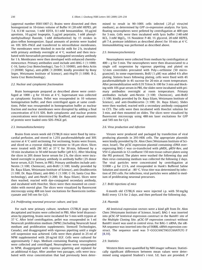
To evaluate the levels of BAG-1 protein in the brain duringmouse development and maturation, we analyzed brain homoge-nate using Western Blot. Protein levels of both BAG-1L (p50) andBAG-1S (p29) isoforms decreased during mice development(Fig. 1). However, the pattern of this decrease was different be-tween the two isoforms. BAG-1L levels decreased significantly be-tween E15 and 1 week postnatal, while BAG-1S levels declinedsuddenly between 1 and 7 weeks postnatal. In additional experi-ments, mice brains were fractionized into cytoplasmic and nuclearfractions using centrifugation. BAG-1S was the dominant isoformin both fractions at E15, while BAG-1L and BAG-1S levels werenearly equal in the nuclear fraction at stages of full development.However, both isoforms decreased in both fractions during mousedevelopment. In conclusion, BAG-1 has a higher expression in theearly development of the brain than in the adult brain.
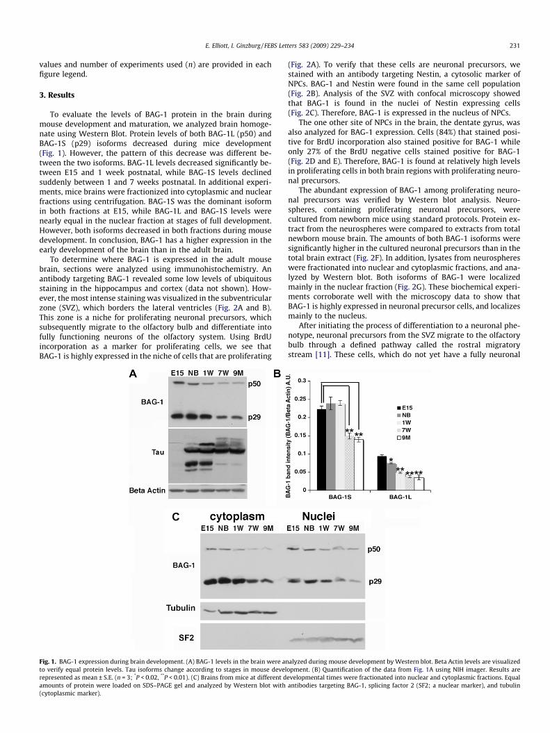
To determine where BAG-1 is expressed in the adult mousebrain, sections were analyzed using immunohistochemistry. Anantibody targeting BAG-1 revealed some low levels of ubiquitousstaining in the hippocampus and cortex (data not shown). How-ever, the most intense staining was visualized in the subventricularzone (SVZ), which borders the lateral ventricles (Fig. 2A and B).This zone is a niche for proliferating neuronal precursors, whichsubsequently migrate to the olfactory bulb and differentiate intofully functioning neurons of the olfactory system. Using BrdUincorporation as a marker for proliferating cells, we see thatBAG-1 is highly expressed in the niche of cells that are proliferating
Fig. 1. BAG-1 expression during brain development. (A) BAG-1 levels in the brain were anto verify equal protein levels. Tau isoforms change according to stages in mouse develrepresented as mean ± S.E. (n = 3; *P < 0.02, **P < 0.01). (C) Brains from mice at different damounts of protein were loaded on SDS–PAGE gel and analyzed by Western blot with(cytoplasmic marker).
(Fig. 2A). To verify that these cells are neuronal precursors, westained with an antibody targeting Nestin, a cytosolic marker ofNPCs. BAG-1 and Nestin were found in the same cell population(Fig. 2B). Analysis of the SVZ with confocal microscopy showedthat BAG-1 is found in the nuclei of Nestin expressing cells(Fig. 2C). Therefore, BAG-1 is expressed in the nucleus of NPCs.
The one other site of NPCs in the brain, the dentate gyrus, wasalso analyzed for BAG-1 expression. Cells (84%) that stained posi-tive for BrdU incorporation also stained positive for BAG-1 whileonly 27% of the BrdU negative cells stained positive for BAG-1(Fig. 2D and E). Therefore, BAG-1 is found at relatively high levelsin proliferating cells in both brain regions with proliferating neuro-nal precursors.
The abundant expression of BAG-1 among proliferating neuro-nal precursors was verified by Western blot analysis. Neuro-spheres, containing proliferating neuronal precursors, werecultured from newborn mice using standard protocols. Protein ex-tract from the neurospheres were compared to extracts from totalnewborn mouse brain. The amounts of both BAG-1 isoforms weresignificantly higher in the cultured neuronal precursors than in thetotal brain extract (Fig. 2F). In addition, lysates from neurosphereswere fractionated into nuclear and cytoplasmic fractions, and ana-lyzed by Western blot. Both isoforms of BAG-1 were localizedmainly in the nuclear fraction (Fig. 2G). These biochemical experi-ments corroborate well with the microscopy data to show thatBAG-1 is highly expressed in neuronal precursor cells, and localizesmainly to the nucleus.
After initiating the process of differentiation to a neuronal phe-notype, neuronal precursors from the SVZ migrate to the olfactorybulb through a defined pathway called the rostral migratorystream [11]. These cells, which do not yet have a fully neuronal
alyzed during mouse development by Western blot. Beta Actin levels are visualizedopment. (B) Quantification of the data from Fig. 1A using NIH imager. Results areevelopmental times were fractionated into nuclear and cytoplasmic fractions. Equalantibodies targeting BAG-1, splicing factor 2 (SF2; a nuclear marker), and tubulin
Fig. 2. BAG-1 expression in niches of neuronal precursor proliferation. (A–D) Mouse brain slices (18 lm) from BrdU injected mice were analyzed by immunohistochemistrywith primary antibodies targeting BAG-1 and BrdU (A and D), or nestin (B andC).. The subventricular zone (A–C) and dentate gyrus (D) were examined by fluorescentmicroscopy. Scale bar sizes are 500 lm(A), 50 lm(B), 10 lm(C), and 50 lm(D). (E) Quantitative analysis of BrdU+ compared to BAG-1+ cells in the dentate gyrus. Results arerepresented as means ± S.E. (*P < 0.01). Ten sections were analyzed from three mice for a total of 30 sections analyzed. All cells in the field were analyzed. (F) Total brain andNCS cultures (neurospheres) were analyzed by Western blot. BAG-1 levels are higher in neurosphere (labeled NS) lysate than in total brain homogenate. (G) Neurospherelysate was separated into nuclear and cytoplasmic fractions, and analyzed by Western blot. Both BAG-1 isoforms are primarily in nuclear fraction.
232 E. Elliott, I. Ginzburg / FEBS Letters 583 (2009) 229–234
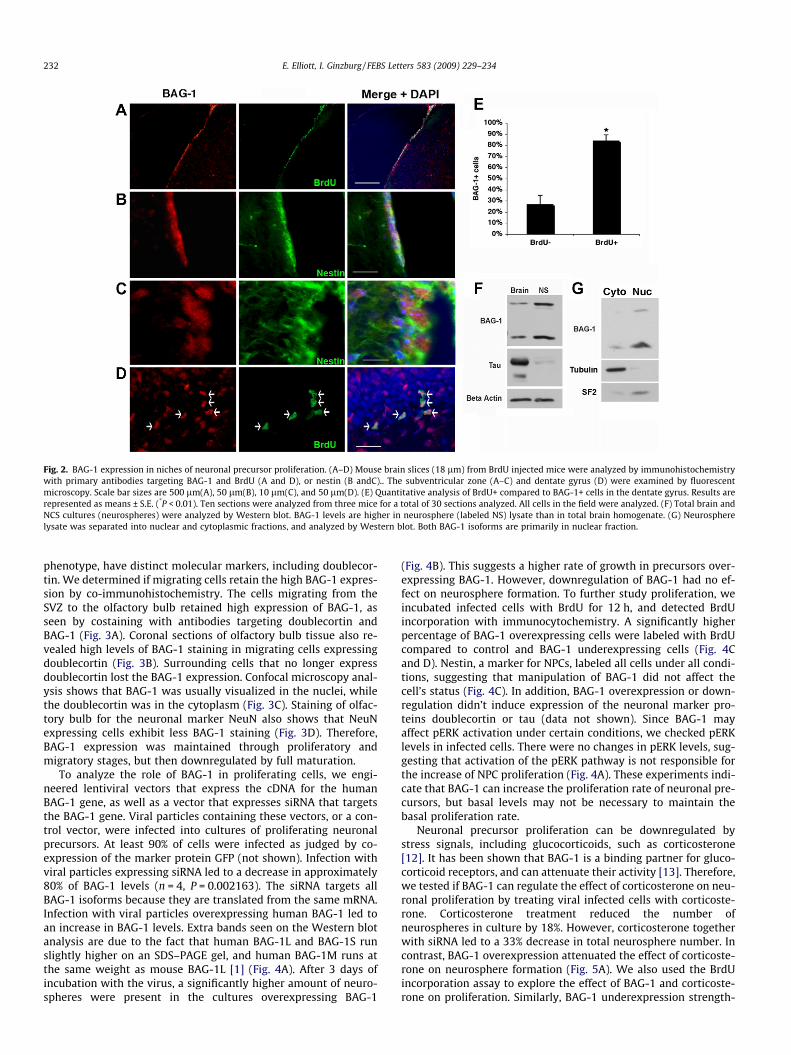
phenotype, have distinct molecular markers, including doublecor-tin. We determined if migrating cells retain the high BAG-1 expres-sion by co-immunohistochemistry. The cells migrating from theSVZ to the olfactory bulb retained high expression of BAG-1, asseen by costaining with antibodies targeting doublecortin andBAG-1 (Fig. 3A). Coronal sections of olfactory bulb tissue also re-vealed high levels of BAG-1 staining in migrating cells expressingdoublecortin (Fig. 3B). Surrounding cells that no longer expressdoublecortin lost the BAG-1 expression. Confocal microscopy anal-ysis shows that BAG-1 was usually visualized in the nuclei, whilethe doublecortin was in the cytoplasm (Fig. 3C). Staining of olfac-tory bulb for the neuronal marker NeuN also shows that NeuNexpressing cells exhibit less BAG-1 staining (Fig. 3D). Therefore,BAG-1 expression was maintained through proliferatory andmigratory stages, but then downregulated by full maturation.
To analyze the role of BAG-1 in proliferating cells, we engi-neered lentiviral vectors that express the cDNA for the humanBAG-1 gene, as well as a vector that expresses siRNA that targetsthe BAG-1 gene. Viral particles containing these vectors, or a con-trol vector, were infected into cultures of proliferating neuronalprecursors. At least 90% of cells were infected as judged by co-expression of the marker protein GFP (not shown). Infection withviral particles expressing siRNA led to a decrease in approximately80% of BAG-1 levels (n = 4, P = 0.002163). The siRNA targets allBAG-1 isoforms because they are translated from the same mRNA.Infection with viral particles overexpressing human BAG-1 led toan increase in BAG-1 levels. Extra bands seen on the Western blotanalysis are due to the fact that human BAG-1L and BAG-1S runslightly higher on an SDS–PAGE gel, and human BAG-1M runs atthe same weight as mouse BAG-1L [1] (Fig. 4A). After 3 days ofincubation with the virus, a significantly higher amount of neuro-spheres were present in the cultures overexpressing BAG-1
(Fig. 4B). This suggests a higher rate of growth in precursors over-expressing BAG-1. However, downregulation of BAG-1 had no ef-fect on neurosphere formation. To further study proliferation, weincubated infected cells with BrdU for 12 h, and detected BrdUincorporation with immunocytochemistry. A significantly higherpercentage of BAG-1 overexpressing cells were labeled with BrdUcompared to control and BAG-1 underexpressing cells (Fig. 4Cand D). Nestin, a marker for NPCs, labeled all cells under all condi-tions, suggesting that manipulation of BAG-1 did not affect thecell’s status (Fig. 4C). In addition, BAG-1 overexpression or down-regulation didn’t induce expression of the neuronal marker pro-teins doublecortin or tau (data not shown). Since BAG-1 mayaffect pERK activation under certain conditions, we checked pERKlevels in infected cells. There were no changes in pERK levels, sug-gesting that activation of the pERK pathway is not responsible forthe increase of NPC proliferation (Fig. 4A). These experiments indi-cate that BAG-1 can increase the proliferation rate of neuronal pre-cursors, but basal levels may not be necessary to maintain thebasal proliferation rate.
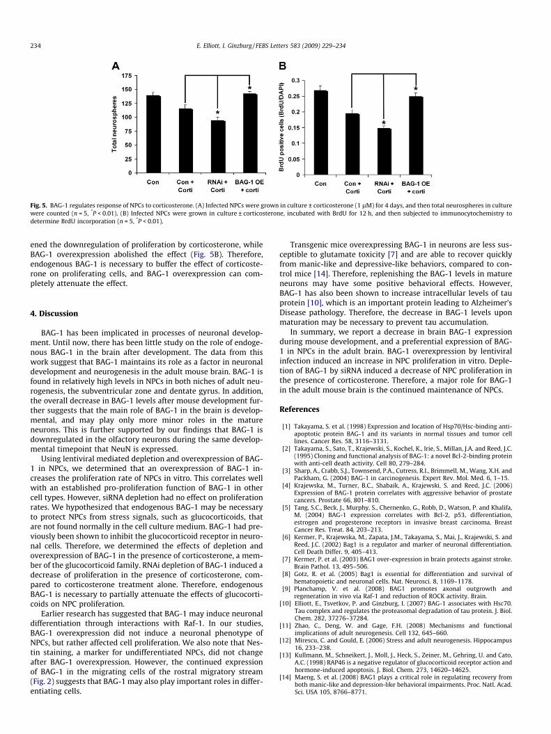
Neuronal precursor proliferation can be downregulated bystress signals, including glucocorticoids, such as corticosterone[12]. It has been shown that BAG-1 is a binding partner for gluco-corticoid receptors, and can attenuate their activity [13]. Therefore,we tested if BAG-1 can regulate the effect of corticosterone on neu-ronal proliferation by treating viral infected cells with corticoste-rone. Corticosterone treatment reduced the number ofneurospheres in culture by 18%. However, corticosterone togetherwith siRNA led to a 33% decrease in total neurosphere number. Incontrast, BAG-1 overexpression attenuated the effect of corticoste-rone on neurosphere formation (Fig. 5A). We also used the BrdUincorporation assay to explore the effect of BAG-1 and corticoste-rone on proliferation. Similarly, BAG-1 underexpression strength-
Fig. 3. BAG-1 expression in migrating neuronal precursors. (A) Sagittal slices of mouse brain were immunostained for BAG-1, and for doublecortin to visualize the migratingneuronal precursors in the rostral migratory stream. (B and C) Coronal section of mouse olfactory bulb was immunostained for BAG-1 and for doublecortin (DCX). Whitearrows show a portion of the cells containing both BAG-1 and doublecortin. BAG-1 is mainly nuclear. (D) Coronal section of mouse olfactory bulb immunostained for BAG-1and for NeuN, a nuclear marker of mature neurons. Scale bar sizes are 250 lm (A), 50 lm (B) 10 lm (C) , and 100 lm (D). Phase contrast overview of the sections are providedfor A, B, and D. Picture from C is taken from area adjacent to area visualized in B. LV, lateral ventricle, OB, olfactory bulb, Gl, glomeruli, M, mitral body layer, Gr, granule celllayer.
Fig. 4. BAG-1 overexpression increases proliferation rates of NPCs. (A) Neuronal precursor cell cultures were infected with lentiviruses containing empty vector, siRNAtargeting BAG-1, or cDNA overexpressing BAG-1 (BAG-1 OE). Western blot analysis shows the levels of BAG-1 after infection. (B) Infected NPCs were grown in culture for fourdays, and then total neurospheres formed were counted from each culture (n = 5, *P < 0.01). (C) Neuronal precursor cells infected with viruses were incubated with BrdU for12 h, and then immunostained for BrdU and the neuronal precursor marker Nestin. (D) Statistical analysis of BrdU+ cells from Fig. 4C (n = 5, *P < 0.01).
E. Elliott, I. Ginzburg / FEBS Letters 583 (2009) 229–234 233
Fig. 5. BAG-1 regulates response of NPCs to corticosterone. (A) Infected NPCs were grown in culture ± corticosterone (1 lM) for 4 days, and then total neurospheres in culturewere counted (n = 5, *P < 0.01). (B) Infected NPCs were grown in culture ± corticosterone, incubated with BrdU for 12 h, and then subjected to immunocytochemistry todetermine BrdU incorporation (n = 5, *P < 0.01).
234 E. Elliott, I. Ginzburg / FEBS Letters 583 (2009) 229–234
ened the downregulation of proliferation by corticosterone, whileBAG-1 overexpression abolished the effect (Fig. 5B). Therefore,endogenous BAG-1 is necessary to buffer the effect of corticoste-rone on proliferating cells, and BAG-1 overexpression can com-pletely attenuate the effect.
4. Discussion
BAG-1 has been implicated in processes of neuronal develop-ment. Until now, there has been little study on the role of endoge-nous BAG-1 in the brain after development. The data from thiswork suggest that BAG-1 maintains its role as a factor in neuronaldevelopment and neurogenesis in the adult mouse brain. BAG-1 isfound in relatively high levels in NPCs in both niches of adult neu-rogenesis, the subventricular zone and dentate gyrus. In addition,the overall decrease in BAG-1 levels after mouse development fur-ther suggests that the main role of BAG-1 in the brain is develop-mental, and may play only more minor roles in the matureneurons. This is further supported by our findings that BAG-1 isdownregulated in the olfactory neurons during the same develop-mental timepoint that NeuN is expressed.
Using lentiviral mediated depletion and overexpression of BAG-1 in NPCs, we determined that an overexpression of BAG-1 in-creases the proliferation rate of NPCs in vitro. This correlates wellwith an established pro-proliferation function of BAG-1 in othercell types. However, siRNA depletion had no effect on proliferationrates. We hypothesized that endogenous BAG-1 may be necessaryto protect NPCs from stress signals, such as glucocorticoids, thatare not found normally in the cell culture medium. BAG-1 had pre-viously been shown to inhibit the glucocorticoid receptor in neuro-nal cells. Therefore, we determined the effects of depletion andoverexpression of BAG-1 in the presence of corticosterone, a mem-ber of the glucocorticoid family. RNAi depletion of BAG-1 induced adecrease of proliferation in the presence of corticosterone, com-pared to corticosterone treatment alone. Therefore, endogenousBAG-1 is necessary to partially attenuate the effects of glucocorti-coids on NPC proliferation.
Earlier research has suggested that BAG-1 may induce neuronaldifferentiation through interactions with Raf-1. In our studies,BAG-1 overexpression did not induce a neuronal phenotype ofNPCs, but rather affected cell proliferation. We also note that Nes-tin staining, a marker for undifferentiated NPCs, did not changeafter BAG-1 overexpression. However, the continued expressionof BAG-1 in the migrating cells of the rostral migratory stream(Fig. 2) suggests that BAG-1 may also play important roles in differ-entiating cells.
Transgenic mice overexpressing BAG-1 in neurons are less sus-ceptible to glutamate toxicity [7] and are able to recover quicklyfrom manic-like and depressive-like behaviors, compared to con-trol mice [14]. Therefore, replenishing the BAG-1 levels in matureneurons may have some positive behavioral effects. However,BAG-1 has also been shown to increase intracellular levels of tauprotein [10], which is an important protein leading to Alzheimer’sDisease pathology. Therefore, the decrease in BAG-1 levels uponmaturation may be necessary to prevent tau accumulation.
In summary, we report a decrease in brain BAG-1 expressionduring mouse development, and a preferential expression of BAG-1 in NPCs in the adult brain. BAG-1 overexpression by lentiviralinfection induced an increase in NPC proliferation in vitro. Deple-tion of BAG-1 by siRNA induced a decrease of NPC proliferation inthe presence of corticosterone. Therefore, a major role for BAG-1in the adult mouse brain is the continued maintenance of NPCs.
References
[1] Takayama, S. et al. (1998) Expression and location of Hsp70/Hsc-binding anti-apoptotic protein BAG-1 and its variants in normal tissues and tumor celllines. Cancer Res. 58, 3116–3131.
[2] Takayama, S., Sato, T., Krajewski, S., Kochel, K., Irie, S., Millan, J.A. and Reed, J.C.(1995) Cloning and functional analysis of BAG-1: a novel Bcl-2-binding proteinwith anti-cell death activity. Cell 80, 279–284.
[3] Sharp, A., Crabb, S.J., Townsend, P.A., Cutress, R.I., Brimmell, M., Wang, X.H. andPackham, G. (2004) BAG-1 in carcinogenesis. Expert Rev. Mol. Med. 6, 1–15.
[4] Krajewska, M., Turner, B.C., Shabaik, A., Krajewski, S. and Reed, J.C. (2006)Expression of BAG-1 protein correlates with aggressive behavior of prostatecancers. Prostate 66, 801–810.
[5] Tang, S.C., Beck, J., Murphy, S., Chernenko, G., Robb, D., Watson, P. and Khalifa,M. (2004) BAG-1 expression correlates with Bcl-2, p53, differentiation,estrogen and progesterone receptors in invasive breast carcinoma. BreastCancer Res. Treat. 84, 203–213.
[6] Kermer, P., Krajewska, M., Zapata, J.M., Takayama, S., Mai, J., Krajewski, S. andReed, J.C. (2002) Bag1 is a regulator and marker of neuronal differentiation.Cell Death Differ. 9, 405–413.
[7] Kermer, P. et al. (2003) BAG1 over-expression in brain protects against stroke.Brain Pathol. 13, 495–506.
[8] Gotz, R. et al. (2005) Bag1 is essential for differentiation and survival ofhematopoietic and neuronal cells. Nat. Neurosci. 8, 1169–1178.
[9] Planchamp, V. et al. (2008) BAG1 promotes axonal outgrowth andregeneration in vivo via Raf-1 and reduction of ROCK activity. Brain.
[10] Elliott, E., Tsvetkov, P. and Ginzburg, I. (2007) BAG-1 associates with Hsc70.Tau complex and regulates the proteasomal degradation of tau protein. J. Biol.Chem. 282, 37276–37284.
[11] Zhao, C., Deng, W. and Gage, F.H. (2008) Mechanisms and functionalimplications of adult neurogenesis. Cell 132, 645–660.
[12] Mirescu, C. and Gould, E. (2006) Stress and adult neurogenesis. Hippocampus16, 233–238.
[13] Kullmann, M., Schneikert, J., Moll, J., Heck, S., Zeiner, M., Gehring, U. and Cato,A.C. (1998) RAP46 is a negative regulator of glucocorticoid receptor action andhormone-induced apoptosis. J. Biol. Chem. 273, 14620–14625.
[14] Maeng, S. et al. (2008) BAG1 plays a critical role in regulating recovery fromboth manic-like and depression-like behavioral impairments. Proc. Natl. Acad.Sci. USA 105, 8766–8771.