+ All Categories
Home > Documents > Basic Human Anatomy - The Brookside Associates

Basic Human Anatomy - The Brookside Associates

Date post: 28-Nov-2021
Category:
Upload: others
View: 3 times
Download: 0 times
Share this document with a friend
48
Basic Human Anatomy Lesson 11: Nervous System Page 1 Basic Human Anatomy Lesson 11: Nervous System Welcome to Lesson 11 of the Basic Human Anatomy Course. Today, we’ll be studying the Human Nervous System. I have 19 goals for you in this lesson: 1. Name and identify two types of nervous tissues. 2. Name three functions for which nervous tissues are specialized. 3. Define neuron, dendrite, and axon. 4. When given the shape, diameter, or function, name the corresponding type of neuron. 5. Describe neuron "connections," including the synapse and the neuromuscular junction. 6. Name and identify the three major divisions of the human nervous system; name the two major subdivisions of the CNS. 7. Name and briefly describe the three major subdivisions of the human brain; name and locate the four ventricles and their connecting channels. 8. Describe the spinal cord, including the two enlargements, elements of its cross section, and the surrounding vertebral canal. 9. Describe the meninges and the skeletal coverings of the CNS. 10. Name and identify the main arteries and veins of the brain and briefly describe the blood supply of the spinal cord. 11. Describe the formation of cerebrospinal fluid (CSF) and the path of CSF flow. 12. Define peripheral nervous system (PNS) and nerve; name and briefly describe two categories of PNS nerves; describe the anatomy of a "typical" spinal nerve; define reflex and reflex arc; briefly describe the components of the general reflex arc.
Transcript

Basic Human Anatomy Lesson 11: Nervous System Page 1

Basic Human Anatomy

Lesson 11: Nervous System

Welcome to Lesson 11 of the Basic Human Anatomy Course. Today, we’ll be

studying the Human Nervous System.

I have 19 goals for you in this lesson:

1. Name and identify two types of nervous tissues.

2. Name three functions for which nervous tissues are specialized.

3. Define neuron, dendrite, and axon.

4. When given the shape, diameter, or function, name the corresponding type

of neuron.

5. Describe neuron "connections," including the synapse and the

neuromuscular junction.

6. Name and identify the three major divisions of the human nervous system;

name the two major subdivisions of the CNS.

7. Name and briefly describe the three major subdivisions of the human brain;

name and locate the four ventricles and their connecting channels.

8. Describe the spinal cord, including the two enlargements, elements of its

cross section, and the surrounding vertebral canal.

9. Describe the meninges and the skeletal coverings of the CNS.

10. Name and identify the main arteries and veins of the brain and briefly

describe the blood supply of the spinal cord.

11. Describe the formation of cerebrospinal fluid (CSF) and the path of CSF

flow.

12. Define peripheral nervous system (PNS) and nerve; name and briefly

describe two categories of PNS nerves; describe the anatomy of a "typical"

spinal nerve; define reflex and reflex arc; briefly describe the components

of the general reflex arc.

Basic Human Anatomy Lesson 11: Nervous System Page 2

13. Define autonomic nervous system (ANS) and visceral organs; briefly

describe efferent pathways of the ANS; name the major divisions of the

human ANS; briefly describe the major activities of the human ANS for the

thoraco-lumbar and cranio-sacral outflows; briefly describe the first and

second neurons, innervations, and effects in each case.

14. Define pathway, neuraxis, sensor pathway, and motor pathway; briefly

describe levels of control, pyramidal and extra-pyramidal motor pathways,

and sensory pathways; and give examples of general senses and special

senses.

15. Briefly describe the sensory receptors and sensory pathways for the special

senses of smell and taste.

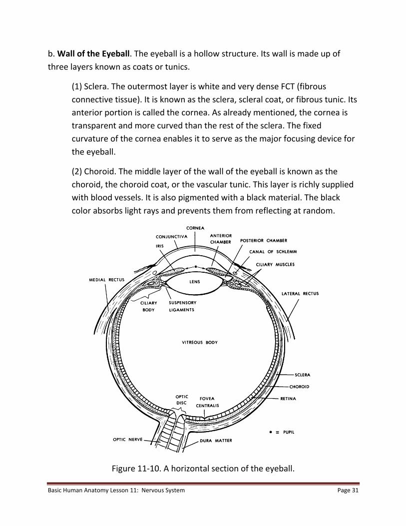
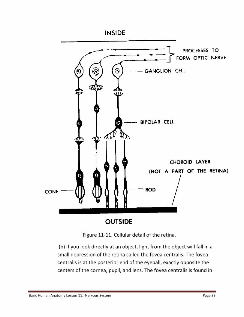
16. Describe the structures of the bulbus oculi, the orbit, and the adnexa.

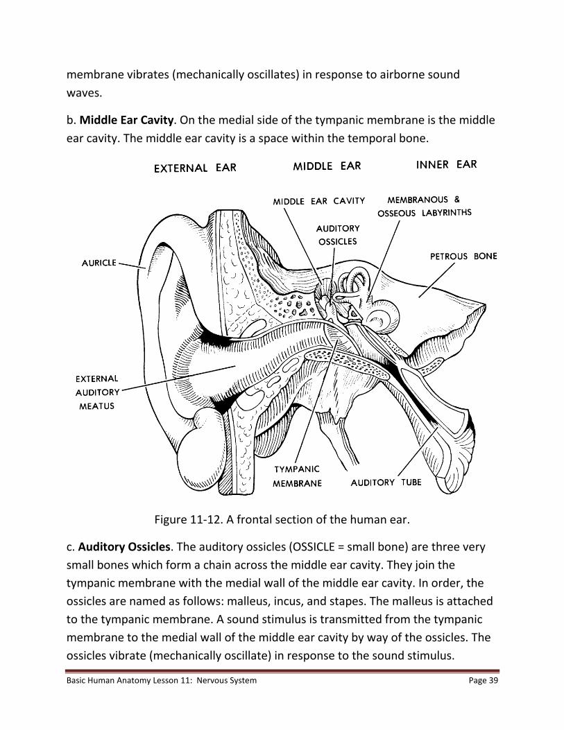
17. Describe the structures of the external ear, the middle ear, and the internal

ear.

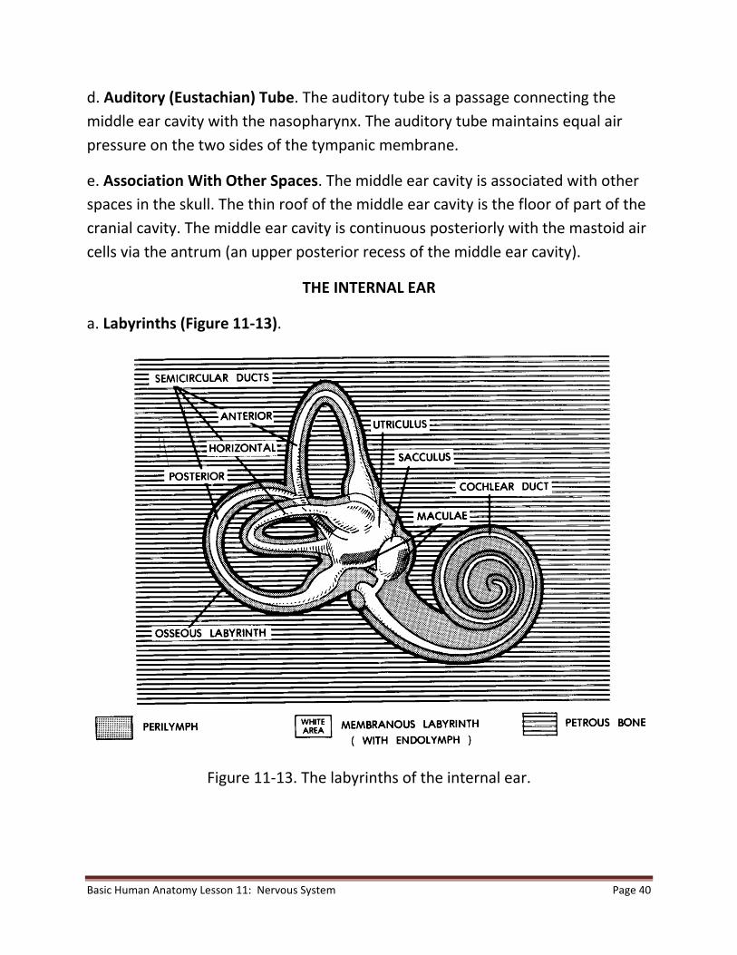
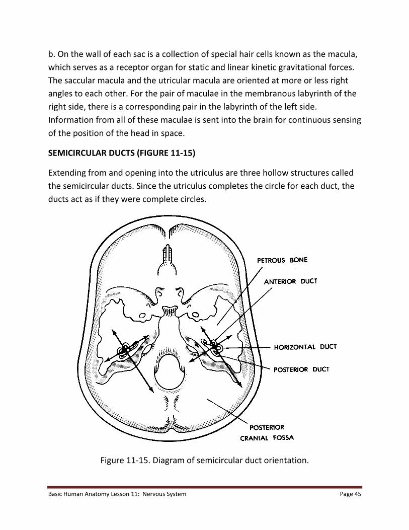
18. Describe the structures of the sacculus, utriculus, semicircular ducts, and

the vestibular nerve.

19. Describe controls in the human nervous system.

Basic Human Anatomy Lesson 11: Nervous System Page 3

INTRODUCTION

NERVOUS TISSUES

There are two types of nervous tissues--the neurons (nerve cells) and glia

(neuroglia). See paragraph 2-17. The neuron is the basic structural unit of the

nervous system. The glia are cells of supporting tissue for the nervous system.

There are several different types of glia, but their general function is support

(physical, nutritive, etc.).

SPECIALIZATION

Nervous tissues are specialized to:

a. Receive Stimuli. Cells receiving stimuli are said to be "irritable" (as are all living

cells to a degree).

b. Transmit Information.

c. "Store" Information. The storing of information is called memory.

THE NEURON AND ITS "CONNECTIONS"

DEFINITION

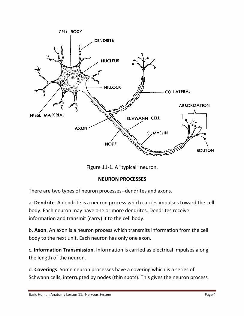
A neuron (figure 11-1) is a nerve cell body and all of its processes (branches).

NEURON CELL BODY

The neuron cell body is similar to that of the "typical" animal cell described in

lesson 1.

Basic Human Anatomy Lesson 11: Nervous System Page 4

Figure 11-1. A "typical" neuron.

NEURON PROCESSES

There are two types of neuron processes--dendrites and axons.

a. Dendrite. A dendrite is a neuron process which carries impulses toward the cell

body. Each neuron may have one or more dendrites. Dendrites receive

information and transmit (carry) it to the cell body.

b. Axon. An axon is a neuron process which transmits information from the cell

body to the next unit. Each neuron has only one axon.

c. Information Transmission. Information is carried as electrical impulses along

the length of the neuron.

d. Coverings. Some neuron processes have a covering which is a series of

Schwann cells, interrupted by nodes (thin spots). This gives the neuron process

Basic Human Anatomy Lesson 11: Nervous System Page 5

the appearance of links of sausage. The Schwann cells produce a lipid (fatty)

material called myelin. This myelin acts as an electrical insulator during the

transmission of impulses.

TYPES OF NEURONS

Neurons may be identified according to shape, diameter of their processes, or

function.

a. According to Shape. A pole is the point where a neuron process meets the cell

body. To determine the type according to shape, count the number of poles.

(1) Multipolar neurons. Multipolar neurons have more than two poles (one

axon and two or more dendrites).

(2) Bipolar neurons. Bipolar neurons have two poles (one axon and one

dendrite).

(3) Unipolar neurons. Unipolar neurons have a single process which

branches into a T-shape. One arm is an axon; the other is a dendrite.

b. According to Diameter (Thickness) of Processes. Neurons may be rated

according to the thickness of myelin surrounding the axon. In order of decreasing

thickness, they are rated A (thickest), B, and C (thinnest). The thickness affects the

rate at which impulses are transmitted. The thickest are fastest. The thinnest are

slowest.

c. According to Function.

(1) Sensory neurons. In sensory neurons, impulses are transmitted from

receptor organs (for pain, vision, hearing, etc.) to the central nervous

system (CNS).

(2) Motor neurons. In motor neurons, impulses are transmitted from the

CNS to muscles and glands (effector organs).

(3) Interneurons. Interneurons transmit information from one neuron to

another. An interneuron "connects" two other neurons.

Basic Human Anatomy Lesson 11: Nervous System Page 6

(4) Others. There are other, more specialized types, for example, in the

CNS.

NEURON "CONNECTIONS"

A neuron may "connect" either with another neuron or with a muscle fiber. A

phrase used to describe such "connections" is "continuity without contact."

Neurons do not actually touch. There is just enough space to prevent the

electrical transmission from crossing from the first neuron to the next. This space

is called the synaptic cleft. Information is transferred across the synaptic cleft by

chemicals called neurotransmitters. Neurotransmitters are manufactured and

stored on only one side of the cleft. Because of this, information flows in only one

direction across the cleft.

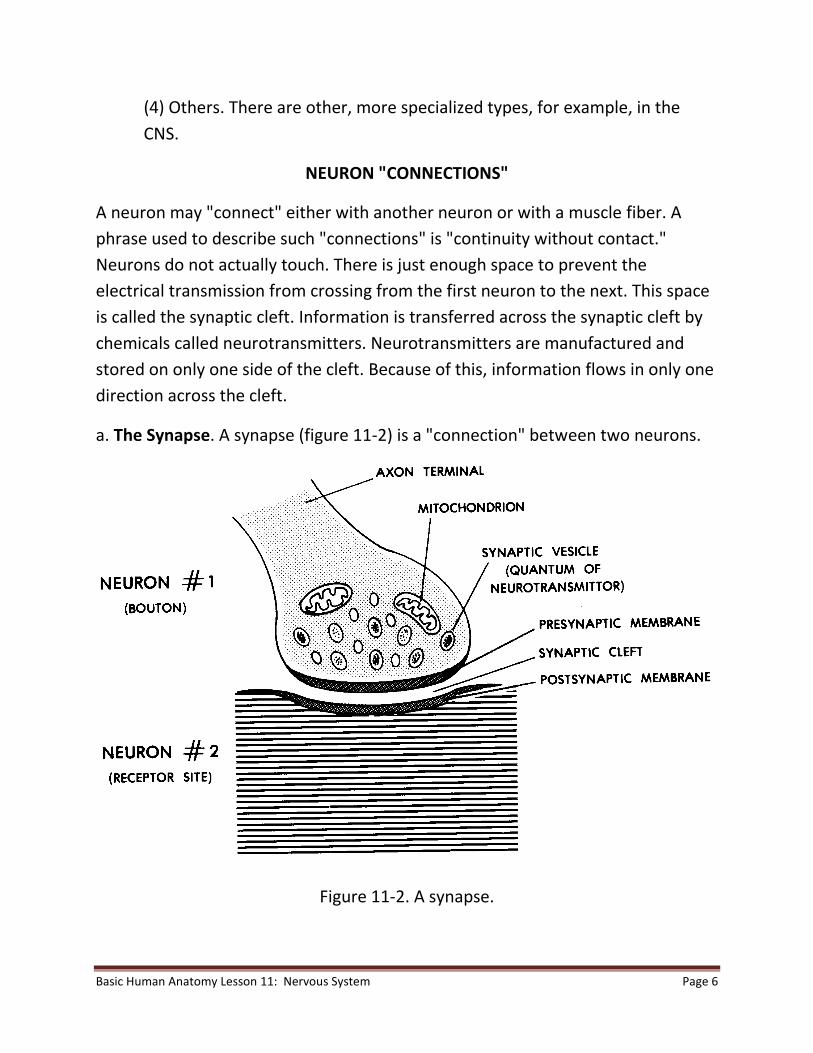
a. The Synapse. A synapse (figure 11-2) is a "connection" between two neurons.

Figure 11-2. A synapse.

Basic Human Anatomy Lesson 11: Nervous System Page 7

(1) First neuron. An axon terminates in tiny branches. At the end of each branch is

found a terminal bulb. Synaptic vesicles (bundles of neurotransmitter) are located

within each terminal bulb. That portion of the terminal bulb which faces the

synaptic cleft is thickened and is called the presynaptic membrane. This is the

membrane through which neurotransmitters pass to enter the synaptic cleft.

(2) Synaptic cleft. The synaptic cleft is the space between the terminal bulb of the

first neuron and the dendrite or cell body of the second neuron.

(3) Second neuron. The terminal bulb of the first neuron lies near a site on a

dendrite or the cell body of the second neuron. The membrane at this site on the

second neuron is known as the postsynaptic membrane. Within the second

neuron is a chemical that inactivates the used neurotransmitter.

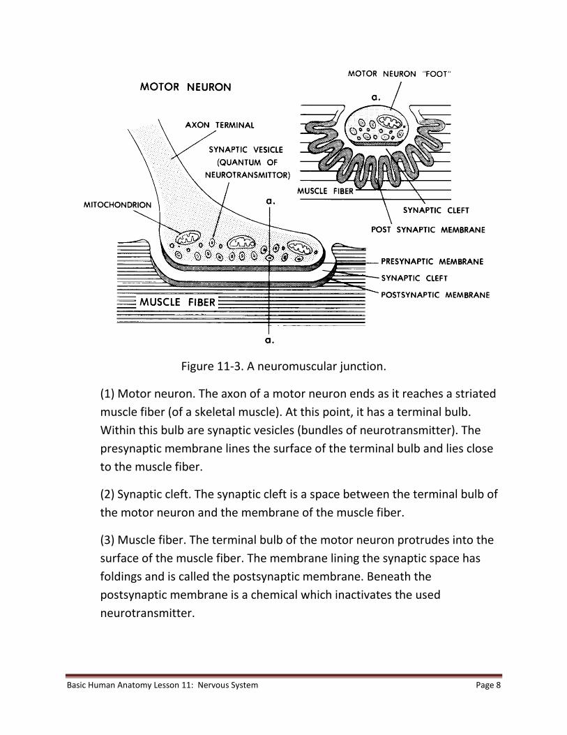
b. The Neuromuscular Junction. A neuromuscular junction (figure 11-3) is a

"connection" between the terminal of a motor neuron and a muscle fiber. The

neuromuscular junction has an organization identical to a synapse. However, the

bulb is larger. The postsynaptic membrane is also larger and has foldings to

increase its surface area.

Basic Human Anatomy Lesson 11: Nervous System Page 8

Figure 11-3. A neuromuscular junction.

(1) Motor neuron. The axon of a motor neuron ends as it reaches a striated

muscle fiber (of a skeletal muscle). At this point, it has a terminal bulb.

Within this bulb are synaptic vesicles (bundles of neurotransmitter). The

presynaptic membrane lines the surface of the terminal bulb and lies close

to the muscle fiber.

(2) Synaptic cleft. The synaptic cleft is a space between the terminal bulb of

the motor neuron and the membrane of the muscle fiber.

(3) Muscle fiber. The terminal bulb of the motor neuron protrudes into the

surface of the muscle fiber. The membrane lining the synaptic space has

foldings and is called the postsynaptic membrane. Beneath the

postsynaptic membrane is a chemical which inactivates the used

neurotransmitter.

Basic Human Anatomy Lesson 11: Nervous System Page 9

THE HUMAN CENTRAL NERVOUS SYSTEM

GENERAL

The major divisions of the human nervous system are the central nervous system

(CNS), the peripheral nervous system (PNS), and the autonomic nervous system

(ANS). The CNS is made up of the brain and spinal cord. Both the PNS and the ANS

carry information to and from the central nervous system. The PNS is generally

concerned with the innervation of skeletal muscles and other muscles made up of

striated muscle tissue, as well as sensory information from the periphery of the

body.

The ANS is that portion of the nervous system concerned with control of smooth

muscle, cardiac muscle, and glands. The CNS (figure 11-4) is known as central

because its anatomical location is along the central axis of the body and because

the CNS is central in function. If we use a computer analogy to understand that it

is central in function, the CNS would be the central processing unit and other

parts of the nervous system would supply inputs and transmit outputs.

Basic Human Anatomy Lesson 11: Nervous System Page 10

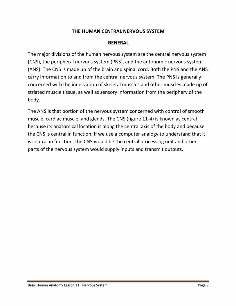
Figure 11-4. The human central nervous system.

Basic Human Anatomy Lesson 11: Nervous System Page 11

a. Major Subdivisions of the CNS. The major subdivisions of the CNS are the brain

and the spinal cord.

b. Coverings of the CNS. The coverings of the CNS are skeletal and fibrous.

c. Cerebrospinal Fluid (CSF). The CSF is a liquid thought to serve as a cushion and

circulatory vehicle within the CNS.
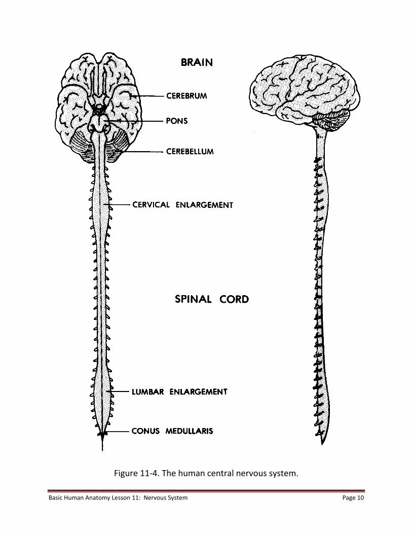
THE HUMAN BRAIN

The human brain has three major subdivisions: brainstem, cerebellum, and

cerebrum. The CNS is first formed as a simple tubelike structure in the embryo.

The concentration of nervous tissues at one end of the human embryo to produce

the brain and head is referred to as cephalization. When the embryo is about four

weeks old, it is possible to identify the early forms of the brainstem, cerebellum,

and cerebrum, as well as the spinal cord. As development continues, the brain is

located within the cranium (Lesson 4) in the cranial cavity. See figures 11-5A and

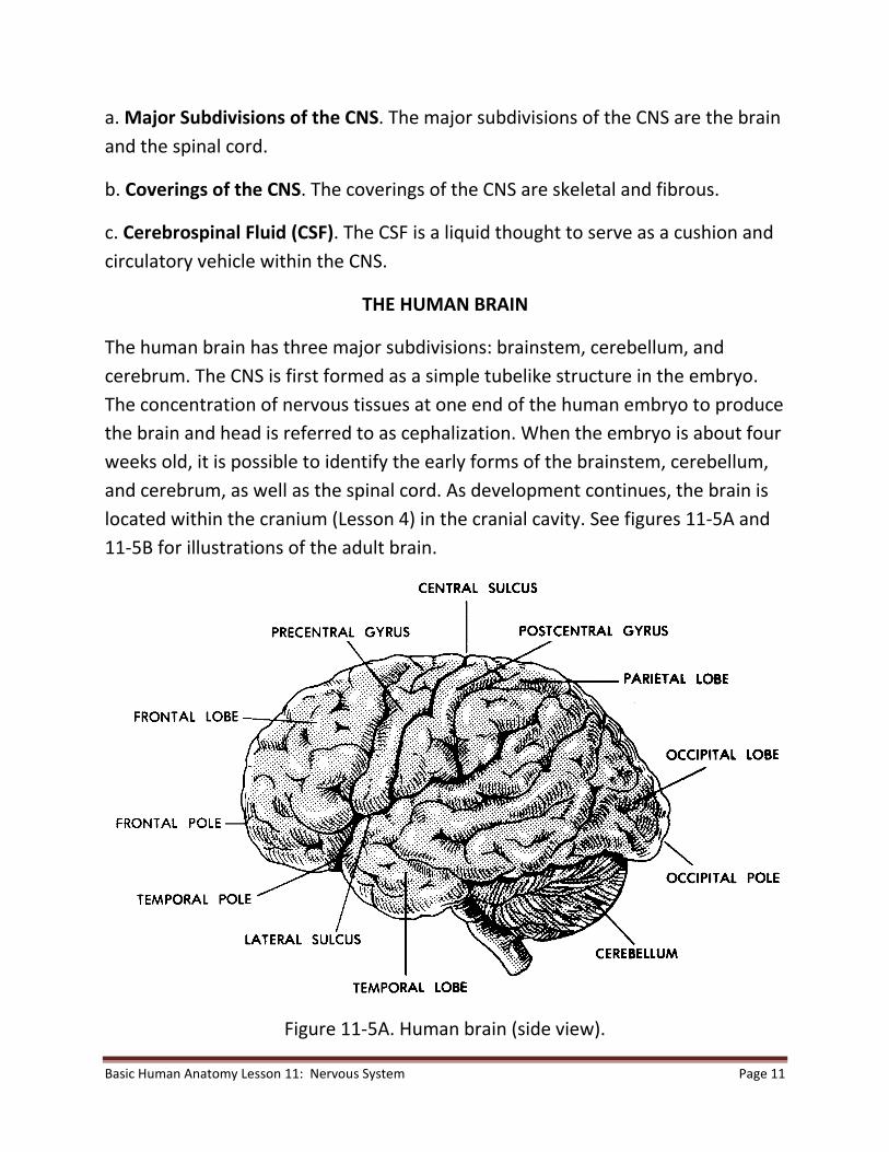
11-5B for illustrations of the adult brain.

Figure 11-5A. Human brain (side view).

Basic Human Anatomy Lesson 11: Nervous System Page 12

Figure 11-5B. Human brain (bottom view).

a. The Brainstem. The term brainstem refers to that part of the brain that

would remain after removal of the cerebrum and cerebellum. The

brainstem is the basal portion (portion of the base) of the brain. The

brainstem can be divided as follows:

FOREBRAINSTEM thalamus

hypothalamus

MIDBRAINSTEM corpora quadrigemina

cerebral peduncles

HINDBRAINSTEM pons

medulla

Basic Human Anatomy Lesson 11: Nervous System Page 13

(1) The brainstem is continuous with the spinal cord. Together, the

brainstem and the spinal cord are sometimes known as the neuraxis.

(2) The brainstem provides major relays and controls for information

passing up or down the neuraxis.

(3) The 12 pairs of cranial nerves connect at the sides of the brainstem.

b. Cerebellum. The cerebellum is a spherical mass of nervous tissue attached to

and covering the hindbrainstem. It has a narrow central part called the vermis and

right and left cerebellar hemispheres.

(1) Peduncles. A peduncle is a stem-like connecting part. The cerebellum is

connected to the brainstem with three pairs of peduncles.

(2) General shape and construction. A cross section of the cerebellum

reveals that the outer cortex is composed of gray matter (cell bodies of

neurons) with many folds and sulci (shallow grooves). More centrally

located is the white matter (myelinated processes of neurons).

(3) Function. The cerebellum is the primary coordinator/integrator of motor

actions of the body.

c. Cerebrum. The cerebrum consists of two very much enlarged hemispheres

connected to each other by a special structure called the corpus callosum. Each

cerebral hemisphere is connected to the brainstem by a cerebral peduncle. The

surface of each cerebral hemisphere is subdivided into areas known as lobes.

Each lobe is named according to the cranial bone under which it lies: frontal,

parietal, occipital, and temporal.

(1) The space separating the two cerebral hemispheres is called the

longitudinal fissure. The shallow grooves in the surface of the cerebrum are

called sulci (sulcus, singular). The ridges outlined by the sulci are known as

gyri (gyrus, singular).

Basic Human Anatomy Lesson 11: Nervous System Page 14

(2) The cerebral cortex is the gray outer layer of each hemisphere. The

occurrence of sulci and gyri helps to increase the amount of this layer.

Deeper within

the cerebral hemispheres, the tissue is white. The "gray matter" represents

cell bodies of the neurons. The "white matter" represents the axons.

(3) The areas of the cortex are associated with groups of related functions.

(a) For example, centers of speech and hearing are located along the

lateral sulcus, at the side of each hemisphere.

(b) Vision is centered at the rear in the area known as the occipital

lobe.

(c) Sensory and motor functions are located along the central sulcus,

which separates the frontal and parietal lobes of each hemisphere.

The motor areas are located along the front side of the central

sulcus, in the frontal lobe. The sensory areas are located along the

rear side of the central sulcus, in the parietal lobe.

d. Ventricles. Within the brain, there are interconnected hollow spaces filled with

cerebrospinal fluid (CSF). These hollow spaces are known as ventricles. The right

and left lateral ventricles are found in the cerebral hemispheres. The lateral

ventricles are connected to the third ventricle via the interventricular foramen (of

Monroe). The third ventricle is located in the forebrainstem. The fourth ventricle

is in the hindbrainstem. The cerebral aqueduct (of Sylvius) is a short tube through

the midbrainstem which connects the third and fourth ventricles. The fourth

ventricle is continuous with the narrow central canal of the spinal cord.

THE HUMAN SPINAL CORD

a. Location and Extent. Referring to figure 4-4, you can see that the typical

vertebra has a large opening called the vertebral (or spinal) foramen. Together,

these foramina form the vertebral (spinal) canal for the entire vertebral column.

The spinal cord, located within the spinal canal, is continuous with the brainstem.

Basic Human Anatomy Lesson 11: Nervous System Page 15

The spinal cord travels the length from the foramen magnum at the base of the

skull to the junction of the first and second lumbar vertebrae.

(1) Enlargements. The spinal cord has two enlargements. One is the cervical

enlargement, associated with nerves for the upper members. The other is

the lumbosacral enlargement, associated with nerves for the lower

members.

(2) Spinal nerves. A nerve is a bundle of neuron processes which carry

impulses to and from the CNS. Those nerves arising from the spinal cord are

spinal nerves. There are 31 pairs of spinal nerves.

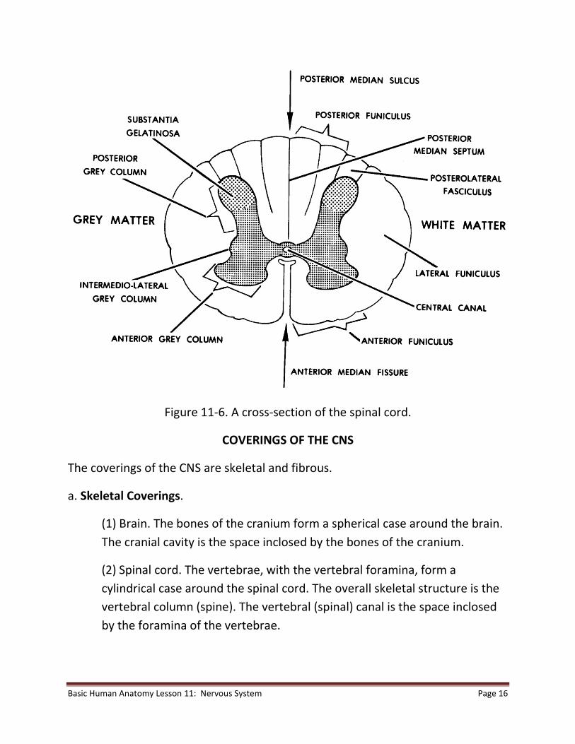
b. A Cross Section of the Spinal Cord (figure 11-6). The spinal cord is a continuous

structure which runs through the vertebral canal down to the lumbar region of

the column. It is composed of a mass of central gray matter (cell bodies of

neurons) surrounded by peripheral white matter (myelinated processes of

neurons). The gray and white matter are thus considered columns of material.

However, in a cross section, this effect of columns is lost.

(1) Central canal. A very narrow canal, called the central canal, is located in

the center of the spinal cord. The central canal is continuous with the

fourth ventricle of the brain.

(2) The gray matter. In the cross section of the spinal cord, one can see a

central H-shaped region of gray matter. Each arm of the H is called a horn,

resulting in two posterior horns and two anterior horns. The connecting link

is called the gray commissure. Since the gray matter extends the full length

of the spinal cord, these horns are actually sections of the gray columns.

(3) The white matter. The peripheral portion of the spinal cord cross section

consists of white matter. Since a column of white matter is a large bundle

of processes, it is called a funiculus. In figure 11-6, note the anterior, lateral,

and posterior funiculi.

Basic Human Anatomy Lesson 11: Nervous System Page 16

Figure 11-6. A cross-section of the spinal cord.

COVERINGS OF THE CNS

The coverings of the CNS are skeletal and fibrous.

a. Skeletal Coverings.

(1) Brain. The bones of the cranium form a spherical case around the brain.

The cranial cavity is the space inclosed by the bones of the cranium.

(2) Spinal cord. The vertebrae, with the vertebral foramina, form a

cylindrical case around the spinal cord. The overall skeletal structure is the

vertebral column (spine). The vertebral (spinal) canal is the space inclosed

by the foramina of the vertebrae.

Basic Human Anatomy Lesson 11: Nervous System Page 17

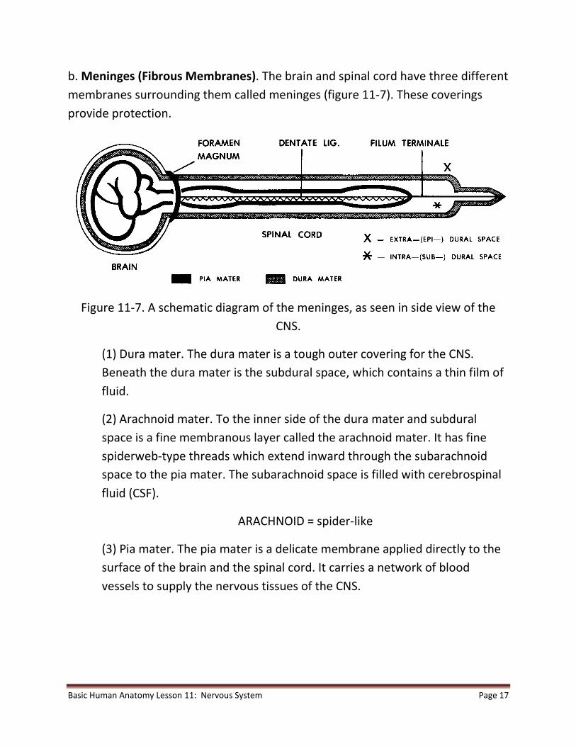
b. Meninges (Fibrous Membranes). The brain and spinal cord have three different

membranes surrounding them called meninges (figure 11-7). These coverings

provide protection.

Figure 11-7. A schematic diagram of the meninges, as seen in side view of the

CNS.

(1) Dura mater. The dura mater is a tough outer covering for the CNS.

Beneath the dura mater is the subdural space, which contains a thin film of

fluid.

(2) Arachnoid mater. To the inner side of the dura mater and subdural

space is a fine membranous layer called the arachnoid mater. It has fine

spiderweb-type threads which extend inward through the subarachnoid

space to the pia mater. The subarachnoid space is filled with cerebrospinal

fluid (CSF).

ARACHNOID = spider-like

(3) Pia mater. The pia mater is a delicate membrane applied directly to the

surface of the brain and the spinal cord. It carries a network of blood

vessels to supply the nervous tissues of the CNS.

Basic Human Anatomy Lesson 11: Nervous System Page 18

BLOOD SUPPLY OF THE CNS

a. Blood Supply of the Brain. The paired internal carotid arteries and the paired

vertebral arteries supply blood rich in oxygen to the brain. Branches of these

arteries join to form a circle under the base of the brain. This is called the cerebral

circle (of Willis). From this circle, numerous branches supply specific areas of the

brain.

(1) A single branch is often the only blood supply to that particular area. Such an

artery is called an end artery. If it fails to supply blood to that specific area, that

area will die (stroke).

(2) The veins and venous sinuses of the brain drain into the paired internal jugular

veins, which carry the blood back toward the heart.

b. Blood Supply of the Spinal Cord. The blood supply of the spinal cord is by way

of a combination of three longitudinal arteries running along its length and

reinforced by segmental arteries from the sides.

CEREBROSPINAL FLUID (CSF)

A clear fluid called cerebrospinal fluid (CSF) is found in the cavities of the CNS. CSF

is found in the ventricles of the brain, the subarachnoid space, and the central

canal of the spinal cord. CSF and its associated structures make up the circulatory

system for the CNS.

a. Choroid Plexuses. Choroid plexuses are special collections of arterial capillaries

found in the roofs of the third and fourth ventricles of the brain. The choroid

plexuses continuously produce CSF from the plasma of the blood.

b. Path of the CSF Flow. Blood flows through the arterial capillaries of the choroid

plexuses. As CSF is produced by the choroid plexuses, it flows into all four

ventricles. CSF from the lateral ventricles flows into the third ventricle and then

through the cerebral aqueduct into the fourth ventricle. By passing through three

small holes in the roof of the fourth ventricle, CSF enters the subarachnoid space.

From the subarachnoid space, the CSF is transported through the arachnoid villi

Basic Human Anatomy Lesson 11: Nervous System Page 19

(granulations) into the venous sinuses. Thus, the CSF is formed from arterial blood

and returned to the venous blood.

THE PERIPHERAL NERVOUS SYSTEM (PNS)

GENERAL

a. Definitions.

(1) The peripheral nervous system (PNS) is that portion of the nervous

system generally concerned with commands for skeletal muscles and other

muscles made up of striated muscle tissue, as well as sensory information

from the periphery of the body. The sensory information is carried to the

CNS where it is processed. The PNS carries commands from the CNS to

musculature.

(2) A nerve is a collection of neuron processes, together and outside the

CNS. (A fiber tract is a collection of neuron processes, together and inside

the CNS.)

b. General Characteristics of the Peripheral Nerves. The PNS is made up of a

large number of individual nerves. These nerves are arranged in pairs. Each pair

includes one nerve on the left side of the brainstem or spinal cord and one nerve

on the right side. The nerve pairs are in a series, each pair resembling the

preceding, from top to bottom.

c. Categories of PNS Nerves. PNS nerves include cranial nerves and spinal nerves.

(1) Cranial nerves. The 12 pairs of nerves attached to the right and left

sides of the brainstem are called cranial nerves. Each cranial nerve is

identified by a Roman numeral in order from I to XII and an individual

name. For example, the Vth ("fifth") cranial nerve is known as the

trigeminal nerve (N.).

TRI = three

GEMINI = alike

Basic Human Anatomy Lesson 11: Nervous System Page 20

TRIGEMINAL = having three similar major branches

(2) Spinal nerves. Attached to the sides of the spinal cord are 31 pairs of

spinal nerves. The spinal nerves are named by:

(a) The region of the spinal cord with which the nerve is associated.

(b) An Arabic numeral within the region. For example, T-5 is the fifth

spinal nerve in the thoracic region.

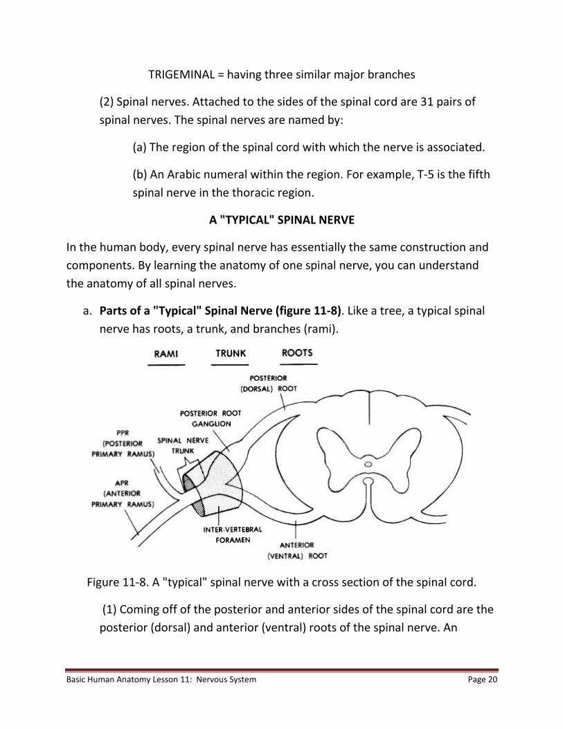
A "TYPICAL" SPINAL NERVE

In the human body, every spinal nerve has essentially the same construction and

components. By learning the anatomy of one spinal nerve, you can understand

the anatomy of all spinal nerves.

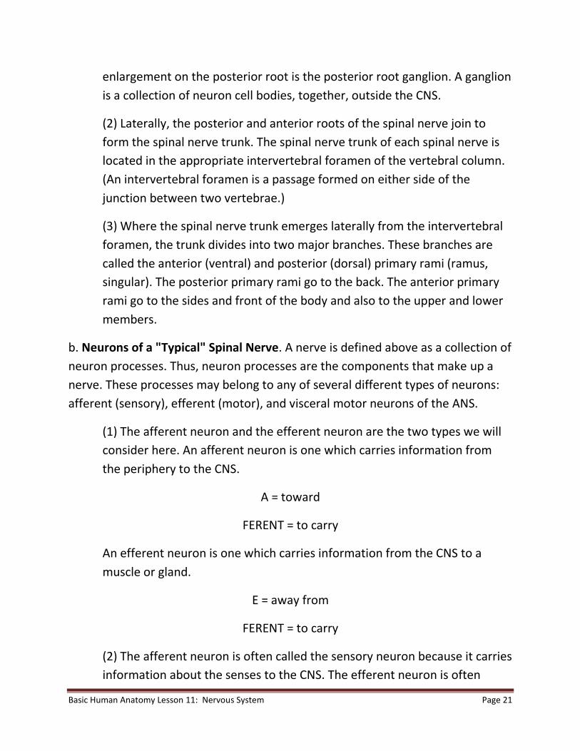
a. Parts of a "Typical" Spinal Nerve (figure 11-8). Like a tree, a typical spinal

nerve has roots, a trunk, and branches (rami).

Figure 11-8. A "typical" spinal nerve with a cross section of the spinal cord.

(1) Coming off of the posterior and anterior sides of the spinal cord are the

posterior (dorsal) and anterior (ventral) roots of the spinal nerve. An

Basic Human Anatomy Lesson 11: Nervous System Page 21

enlargement on the posterior root is the posterior root ganglion. A ganglion

is a collection of neuron cell bodies, together, outside the CNS.

(2) Laterally, the posterior and anterior roots of the spinal nerve join to

form the spinal nerve trunk. The spinal nerve trunk of each spinal nerve is

located in the appropriate intervertebral foramen of the vertebral column.

(An intervertebral foramen is a passage formed on either side of the

junction between two vertebrae.)

(3) Where the spinal nerve trunk emerges laterally from the intervertebral

foramen, the trunk divides into two major branches. These branches are

called the anterior (ventral) and posterior (dorsal) primary rami (ramus,

singular). The posterior primary rami go to the back. The anterior primary

rami go to the sides and front of the body and also to the upper and lower

members.

b. Neurons of a "Typical" Spinal Nerve. A nerve is defined above as a collection of

neuron processes. Thus, neuron processes are the components that make up a

nerve. These processes may belong to any of several different types of neurons:

afferent (sensory), efferent (motor), and visceral motor neurons of the ANS.

(1) The afferent neuron and the efferent neuron are the two types we will

consider here. An afferent neuron is one which carries information from

the periphery to the CNS.

A = toward

FERENT = to carry

An efferent neuron is one which carries information from the CNS to a

muscle or gland.

E = away from

FERENT = to carry

(2) The afferent neuron is often called the sensory neuron because it carries

information about the senses to the CNS. The efferent neuron is often

Basic Human Anatomy Lesson 11: Nervous System Page 22

called the motor neuron because it carries commands from the CNS to

cause a muscle to act.

(3) A stimulus acts upon a sensory receptor organ in the skin or in another

part of the body. The information is carried by an afferent (sensory) neuron

through merging branches of the spinal nerve to the posterior root

ganglion. The afferent (sensory) neuron's cell body is located in the

posterior root ganglion. From this point, information continues in the

posterior root to the spinal cord. The efferent (motor) neuron carries

command information from the spinal cord to the individual muscle of the

human body.

(4) Visceral motor neurons of the ANS (see section V), which innervate

visceral organs of the body's periphery, are distributed along with the

peripheral nerves.

Basic Human Anatomy Lesson 11: Nervous System Page 23

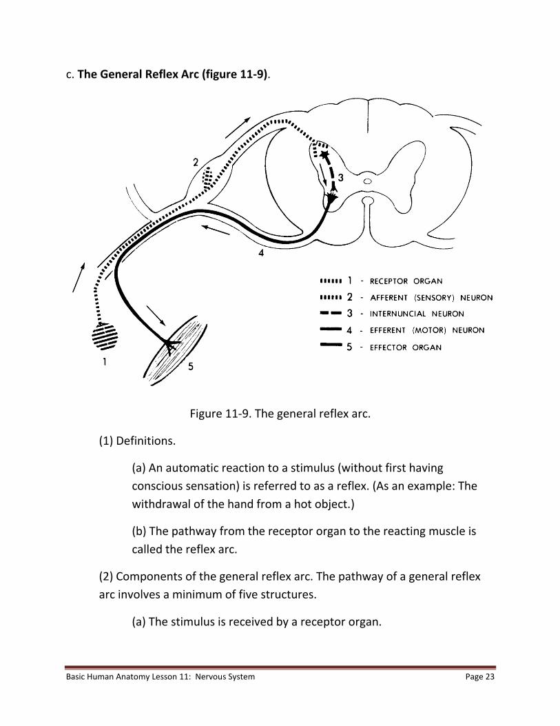
c. The General Reflex Arc (figure 11-9).

Figure 11-9. The general reflex arc.

(1) Definitions.

(a) An automatic reaction to a stimulus (without first having

conscious sensation) is referred to as a reflex. (As an example: The

withdrawal of the hand from a hot object.)

(b) The pathway from the receptor organ to the reacting muscle is

called the reflex arc.

(2) Components of the general reflex arc. The pathway of a general reflex

arc involves a minimum of five structures.

(a) The stimulus is received by a receptor organ.

Basic Human Anatomy Lesson 11: Nervous System Page 24

(b) That information is transmitted to the CNS by the afferent

(sensory) neuron.

(c) Within the spinal cord, there is a special neuron connecting the

afferent neuron to the efferent neuron. This special connecting

neuron is called the internuncial neuron, or interneuron.

INTER = between

NUNCIA = messenger

INTERNUNCIAL = the carrier of information between

(d) The efferent (motor) neuron carries the appropriate command

from the spinal cord to the reacting muscle.

(e) The reacting muscle is called the effector organ.

THE AUTONOMIC NERVOUS SYSTEM (ANS)

GENERAL

The autonomic nervous system (ANS) is that portion of the nervous system

generally concerned with commands for smooth muscle tissue, cardiac muscle

tissue, and glands.

a. Visceral Organs.

(1) Definition. The term visceral organs may be used to include:

(a) The various hollow organs of the body whose walls have smooth

muscle tissue in them. Examples are the blood vessels and the gut.

(b) The glands.

(2) Distribution. The visceral organs are located in the central cavity of the

body (example: stomach) and throughout the periphery of the body

(example: sweat glands of the skin).

Basic Human Anatomy Lesson 11: Nervous System Page 25

(3) Control. It has always been thought that the control of visceral organs

was "automatic" and not conscious. However, recent researches indicate

that proper training enables a person to consciously control some of the

visceral organs.

b. Efferent Pathways. Earlier, we said that each neuron in the PNS extended the

entire distance from the CNS to the receptor or effector organ. In the ANS, there

are always two neurons (one after the other) connecting the CNS with the visceral

organ. The cell bodies of the second neurons form a collection outside the CNS,

called a ganglion.

(1) The first neuron extends from the CNS to the ganglion and is therefore

alled the preganglionic neuron.

(2) Cell bodies of the second neuron make up the ganglion. The second

neuron's processes extend from the ganglion to the visceral organ. Thus,

the second neuron is called the post-ganglionic neuron.

c. Major Divisions of the Human ANS. The efferent pathways of the ANS fall into

two major divisions:

(1) The thoraco-lumbar outflow (sympathetic nervous system).

(2) The cranio-sacral outflow (parasympathetic nervous system).

d. Major Activities of the Human ANS.

(1) The ANS maintains visceral activities in a balanced or stable state. This is

called homeostasis.

(2) When subjected to stress, such as a threat, the body responds with the

"fight-or-flight reaction." That is, those activities of the body necessary for

action in an emergency are activated and those not necessary are

deactivated. This is the primary function of the sympathetic portion of the

ANS.

Basic Human Anatomy Lesson 11: Nervous System Page 26

THE THORACO-LUMBAR OUTFLOW (SYMPATHETIC NERVOUS SYSTEM)

a. Remember the H-shaped region of gray matter in the cross section of the spinal

cord. Imagine extending the cross link of the H slightly to the left and right of the

vertical arms; the extended ends would correspond to the intermediolateral gray

columns. Cell bodies of the first neurons of the sympathetic NS make up those

columns between the T-1 and L-2 levels of the spinal cord, a total of 14 levels.

Here, we are speaking of preganglionic sympathetic neurons.

b. Cell bodies of the second neurons make up various sympathetic ganglia of the

body. These ganglia include the trunk or chain ganglia and the pre-aortic or

"central" ganglia. Here, we are speaking of post- ganglionic sympathetic neurons.

c. The sympathetic NS innervates:

(1) Peripheral visceral organs (example: sweat glands).

(2) Central visceral organs (examples: lungs and stomach).

d. The neurons innervating the peripheral visceral organs are distributed to them

by being included in the nerves of the PNS.

e. The sympathetic NS activates those visceral organs needed to mobilize energy

for action (example: heart) and deactivates those not needed (example: gut).

THE CRANIO-SACRAL OUTFLOW (PARASYMPATHETIC NERVOUS SYSTEM)

a. Cell bodies of the first neurons of the parasympathetic NS make up the

intermediolateral gray columns in the sacral spinal cord at the S-2, S-3, and S-4

levels. Cell bodies of the first neurons also make up four pairs of nuclei in the

brainstem; these nuclei are associated with cranial nerves III, VII, IX, and X. Here,

we are speaking of preganglionic parasympathetic neurons.

b. Cell bodies of the second neurons make up intramural ganglia within the walls

of the visceral organs. These second neurons innervate the central visceral

organs. They do NOT innervate peripheral visceral organs. Here, we are speaking

of the post-ganglionic parasympathetic neurons.

Basic Human Anatomy Lesson 11: Nervous System Page 27

c. The parasympathetic NS has the opposite effect on visceral organs from that of

the sympathetic NS. (Example: The heart is accelerated by the sympathetic NS

and decelerated by the parasympathetic NS.)

PATHWAYS OF THE HUMAN NERVOUS SYSTEM

GENERAL

a. Definitions.

(1) A pathway is the series of nervous structures utilized in the transmission

of an item of information. An example of a pathway is the reflex arc we

previously discussed.

(2) The brainstem is continuous with the spinal cord. Together, the

brainstem and the spinal cord are sometimes known as the neuraxis.

b. General Categories of Neural Pathways.

(1) Sensory pathways. A sensory pathway is a series of nervous structures

used to transmit information from the body to the CNS. Upon arrival in the

CNS, these pathways ascend (go up) the neuraxis to the brain.

(2) Motor pathways. A motor pathway is a series of nervous structures

used to transmit information from the CNS to the body. The commands for

motor action originate in the brain and descend (go down) the neuraxis to

the appropriate spinal levels. From this point, the commands pass through

the nerves to the effector organs.

c. Controls. The human nervous system has several levels for control. The lowest

level is the simple reflex arc (see para 11-15c). The highest level of control is the

conscious level. From the lowest to the highest levels are several progressively

higher levels, such as the righting reflex. Thus, the processing of information and

the transmission of commands are not haphazard but very carefully monitored

and controlled. All information input and all information output are monitored

and evaluated.

Basic Human Anatomy Lesson 11: Nervous System Page 28

THE MOTOR PATHWAYS

Motor pathways begin in the brain. They descend the neuraxis in bundles of a

number of specific neuron processes called motor fiber tracts. Commands

originating in the right half of the brain leave the CNS through peripheral nerves

on the left side. Commands from the left half of the brain leave the CNS on the

right side. Therefore, the right half of the brain controls the left side of the body

and the left half of the brain controls the right side of the body. For example, the

actions of the right hand are controlled by the left half of the brain. (In those

people who are right-handed, we refer to the left half of the brain as being

dominant.)

a. Pyramidal Motor Pathways. A pyramidal motor pathway is primarily concerned

with volitional (voluntary) control of the body parts, in particular the fine

movements of the hands. Because control is volitional, the pathways can be used

for neurological screening and testing. These pathways are called pyramidal

because their neuron processes contribute to the makeup of a pair of structures

in the base of the brain known as the pyramids.

b. Extrapyramidal Motor Pathways. An extrapyramidal pathway is primarily

concerned with automatic (nonvolitional) control of body parts for purposes of

coordination. Extrapyramidal pathways use many intermediate relays before

reaching the effector organs. The cerebellum of the brain plays a major role in

extrapyramidal pathways; the cerebellum helps to integrate patterned

movements of the body.

THE SENSORY PATHWAYS

a. The body is continuously bombarded by types of information called stimuli

(stimulus, singular). Those few stimuli which are consciously perceived (in the

cerebral hemispheres) are called sensations.

b. Those stimuli received throughout the body are called the general senses.

Stimuli received by only single pairs of organs in the head (for example, the eyes)

are called special senses (for example, smell and taste).

Basic Human Anatomy Lesson 11: Nervous System Page 29

c. The general senses in humans include pain, temperature (warm and cold),

touch (light and deep), and proprioception ("body sense": posture, tone, tension).

d. The special senses in humans include smell (olfaction), taste (gustation), vision,

hearing (auditory), and equilibrium.

e. The input from each special sensory receptor goes to its own specific area of

the opposite cerebral hemisphere. The general sensory pathway is from the

receptor organ, via the PNS nerves, to the CNS. This general pathway then

ascends fiber tracts in the neuraxis. The pathway ends in the central area of the

cerebral hemisphere (on the side opposite to the input).

THE SPECIAL SENSE OF SMELL (OLFACTION)

SENSORY RECEPTORS

Molecules of various materials are dispersed (spread) throughout the air we

breathe. A special olfactory epithelium is located in the upper recesses of the

nasal chambers in the head. Special hair cells in the olfactory epithelium are

called chemoreceptors, because they receive these molecules in the air.

OLFACTORY SENSORY PATHWAY

The information received by the olfactory hair cells is transmitted by way of the

olfactory nerves (cranial nerves I). It passes through these nerves to the olfactory

bulbs and then into the opposite cerebral hemisphere. Here, the information

becomes the sensation of smell.

THE SPECIAL SENSE OF TASTE (GUSTATION)

SENSORY RECEPTORS

Molecules of various materials are also dispersed or dissolved in the fluids (saliva)

of the mouth. These molecules are from the food ingested (taken in). Organs

known as taste buds are scattered over the tongue and the rear of the mouth.

Special hair cells in the taste buds are chemoreceptors to react to these

molecules.

Basic Human Anatomy Lesson 11: Nervous System Page 30

SENSORY PATHWAY

The information received by the hair cells of the taste buds is transmitted to the

opposite side of the brain by way of three cranial nerves (VII, IX, and X). This

information is interpreted by the cerebral hemispheres as the sensation of taste.

THE SPECIAL SENSE OF VISION (SIGHT)

GENERAL

a. Stimulus. Rays of light stimulate the receptor tissues of the eyeballs (bulbus

oculi) to produce the special sense of vision. This includes both the sensation of

vision or seeing and a variety of reactions known as the light reflexes. The actual

reception of the light energy is a chemical reaction which in turn stimulates the

neuron endings.

b. Optical Physics. To appreciate the functioning of the bulbus oculi, some simple

principles of optical physics must be understood.

(1) By means of a lens system, light rays are bent and brought to the focal

point for acute vision. This process is referred to as focusing.

(2) The focal length is the distance from the focal point to the center of the

lens. The amount of bending or focusing depends upon the exact

curvatures of the lens system.

c. Sense Organ. The eyeball is the special sense organ which contains the receptor

tissues. The eyeball is suspended in the orbit. The orbit is a skeletal socket of the

skull which helps protect the eyeball. Various structures associated with the

functioning of the eyeball are called the adnexa. The adnexa include the eyelids,

the lacrimal system, etc.

THE EYEBALL (FIGURE 11-10)

a. Shape. In the main, the eyeball is a spherical bulb-like structure. Its anterior

surface, transparent and more curved, is known as the cornea of the eyeball.

Basic Human Anatomy Lesson 11: Nervous System Page 31

b. Wall of the Eyeball. The eyeball is a hollow structure. Its wall is made up of

three layers known as coats or tunics.

(1) Sclera. The outermost layer is white and very dense FCT (fibrous

connective tissue). It is known as the sclera, scleral coat, or fibrous tunic. Its

anterior portion is called the cornea. As already mentioned, the cornea is

transparent and more curved than the rest of the sclera. The fixed

curvature of the cornea enables it to serve as the major focusing device for

the eyeball.

(2) Choroid. The middle layer of the wall of the eyeball is known as the

choroid, the choroid coat, or the vascular tunic. This layer is richly supplied

with blood vessels. It is also pigmented with a black material. The black

color absorbs light rays and prevents them from reflecting at random.

Figure 11-10. A horizontal section of the eyeball.

Basic Human Anatomy Lesson 11: Nervous System Page 32

(3) Retina. The inner layer of the wall of the eyeball is known as the retina,

retinal coat, or internal tunic. The actual photoreceptor elements are

located in the retina at the back and sides of the eyeball. These elements

are the rods and cones. They constitute the nervous portion of the retina.

In the anterior part of the eyeball, the retina continues as a nonnervous

portion.

c. Internal Structures of the Eyeball.

(1) The nervous retina.

(a) The photoreceptors of the nervous portion of the retina (figure

11-11) contain chemicals known as visual pigments (rhodopsin). The

cones are more concentrated in the center at the back of the eyeball.

The cones can register colors and are used for acute vision. However,

cones require more intense light than do rods. The rods are

distributed more toward the sides of the nervous retina. Although

the rods are capable of registering less intense light, rods perceive

only black and white.

Basic Human Anatomy Lesson 11: Nervous System Page 33

Figure 11-11. Cellular detail of the retina.

(b) If you look directly at an object, light from the object will fall in a

small depression of the retina called the fovea centralis. The fovea

centralis is at the posterior end of the eyeball, exactly opposite the

centers of the cornea, pupil, and lens. The fovea centralis is found in

Basic Human Anatomy Lesson 11: Nervous System Page 34

a small yellow area of the retina called the macula lutea. The macula

lutea is the area of the retina where vision is sharpest.

FOVEA = small depression

CENTRALIS = center

MACULA = spot

LUTEA = yellow

(c) Associated with the rods and cones are the beginnings of neurons

of the optic nerve. These neurons pass out of the eyeball at the

posterior end (in a point medial and superior to the fovea centralis).

At the point of exit, there are no rods or cones. Therefore, it is called

the blind spot (optic disc).

(2) Ciliary body. The anterior end of the choroid layer thickens to form a

circular "picture frame" around the lens of the eyeball. This is also near the

margin of the base of the cornea. The framelike structure is called the

ciliary body. It includes mostly radial muscle fibers, which form the ciliary

muscle.

(3) Ligaments. The lens is suspended in place by ligaments (fibers of the

ciliary zonule). These ligaments connect the margin (equator) of the lens

with the ciliary body.

(4) Lens. The lens is located in the center of the anterior of the eyeball, just

behind the cornea.

(a) The lens is biconvex. This means that it has two outwardly curved

surfaces. The anterior surface is flatter (less curved) than the

posterior surface.

(b) The lens is transparent and elastic. (As one grows older, the lens

becomes less and less elastic.) The ligaments maintain a tension

upon the lens. This tension keeps the lens flatter and allows the lens

to focus on distant objects. When the ciliary muscle contracts, the

Basic Human Anatomy Lesson 11: Nervous System Page 35

tension on the lens is decreased. The decreased tension allows the

lens to thicken. The greater thickness increases the anterior

curvature and allows close objects to be seen clearly.

(c) The process of focusing the lens for viewing close objects clearly is

called accommodation. The process of accommodation is

accompanied by a reduction in the pupil size as well as a convergence

of the two central lines of sight (axes of eyeball).

(5) Iris. Another structure formed from the anterior portion of the choroid

layer is the iris. The iris is located between the lens and the cornea.

(a) The pupil is the hole in the middle of the iris. The size of the pupil

is controlled by radial and circular muscles in the iris. The radial

muscles are dilators. The circular muscles are constrictors. By

changing the size of the pupil, the iris controls the amount of light

entering the eyeball.

(b) The iris may have many different colors. The actual color is

determined by multiple genes.

(6) Chambers. The space between the cornea and the lens is called the

anterior cavity. The space between the cornea and the iris is called the

anterior chamber. The space between the iris and the lens is called the

posterior chamber (see fig 11-10). Both chambers of the anterior cavity are

filled with a fluid called the aqueous humor. The aqueous humor is

secreted into the chambers by the ciliary body. It drains into the encircling

canal of Schlemm, located in the angle between the cornea and the iris.

This angle is called the iridiocornealis angle.

(7) Vitreous body. Behind the lens is a jellylike material called the vitreous

Basic Human Anatomy Lesson 11: Nervous System Page 36

THE ORBIT

The orbit is the cavity in the upper facial skull which contains the eyeball and its

adnexa. The orbit is open anteriorly.

a. The floor of the orbit is generally horizontal. Its medial wall is generally vertical

and straight from front to back. Since the lateral wall and roof converge to the

rear, the orbit is a conelike cavity.

b. In the facial skull, the orbit is surrounded by a number of specific spaces.

Superiorly, the roof of the orbit is also the floor of the anterior cranial cavity,

where the frontal portion of the brain is. Just medial to the medial wall are the

structures of the nasal chamber. Inferiorly, the floor of the orbit is also the roof of

the maxillary sinus. Laterally, the wall of the orbit is the inner wall of the temporal

fossa, a depression on each side of the skull where a fan-shaped chewing muscle

(temporalis M.) is attached.

THE ADNEXA

The adnexa are the various structures associated with the eyeball.

a. Extrinsic Ocular Muscles. Among the adnexa are the extrinsic ocular muscles,

which move the eyeball within the orbit. Each eyeball has associated with it six

muscles made up of striated muscle fibers.

(1) Four recti. Four of these muscles are straight from the rear of the orbit

to the eyeball. They are therefore known as the recti muscles (RECTUS =

straight). Each name indicates the position of the muscles in relationship to

the eyeball as follows:

lateral rectus M. (on the outer side)

superior rectus M. (above)

medial rectus M. (on the inner side)

inferior rectus M. (below)

Basic Human Anatomy Lesson 11: Nervous System Page 37

(2) Two obliques. Two muscles approach the eyeball from the medial side

and are known as the superior oblique and inferior oblique muscles.

b. Eyelids. Attached to the margins of the orbit, in front of the eyeball, are the

upper and lower eyelids (palpebra (Latin), blepharon (Greek)). These have

muscles for opening and closing the eyelids. The eyelashes (cilia) are special hairs

of the eyelids which help protect the eyeball. The margins of the eyelids have

special oil to prevent the loss of fluids from the area. The inner lining of the

eyelids is continuous with the conjunctiva, a membrane over the anterior surface

of the eyeball.

c. Lacrimal Apparatus. The conjunctiva must be kept moist and clean at all times.

To do this, a lacrimal apparatus is associated with the eyelids. In the upper outer

corner of the orbit is a lacrimal gland, which secretes a lacrimal fluid (tears) into

the junction between the upper eyelid and the conjunctiva. By the motion of the

eyeball and the eyelids (blinking), this fluid is moved across the surface of the

conjunctiva to the medial inferior aspect. Here, the lacrimal fluid is collected and

delivered into the nasal chamber by the nasolacrimal duct.

d. Eyebrow. The eyebrow (supercilium) is a special group of hairs above the orbit.

The eyebrow serves to keep rain and sweat away from the eyeball.

e. Optic Nerve (Cranial Nerve II). Neurons carry information from the

photoreceptors of the nervous retina. They leave the eyeball at the blind spot. At

the optic nerve, or second cranial nerve, the neurons pass to the rear of the orbit.

There, the optic nerve exits through the optic canal into the cranial cavity.

Beneath the brain, the optic nerves from both sides join to form the optic

chiasma, in which half of the neurons from each optic nerve cross to the opposite

side. From the optic chiasma, the right and left optic tracts proceed to the brain

proper.

Basic Human Anatomy Lesson 11: Nervous System Page 38

THE SPECIAL SENSE OF HEARING (AUDITORY)

GENERAL

The human ear serves two major special sensory functions--hearing (auditory)

and equilibrium (balance). The stimulus for hearing is sound waves. The stimulus

for equilibrium is gravitational forces.

a. Methods of Sound Transmission. The sound stimulus is transmitted in a variety

of ways. Regardless of the actual transmission method, the sound stimulus is

unchanged. Sound may be transmitted as:

(1) Airborne waves. These airborne waves have frequency (pitch) and

amplitude (loudness or intensity).

(2) Mechanical oscillations (vibrations) of structures.

(3) Fluid-borne pressure pulses.

(4) Electrical impulses along the neurons to and in the brain.

b. Sections of the Human Ear (figure 11-12). The human ear has three major

parts. Each part serves a specific function in the transmission and reception of the

sound stimulus. The three parts are known as the external (outer) ear, the middle

ear, and the internal (inner) ear.

THE EXTERNAL EAR

The external ear begins on the outside of the head in the form of a funnel-shaped

auricle (pinna). Actually serving as a funnel, the auricle directs airborne sound

waves into the external auditory meatus. The external auditory meatus is a

tubular canal extending into the temporal portion of the skull.

THE MIDDLE EAR

a. Tympanic Membrane. At the inner end of the external auditory meatus is the

tympanic membrane. The tympanic membrane (eardrum) is a circular membrane

separating the external auditory meatus from the middle ear cavity. The tympanic

Basic Human Anatomy Lesson 11: Nervous System Page 39

membrane vibrates (mechanically oscillates) in response to airborne sound

waves.

b. Middle Ear Cavity. On the medial side of the tympanic membrane is the middle

ear cavity. The middle ear cavity is a space within the temporal bone.

Figure 11-12. A frontal section of the human ear.

c. Auditory Ossicles. The auditory ossicles (OSSICLE = small bone) are three very

small bones which form a chain across the middle ear cavity. They join the

tympanic membrane with the medial wall of the middle ear cavity. In order, the

ossicles are named as follows: malleus, incus, and stapes. The malleus is attached

to the tympanic membrane. A sound stimulus is transmitted from the tympanic

membrane to the medial wall of the middle ear cavity by way of the ossicles. The

ossicles vibrate (mechanically oscillate) in response to the sound stimulus.

Basic Human Anatomy Lesson 11: Nervous System Page 40

d. Auditory (Eustachian) Tube. The auditory tube is a passage connecting the

middle ear cavity with the nasopharynx. The auditory tube maintains equal air

pressure on the two sides of the tympanic membrane.

e. Association With Other Spaces. The middle ear cavity is associated with other

spaces in the skull. The thin roof of the middle ear cavity is the floor of part of the

cranial cavity. The middle ear cavity is continuous posteriorly with the mastoid air

cells via the antrum (an upper posterior recess of the middle ear cavity).

THE INTERNAL EAR

a. Labyrinths (Figure 11-13).

Figure 11-13. The labyrinths of the internal ear.

Basic Human Anatomy Lesson 11: Nervous System Page 41

(1) Bony labyrinth. The bony labyrinth (LABYRINTH = a maze) is a complex

cavity within the temporal bone. It has three semicircular canals, a

vestibule (hallway), and a snail-shaped cochlear portion.

(2) Membranous labyrinth. The membranous labyrinth is a hollow tubular

structure suspended within the bony labyrinth.

b. Fluids of the Internal Ear. The endolymph is a fluid filling the space within the

membranous labyrinth. The perilymph is a fluid filling the space between the

membranous labyrinth and the bony labyrinth. These fluids are continuously

formed and drained away.

ENDO = within

PERI = around

c. The Cochlea. The cochlea is a spiral structure associated with hearing. It has 2-

1/2 turns. Its outer boundaries are formed by the snail- shaped portion of the

bony labyrinth.

(1) The central column or axis of the cochlea is called the modiolus.

Extending from this central column is a spiral shelf of bone called the spiral

lamina. A fibrous membrane called the basilar membrane (or basilar

lamina) connects the spiral lamina with the outer bony wall of the cochlea.

The basilar membrane forms the floor of the cochlear duct, the spiral

portion of the membranous labyrinth. Within the cochlear duct, there is a

structure on the basilar membrane called the organ of Corti. The organ of

Corti has hairs which are the sensory receptors for the special sense of

hearing.

LAMINA = thin plate

(2) Within the bony cochlea, the space above the cochlear duct is known as

the scala vestibuli and the space below is known as the scala tympani. (See

figure 11- 14.) Since the scalae are joined at their apex, they form a

continuous channel and the connection between them is called the

helicotrema.

Basic Human Anatomy Lesson 11: Nervous System Page 42

(3) Between the scalae and the middle ear cavity are two windows.

(a) Fenestra vestibuli (oval window). Between the middle ear cavity

and the scala vestibuli is an oval window called the fenestra vestibuli.

It is filled with the foot plate of the stapes.

(b) Fenestra cochleae (round window). Between the middle ear

cavity and the scala tympani is a round window called the fenestra

cochleae. It is covered or closed by a membrane.

d. Transmission.

(1) The sound stimulus is transferred from the stapes to the perilymph of the

scala vestibuli. Here the stimulus is transmitted as a pressure pulse in the fluid.

(2) In response, the basilar membrane of the cochlea vibrates (mechanically

oscillates). Only selected portions of the basilar membrane vibrate at any one

time, depending on the frequency of the sound stimulus.

(3) The hair cells of the organ of Corti at that particular location are mechanically

stimulated. This stimulation is transferred to the neurons of the acoustic nerve

(cranial nerve VIIIa). The acoustic nerve passes out of the modiolus into the

internal auditory meatus of the temporal bone. From here, it enters into the

cranial cavity and goes to the brain.

Basic Human Anatomy Lesson 11: Nervous System Page 43

Figure 11-14. Diagram of the scalae.

Basic Human Anatomy Lesson 11: Nervous System Page 44

THE SPECIAL SENSE OF EQUILIBRIUM (BALANCE)

GENERAL

a. Posture. Posture is the specific alinement of the body parts at any given time.

Humans can assume an infinite variety of postures. However, the truly erect

posture is unique to humans.

b. Equilibrium. Equilibrium is the state of balance of the body. An erect standing

human has a highly unstable equilibrium and therefore can easily fall. Through a

variety of sensory inputs (visual, etc.) and postural reflexes, the body is

maintained in its erect posture.

c. Stimulus-Gravitational Forces. A primary sensory input for equilibrium consists

of gravitational forces. This input is received by the membranous labyrinth within

the internal ear. The gravitational forces are of two types: static, when the body is

standing still, and kinetic, when the body is moving in either linear (straight) or

angular directions.

d. Membranous Labyrinth. The specific portions of the membranous labyrinth

involved are the two sac-like structures--the sacculus and the utriculus. Each of

these two structures has an area of special hair cells called the macula. In

addition, there are three semicircular ducts located within the osseous

semicircular canals of the temporal bone of the skull. Each semicircular duct has a

crista, a little ridge of hair cells across the axis of the duct.

e. "Body Sense." All of the various sensory inputs related to the maintenance of

equilibrium and posture are integrated within the brain as "body sense." Correct

information is sent to the muscles of the body by means of specific postural

reflexes in order to maintain the proper posture.

SACCULUS AND UTRICULUS

a. The sacculus and the utriculus are two sac-like portions of the membranous

labyrinth. They are filled with endolymph.

Basic Human Anatomy Lesson 11: Nervous System Page 45

b. On the wall of each sac is a collection of special hair cells known as the macula,

which serves as a receptor organ for static and linear kinetic gravitational forces.

The saccular macula and the utricular macula are oriented at more or less right

angles to each other. For the pair of maculae in the membranous labyrinth of the

right side, there is a corresponding pair in the labyrinth of the left side.

Information from all of these maculae is sent into the brain for continuous sensing

of the position of the head in space.

SEMICIRCULAR DUCTS (FIGURE 11-15)

Extending from and opening into the utriculus are three hollow structures called

the semicircular ducts. Since the utriculus completes the circle for each duct, the

ducts act as if they were complete circles.

Figure 11-15. Diagram of semicircular duct orientation.

Basic Human Anatomy Lesson 11: Nervous System Page 46

a. Orientation. Two of the ducts are vertically oriented (one anterior and one

posterior). The third duct is essentially horizontal. The three ducts are all oriented

at right angles to each other. In addition, the three ducts of one membranous

labyrinth are matched or paired by the three ducts of the opposite membranous

labyrinth.

b. Ampullae and Cristae. Each semicircular duct ends with an enlargement where

it opens into the utriculus. This enlargement or swelling is called an ampulla. The

crista is at a right angle to the axis of the duct. Movement of the endolymph

within the duct--caused by movement of the head in space--deforms (bends) the

hairs of the crista in specific directions. These are responses to linear and/or

angular kinetic gravitational forces.

THE VESTIBULAR NERVE

The vestibular nerve (cranial nerve VIII) carries all this information from the

maculae and cristae to the brain. The vestibular and auditory nerves are

contained in the same fibrous sheath from the membranous labyrinth to the

brain. Within the brain, the vestibular and auditory nerves separate into different

pathways.

CONTROLS IN THE HUMAN NERVOUS SYSTEM

GENERAL CONCEPT

The human nervous system can be thought of as a series of steps or levels. Each

level is more complex than the level just below. No level is completely

overpowered by upper levels, but each level is controlled or guided by the next

upper level as it functions.

LEVELS OF CONTROL

a. Reflex Arc. The simplest and lowest level of control is the reflex arc. The reflex

arc operates essentially on the level of the sensory input.

b. Segmental Reflexes. Segmental reflexes produce a wider reaction to a stimulus

than the reflex arc. For this purpose, the nervous system is organized more

Basic Human Anatomy Lesson 11: Nervous System Page 47

complexly. Thus, information spreads to a wider area of the CNS. We can observe

a greater reaction to the stimulus.

c. Medullary Hindbrain. In the hindbrainstem are to be found a number of nuclei

(collections of neuron cell bodies) which monitor and control the activities of the

visceral functions of the body, such as respiration, heartbeat, etc.

d. Reticular Formation. Within the substance of the brainstem is a diffuse system

called the reticular formation.

RETICULAR = network

This reticular formation has a facilitatory (excitatory) area and an inhibitory area.

These areas monitor and control general body functions, including sleep.

e. Thalamus. In the forebrainstem is a major collection of nuclei, all together

called the thalamus. The thalamus is a primary relay for information going to and

from the cerebrum and cerebellum. In the lowest animals, the thalamus

represents the highest level of nervous control.

f. Cerebellum. The cerebellum has been greatly developed with many functional

subdivisions. All together, it is one of the most important integrators of motor

activity of the body.

g. Cerebrum. In humans, the highest level of nervous control is localized in the

cerebrum. It is at this level that conscious sensation and volitional motor activity

are localized. Even so, we can clearly designate three levels of control within the

cerebrum:

(1) Visceral (vegetative) level. This level is concerned primarily with visceral

activities of the body as related to fight-or-flight, fear, and other emotions.

(2) Patterned (stereotyped) motor actions. Here, activities of the body are

standardized and repetitive in nature. An example of a stereo- typed

pattern of muscle activity would be the sequence of muscle actions

involved in walking.

Basic Human Anatomy Lesson 11: Nervous System Page 48

(3) Volitional level. The volitional level is the highest and newest level of

control. Here, unique, brand-new solutions can be created.

Introduction to Basic Human Anatomy is a distance learning product that is based on the Correspondence Subcourse MD0006 of the U.S. Army Medical Department Center and School. This presentation was produced by the Brookside Associates, Ltd., which is privately-held and not connected to any governmental agency. The views expressed here are those of the authors, and unless otherwise noted, do not necessarily reflect the views of the Brookside Associates, Ltd., any governmental agencies or private organizations. This presentation is unclassified, and © 2009, with all rights reserved.


Recommended