110
Basic science of Bone Dr.Amanj Gardi Supervision by Dr.Abdulqadir Alani
| Date post: | 24-Jul-2015 |
| Category: |
Documents |
| Upload: | amanj-gardi |
| View: | 61 times |
| Download: | 1 times |
Basic science of
BoneDr.Amanj Gardi
Supervision by
Dr.Abdulqadir Alani
Outlines•Histologic features of bone
•Bone injury and Repair
•Conditions of bone mineralization, bone mineral density and bone viability
•
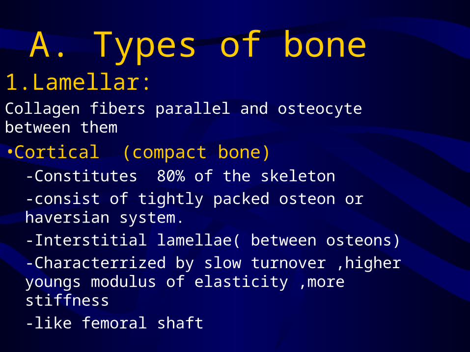
A. Types of bone 1.Lamellar:Collagen fibers parallel and osteocyte between them
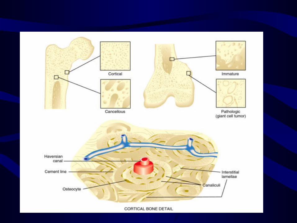
•Cortical (compact bone) -Constitutes 80% of the skeleton-consist of tightly packed osteon or haversian system.-Interstitial lamellae( between osteons)-Characterrized by slow turnover ,higher youngs modulus of elasticity ,more stiffness-like femoral shaft

• Cancellous bone (spongy or trabecular)(honey comb app)
• less dense and more remodelling according to lines of stress (Wolffs law)
• Characterized by high turnover rate , smaller yaoungs modulus and more elacsticity
• like distal femoral metaphysis

2.Woven -immature which is non stress oriented ,like embryonic skeleton and fracture callus
-pathologic Rondom organization ,increased turnover,weak and flexible Like fibrous dysplasia , Osteogenic Sarcoma
B.Cellular Biology 1.Osteoblast :-Derived from undifferentiated mesenchymal stem cells (more ER ,golgi apparatus and Mitiochondia )
-Osteoblast differentiation in vivo effected by Interleukin ,PDGF and IDGF

Receptors-effector interactions in osteoblasts

Osteoblast produce the following
• ALP ase• Osteocalcin• Type 1 collagen• Bone sialoprotein• Receptor activator of nuclear
factor B ligand RANK

• Osteoblast activity stimulated by intermittent (pulsatile) exposure to parathyroid hormone
• Inhibited by TNF- alpha
• Certain antiseptics toxic to cultured osteoblasts
1.Hydrogen peroxide2.Povidone-iodine(betadine)3.Bacitracin ( believed to be less
toxic)
2.Osteocyte• Maintain bone• Constitute 90% of the cells in the mature
skeleton • Less active in matrix production than are
osteoblast• Important in control of EC Ca and Ph conc.• Participate in bone resorption (osteocytic
osteolysis)• Directly stimulated by Calcitonin • Inhibited by PTH
3.Osteoclast
• Reasorb bone• Acts normally and pathologically • Derived from hematopoietic cells
in macrophage linage• Possess ruffled border • Have receptor for calcitonin which
inhibit osteoclastic resorption
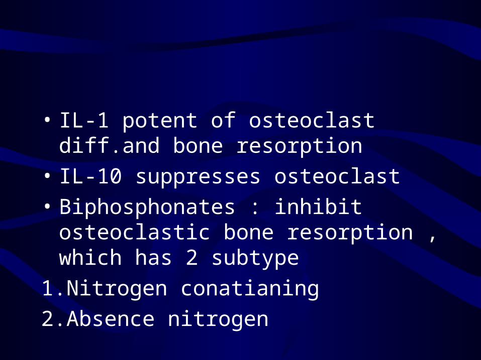
• IL-1 potent of osteoclast diff.and bone resorption
• IL-10 suppresses osteoclast• Biphosphonates : inhibit
osteoclastic bone resorption , which has 2 subtype
1.Nitrogen conatianing 2.Absence nitrogen
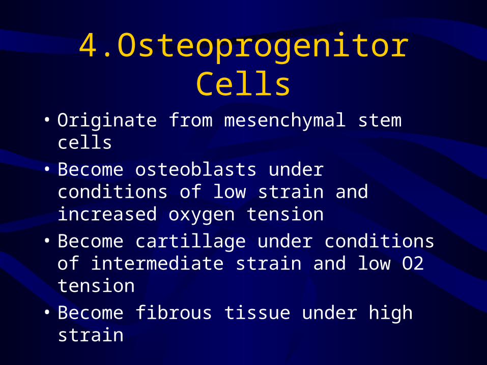
4.Osteoprogenitor Cells
• Originate from mesenchymal stem cells • Become osteoblasts under conditions
of low strain and increased oxygen tension
• Become cartillage under conditions of intermediate strain and low O2 tension
• Become fibrous tissue under high strain
5.Lining Cells
• Narrow, flattened, osteoblast that form an envelop around bone.
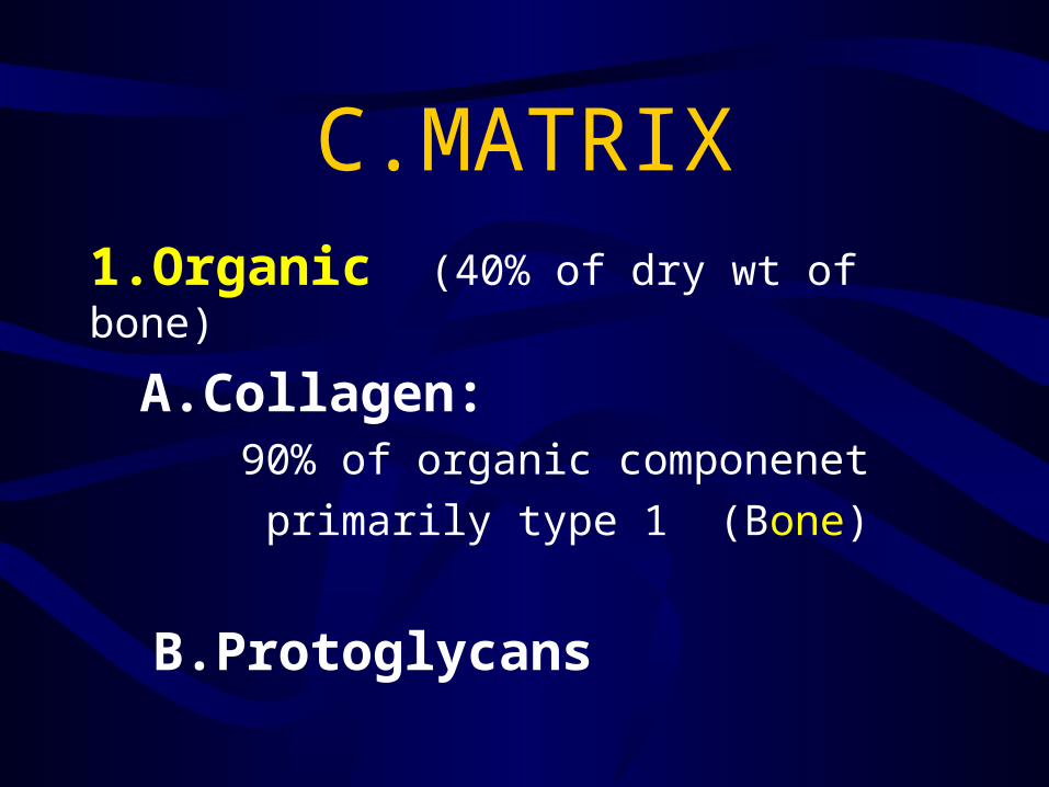
C.MATRIX1.Organic (40% of dry wt of bone)
A.Collagen: 90% of organic componenet primarily type 1 (Bone)
B.Protoglycans
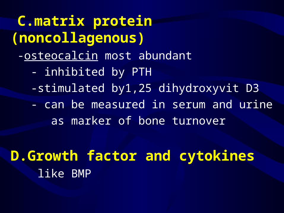
C.matrix protein (noncollagenous) -osteocalcin most abundant - inhibited by PTH -stimulated by1,25 dihydroxyvit D3 - can be measured in serum and urine as marker of bone turnover
D.Growth factor and cytokines like BMP

Inorganic (mineral)componenet
• 60% of the dry weight of bone
- Calcium hydroxyapetide - Calcium phosphate

D.Remodeling - wolffs law
- piezoelectrical remodeling
- Hueter-Volkamn Law
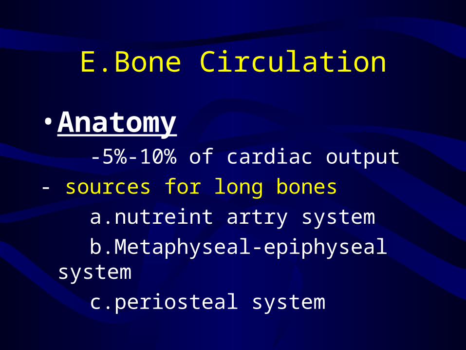
E.Bone Circulation
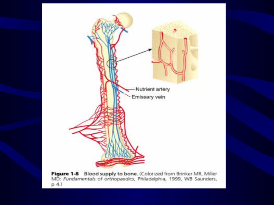
•Anatomy -5%-10% of cardiac output- sources for long bones a.nutreint artry system b.Metaphyseal-epiphyseal system c.periosteal system

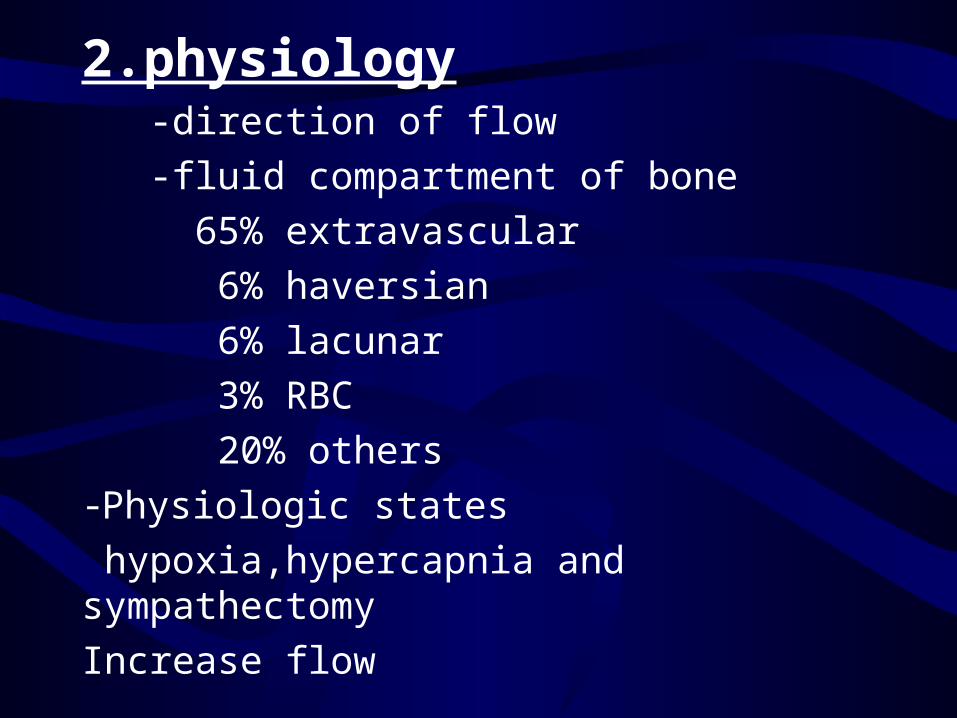
2.physiology -direction of flow -fluid compartment of bone 65% extravascular 6% haversian 6% lacunar 3% RBC 20% others -Physiologic states hypoxia,hypercapnia and sympathectomy Increase flow
•3.fracture Healing -Blood flow -Nutrient -Decrease blood flow -Increase blood flow
4.Regulation of bone blood flow

F.Tissue surrounding bone
• 1.periosteum -inner cambium -outer ( fibrous)2.Bone marrow -red marrow -yellow marrow
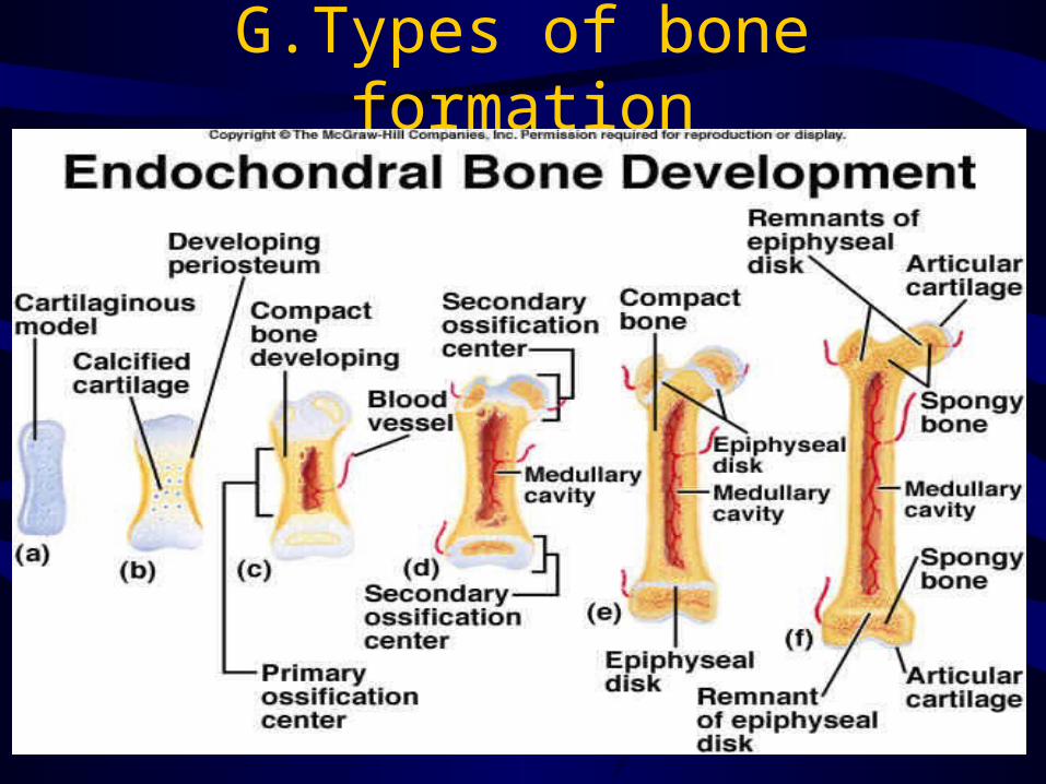
G.Types of bone formation

Intramembranous ossification
Appositional ossification
– Osteoblasts align on the existing bone surface and lay down new bone
– Periosteal bone enlargement– Bone formation phase of bone
remodeling
Bone injury and repair
–Fracture repair 1.A continum from inflammation to
repair 2.Blood supply3.Stages of fracture repair -inflammation -Repair -Remodelling
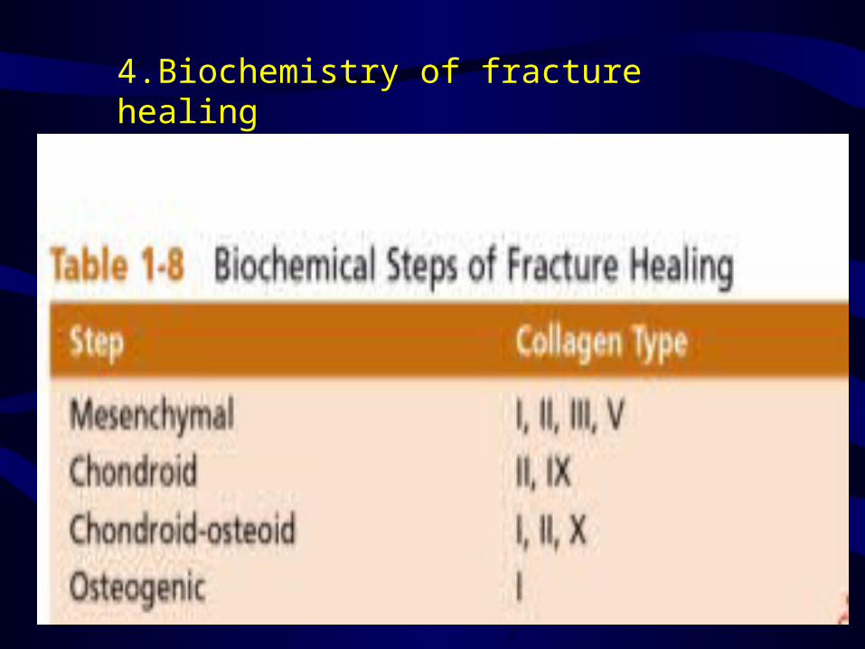
4.Biochemistry of fracture healing
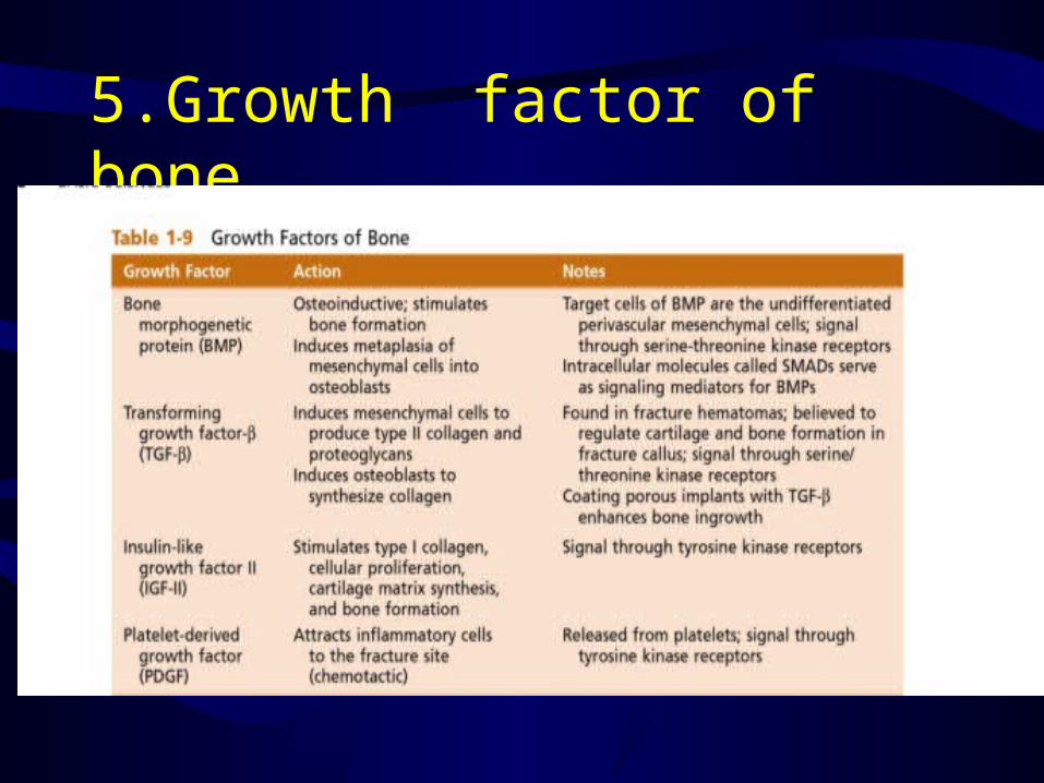
5.Growth factor of bone
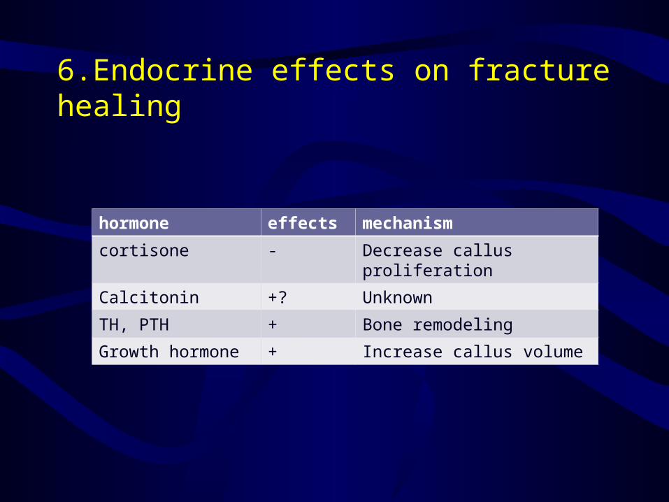
6.Endocrine effects on fracture healing
hormone effects mechanism
cortisone - Decrease callus proliferation
Calcitonin +? Unknown
TH, PTH + Bone remodeling
Growth hormone + Increase callus volume

7.head injury8.Nicotine (smoking)9.NSAIDs10.Quinolone Antibiotics11.Ultrasonography and fracture healing12.Effect of radiation on bone13.Diet and fracture healing14.Electricity and fracture healing15.Pathalogic fracture
Conditions of Bone
Mineralization, Bone Mineral Density and Bone Viability
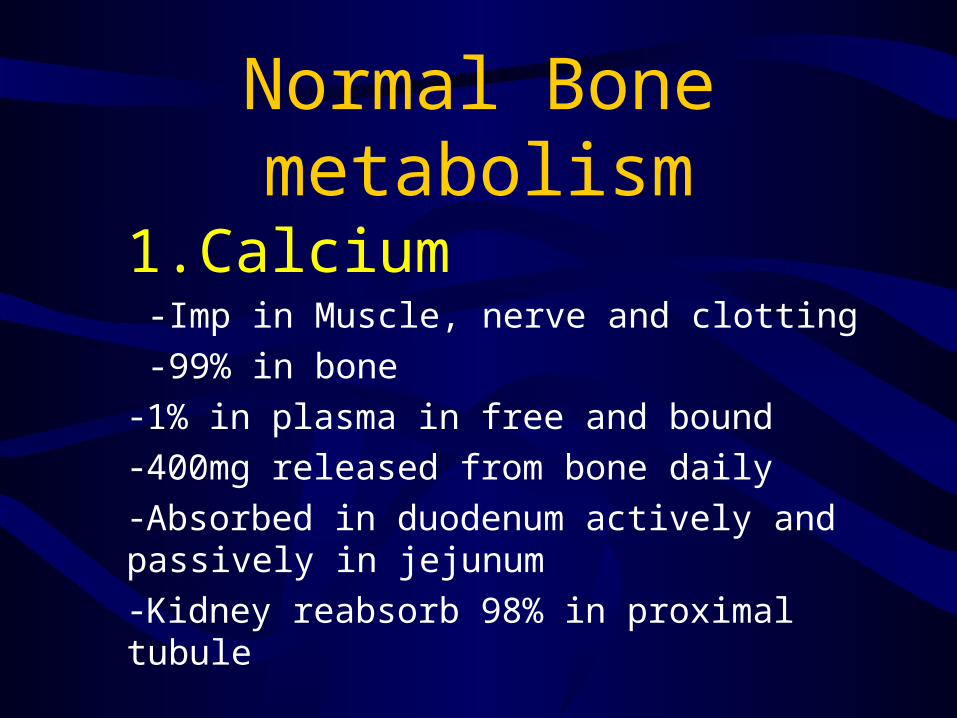
Normal Bone metabolism
1.Calcium
-Imp in Muscle, nerve and clotting -99% in bone-1% in plasma in free and bound -400mg released from bone daily-Absorbed in duodenum actively and passively in jejunum -Kidney reabsorb 98% in proximal tubule
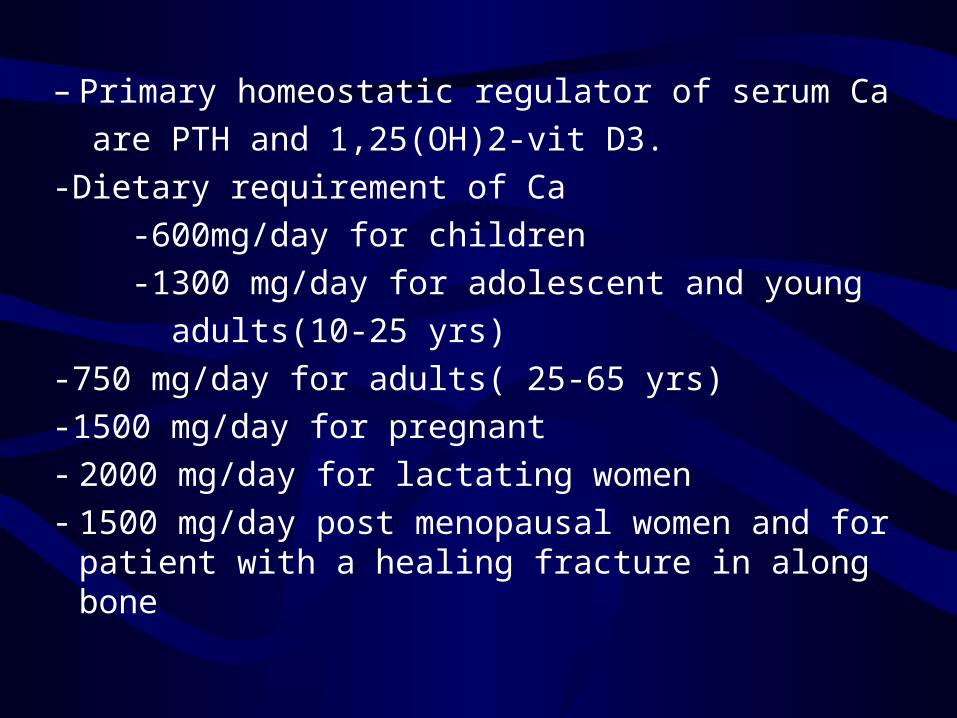
– Primary homeostatic regulator of serum Ca are PTH and 1,25(OH)2-vit D3.-Dietary requirement of Ca -600mg/day for children -1300 mg/day for adolescent and young adults(10-25 yrs)-750 mg/day for adults( 25-65 yrs)-1500 mg/day for pregnant- 2000 mg/day for lactating women- 1500 mg/day post menopausal women and for
patient with a healing fracture in along bone
• Calcium balance is +ve in the 1st three decades of life and –ve after the fourth decade.
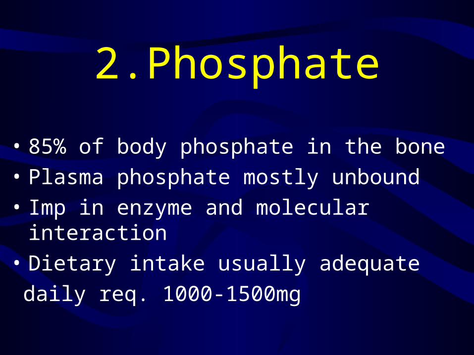
2.Phosphate
• 85% of body phosphate in the bone• Plasma phosphate mostly unbound• Imp in enzyme and molecular interaction• Dietary intake usually adequate daily req. 1000-1500mg

3.PTH• PTH is 84-amino acid peptide• Synthesized and secreted in chief
cells of four parathyroid gland• PTH regulate plasma calciumDecreased Ca level in the ECF
stimulate B2 receptors to release PTH which acts at the intestines, kidney and bones
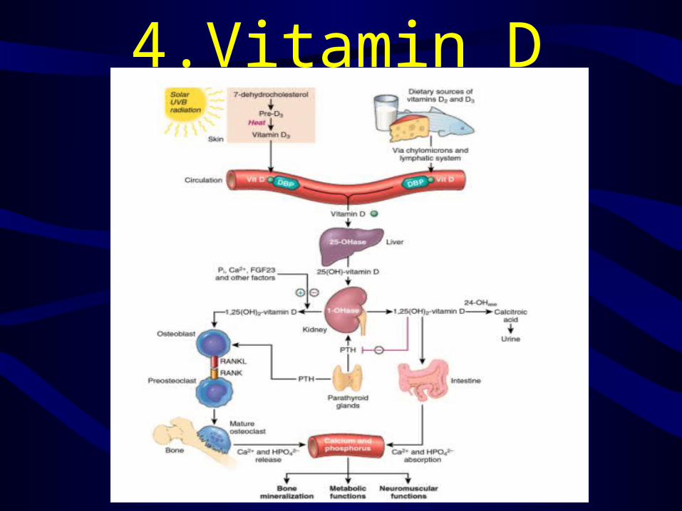
4.Vitamin D


5.Calcitonin32-amino acid peptide hormoneProduced by parafollicles of the thyroid glandHas limited role in calcium regulationIncrease ECF Ca level cause secretion of
calcitoninControlled by B2 rec.Inhibit osteoclastic bone resorption 1.Osteoclasts have Calcitonin rec2.Calcitonin decrease osteoclast No. and activity3.Decrease Ca serum level
6.Other hormones affecting metabolismA.Estrogen Inhibit bone loss by inhibiting bone resorption Decrease in urinary pyridinoline cross linksBecause bone formation and resorption is a couple mechanism that is why estrogen also decrease bone formationSupplemantaion

B.CorticosteroidIncrease bone lossDecrease gut absorption of Ca by decreasing binding proteinDecrease bone formation (cancellous more than cortical) by inhibiting collagen synthesis and osteoblast productivityDo not affect mineralizationAlternate day therapy may reduce effects
C. Thyroid hormoneAffect bone resorption more than
formationThyroxin can lead to osteoporosisRegulate skeletal growth at physis ,
stimulate chondrocyte growth , type X collagen syn. & ALP activity
D. Growth Hormone
cause positive Ca balance -increase gut absorption of calcium
more than it increases urinary excretion
insulin and somatostatin participate in this effect
E. Grwoth FactorTGF-B ,PDGF , monokines & lymphokines Have roles in bone and cartillage repair
7.Bone AgingPeak bone mass between 16-25 yrsAfter peak bone loss occurs at a rate
0.3% to 0.5% per year Rate of bone loss 2-3% per year in
untreated women during ten year after menopause
Affect trabecular more than cortical bone

Cortical bone becomes thinner & intracortical porosities increase
Cortical bone becomes more brittle , less strong and less stiff.
Long bones have increased inner and outer diameter

Conditions Of Bone
Mineralization
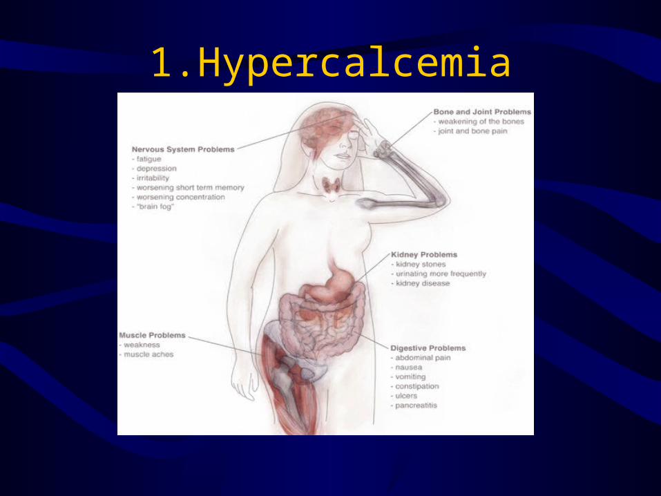
1.Hypercalcemia
1.HypercalcemiaCauses :•Primary hyperparathyroidism
Lab investigation
Increase Ca Increase PTHIncrease Urinary phosphate
Decrease serum phosphate
Bony changes-osteopenia-osteitis cystica (fibrous replacement of marrow)-Brown tumor-chondrocalcinosis
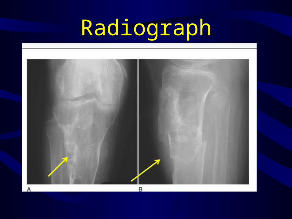
Radiograph
Other causes-familial syndrome(pit adenoma) or ( familial hypocalciuric hypercalcemia)-malignancy (most common)
Treatment• Hydration• Loop diuretics• Dialysis• Mobilization• Specific drugs ( biphosphonate,
mithramycin, calcitonin)
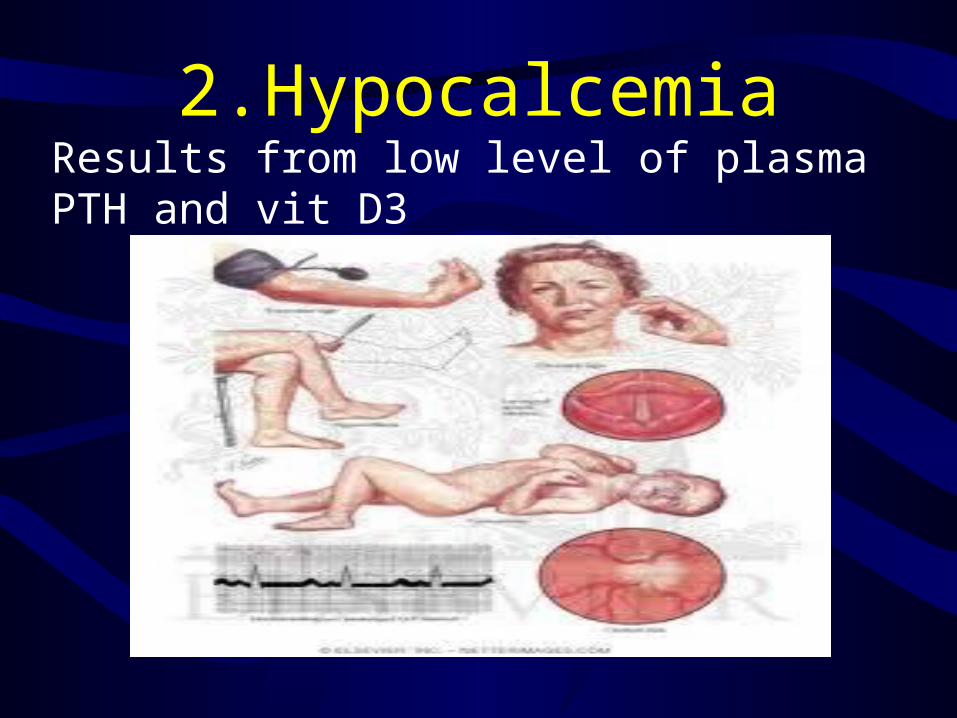
2.HypocalcemiaResults from low level of plasma PTH and vit D3

-hypoparathyroidismLead to decrease in plasma calcium level and increase phosphate level in serum-Psuedohypoparathyroidism-
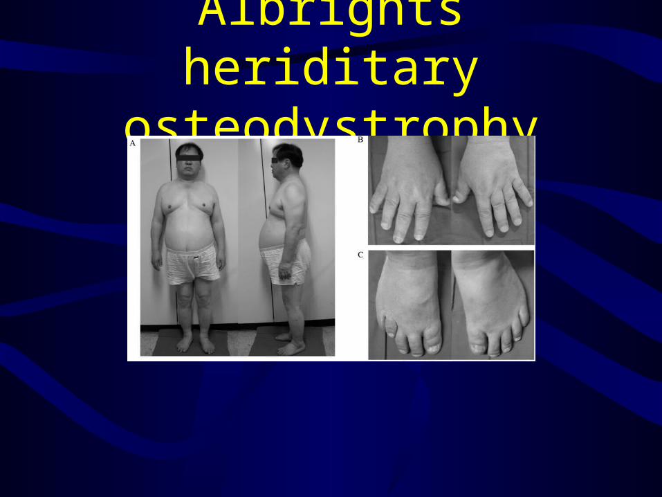
Albrights heriditary osteodystrophy
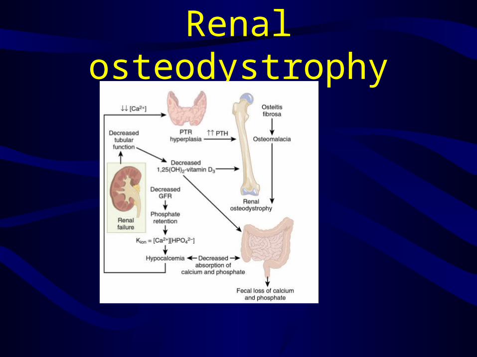
Renal osteodystrophy
Rickets (osteomalacia in adult)
• Is failure of mineralization , leading to changes in the physis in the zone of provisional calcification ( increase width and disoreintation ) and bone ( thinning and bowing)
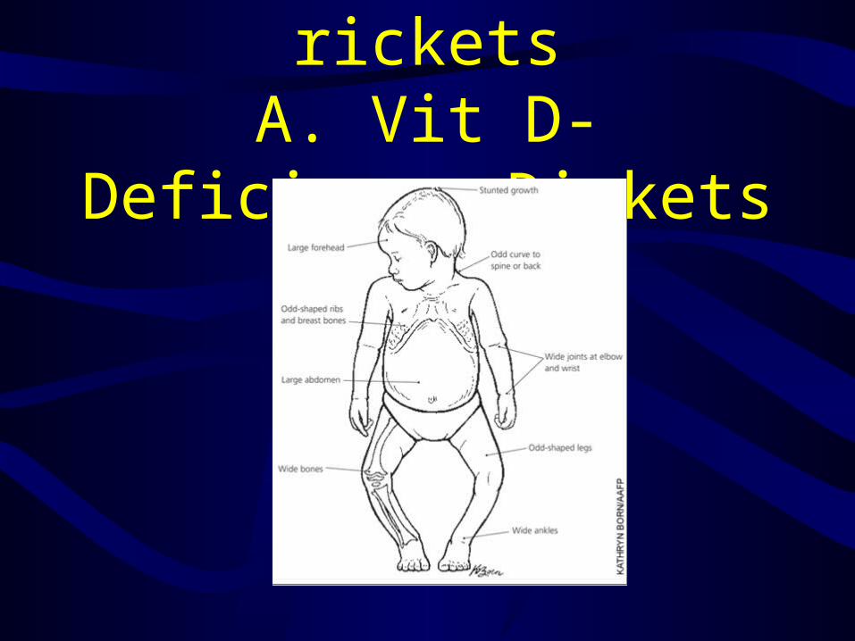
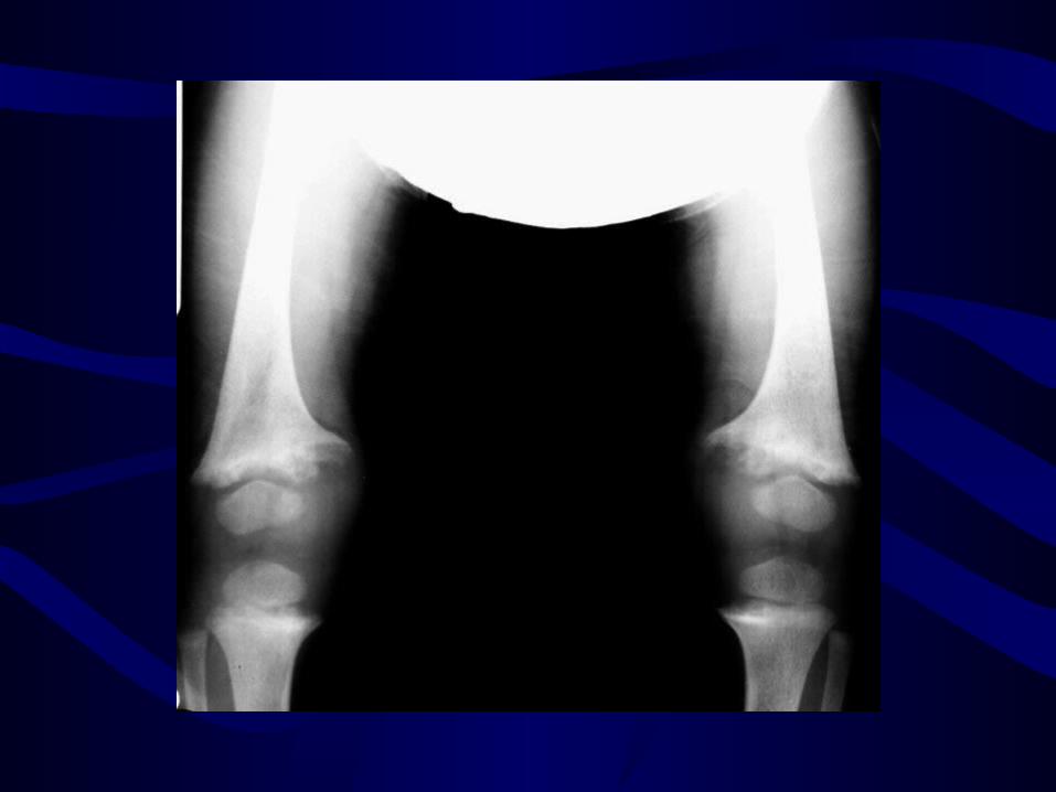
1.Nutritional ricketsA. Vit D-Deficiency
Rickets

Tratment Vit D 5000 IU daily resolve most deformities
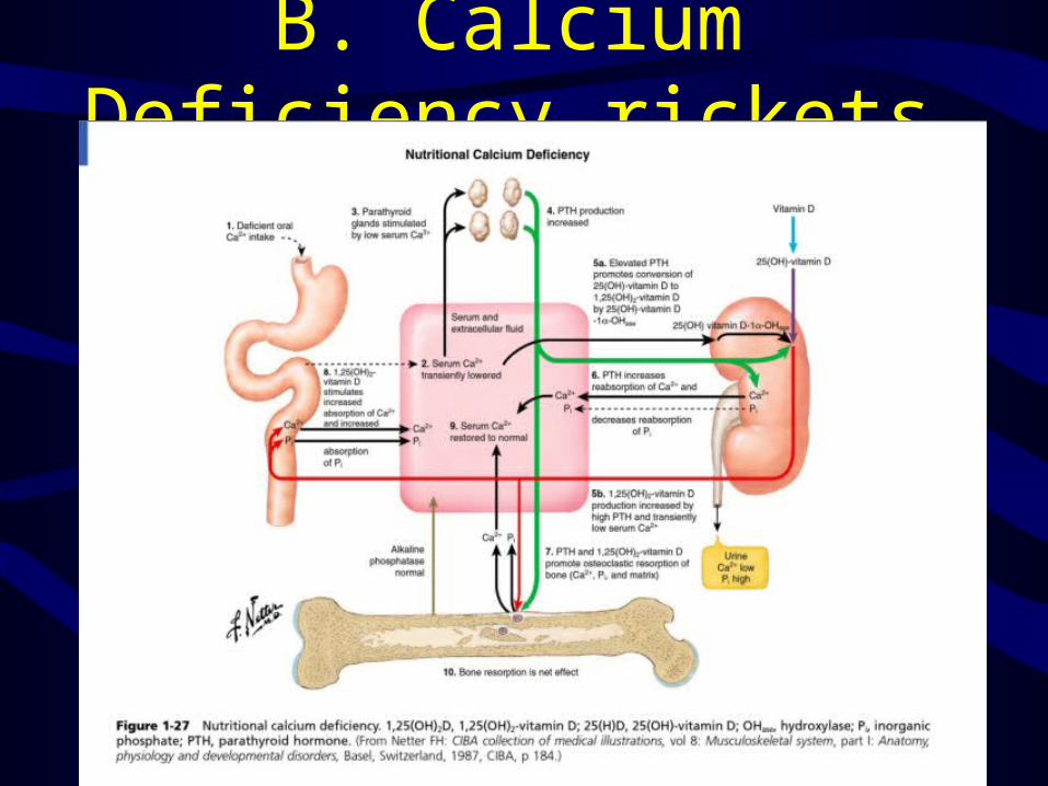
B. Calcium Deficiency rickets

C. Phosphate –deficiency rickets
• May be due to renal tubular disease• Ca serum level normal• There is no any sign of
hyperparathyroidism
2.Heriditory Vit –D dependent rickets
Less common and same like nutritional and more severe , may have total baldness1.Type 1: 25(OH)-vit D 1 alpha hydroxylase 2.Type 2:defect in intracellular 1,25(OH) vit D3
3.Familial Hypophosphatemic rickets
• Most common type encountered• X-linked dominant• Imp. Renal tubular ph reabsorption• Normal GFR with an impaired vit D3
response• Treatment -phosphate replacement (1-3 g daily) - high dose vit D3

Hypophosphatasia• Autosomal reciessive• Error in isoenzyme of ALK• Similar to rickets• Increased urinary
phosphoethanolamine diagnostic• Treatment may include phosphate

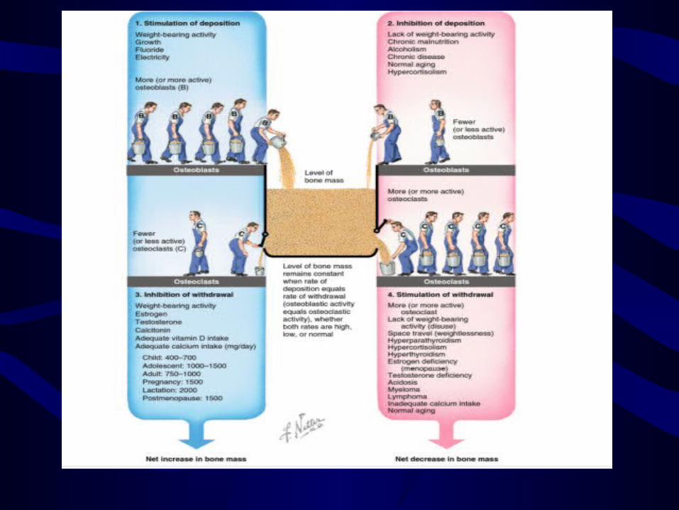
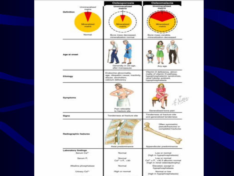
Conditions of bone mineral density
Bone mass is regulated by relative rates of deposition and withdrawal

1.osteoporosis• Age related decrease in bone mass• Is quantitative not qualitative defect• WHO (L2-L4) density is 2.5 or more
standard deviations less than mean peak bone mass of healthy 25 yrs old (T-score)
• Osteopenia: 1.0 – 2.5 deviatioans
• Responsible for more than 1 million fracture/year
- vertebral # more common -after initial vertebral # the risk for
second vert. # 20%• vertebral compression # associated
with increase mortality rate• More higher incidence in Men than
women• Life time risk of # in white women after
50 yrs of age 75%• Risk of hip # 15% - 20%

Risk factor White, female gender, northern EuropeanSedentary lifeThinnerSmokingHeavy drinkingPhenytoinDiet low in Ca and Vit. DHistory of breast feedingPositive family historyPremature menopause

Sign and symptom 1.Distal radius #2.Hip #3.Vertebral # codfish sign
Types of Osteoporosis1. Type 1 : Primarily affect trabecular bone Vertebral and distal radius more common 2.Type 2: In pt more than 75 yrs old Affect both trabecular and cortical Relared to poor Ca absorption Hip and pelvic # more common

Diagnosis • Obtained to role out secondary causes of
low bone mass like hyperthyroidism, vit D def. , HPTH, cushing syndrome, haematologis disorder, malignancy
• CBC , serum Ca, Vit D , ALK, creatinine and total albumin leverl . Results of these studies usually unremarkable in osteoporosis
• Plain radiograph not helpful unless bone loss exceeds 30%
Special study1.Single photon ( appendicular) absorptiometry2.Double photon (axial) absorptiometry3.Quantitative computed tomography (CT)
4.Dual-energey X-ray absorptiometry (DEXA)
BiopsyHistologic changes
Prophylaxis for pt with risk of osteoporosis
1.Diet with adequate Ca.2.Wt bearing exercise program3.Estrogen therapy evaluation at menopause
Treatment
1.Physical activity2.Ca supplement 1000-1500mg + 400-800IU vit D per day3.Fluoride4.Biphosphonate5.Other like calcitonin IM

2. Idiopathic transient osteoporosis of the hip
• Uncommon • Most common during 3rd trimester• Groin pain, limited ROM, localized
osteopenia without history of trauma
• Self limiting and tend to resolve spontaneously after 6 – 8 months
• Traetmant : analgesia and limiting wt bearing
• Stress # may occure

3.Osteomalacia• Qualitative defect defect of mineralization result in large
amount of unmineralized osteoid• Causes : - vit D def. -GIT dis -Renal dystrophy -Drugs -Alcoholism
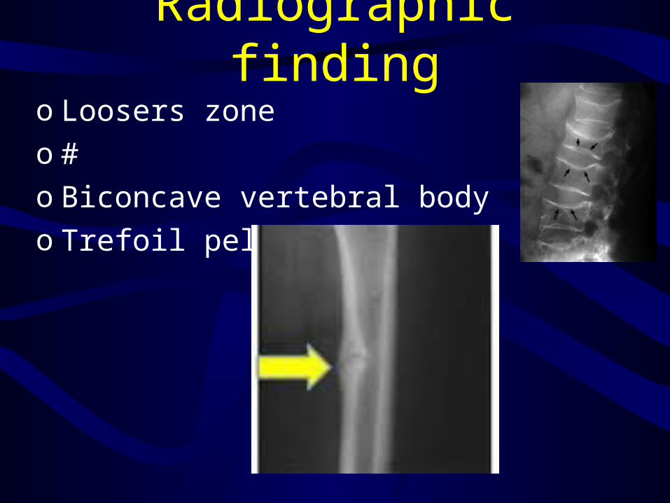
Radiographic findingo Loosers zoneo #o Biconcave vertebral bodyo Trefoil pelvis

TreatmentLarge doses of vit D
4.Scurvey
Vit C defProduce a decrease in chondroitin
sulfate synthesis
Sign and symptomsFatigueGum bleedingEcchymosisJoint effusionIron def
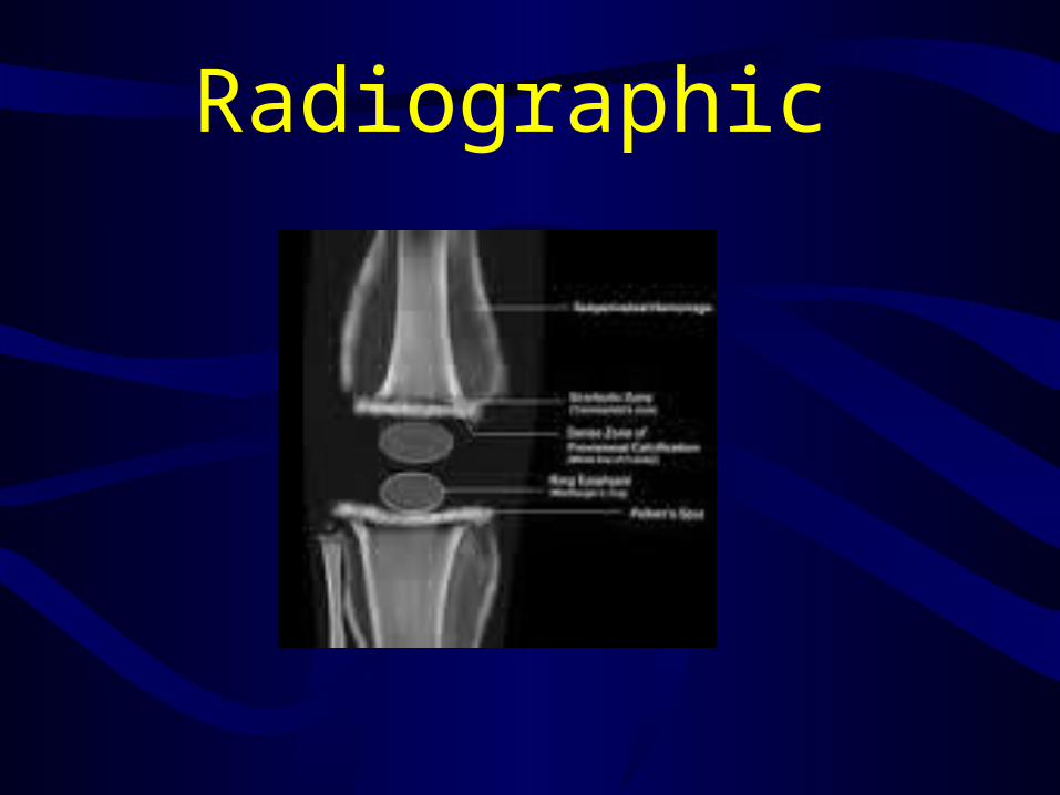
Radiographic
Histologic feature• Primary trabeculae replaced with
granulation tissue• Areas of haemorrhage• Widening of the zone of provisional
calcification in the physis
5.Marrow packing disorder
MyelomaLeukemia
Osteogenesis imperfecta
• Caused by abnormal collagen synthesis
• Type 1 collagen• Increased bone turn over
Lead poisoning
• Result in short stature and reduced bone density
• Lead alters chondrocyte response to PTH related protein and to TGF-B
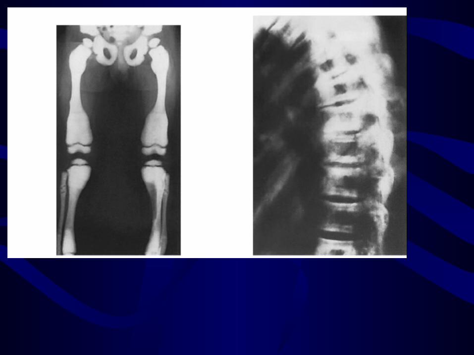
4.Increased OsteodensityA. Osteopetrosis (marable bone disease) -due to decrease osteoclast function - may be due to immune sys - abnormal osteoclast - one of them infantile autosommal
recessive dis (malignant)- Another one AD (tarda) benign (albers –
schonberg dis), typical rugger jersy spine
B. Osteopoikilosis• Spotted bone disease• Islands of deep cortical bone appear
within the medullary cavity and the cancellous of the long bone
• Especially in the hands and feet• No malignancy incidence
5.Pagets disease-virus inclusion like bodies in osteoclast-both decrease and oncrease osteodensity may be present 1.active phase: lytic mixed sclerotic2. Inactive phase
-Elevated Serum ALK-urinary hydroxyproline level increase
Conditions of viability1.Osteonecrosis• death of bony tissue other than infection•Usually adjacent to a joint surface•Caused by loss of blood supply•Like SCFE, perths disease

• Associated with -steroid therapy - blood dyscrasias (SCD) -Dysbarism ( Caissons disease) -excessive radiation therapy -gaucher disease

causes1.Thiories vary2.Enlarged space occupying fat marrow3.Vascular insult4.Idiopathic : like chandlers disease medial femoral condyle osteonecrosis5.Secondary to blood dis

DiagnosisMRI is earliest study to yield positive results, highest sensitivity and specificity

Traetment:1.Arthroplasty associated with increased loosening.2.Non traumatic necrosis of femoral condyle and proximal humerus may improve spont. With out surgery3.Precise role Core decompression is still unresolved4.Core decompression is benefit in early hip disease
Osteochondrosis1.This condition can oocure at traction apophysis in children2.It may or not associated with trauma,joint capsule inflammation, vascular insult or secondary thrombosis3.The pathologic process is similar to that ofOsteonecrosis in the adult.


Reference1.Review of orthopaedics 6th edition Mark D.Miller2.Apleys Ninth edition System Of Orthopaedics and Fractures3.Internet for pictures