BB0172, a Borrelia burgdorferi Outer Membrane Protein That BindsIntegrin �3�1
Elaine Wood,a Silvia Tamborero,b Ismael Mingarro,b Maria D. Esteve-Gassenta
Department of Veterinary Pathobiology, College of Veterinary Medicine and Biomedical Sciences, Texas A&M University, College Station, Texas, USAa; Department ofBiochemistry and Molecular Biology, Faculty of Biology, Burjassot, Valencia, Spainb
Lyme disease is a multisystemic disorder caused by Borrelia burgdorferi infection. Upon infection, some B. burgdorferi genes areupregulated, including members of the microbial surface components recognizing adhesive matrix molecule (MSCRAMM) pro-tein family, which facilitate B. burgdorferi adherence to extracellular matrix components of the host. Comparative genome anal-ysis has revealed a new family of B. burgdorferi proteins containing the von Willebrand factor A (vWFA) domain. In the presentstudy, we characterized the expression and membrane association of the vWFA domain-containing protein BB0172 by using invitro transcription/translation systems in the presence of microsomal membranes and with detergent phase separation assays.Our results showed evidence of BB0172 localization in the outer membrane, the orientation of the vWFA domain to the extracel-lular environment, and its function as a metal ion-dependent integrin-binding protein. This is the first report of a borrelial adhe-sin with a metal ion-dependent adhesion site (MIDAS) motif that is similar to those observed in eukaryotic integrins and has asimilar function.
Lyme disease is a multisystemic disorder that leads to arthritis in60% of cases, carditis in 10% of untreated adults, and other
neurological symptoms. The causative agent of Lyme disease is thespirochetal pathogen Borrelia burgdorferi, which is transmitted tohumans through the bite of an infected Ixodes sp. tick (1). There iscurrently very little information available on the tissue-specifichost-pathogen interactions that lead to pathological manifesta-tions of B. burgdorferi infection. This pathogen’s ability to colo-nize mammals is dependent on its capacity to rapidly alter geneexpression in response to highly disparate environmental signalsfollowing transmission from infected ticks (2). The open readingframes that are upregulated upon infection include members ofthe microbial surface components recognizing adhesive matrixmolecules (MSCRAMM) protein family, and they facilitate theadherence of B. burgdorferi to extracellular matrix (ECM) compo-nents of the host (3).
Comparative genome analysis has also identified a family ofvon Willebrand factor A (vWFA) domain-containing proteins inB. burgdorferi, including BB0172, BB0173, BB0175, and BB0325(4–6). The vWFA domains present in ECM proteins and on eu-karyotic cells are involved in cell adhesion and protein-proteininteractions; they play key roles in the adhesion of platelets toareas of vascular damage by binding to glycoproteins on the plate-let surface, to exposed ECM components (5, 7, 8), and to metal-loproteases (ADAMTS 13) (7–12). Therefore, the vWFA domain-containing borrelial proteins might be involved in the adhesion ofB. burgdorferi to eukaryotic cells, ECM components, and activatedplatelets, and they may thus play a role in the virulence mecha-nisms of B. burgdorferi. In humans, absence of vWF results insevere bleeding disorders due to the removal of blood clottingfactor VIII from the circulation (12–15). vWF-binding proteins(vWFbp) have been identified in several bacterial species, such asHelicobacter pylori and Staphylococcus aureus (16–19), and thesesecreted or surface-exposed proteins are involved in the binding ofthese pathogens to ECM components, platelets, and endothelialcells, thus playing an important role in pathogen colonization anddissemination in the mammalian host. In silico sequence analysis
has shown that B. burgdorferi vWFA domain-containing proteinshave a sequence domain (DXSXS) that is very similar to the metalion-dependent adhesion site (MIDAS) found in integrins (20, 21).These proteins also show similarity to the Plasmodium spp. extra-cellular adhesion molecule TRAP and the LFA-1 integrin (Fig. 1)(6, 22).
The Lyme disease agent binds to a variety of ECM componentsand integrins, which are metal ion-dependent heterodimeric re-ceptors that mediate cell-to-cell and cell-to-ECM interactions(23). The present study used a well-established in vitro model toinvestigate the localization and function of the vWFA domain-containing BB0172 protein of B. burgdorferi and to determine itsfunction in adherence to different tissues during infection. Ourfindings established the topology of the BB0172 protein in biolog-ical membranes and its adherence to different ECM componentsand integrins, emphasizing the complexity of host-pathogen in-teractions in Lyme disease.
MATERIALS AND METHODSBacterial strains and growth conditions. Borrelia burgdorferi B31 iso-late A3 (24) was used throughout this study. To mimic the temperatureand pH conditions during the transition of this bacterium from theunfed to the fed tick, the strain was grown in BSK-II medium pH 7.6complemented with 6% inactivated normal rabbit serum at room tem-perature (RT) until reaching a density of 107 cells/ml. Then an aliquotof this culture was transferred to BSK-II medium (pH 6.8) and incu-bated at 37°C with 1% CO2 until reaching a density of 5 � 107 cells/ml(25). Escherichia coli OneShot Top10 cells (Invitrogen, CA) were usedfor all cloning steps, and Rosetta-gami(DE3)pLysS cells (Novagen,
Received 14 February 2013 Accepted 14 May 2013
Published ahead of print 17 May 2013
Address correspondence to Marie D. Esteve-Gassent,[email protected].
Madison, WI) were used for BB0172 recombinant protein expression.All E. coli strains were grown in LB (Difco) broth with the appropriateantibiotics.
RNA and genomic DNA purification for detecting bb0172 tran-scripts by PCR. RNA was extracted as previously described (25, 26).Briefly, B. burgdorferi cultures were grown to a density of 2 � 107 to 3 �107 spirochetes/ml under the shifting conditions outlined above. RNAwas extracted by resuspending the bacterial pellets with 0.2 ml RNA-Bee(Tel-Test, Inc., Friendswood, TX) for every 106 cells. Following extractionwith chloroform, RNA was precipitated with isopropanol, washed with75% ethanol, air dried, and resuspended in RNase-free water. To removecontaminating DNA, the RNA was treated twice with DNase I at 37°C for45 min. Then, the total RNA was quantified spectrophotometrically andreverse transcribed to cDNA by using TaqMan reverse transcription re-agents (Applied Biosystems, Foster City, CA). From B. burgdorferi cul-tures growing under tick-feeding conditions (pH 6.8, 37°C) or regulargrowing conditions (pH 7.6, 32°C), genomic DNA was obtained by gen-eral phenol-chloroform extraction.
RNA, cDNA, and genomic DNA (positive control) samples fromeach growing condition were used to detect when bb0172 was ex-pressed. A 500-bp fragment of bb0172 was amplified using primersBB0172cDNA-F (B. burgdorferi nucleotides 174705 to 174728) andBB0172cDNA-R (B. burgdorferi nucleotides 174225 to 174249) (Table1). Primers specific to the flaB, ospC, and p66 genes were also includedas controls for the temperature and pH shift (Table 1) (27, 28). PCRproducts were separated on 0.8% agarose gels and imaged using theBio-Rad Gel Doc XR system.
Computer-assisted analysis of BB0172 transmembrane (TM) re-gions. Membrane-spanning regions in the BB0172 sequence were predictedby using six of the most commonly used prediction methods available online:DAS-TMfilter (29; http://mendel.imp.univie.ac.at/sat/DAS/DAS.html), �GPrediction (30; http://dgpred.cbr.su.se/), MEMSAT3 (31; http://bioinf.cs.ucl.ac.uk/psipred/submit), OCTOPUS (32; http://octopus.cbr.su.se/index.php), SOSUI (33; http://bp.nuap.nagoya-u.ac.jp/sosui/sosui_submit.html),and TMHMM (34; http://www.cbs.dtu.dk/services/TMHMM/). The poten-tial presence of a putative signal sequence was also investigated using the
FIG 1 In silico analysis of BB0172. (A) Schematic representation of BB0172. TM1 and TM2, transmembrane domains 1 (amino acids 17 to 35) and 2 (aminoacids 264 to 281), respectively. The vWFA domain includes amino acids 51 to 256. The recombinant BB0172 protein (BB0172T) amino acid sequence spansresidues 38 to 291. The anti-BB0172pep antibody (Ab) was generated against a 30-mer peptide, from amino acids 150 to 180. The MIDAS motif is located atresidues 57 to 61. (B) Hydrophobic amino acid sequences (HRs) were cloned to study their insertion into membranes. HR1, amino acids 16 to 38, HR2, aminoacids 263 to 284. (C) Clustal W (v1.83) alignment of BB0172 of B. burgdorferi B31 (in bold) and ZS7 strains to its homologues in Borrelia garinii (NTL01BG0170),Borrelia afzelii (NTL07BA0169), and the relapsing fever species Borrelia hermsii (NT03BH0168) and Borrelia tunicate (NT06BT0168), as well as to the humanadhesins LFA-1 and CD11b and the Plasmodium falciparum membrane protein TRAP. Bold text with gray shadowing indicates the MIDAS domain (DXSXS) andthe aspartic acid (D) necessary to complete the metal binding (20, 21). The threonine (T) that is highlighted in gray and underlined is also involved in the MIDASmotif function; it is present in a different location in the bacterial species compared to eukaryotic counterparts. MacVector version 12.6 (MacVector, Inc.) wasused for sequence analyses and production of schematics.
SignalP 4.0 server (35; http://www.cbs.dtu.dk/services/SignalP/). All user-ad-justable parameters were left at their default values.
Cloning of transmembrane domain regions. To study the insertionof BB0172 hydrophobic regions into membranes, the segments to betested (Fig. 1A and B) were engineered into the luminal P2 domain of theintegral membrane protein Lep from E. coli (leader peptidase), where itwas flanked by two N-glycosylation sites that were used as reporters (seeFig. 5, top). To further clone these regions, the two BB0172 putative TMdomains were PCR amplified from total genomic DNA obtained from theB31A3 Borrelia strain by using the primers described in Table 1. PCRproduct size was verified on 2% agarose gels, and then amplicons werecleaned using the Wizard SV gel and PCR cleanup system (Promega,Madison, WI), following the manufacturer’s recommendations. The PCRproducts were then double digested using SpeI/KpnI enzymes (NEB, Ip-swich, MA) and cloned into pGEM-Lep as previously described (36–38).Ligation reaction mixtures were precipitated overnight and electropo-rated into TOP10 cells. Positive clones were selected on ampicillin plates(100 �g/ml) and verified by sequencing (Eton Biosciences, San Diego,CA). Clones showing the hydrophobic TMs in frame with Lep were usedfor the in vitro transcription-translation experiments.
In vitro transcription-translation. In vitro translation of in vitro-transcribed mRNA was performed in the presence of reticulocyte lysate,[35S]Met-Cys, and dog pancreas microsomes, as described previously(39). Lep constructs with hydrophobic region (HR)-tested segments fromthe BB0172 sequence (residues 16 to 38 and 263 to 284) were transcribedand translated as previously reported (36). After translation, membraneswere collected by ultracentrifugation and analyzed by sodium-dodecylsulfate-polyacrylamide gel electrophoresis (SDS-PAGE), and the gelswere visualized on a Fuji FLA3000 PhosphorImager with ImageGaugesoftware.
The proteinase K protection assay was performed as previously de-scribed (40). Briefly, the translation mixture was supplemented with 1 �lCaCl2 (50 mM) and 1 �l proteinase K (4 mg/ml) and then digested for 40
min on ice. The reaction was stopped by adding 1 mM phenylmethylsul-fonyl fluoride (PMSF), followed by SDS-PAGE analysis. This process isshown schematically in Fig. 5, below.
Expression and purification of BB0172T. For this experiment, a trun-cated BB0172 (BB0172T) protein was purified to avoid insertion of theprotein into E. coli membranes and to ensure purification of BB0172 as asoluble protein. Total genomic DNA obtained from the B31A3 strain(Table 2) was used as a template to PCR amplify bb0172 from nucleotides150 to 873 (amino acids 50 to 290), using the primers listed in Table 1. Theamplicons were cloned into the pCR2.1-TOPO vector (Invitrogen), trans-formed into E. coli TOP 10 cells, and subjected to blue/white colonyscreening in the presence of ampicillin (100 mg/ml) and kanamycin (50mg/ml). The insert was digested with NdeI/XhoI and ligated into thepET23a expression vector. The ligated products were electrotransformedinto E. coli TOP10 cells and screened for the presence of the insert byrestriction enzyme digestion. The junctions of plasmids containing insertsof the expected sizes were sequenced and used to transform the E. coliexpression host. Recombinant BB0172 (rBB0172) with a C-terminal6�His tag was overexpressed by inducing the E. coli strain containingpET23a-bb0172T with 1 mM isopropyl-�-D-thiogalactopyranoside for 3h. The bacterial pellets were disrupted by using a French press and dena-turing lysis buffer (8 M urea, 20 mM imidazole; pH 7.4). The supernatantswere collected, clarified by centrifugation, and subjected to affinity puri-fication with a His60 Ni Superflow resin (Clontech, Mountain View, CA),following the manufacturer’s instructions. The bound 6�His-tagged pro-teins were eluted as 0.5-ml fractions with elution buffer (8 M urea, 300mM imidazole; pH 7.4) and then analyzed by SDS–12.5% PAGE. Selectfractions with the largest concentrations of eluted proteins were furtherpurified using dialysis against 50 mM sodium phosphate and 300 mMNaCl (pH 7.4; Slide-A Lyze G2 dialysis cassette; Thermo Scientific). Afterdialysis, Amicon centrifugal filters (Millipore) were used to concentratethe proteins. A 27.5-kDa protein was purified to homogeneity (data not
TABLE 1 Oligonucleotide primers used in this study
Primer pair RSa Sequence (5=¡3=)b Application
BB0172cDNA-F GCTTGATTTTTTTAATTTTATCC Amplification of bb0172 500-bp fragmentfrom B. burgdorferi cDNABB0172cDNA-R CGGGATTACTCCCGCCAATCCCAA
FlaBcDNA-F AACACACCAGCATCACTTTCAGG Amplification of flab 234-bp fragmentfrom B. burgdorferi cDNAFlaBcDNA-R GAGAATTAACTCCGCCTTGAGAAGG
P66cDNA-F CAAAAAAGAAACACCCTCAGATCC Amplification of p66 683-bp fragmentfrom B. burgdorferi cDNAP66cDNA-R CCTGTTTTTAAATAAATTTTTGTAGCATC
OspCcDNA-F TATTAATGACTTTATTTTTATTTATATCT Amplification of ospC 593-bp fragmentfrom B. burgdorferi cDNAOspCcDNA-R TTGATTTTAATTAAGGTTTTTTTGG
BB0172TF NdeI ACGCCATATGTGGGGGCAAAGAGCTGTTGAGGA Amplification of bb0172 for cloning intopCR2.1 and pET23a for proteinexpression and purification
BB0172TR XhoI ACGCCTCGAGCTAAAAAGTTTCGTCCCA
BB0172TM1-F SpeI ACGCACTAGTGGAGGACCAGGAGTTCTAATGTCAATGTTT Amplification of the first bb0172hydrophobic region in frame with Lepsequence
BB0172-T ACAGATGGCGATGATTGGGGGGAA SequencingSP6 ATTTAGGTGACACTATAG SequencingBB0172 SDM MIDAS-1F GAGAATTTCTTTTATTTTTGCTATTTCTCGTAGCATG Site-directed mutagenesis of the first
BB0172 SDM MIDAS-3X-F GAGAATTTCTTTTATTTTTGCTATTGCTCGTGCTATGTTAAGTGTAGATG Site-directed mutagenesis of the threeamino acids of the MIDAS motif,DXSXS¡AXAXA
shown), quantified in a bicinchoninic acid (BCA; Thermo Scientific, Inc.)assay, and stored at �80°C until further use.
Site-directed mutagenesis of DXSXS metal-binding domain.BB0172 contains a MIDAS motif (Fig. 1C) comprising amino acids 57 to61 (DXSXS). To determine which amino acids are essential for maintain-ing this protein’s function, we performed site-directed mutagenesis tochange the motif-relevant amino acids to alanine, creating the mutantsD57A, S59A, and S61A and the triple mutant (21). For this process, weused the QuikChange II XL site-directed mutagenesis kit (Agilent Tech-nologies), following the manufacturer’s recommendations. Primers (Ta-ble 1) were designed as recommended by the manufacturer and usingtheir primer design tool for site-directed mutagenesis. Positive colonieswere screened by sequencing using the universal T7 promoter and termi-nator primers (Eton Biosciences, Ltd., San Diego, CA). Mutant MIDASmotif clones were stored at �80°C for further use. For their expressionand purification, 2 �l of purified plasmid for each mutation was electro-porated into Rosetta-gami E. coli strains (Novartis). Further binding as-says were performed with all isolated mutants, as described below.
Generation of specific BB0172 antibodies. To generate antibodiesspecific to the extracellular domain of BB0172, we design a 30-mer-longpeptide corresponding to amino acids 150 to 180 (Fig. 1A) with the se-quence YNFLVILTDG DDWGENNYYR FSKFVNNLKL conjugated toKLH (Peptide 2.0, Chantilly, VA). The synthetic peptide was dissolved in10% dimethyl sulfoxide until reaching homogeneity and administered tothree BALB/c mice at a dose of 50 �g/mouse in conjugation with TiterMaxGold (Sigma-Aldrich, St. Louis, MO) on days 0, 14, and 21. At 28 days, themice were euthanized and exsanguinated. Serum was collected and usedin the following immunoblot and binding assays; this specific antiserum ishenceforth referred to as anti-BB0172pep. Specific serum to the wholeBB0172 protein was obtained following the same immunization protocoland using 50 �g of rBB0172T per mouse, but we detected no BB0172 in theB. burgdorferi whole-cell lysates or from purified rBB0172T. Therefore, weused the anti-BB0172pep developed in this study.
Triton X-114 partitioning of B. burgdorferi outer membrane pro-teins. To evaluate the localization of BB0172 in B. burgdorferi, we sepa-rated the outer membrane proteins (OMPs) from the protoplasmic cyl-inders (PCs) by Triton X-114 partitioning as previously described (40–43). Briefly, B. burgdorferi was grown at RT and pH 7.6 and then shifted to37°C, pH 6.8 and grown to a density of 5 � 107 spirochetes/ml. Cells werewashed in phosphate-buffered saline (PBS), resuspended in PBS contain-ing 1% Triton X-114, and incubated overnight at 4°C with gentle rockingto ensure separation of the outer membrane proteins without disruptionof the protoplasmic cylinders. After the overnight partitioning, PCs were
pelleted by centrifugation at 15,000 � g for 15 min at 4°C. Supernatantscontaining OMPs were incubated at 37°C for 15 min in the presence of 2%Triton X-114. The OMP-containing organic phase was separated by cen-trifugation at 2,000 � g for 10 min at RT and then washed three times withPBS. Finally the OMPs were precipitated out of the organic phase byadding 10� ice-cold acetone at �20°C for 2 h, followed by centrifugationat 15,000 � g for 30 min at 4°C. PCs, the aqueous phase, and the detergentphase containing the OMPs from different growing conditions were sep-arated in SDS–12% PAGE gels and silver stained (Silver Staining Plus;Bio-Rad Laboratories, Inc., Hercules, CA), following the manufacturer’srecommendations, or transferred to polyvinylidene difluoride (PVDF)membranes to determine the BB0172 localization by immunoblot assay asdescribed below. OMPs OspC and P66, the periplasmic protein superox-ide dismutase A (SodA), and the cytosolic proteins carbon storage regu-lator A (CsrA) and the Borrelia oxidative stress regulator (BosR) were usedas controls.
SDS-PAGE gels and immunoblot analysis. Borrelia burgdorferiwhole-cell lysates (prepared from cultures grown at RT and pH 7.6, thenshifted to pH 6.8 and 37°C) were treated with proteinase K (20 �g/ml) andseparated by SDS–12.5% PAGE. The separated proteins were either visu-alized by Coomassie brilliant blue staining or transferred onto a PVDFmembrane (Hybond-P; GE Healthcare, Piscataway, NJ) and subjected toimmunoblot analysis. The PVDF membranes were blocked overnight at4°C in Tris-buffered saline containing 0.2% Tween 20 (TBS-T) and 10%skim milk. After blocking, membranes were probed with mouse anti-BB0172pep. Polyclonal mouse anti-P66 and anti-OspC (25) sera were usedas controls for proteinase K activity on outer membrane proteins andcontrols for the fractioning of outer membrane proteins in the Triton-X114 partitioning assay. Polyclonal mouse anti-SodA and rabbit anti-BosR sera were used as controls of cytoplasm-located proteins (44). Theblots were developed following incubation with appropriate dilutions ofhorseradish peroxidase (HRP)-conjugated secondary antibodies and us-ing enhanced chemiluminescence Western blotting reagents (GE Health-care, Piscataway, NJ) as previously described (25, 26, 44, 45).
Binding assays. To understand the possible capability of BB0172 tobind to ECMs, we purified a truncated BB0172 protein lacking the Nterminus and the first TM segment (BB0172T) (Fig. 1), and we analyzed itsbinding to collagen type I, collagen type IV, laminin, and human fibronec-tin (BD BioCoat, Bedford, MA). All incubations were performed usingHBS buffer (HEPES-buffered saline) containing 0.25 mM Ca2�, 1 mMMn2�, 1 mM Mg2�, and 1% bovine serum albumin (BSA) (46, 47). Afterblocking plates with 3% BSA in HBS containing 1% Tween 20 (HBS-T),we added 500 ng/well of rBB0172T or rBBK32 (positive binding control)
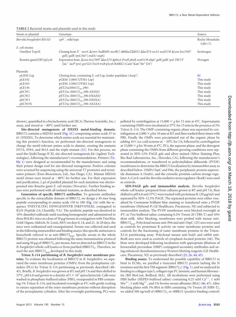
TABLE 2 Bacterial strains and plasmids used in this study
Strain or plasmid Genotype Source
Borrelia burgdorferi B31A3 cp9�, wild type Rocky MountainLabs (1)
PlasmidspGEM-Lep Cloning host, containing E. coli Lep, leader peptidase (Ampr) 82pLE102 pGEM-1(bb0172TM1-Lep) This studypLE103 pGEM-1(bb0172TM2-Lep) This studypLE136 pET23a(bb017250–290) This studypECW1 pET23a (bb017250–290-AXSXS) This studypECW2 pET23a (bb017250–290-DXAXS) This studypECW3 pET23a (bb017250–290-DXSXA) This studypECW3X pET23a (bb017250–290-AXAXA) This study
to the plates in quadruplicate wells and incubated them at RT for 1 h(47–49). The plates were washed three times with HBS-T to remove un-bound BB0172T protein. Then, plates were incubated with a 1:1,000 dilu-tion of mouse-anti-rBB0172pep or 1:2,000 dilution of the mouse anti-rBBK32 antibody for 1 h at RT, followed by a 1-hour incubation with a1:5,000 dilution of anti-mouse HRP-conjugated antibody (GE Health-care, Piscataway, NJ). Plates were washed and incubated with o-phenyl-diamine dihydrochloride (OPD) tablets (Thermo Scientific), and the ab-sorbency at 450 nm was measured by using a FLUOstar Omega platereader (BMG LabTech, Cary, NC). A blank control (BK) was included inquadruplicate in all plates.
To evaluate the binding of rBB0172T to a wider range of ECMs andintegrins, and to prevent any interference of the anti-rBB0172 antibodywith binding capabilities to the substrates, 96-well flat-bottom plates(Nunc) were coated with 500 ng/well of each of the recombinant proteins:rBB0172, rBBK32, and rP66. All incubations used HBS buffer containing0.25 mM Ca2�, 1 mM Mn2�, and 1 mM Mg2� (46, 47). After coatingovernight at 4°C in carbonate buffer, pH 9.2, plates were washed andblocked using HBS-T with 3% BSA buffer for 2 h at RT. Blocked plateswere washed with HBS-T and incubated for 1 h at RT with 500 ng/well ofeach of the ECM components (human plasma fibronectin, human tissuefibronectin, aggrecan, decorin, vitronectin, elastin, and fibrinogen; Sig-ma-Aldrich, Co., LLC) and integrins (�1�1, �3�1, �V�3, �V�5, and �5�1;EMD Millipore Corporation) (46, 47). After washing, plates were incu-bated for 1 h at RT with each respective anti-ECM component or anti-integrin antibody. The plates were washed again, and a secondary HRP-conjugated antibody was applied for 1 h at RT, followed by developmentwith OPD tablets (Thermo Scientific) and measurement of the A450 asdescribed above. Wells without ECM components or integrins were usedas blanks. The effects of the mutations in the MIDAS motif of BB0172were also evaluated by analyzing their binding to �3�1 integrin followingthe same protocol described above.
Statistical analysis. Two-way analysis of variance was used to deter-mine significant differences in the binding of BB0172T to the differentintegrins and extracellular matrix components, along with the Sidak mul-tiple comparison test with a 95% confidence interval. Analyses were per-formed using Prism 6.0d (GraphPad Software, Inc.).
RESULTSB. burgdorferi expression and localization of BB0172 protein.We found that the bb0172 gene was not expressed when B. burg-dorferi was grown at RT and pH 7.6, while an mRNA product wasdetected when cultures were shifted from RT/pH 7.6 to either37°C/pH 6.8 or 32°C/pH 7.6 (Fig. 2, compare lanes 1 and 3 in A tolanes 1 and 4 in B). Evaluation of flaB, p66, and ospC gene expres-sion at the mRNA level showed the presence of flaB and p66 underboth the RT/pH 7.6 and the temperature-pH (T/pH)-shiftingconditions, while ospC was expressed only under T/pH-shiftingconditions (Fig. 2A). These results suggested that bb0172 geneexpression is selectively triggered by a change in temperature.Therefore, in subsequent studies, we adopted the temperature andpH shift strategy to induce BB0172 expression on borrelial cellsand to detect the protein.
We next evaluated the cellular localization of the BB0172 pro-tein in B. burgdorferi based on proteinase K sensitivity and TritonX-114 partitioning of OMPs, followed by immunoblot analysis(Fig. 3). The BB0172 protein was only present in cultures that wereshifted to 37°C/pH 6.8, not in RT cultures (Fig. 3B, lane 1). Thisresult correlates with the RNA data, supporting the hypothesisthat this protein is expressed during the change from RT condi-tions to warmer conditions (32°C to 37°C). Furthermore, the pro-tein was not observed after proteinase K treatment, suggesting thatthe vWFA domain of this protein (against which the antibodieswere generated) is accessible from outside the cell.
We also evaluated the presence of BB0172 in the outer mem-brane fraction of B. burgdorferi by using Triton X-114 partitioningof OMPs after shifting the cultures from RT/pH 7.6 to 37°C/pH6.8. BB0172 was only detected in the OMP fraction of the shiftedB. burgdorferi cultures, not in the PC or OMP fractions extractedfrom cultures grown at RT/pH 7.6 (Fig. 4B). In addition, when B.burgdorferi was grown at 37°C/pH 6.8, the borrelial outer mem-brane lipoprotein OspC was detected at larger amounts in theOMP fraction than in the PC fraction. Very small amounts ofOspC were detected in the OMP fraction isolated from cells grow-
FIG 2 BB0172 expression upon temperature shift. B. burgdorferi B31A3 strainwas grown at RT/pH 7.6 and shifted to 37°C/pH 6.8 (T/pH shift) or to32°C/pH 7.6 (T shift). The purified mRNA was reverse transcribed to cDNA,and PCR was performed to detect bb0172, flaB, p66, and ospC. Water was usedas a negative control (-). (A) RNA samples from all purifications were checkedfor DNA contamination and applied in lanes 2 and 4. Genomic DNA wasincluded as a positive control in lane 5. cDNA results are shown in lanes 1 and3. (B) cDNA samples of cultures growing after T/pH shift or T shift are repre-sented in lanes 1 and 4, RNA is in lanes 2 and 5, and genomic DNA is in lanes3 and 6. The DNA ladder is shown on the left side of each panel, with the sizesindicated in base pairs.
FIG 3 BB0172 localizes on the B. burgdorferi surface. B. burgdorferi B31A3 wascultured at RT/pH 7.6 (lane 1), shifted to 37°C/pH 6.8 (lane 2), and shifted to37°C/pH 6.8 and treated with proteinase K (lane 3). Whole-cell lysates of alltreatments were separated on a 12% SDS-PAGE gel and stained with Coomas-sie blue (A) or transferred to PVDF membranes and probed with mouse anti-BB0172pep followed by anti-mouse HRP-conjugated antibody (B). OspC(outer membrane lipoprotein), SodA (intracellular), and P66 (outer mem-brane integral protein) were used as controls for the proteinase K treatment.T/pH shift denotes the shift from RT/pH 7.6 to 37°C/pH 6.8.
ing at RT/pH 7.6, and it was only detectable after overexposing theimmunoblot (data not shown). Moreover, SodA was only de-tected in the Borrelia aqueous phase, regardless of the growingconditions used, while BosR was only detected in the PC fractionfrom cells growing at 37°C/pH 7.6. Altogether, these results con-firmed the presence of the BB0172 protein in the outer membraneof B. burgdorferi, with its vWFA domain oriented toward the ex-tracellular milieu.
BB0172 is anchored to biological membranes through twotransmembrane segments. The BB0172 amino acid sequence wasparsed to test the performance of several commonly used algorithmsfor predicting TM regions of integral membrane proteins. We sub-mitted the BB0172 sequence to the most current online versions of sixwidely used prediction methods: DAS-TMfilter (29), �G Predictor(30), MEMSAT3 (31), OCTOPUS (32), SOSUI (33), and TMHMM(34). The predicted outcomes significantly coincided across the dif-ferent methods used, with all algorithms predicting the presence oftwo TM domains at similar positions in the protein sequence (Table3). It has been established that the reliability of a given prediction isvery high when many different methods agree (40). Additionally, theSignalP 4.0 (35) results predicted that it is unlikely that BB0172 con-tains a signal sequence.
The membrane insertion of the two BB0172 hydrophobic re-gions was investigated by using an in vitro experimental systemthat accurately reports the integration of TM helices into micro-somal membranes (Fig. 5, top) (37, 50). The translation of thechimeric constructs harboring the predicted BB0172 TM regionsas HR-tested sequences mainly resulted in single glycosylatedforms (Fig. 5, lanes 4 to 9), suggesting membrane integration ofthese two regions. Lanes 1 to 3 in Fig. 5 show the control construct(38, 40, 51) with a previously tested computer-designed translo-cated (nonintegrated) sequence. Proteinase K treatment of thesesamples rendered complete loss of detectable fragments in the caseof the BB0172 tested sequences, while a clear protected band wasobserved in the translocated control construct (Fig. 5, comparelane 3 to lanes 6 and 9), confirming membrane insertion of theBB0172 hydrophobic regions. Although these in vitro experimentsare not entirely representative of in vivo conditions, the resultsstrongly suggest that the translocon machinery can recognize theassayed regions, ultimately ensuring their proper integration intothe intended target membrane.
Binding of BB0172 to human ECM components and integ-rins. After characterizing the orientation in the membrane of theBB0172 vWFA domain, the recombinant BB0172 protein wasused to perform a number of binding assays to different humanECM components and integrins. Recombinant expression of full-length BB0172 protein in Escherichia coli cells was not detected onCoomassie-stained gels or Western blots (data not shown). Sinceit was previously reported that the first TM segment can preventtranslation of membrane proteins in E. coli cells (52), we thenexpressed a truncated version of the BB0172 protein lacking the Nterminus and the first TM segment (BB0172T) (Fig. 1). This strat-egy enabled expression and purification of the truncated BB0172as a recombinant protein in sufficient concentrations for furthercharacterization. The first set of binding experiments showed that
FIG 4 B. burgdorferi BB0172 anchors to the outer membrane. The B. burgdorferi B31A3 strain was cultured at RT/pH 7.6 and shifted to 37°C/pH 6.8, and the PCsand OMPs were separated by Triton X-114 partitioning. PC and OPM fractions from all treatments were separated on 12% SDS-PAGE gels and stained withSilver Staining Plus (Bio-Rad) (A) or transferred to PVDF membranes and probed with mouse anti-BB0172pep followed by anti-mouse HRP-conjugatedantibody (B). Serum specific for OspC (outer membrane lipoprotein), SodA (periplasmic), BosR (cytoplasmic), and CsrA (cytoplasmic) were used as a controls.T/pH shift denotes the shift from RT/pH 7.6 to 37°C/pH 6.8. MW, molecular weight marker; lane 1, PCs from cultures at RT/pH 7.6; lane 2, OMPs from culturesat RT/pH 7.6; lane 3, PCs from cultures at 37°C/pH 6.8; lane 4, OMPs from cultures at 37°C/pH 6.8; lane a, aqueous phase from cultures at RT/pH 7.6; lane b,aqueous phase from cultures at 37°C/pH 6.8.
TABLE 3 In silico analysis of the BB0172 amino acid sequence
AlgorithmMembraneprotein?
Resulting no. of TM segments (startingamino acids/ending amino acids)
DAS-TMfilter Yes 2 (17/35 and 264/281)�G Prediction Yes 2 (16/38 and 263/284)MEMSAT3 Yes 2 (21/39 and 264/283)OCTOPUS Yes 2 (17/38 and 263/283)SOSUI Yes 4 (16/38, 177/198, 219/241, and 258/280)a
the BB0172 protein was slightly bound to human plasma fibronec-tin in the presence of the metals Ca2�, Mn2�, and Mg2�; bindingto other ECM components was not observed (Fig. 6A). The bind-ing of the BB0172 protein to human plasma fibronectin was veryweak and significantly lower than that observed for BBK32, anextensively characterized plasma fibronectin-binding protein ofB. burgdorferi (48, 53–55). We next evaluated the binding capabil-ity of the BB0172 protein to integrins. The BB0172 protein signif-icantly bound to �3�1 integrin in the presence of divalent cations,while P66 (a �3 integrin-binding protein from B. burgdorferi)showed very low binding (Fig. 6B). This result was consistentlyobserved, suggesting that the BB0172 protein could be a �3�1
integrin-binding protein.Ligand binding to integrins is usually dependent upon divalent
cations. The presence of a MIDAS motif in the BB0172 amino acidsequence (Fig. 1C) suggests that this region may accomplish thisfunction by adopting a specific fold. To investigate whether thisregion of BB0172 is functionally equivalent to a MIDAS domain,we performed an alanine-scanning analysis by replacing critical
residues of the MIDAS motif (DXSXS) (Table 2) with alaninethrough site-directed mutagenesis. Each single-point mutantstrain (AXSXS, DXAXS, and DXSXA) plus a triple mutant strainharboring all three replacements (AXAXA) was used in a �3�1
integrin-binding assay. The results showed that all three individ-ual mutations significantly reduced the binding of BB0172 to �3�1
integrin compared to the wild-type sequence (Fig. 6C), suggestingthat all three amino acid residues are important for the coordina-tion of the metal required to maintain the function of the BB0172protein. As expected, the triple mutation induced the highest re-duction of binding, indicating that these amino acids play a criticalrole in maintaining the coordination of metals in the BB0172 pro-tein.
DISCUSSION
vWFA domain-containing proteins are found in a broad array ofsystems, from integrins in mammalian organisms to bacteria (5,13, 56–58). To be functional, these proteins must exist on the cellsurface to interact with their receptors. The chromosome of Bor-relia burgdorferi encodes a series of vWFA domain-containingproteins (BB0172, BB0173, BB0175, and BB0325). Of all thesehypothetical proteins, in silico analysis only predicts those en-coded by the bb0172 and bb0173 genes to be membrane proteins.The main objectives of the present work were to determine theBB0172 protein expression pattern, to demonstrate its membraneassociation, and to characterize its binding to potential host com-ponents. Furthermore, we evaluated the cellular localization of theBB0172 vWFA domain by using proteinase K treatment and Tri-ton X-114 partitioning, together with immunoblot analyses. Alto-gether, the results showed that BB0172 is a helical membrane pro-tein with two TM segments expanding from amino acids 16 to 38and 263 to 284, suggesting a specific membrane disposition (Fig.7). Immunoblot analysis with proteinase K-treated whole cellsconsistently revealed that the vWFA domain of BB0172 was notvisible due to its degradation during treatment, indicating that thevWFA domain was exposed to the outer cell surface. Triton X-114partitioning experiments also demonstrated the localization ofBB0172 in the outer membrane of B. burgdorferi.
This is the first study demonstrating that a vWFA domain-containing borrelial protein is anchored to the cell surfacethrough two TM helices. We could not obtain the full-length pro-tein by in vitro translation techniques, probably due to differencesin the codon usage between B. burgdorferi and the rabbit systemutilized in these experiments. For instance, the codons for Arg(AGA), Gln (CAA), Ile (AUU), Leu (UUA) Pro (CCU), Ser(UCU), and Thr (ACU) that are preferentially used by B. burgdor-feri are poorly used in rabbit (Oryctolagus cuniculus). In addition,previous experiments conducted in our laboratory showed thatsome borrelial proteins are only recombinantly expressed in E. colistrains (Rosetta strain from Novagen) that are engineered to rec-ognize rare codons for Leu and Ile (data not shown). Due to theselimitations, here we evaluated short polypeptide regions by usingin vitro translocation/insertion of putative TM domains in thepresence of canine pancreatic microsomes and rabbit reticulo-cytes. While the hydrophobic regions tested here came from pro-karyotic proteins, the high degree of sequence conservation in thetranslocon components between prokaryotes and eukaryotes (59)suggests that general conclusions can be drawn based on studiesusing the microsomal assay system and that the present results arerelevant to the study of the BB0172 surface-exposed regions of B.
FIG 5 Hydrophobic regions of the BB0172 insert into microsomal mem-branes. (Top) Schematic representation of the Lep construct used to reportinsertion into the endoplasmic reticulum (ER) membrane of BB0172 hydro-phobic regions. The TM segment under investigation (HR-tested) was in-serted into the P2 domain of Lep, flanked by two artificial glycosylation accep-tor sites (G1 and G2). Recognition of the tested sequence as a TM domain bythe translocon machinery results in the location of only G1 in the luminal sideof the ER membrane, preventing G2 glycosylation (left). The Lep chimera isdoubly glycosylated when the sequence being tested is translocated into thelumen of the microsomes (right). (Bottom) In vitro translation of different Lepconstructs containing BB0172 HR1 (residues 16 to 38; lanes 4 to 6) and HR2(residues 263 to 284, lanes 7 to 9) sequences in the presence of membranes. Acontrol construct was used to verify sequence translocation (lanes 1 to 3).Constructs were transcribed and translated in the presence or absence of roughmicrosomal membranes (RM) and proteinase K (PK), as indicated. Bands ofnonglycosylated protein are indicated by a white dot; singly and doubly glyco-sylated proteins are indicated by one and two black dots, respectively. Thearrowhead identifies protected a double-glycosylated P2 domain after PKtreatment.
burgdorferi. This contention is further supported by previous ob-servations of prokaryotic sequences based on measurements ob-tained using this system (60). Based on our present results, thisstrategy appears to be a valuable tool for studying complex mem-
brane proteins from prokaryotic organisms that are difficult toculture in vitro but that have a sequenced genome. Further mod-ifications of the current system utilizing a synthetic bb0172 generecoded with a more appropriate codon combination are in prog-ress.
In our study, both bb0172 RNA and protein were only detectedwhen the pathogenic B. burgdorferi B31A3 strain was shifted fromRT/pH 7.6 to 37°C/pH 6.8 or 32°C/pH 7.6. Additionally, bb0172expression was inhibited when cells were adapted to any of thestudied temperature/pH combinations. These results suggest thatBB0172 is only present under the conditions found during thetemperature shift that is part of the transition of B. burgdorferifrom the unfed tick to the fed tick, indicating that BB0172 mightbe of great importance during the migration of this pathogen fromthe tick to the mammalian host. It should be noted that a recentproteomic analysis of B. burgdorferi in response to culture condi-tion changes detected two BB0172-derived peptides at RT/pH 7.6but not at 34°C/pH 6.6 (61). This discrepancy with the presentdata could have arisen from the differences in culture shiftingconditions between studies and from the low total number ofspectral counts found with the proteomic approach, which indi-cates a very low abundance of BB0172 under all the conditionsstudied. Taking all these observations together, we hypothesize
FIG 6 BB0172 is an integrin-binding protein. (A) Purified BB0172T was incubated with different ECM components: aggrecan, collagen I and IV, decorin, elastin,laminin, fibrinogen, human plasma fibronectin, human tissue fibronectin, and vitronectin. Recombinant BBK32 was used as a borrelial positive-control protein for thebinding to human plasma fibronectin. (B) The binding of BB0172 to different integrins by using HBS buffer containing Ca2�, Mg2�, and Mn2�. Recombinant P66 wasused as a positive control for the binding to human integrins. BB0172 bound more strongly to �3�1 integrin compared with the P66 binding. (C) Purified recombinantBB0172 proteins containing mutations in the MIDAS motif (AXSXS, DXAX, DXSXA, and AXAXA) were incubated with �3�1 integrin. BB0172T corresponds to thewild-type sequence. Each mutation of the MIDAS motif altered efficacy of the binding of BB0172 to �3�1 integrin. *, P 0.05; **, P 0.01; ***, P 0.001 (comparedwith the control protein). All binding assays entailed four replicates for each protein in each ECM component or integrin.
FIG 7 Schematic representation of B. burgdorferi BB0172 topology. We pro-pose that BB0172 is anchored to the borrelial outer membrane through twoTM segments located at the N terminus and C terminus of the protein and thatthe vWFA domain will be exposed to the exterior of the cell. The MIDAS motifis also present in the external segment of this protein, actively participating inthe adhesion of BB0172 to �3�1 integrin.
that BB0172 plays an important role as an adhesin during the firsthours postinfection. Therefore, we performed a number of bind-ing assays to ECM components as well as to several commerciallyavailable integrins. Our findings suggested that the BB0172 pro-tein is a metal-dependent adhesin to the human �3�1 integrin.
Integrins are heterodimeric receptors that mediate cell-to-celland cell-to-ECM interactions (23, 62). Pathogenic B. burgdorferican target integrins and ECM components to local sites of damagein host cells to promote bacterial dissemination (23, 63). For in-stance, the outer membrane protein P66 interacts with integrin�IIb�3 on the surface of activated platelets, as well as with integrin�v�3 on endothelial cells (47, 64, 65). Similarly, B. burgdorferiinfection induces the expression of selected integrins in the C3H/HeN mouse model, which can influence the severity of Lyme dis-ease-derived carditis and arthritis (66, 67). Moreover, different B.burgdorferi proteins can bind to specific ECM components; forexample, the BBK32 protein interacts with fibronectin (48, 53–55), and decorin-binding proteins A and B (DbpA and DbpB)interact with decorin and chondroitin (68–71). However, analysisof borrelial mutants deficient in BBK32 or DbpA/B did not showcomplete abrogation of the binding to extracellular products, sug-gesting that this binding might be mediated by unknown func-tionally and structurally related determinants (49).
Behera and collaborators (72) previously described BBB07, anintegrin �3�1-binding protein of B. burgdorferi. They showed thatthe RGD-dependent binding of the BBB07 protein to integrinsspecifically stimulated the generation of proinflammatory cyto-kines in primary human chondrocyte cultures. In the presentstudy, we observed a similar level of binding between the BB0172protein and the same �3�1 integrin, but in an RGD-independentmanner, since BB0172 lacks this motif. Integrin �3�1 binds tocollagen I and IV, laminin, fibronectin, and nidogen, and in somecell types to thrombospondin 1 (73–76). Integrin �3�1 is also crit-ical for the formation of kidneys and epidermis, and also in woundhealing, and it is significantly expressed by neurons in the brain(73–76).
Interestingly, we found that BB0172 has a MIDAS sequencemotif at residues 57 to 61 (DXSXS), which is highly conservedamong Borrelia species. Mutations of these amino acid residuesreduced recombinant BB0172 binding to integrin �3�1, with thestrongest impact seen with the triple mutant (AXAXA), whichreduced the binding to near background levels. We hypothesizedthat these mutations reduced the ability of BB0172 to coordinatemetals, thus diminishing capability of the protein to bind integrin�3�1. Consequently, we speculate that the vWFA domain ofBB0172 includes a MIDAS domain similar to that found in theI-domain of integrins (77–81), which is essential to maintain theprotein’s function. Further studies are in progress with the aim ofdeleting bb0172 from B. burgdorferi and complementing it with itsnative form and the mutated versions. We propose that this mu-tation might reduce the ability of B. burgdorferi to bind to tissueswhere integrin �3�1 is highly expressed, e.g., the basolateral mem-brane in epidermis, glomerular podocytes in the kidney, and col-lecting tubes and neurons in the brain (74).
In summary, the results of the present study prove that BB0172is a surface-exposed membrane protein that displays a significantcapacity to bind integrin �3�1. Furthermore, the in vitro transcrip-tion/translation strategy utilized here could be useful for furtherstudies of the helical membrane proteins of fastidiously growingbacteria. The technique developed in this paper was demonstrated
to correlate well with studies of whole-protein localization on theborrelial surface, validating the potential use of this technologywith other hypothetical membrane proteins of B. burgdorferi. Fi-nally, this is the first report of a metal-dependent borrelial adhesinwith an active MIDAS motif that is essential for maintaining pro-tein function.
ACKNOWLEDGMENTS
This work was supported by an American Heart Association postdoc-toral fellowship (AHA-0825175F), a Scientist Developing Grant(11SDG4990006), a Texas A&M International Research Travel AwardGrant (IRTAG) (to M.D.E.-G.), the Texas A&M University Under-graduate Scholar Program, the Undergraduate Biology and Mathemat-ics Program (to E.C.W.), and the Spanish Ministry of Science and Inno-vation (BFU2009-08401 and BFU2012-38482) to I.M. S.T. received apredoctoral fellowship from the University of Valencia (V Segles pro-gram).
We thank Patricia Rosa at Rocky Mountain Labs for kindly providingB. burgdorferi B31A3 strain and J. Seshu at the University of Texas SanAntonio for providing the plasmids used for expression of the BB0172recombinant protein.
REFERENCES1. Radolf JD, Caimano MJ, Stevenson B, Hu LT. 2012. Of ticks, mice and
men: understanding the dual-host lifestyle of Lyme disease spirochaetes.Nat. Rev. Microbiol. 10:87–99.
2. Samuels DS. 2011. Gene regulation in Borrelia burgdorferi. Annu. Rev.Microbiol. 65:479 – 499.
3. Kenedy MR, Lenhart TR, Akins DR. 2012. The role of Borrelia burgdor-feri outer surface proteins. FEMS Immunol. Med. Microbiol. 66:1–19.
4. Ponting CP, Aravind L, Schultz J, Bork P, Koonin EV. 1999. Eukaryoticsignalling domain homologues in archaea and bacteria. Ancient ancestryand horizontal gene transfer. J. Mol. Biol. 289:729 –745.
5. Whittaker CA, Hynes RO. 2002. Distribution and evolution of von Wil-lebrand/integrin A domains: widely dispersed domains with roles in celladhesion and elsewhere. Mol. Biol. Cell 13:3369 –3387.
6. Subramanian G, Koonin EV, Aravind L. 2000. Comparative genomeanalysis of the pathogenic spirochetes Borrelia burgdorferi and Treponemapallidum. Infect. Immun. 68:1633–1648.
7. Nishio K, Anderson PJ, Zheng XL, Sadler JE. 2004. Binding of plateletglycoprotein Ib� to von Willebrand factor domain A1 stimulates thecleavage of the adjacent domain A2 by ADAMTS13. Proc. Natl. Acad. Sci.U. S. A. 101:10578 –10583.
8. Porter S, Clark IM, Kevorkian L, Edwards DR. 2005. The ADAMTSmetalloproteinases. Biochem. J. 386:15–27.
9. Zheng X, Nishio K, Majerus EM, Sadler JE. 2003. Cleavage of vonWillebrand factor requires the spacer domain of the metalloproteaseADAMTS13. J. Biol. Chem. 278:30136 –30141.
11. Xie L, Chesterman CN, Hogg PJ. 2001. Control of von Willebrand factormultimer size by thrombospondin-1. J. Exp. Med. 193:1341–1349.
12. Pimanda JE, Ganderton T, Maekawa A, Yap CL, Lawler J, Kershaw G,Chesterman CN, Hogg PJ. 2004. Role of thrombospondin-1 in control ofvon Willebrand factor multimer size in mice. J. Biol. Chem. 279:21439 –21448.
13. Bernardo A, Ball C, Nolasco L, Choi H, Moake JL, Dong JF. 2005.Platelets adhered to endothelial cell-bound ultra-large von Willebrandfactor strings support leukocyte tethering and rolling under high shearstress. J. Thromb. Haemost. 3:562–570.
14. Castaman G, Federici AB, Rodeghiero F, Mannucci PM. 2003. VonWillebrand’s disease in the year 2003: towards the complete identificationof gene defects for correct diagnosis and treatment. Haematologicaae 88:94 –108.
15. Sadler JE. 2005. von Willebrand factor: two sides of a coin. J. Thromb.Haemost. 3:1702–1709.
16. Bjerketorp J, Jacobsson K, Frykberg L. 2004. The von Willebrand factor-binding protein (vWbp) of Staphylococcus aureus is a coagulase. FEMSMicrobiol. Lett. 234:309 –314.
17. Byrne MF, Kerrigan SW, Corcoran PA, Atherton JC, Murray FE,Fitzgerald DJ, Cox DM. 2003. Helicobacter pylori binds von Willebrandfactor and interacts with GPIb to induce platelet aggregation. Gastroen-terology 124:1846 –1854.
18. Grewal PK, Uchiyama S, Ditto D, Varki N, Le DT, Nizet V, Marth JD.2008. The Ashwell receptor mitigates the lethal coagulopathy of sepsis.Nat. Med. 14:648 – 655.
19. Nilsson M, Bjerketorp J, Wiebensjo A, Ljungh A, Frykberg L, Guss B.2004. A von Willebrand factor-binding protein from Staphylococcus lug-dunensis. FEMS Microbiol. Lett. 234:155–161.
21. Lee JO, Rieu P, Arnaout MA, Liddington R. 1995. Crystal structure ofthe A domain from the alpha subunit of integrin CR3 (CD11b/CD18). Cell80:631– 638.
22. Gross DM, Forsthuber T, Tary-Lehmann M, Etling C, Ito K, Nagy ZA,Field JA, Steere AC, Huber BT. 1998. Identification of LFA-1 as a can-didate autoantigen in treatment-resistant Lyme arthritis. Science 281:703–706.
23. Coburn J, Fischer JR, Leong JM. 2005. Solving a sticky problem: newgenetic approaches to host cell adhesion by the Lyme disease spirochete.Mol. Microbiol. 57:1182–1195.
24. Elias AF, Stewart PE, Grimm D, Caimano MJ, Eggers CH, Tilly K, BonoJL, Akins DR, Radolf JD, Schwan TG, Rosa P. 2002. Clonal polymor-phism of Borrelia burgdorferi strain B31 MI: implications for mutagenesisin an infectious strain background. Infect. Immun. 70:2139 –2150.
25. Karna SL, Sanjuan E, Esteve-Gassent MD, Miller CL, Maruskova M,Seshu J. 2011. CsrA modulates levels of lipoproteins and key regulators ofgene expression critical for pathogenic mechanisms of Borrelia burgdor-feri. Infect. Immun. 79:732–744.
26. Sanjuan E, Esteve-Gassent MD, Maruskova M, Seshu J. 2009. Overex-pression of CsrA (BB0184) alters the morphology and antigen profiles ofBorrelia burgdorferi. Infect. Immun. 77:5149 –5162.
27. Bunikis J, Garpmo U, Tsao J, Berglund J, Fish D, Barbour AG. 2004.Sequence typing reveals extensive strain diversity of the Lyme borreliosisagents Borrelia burgdorferi in North America and Borrelia afzelii in Europe.Microbiology 150:1741–1755.
28. Jaulhac B, Heller R, Limbach FX, Hansmann Y, Lipsker D, Monteil H,Sibilia J, Piemont Y. 2000. Direct molecular typing of Borrelia burgdorferisensu lato species in synovial samples from patients with lyme arthritis. J.Clin. Microbiol. 38:1895–1900.
29. Cserzo M, Eisenhaber F, Eisenhaber B, Simon I. 2002. On filtering falsepositive transmembrane protein predictions. Protein Eng. 15:745–752.
30. Hessa T, Meindl-Beinker NM, Bernsel A, Kim H, Sato Y, Lerch-BaderM, Nilsson I, White SH, von Heijne G. 2007. Molecular code for trans-membrane-helix recognition by the Sec61 translocon. Nature 450:1026 –1030.
31. Nugent T, Jones DT. 2009. Transmembrane protein topology predictionusing support vector machines. BMC Bioinformatics 10:159. doi:10.1186/1471-2105-10-159.
32. Viklund H, Elofsson A. 2008. OCTOPUS: improving topology predictionby two-track ANN-based preference scores and an extended topologicalgrammar. Bioinformatics 24:1662–1668.
33. Hirokawa T, Boon-Chieng S, Mitaku S. 1998. SOSUI: classification andsecondary structure prediction system for membrane proteins. Bioinfor-matics 14:378 –379.
34. Krogh A, Larsson B, von Heijne G, Sonnhammer EL. 2001. Predictingtransmembrane protein topology with a hidden Markov model: applica-tion to complete genomes. J. Mol. Biol. 305:567–580.
35. Petersen TN, Brunak S, von Heijne G, Nielsen H. 2011. SignalP 4.0:discriminating signal peptides from transmembrane regions. Nat. Meth-ods 8:785–786.
36. Martinez-Gil L, Johnson AE, Mingarro I. 2010. Membrane insertion andbiogenesis of the Turnip crinkle virus p9 movement protein. J. Virol.84:5520 –5527.
37. Tamborero S, Vilar M, Martinez-Gil L, Johnson AE, Mingarro I. 2011.Membrane insertion and topology of the translocating chain-associatingmembrane protein (TRAM). J. Mol. Biol. 406:571–582.
38. Saaf A, Wallin E, von Heijne G. 1998. Stop-transfer function of pseudo-random amino acid segments during translocation across prokaryotic andeukaryotic membranes. Eur. J. Biochem. 251:821– 829.
39. Martinez-Gil L, Bano-Polo M, Redondo N, Sanchez-Martinez S, Nieva
JL, Carrasco L, Mingarro I. 2011. Membrane integration of poliovirus 2Bviroporin. J. Virol. 85:11315–11324.
40. Martinez-Gil L, Sauri A, Vilar M, Pallas V, Mingarro I. 2007. Membraneinsertion and topology of the p7B movement protein of Melon necroticspot virus (MNSV). Virology 367:348 –357.
42. Bryksin AV, Tomova A, Godfrey HP, Cabello FC. 2010. BmpA is asurface-exposed outer-membrane protein of Borrelia burgdorferi. FEMSMicrobiol. Lett. 309:77– 83.
43. Skare JT, Shang ES, Foley DM, Blanco DR, Champion CI, MirzabekovT, Sokolov Y, Kagan BL, Miller JN, Lovett MA. 1995. Virulent strainassociated outer membrane proteins of Borrelia burgdorferi. J. Clin. Invest.96:2380 –2392.
44. Esteve-Gassent MD, Elliott NL, Seshu J. 2009. sodA is essential forvirulence of Borrelia burgdorferi in the murine model of Lyme disease.Mol. Microbiol. 71:594 – 612.
45. Raju BV, Esteve-Gassent MD, Karna SL, Miller CL, Van Laar TA, SeshuJ. 2011. Oligopeptide permease A5 modulates vertebrate host-specific ad-aptation of Borrelia burgdorferi. Infect. Immun. 79:3407–3420.
46. Antonara S, Ristow L, Coburn J. 2011. Adhesion mechanisms of Borreliaburgdorferi. Adv. Exp. Med. Biol. 715:35– 49.
47. Coburn J, Chege W, Magoun L, Bodary SC, Leong JM. 1999. Charac-terization of a candidate Borrelia burgdorferi �3-chain integrin ligandidentified using a phage display library. Mol. Microbiol. 34:926 –940.
48. Li X, Liu X, Beck DS, Kantor FS, Fikrig E. 2006. Borrelia burgdorferilacking BBK32, a fibronectin-binding protein, retains full pathogenicity.Infect. Immun. 74:3305–3313.
49. Seshu J, Esteve-Gassent MD, Labandeira-Rey M, Kim JH, Trzecia-kowski JP, Hook M, Skare JT. 2006. Inactivation of the fibronectin-binding adhesin gene bbk32 significantly attenuates the infectivity poten-tial of Borrelia burgdorferi. Mol. Microbiol. 59:1591–1601.
50. Hessa T, Kim H, Bihlmaier K, Lundin C, Boekel J, Andersson H,Nilsson I, White SH, von Heijne G. 2005. Recognition of transmem-brane helices by the endoplasmic reticulum translocon. Nature 433:377–381.
51. Bano-Polo M, Baldin F, Tamborero S, Marti-Renom MA, Mingarro I.2011. N-glycosylation efficiency is determined by the distance to the C-terminus and the amino acid preceding an Asn-Ser-Thr sequon. ProteinSci. 20:179 –186.
52. Kiefer H, Krieger J, Olszewski JD, von Heijne G, Prestwich GD, BreerH. 1996. Expression of an olfactory receptor in E. coli: purification, recon-stitution, and ligand binding. Biochemistry 35:16077–16084.
53. Kim JH, Singvall J, Schwarz-Linek U, Johnson BJ, Potts JR, Hook M.2004. BBK32, a fibronectin binding MSCRAMM from Borrelia burgdor-feri, contains a disordered region that undergoes a conformational changeon ligand binding. J. Biol. Chem. 279:41706 – 41714.
54. Probert WS, Kim JH, Hook M, Johnson BJ. 2001. Mapping the ligand-binding region of Borrelia burgdorferi fibronectin-binding protein BBK32.Infect. Immun. 69:4129 – 4133.
55. Raibaud S, Schwarz-Linek U, Kim JH, Jenkins HT, Baines ER, Gurusid-dappa Hook S M, Potts JR. 2005. Borrelia burgdorferi binds fibronectinthrough a tandem beta-zipper, a common mechanism of fibronectinbinding in staphylococci, streptococci, and spirochetes. J. Biol. Chem.280:18803–18809.
56. Emsley J, Cruz M, Handin R, Liddington R. 1998. Crystal structure ofthe von Willebrand factor A1 domain and implications for the binding ofplatelet glycoprotein Ib. J. Biol. Chem. 273:10396 –10401.
57. Pinto AF, Terra RM, Guimaraes JA, Fox JW. 2007. Mapping von Wil-lebrand factor A domain binding sites on a snake venom metalloprotei-nase cysteine-rich domain. Arch. Biochem. Biophys. 457:41– 46.
58. Plow EF, Haas TA, Zhang L, Loftus J, Smith JW. 2000. Ligand bindingto integrins. J. Biol. Chem. 275:21785–21788.
59. Martinez-Gil L, Sauri A, Marti-Renom MA, Mingarro I. 2011. Mem-brane protein integration into the endoplasmic reticulum. FEBS J. 278:3846 –3858.
60. Hedin LE, Ojemalm K, Bernsel A, Hennerdal A, Illergard K, Enquist K,Kauko A, Cristobal S, von Heijne G, Lerch-Bader M, Nilsson I, ElofssonA. 2010. Membrane insertion of marginally hydrophobic transmembranehelices depends on sequence context. J. Mol. Biol. 396:221–229.
61. Angel TE, Luft BJ, Yang X, Nicora CD, Camp DG, Jacobs JM, SmithRD. 2010. Proteome analysis of Borrelia burgdorferi response to environ-mental change. PLoS One 5:e13800. doi:10.1371/journal.pone.0013800.
62. Campbell ID, Humphries MJ. 2011. Integrin structure, activation, andinteractions. Cold Spring Harb. Perspect. Biol. 3:pii a004994. doi:10.1101/cshperspect.a004994.
63. Fikrig E, Narasimhan S. 2006. Borrelia burgdorferi: traveling incognito?Microbes Infect. 8:1390 –1399.
64. Coburn J. 2001. Adhesion mechanisms of the Lyme disease spirochete,Borrelia burgdorferi. Curr. Drug Targets Infect. Disord. 1:171–179.
65. Coburn J, Cugini C. 2003. Targeted mutation of the outer membraneprotein P66 disrupts attachment of the Lyme disease agent, Borrelia burg-dorferi, to integrin �v�3. Proc. Natl. Acad. Sci. U. S. A. 100:7301–7306.
66. Glasner J, Blum H, Wehner V, Stilz HU, Humphries JD, Curley GP,Mould AP, Humphries MJ, Hallmann R, Rollinghoff M, Gessner A.2005. A small molecule alpha 4 beta 1 antagonist prevents development ofmurine Lyme arthritis without affecting protective immunity. J. Immu-nol. 175:4724 – 4734.
67. Guerau-de-Arellano M, Alroy J, Huber BT. 2005. �2 integrins controlthe severity of murine Lyme carditis. Infect. Immun. 73:3242–3250.
68. Brown EL, Guo BP, O’Neal P, Hook M. 1999. Adherence of Borreliaburgdorferi. Identification of critical lysine residues in DbpA required fordecorin binding. J. Biol. Chem. 274:26272–26278.
69. Fischer JR, Parveen N, Magoun L, Leong JM. 2003. Decorin-bindingproteins A and B confer distinct mammalian cell type-specific attachmentby Borrelia burgdorferi, the Lyme disease spirochete. Proc. Natl. Acad. Sci.U. S. A. 100:7307–7312.
70. Guo BP, Brown EL, Dorward DW, Rosenberg LC, Hook M. 1998.Decorin-binding adhesins from Borrelia burgdorferi. Mol. Microbiol. 30:711–723.
71. Pikas DS, Brown EL, Gurusiddappa S, Lee LY, Xu Y, Hook M. 2003.
Decorin-binding sites in the adhesin DbpA from Borrelia burgdorferi: asynthetic peptide approach. J. Biol. Chem. 278:30920 –30926.
72. Behera AK, Durand E, Cugini C, Antonara S, Bourassa L, HildebrandE, Hu LT, Coburn J. 2008. Borrelia burgdorferi BBB07 interaction withintegrin �3�1 stimulates production of pro-inflammatory mediators inprimary human chondrocytes. Cell. Microbiol. 10:320 –331.
73. Kreidberg JA, Symons JM. 2000. Integrins in kidney development, func-tion, and disease. Am. J. Physiol. Renal Physiol. 279:F233–F242.
76. DiPersio CM, van der Neut R, Georges-Labouesse E, Kreidberg JA,Sonnenberg A, Hynes RO. 2000. �3�1 and �6�4 integrin receptors forlaminin-5 are not essential for epidermal morphogenesis and homeostasisduring skin development. J. Cell Sci. 113:3051–3062.
78. Askari JA, Buckley PA, Mould AP, Humphries MJ. 2009. Linkingintegrin conformation to function. J. Cell Sci. 122:165–170.
79. Ruoslahti E. 1991. Integrins. J. Clin. Invest. 87:1–5.80. Takada Y, Ye X, Simon S. 2007. The integrins. Genome Biol. 8:215.81. Takagi J. 2007. Structural basis for ligand recognition by integrins. Curr.
Opin. Cell Biol. 19:557–564.82. Vilar M, Sauri A, Monne M, Marcos JF, von Heijne G, Perez-Paya E,
Mingarro I. 2002. Insertion and topology of a plant viral movement pro-tein in the endoplasmic reticulum membrane. J. Biol. Chem. 277:23447–23452.