BCL-2 Is Phosphorylated and Inactivated by an ASK1/Jun N-TerminalProtein Kinase Pathway Normally Activated at G2/M
KAZUHITO YAMAMOTO,1 HIDENORI ICHIJO,2 AND STANLEY J. KORSMEYER1*
Departments of Pathology and Medicine, Harvard Medical School and Dana-Farber CancerInstitute, Boston, Massachusetts 02115,1 and Department of Biomaterials Science, Facultyof Dentistry, Tokyo Medical and Dental University, Bunkyo-ku, Tokyo 113-8549, Japan2
Received 21 July 1999/Accepted 14 September 1999
Multiple signal transduction pathways are capable of modifying BCL-2 family members to reset suscepti-bility to apoptosis. We used two-dimensional peptide mapping and sequencing to identify three residues (Ser70,Ser87, and Thr69) within the unstructured loop of BCL-2 that were phosphorylated in response to microtubule-damaging agents, which also arrest cells at G2/M. Changing these sites to alanine conferred more antiapoptoticactivity on BCL-2 following physiologic death signals as well as paclitaxel, indicating that phosphorylation isinactivating. An examination of cycling cells enriched by elutriation for distinct phases of the cell cycle revealedthat BCL-2 was phosphorylated at the G2/M phase of the cell cycle. G2/M-phase cells proved more susceptibleto death signals, and phosphorylation of BCL-2 appeared to be responsible, as a Ser70Ala substitution restoredresistance to apoptosis. We noted that ASK1 and JNK1 were normally activated at G2/M phase, and JNK wascapable of phosphorylating BCL-2. Expression of a series of wild-type and dominant-negative kinases indicatedan ASK1/Jun N-terminal protein kinase 1 (JNK1) pathway phosphorylated BCL-2 in vivo. Moreover, thecombination of dominant negative ASK1, (dnASK1), dnMKK7, and dnJNK1 inhibited paclitaxel-inducedBCL-2 phosphorylation. Thus, stress response kinases phosphorylate BCL-2 during cell cycle progression asa normal physiologic process to inactivate BCL-2 at G2/M.
Programmed cell death plays an indispensable role in thedevelopment and maintenance of homeostasis within all mul-ticellular organisms (62, 65). Genetic and molecular analysis ofCaenorhabditis elegans to humans has indicated that the path-way of cellular suicide is highly conserved (14, 56). Althoughthe capacity to carry out apoptosis appears to be inherent to allcells, their susceptibility varies markedly and is influenced byexternal and cell-autonomous events (49). Multiple steps inthis process are subject to regulation, from cell surface deathreceptors to the BCL-2 family of proteins, to Apaf-1 and finallythe caspases, which proteolytically cleave death substrates (10).Apoptosis has two predominant effector pathways, the activa-tion of caspases and organelle dysfunction, of which mitochon-drial dysfunction is best characterized (16). The BCL-2 familyof proteins resides at a critical decisional point upstream toirreversible cellular damage, and the proteins focus much oftheir activities at the level of the mitochondria. The BCL-2family possesses both pro- and antiapoptotic molecules, andtheir ratio determines, in part, the response to a death signal(44). Evidence that many of these molecules have inactive andactive conformations has emerged. These transitions mediatedby posttranslational modifications in response to death or sur-vival signals are perhaps best characterized for the prodeathmembers.
Modifications to proapoptotic members include phosphory-lation, dimerization, and proteolytic cleavage and often resultin subcellular translocation. BAD belongs to a divergent “BH3domain only” subset of the BCL-2 family, which possessessubstantial sequence homology only within the BH3 amphi-pathic a helix (46, 69). In the presence of survival factor, BAD
is inactivated by phosphorylation on two serine residues(Ser112 and Ser136) and sequestered in the cytosol bound to14-3-3 (70). Upon factor deprivation, BAD is activated bydephosphorylation and found associated with BCL-XL/BCL-2at membrane sites including mitochondria. AKT/PKB/RAC, aSer/Thr kinase downstream of phosphatidylinositol 3-kinase, issite specific for Ser136 of BAD (3, 11, 12). PKA is a BADSer112 site-specific kinase tethered to the outer mitochondrialmembrane by an A-kinase anchoring protein which focusesthis kinase-substrate interaction at the organelle where activeBAD functions (22). Activation of the proapoptotic moleculeBAX appears to involve subcellular translocation and dimer-ization (17, 24, 66). In viable cells, a substantial portion ofBAX is monomeric and found either in the cytosol or looselyattached to membranes. Death stimuli result in the transloca-tion of BAX to the mitochondria, where it is membrane inte-gral and cross-linkable as a homodimer. Cytosolic BID is ac-tivated by caspase-8-mediated cleavage following tumornecrosis factor receptor 1/Fas death signals (18, 30, 34). Thetruncated p15 (tBID) targets mitochondria, and immunodeple-tion studies suggest that tBID is required for the release ofcytochrome c. Structural insight into the active versus inactiveconformation of BCL-2 family molecules has come from com-parison of the three-dimensional structure of BID with that ofBCL-XL (7, 38). One model posits that molecules with a buriedhydrophobic face of the BH3 domain appear to be eitherantiapoptotic or inactive proapoptotic proteins. Active confor-mations can result from exposure of the BH3 domain andpotentially other hydrophobic faces.
The founder of this family, antiapoptotic BCL-2, was discov-ered as a proto-oncogene at the chromosomal breakpoint oft(14;18)-bearing human B-cell lymphomas (1, 8, 60). BCL-2 isphosphorylated in response to multiple treatments, yet reportsvary as to whether this modification activates or inactivates themolecule. BCL-2 is phosphorylated following exposure to thechemotherapeutic taxanes paclitaxel and taxotere, which pro-
* Corresponding author. Mailing address: Departments of Pathol-ogy and Medicine, Harvard Medical School and Dana-Farber CancerInstitute, One Jimmy Fund Way, Boston, MA 02115. Phone: (617)632-6402. Fax: (617) 632-6401. E-mail: [email protected].
8469
Dow
nloa
ded
from
http
s://j
ourn
als.
asm
.org
/jour
nal/m
cb o
n 30
Jan
uary
202
2 by
126
.130
.153
.41.
mote microtubule assemblage (21). Phosphorylation appearsto occur in an unstructured loop of BCL-2, as deletion of thisloop eliminates paclitaxel-induced phosphorylation (5, 15, 54)and mutation of select serines reduces it (19). Deletion of thisloop enhanced BCL-2 antiapoptotic ability, arguing that phos-phorylation of BCL-2 inactivates the molecule. Paclitaxel bindspeptides resembling the BCL-2 loop, suggesting a specificphysical interaction among taxanes, microtubules, and a kinasefor BCL-2 (51). However, the capacity of drugs including vin-cristine and vinblastine, which damage microtubules by othermechanisms, and even nocodazole to result in BCL-2 phos-phorylation raises the possibility that this modification is re-lated to a G2/M cell cycle block (32, 53). In contrast, interleu-kin-3 (IL-3), a growth and survival factor used in another cellsystem, results in the phosphorylation of BCL-2 (36, 47). Thisphosphorylation has been proposed to be either required forantiapoptotic function (27) or needed to relax the antiprolif-erative effect of BCL-2 (47). Multiple kinases have been pro-posed to mediate the phosphorylation of BCL-2 followingthese varied stimuli. These include paclitaxel-activated Raf-1(2), bryostatin-1-induced mitochondrion-localized PKCa (52),paclitaxel or vincristine-induced PKA (55), or Jun N-terminalkinase/stress-activated protein kinase (JNK/SAPK) when over-expressed or activated by paclitaxel (35, 54).
When we initiated this study, we believed it important todetermine the precise sites of phosphorylation on BCL-2 in-duced by paclitaxel by using a combination of tryptic phos-phopeptide mapping and sequencing. Three residues withinthe unstructured loop (Ser70, Ser87, and Thr69) were phos-phorylated, and alteration of these sites to alanine conferredresistance to physiologic death signals as well as microtubule-damaging agents. We examined normal cycling cells enrichedby elutriation for distinct phases of the cell cycle and foundthat BCL-2 is normally phosphorylated at G2/M. Moreover,G2/M-phase cells proved more susceptible to a Fas death sig-nal, and a BCL-2 Ser70Ala mutant restored resistance. Exam-ination of candidate kinases eliminated the cyclin B1-Cdc2complex, revealing instead that the ASK1/JNK1 path was nor-mally activated at G2/M phase. Expression of ASK1/JNK1 cor-rectly phosphorylated BCL-2 in vivo, while dominant negativeforms of ASK1, MKK7, and JNK1 (dnASK1, dnMKK7, anddnJNK1) inhibited paclitaxel-induced BCL-2 phosphorylation.Thus, stress-activated kinase inactivates BCL-2 at the G2/Mphase during normal cell cycle progression as a physiologicprocess. Taxane treatment represents a convergence of G2/Marrest, microtubule polymerization, and JNK activation thatalso phosphorylates and inactivates BCL-2.
MATERIALS AND METHODS
Plasmids. Mammalian expression plasmid for human BCL-2 (hBCL-2) inpSFFV was described previously (67). To replace Ser70, Ser87, or Thr69 withAla, a PCR-based site-directed mutagenesis kit (Stratagene) was used. Theexpression vectors for the wild type (WT) and mutants were constructed inpSFFV or pcDNA3. The Ser70 Ser87 double mutant and Thr69 Ser70 Ser87triple mutant (AA/A) were constructed by subcloning. The hBCL-2-His bacterialexpression vector was made by replacing the C-terminal hydrophobic region ofhBCL-2 with a synthetic oligonucleotide-encoding hexahistidine tag and sub-cloned in pET-25(1) vector. ASK1-hemagglutinin epitope (HA) and dnASK1-HA(K709R) in pcDNA3 were described elsewhere (25). MKK4 and Flag-p38(AGF) expression vectors were provided by R. J. Davis (13). HA-JNK1expression vector was a gift from J. S. Gutkind (9). pSRa-SEK1-K116R andpSRa-JNK1-APF vectors were from G. L. Johnson (28). Plasmid pMT3-HA-p38was a gift from J. Kyriakis (50). pSRa-MKK7 and pSRa-MKK7KL expressionvectors were provided by E. Nishida (41). Bacterial glutathione S-transferase(GST)–MKK6(K/A) expression vector was kindly provided by H. Saito (58).
Stable transfections. Jurkat human T cells or WEHI-231 murine B cells werecultured in RPMI 1640 supplemented with 10% fetal bovine serum (FBS), 10mM HEPES, 50 mM 2-mercaptoethanol, L-glutamine, penicillin, and streptomy-cin. WEHI-231JM cells were kindly provided by C. B. Thompson (5). Transfec-
tions with various constructs in pSFFV-neo were performed by electroporationof 20 mg of linearized plasmid into 107 cells at 950 mF and 200 V. After 48 h,selection with 1 mg of G418 per ml was started in 96-well plates. Neomycin(Neo)-resistant single-cell-originated clones were selected after 14 days.
Transient transfection assay. 293 cells were maintained in Iscove’s modifiedDulbecco’s medium (GIBCO BRL) supplemented with 10% FBS, penicillin, andstreptomycin. 293 cells grown in six-well dishes were transfected with the appro-priate combinations of expression plasmids, using 8 ml of LipofectAMINE(GIBCO BRL). The total amount of plasmid DNA was kept at 1.75 mg by addingempty vector pcDNA3. Cells were harvested 24 h after transfection and, whereindicated, treated with 1 mM paclitaxel for 8 h. The cell lysates were subjected toWestern blot analysis.
Western blot analysis. Cells were lysed in 50 mM Tris (pH 7.5)–150 mMNaCl–2 mM EDTA–1 mM EGTA–1% Triton X-100 containing proteinase in-hibitors (1 mM phenylmethylsulfonyl fluoride [PMSF], 2 mg of aprotinin per ml,and 2 mg of leupeptin per ml) and phosphatase inhibitors (50 mM NaF and 1 mMsodium orthovanadate), and cellular debris was removed by centrifugation at14,000 3 g. Samples were separated by sodium dodecyl sulfate-polyacrylamidegel electrophoresis (SDS-PAGE) and transferred to a membrane. After blockingof the membrane with 3% milk and 2% bovine serum albumin in Dulbecco’sphosphate-buffered saline (PBS) for 1 h, the membrane was incubated withprimary and secondary antibodies (Abs) for 1 h each and developed with en-hanced chemiluminescence (Amersham). Anti-hBCL-2 Abs 6C8 and Bcl-2/100(Pharmingen) were used at 1 mg/ml, anti-HA Ab (12CA5; Boehringer Mann-heim) was used at 2 mg/ml, anti-Flag Ab (Sigma) was used at 5 mg/ml, anti-JNK1(C-17) and anti-SEK1/MKK4 (K-18) Abs (Santa Cruz Biotechnology) were usedat 1:200, anti-human JNK1/JNK2 (G151-666) Ab (Pharmingen) was used at 1.5mg/ml, and anti-phospho-SAPK/JNK Ab and anti-phospho-p38 mitogen-acti-vated protein (MAP) kinase Ab (New England Biolabs) were used at 1:1,000.
Cell viability assay. Jurkat cells were treated with 0.001, 0.01, 0.1, or 1 mMpaclitaxel (Sigma) or dimethyl sulfoxide (DMSO) (control) for 24 h, collected,and resuspended in 1 mg of propidium iodide (PI) per ml in PBS. Cell popula-tions negative for PI measured with a FACScan (Becton Dickinson) were scoredas viable. Jurkat cells were also treated with 100 ng of anti-Fas antibody CH-11(Upstate Biotechnology) per ml, and cell viability was assayed by PI exclusion atindicated time points. WEHI-231 murine B cells were treated with the anti-immunoglobulin M (IgM) antibody BET-2 supernatant at 1:100 dilution.
Metabolic labeling and immunoprecipitation. Cells (107) were incubated inphosphate-free 10% FBS–RPMI 1640 medium and treated with 1 mM paclitaxelfor Jurkat cells or 1 mM vincristine for WEHI-231 cells for 12 h, followed by theaddition of 32P-orthophosphate at 2 mCi/ml. After 4 h of labeling, cells werewashed with phosphate-free medium twice and lysed in radioimmunoprecipita-tion assay (RIPA) buffer (20 mM Tris [pH 7.5], 150 mM NaCl, 1% NP-40, 0.5%deoxycholic acid, 0.1% SDS, 2 mM EDTA, 1 mM EGTA) with proteinaseinhibitors (1 mM PMSF, 2 mg of aprotinin per ml, and 2 mg of leupeptin per ml)and phosphatase inhibitors (50 mM NaF and 1 mM sodium orthovanadate). Thelysate was spun at 14,000 3 g, and the supernatant was cleared with protein Abeads for 30 min, followed by incubation with anti-hBCL-2 Ab 6C8 at 4°C for1.5 h. The immune complex was captured with protein A beads for 1 h, washedwith RIPA buffer four times, resuspended in loading buffer, and separated bySDS-PAGE. The separated samples were transferred to a membrane and visu-alized by autoradiography. The same membrane was also subjected to immuno-blot analysis with Ab Bcl-2/100 (Pharmingen).
Phosphatase treatment of immunoprecipitated BCL-2. BCL-2 was immuno-precipitated with Ab 6C8 from 5 3 106 BCL-2-expressing Jurkat (Jurkat-BCL-2)cells treated with 1 mM paclitaxel. The immune complex was incubated with 400U of l protein phosphatase (lPPase) in 13 lPPase (50 mM Tris [pH 7.5], 0.1mM EDTA, 5 mM dithiothreitol, 0.01% Brij 35) with or without phosphataseinhibitors (50 mM NaF, 2 mM sodium orthovanadate, 5 mM EDTA, 5 mMEGTA) at 30°C for 30 min. The reaction was terminated by adding SDS-PAGEloading buffer and boiling for 5 min. The resultant samples were separated on anSDS–14% polyacrylamide gel and subjected to immunoblot analysis.
Phosphoamino acid analysis and two-dimensional (2D) mapping. Immuno-precipitated BCL-2 from 32P-labeled Jurkat-BCL-2 cells was separated by SDS-PAGE and transferred to a membrane. The portion containing phosphorylatedBCL-2 was excised after autoradiography, and the membrane was washed fivetimes with deionized water. The analysis was performed by the method describedby Boyle et al. (4), with minor modification.
For phosphoamino acid analysis, the BCL-2 on Immobilon-P (Millipore) wasincubated in 200 ml of 6 N HCl (Pierce) at 110°C for 90 min. After evaporationof HCl, the samples were mixed with phosphoamino acid standard (1 mg each ofcold phosphoserine, phosphothreonine, and phosphotyrosine [Sigma]) in pH 1.9buffer (H2O–88% formic acid–glacial acetic acid, 897:25:78), and the mixtureswere applied onto a thin-layer chromatography (TLC) plate (J. T. Baker) andseparated with an HTLE-7000 electrophoresis system (CBS, Del Mar, Calif.).Electrophoresis was carried out in pH 1.9 buffer at 1,500 V for 30 min for thefirst-dimension separation and then in pH 3.5 buffer (H2O-acetic acid-pyridine,945:50:5) at 1,300 V for 20 min for the second-dimension separation. Positions ofphosphoamino acids were determined by ninhydrin (0.25% in acetone) staining.
For 2D mapping, a nitrocellulose membrane containing each phosphorylatedBCL-2 species was treated with 0.5% PVP-360 (Sigma) in 100 mM acetic acid for30 min at 37°C and then washed with deionized water five times and twice with
8470 YAMAMOTO ET AL. MOL. CELL. BIOL.
Dow
nloa
ded
from
http
s://j
ourn
als.
asm
.org
/jour
nal/m
cb o
n 30
Jan
uary
202
2 by
126
.130
.153
.41.
50 mM NH4HCO3. The proteins on the membrane were digested with 10 mg oftosylsulfonyl phenylalanyl chloromethyl ketone (TPCK)-trypsin (WorthingtonBiochemicals) in 50 mM NH4HCO3 overnight at 37°C. The digestion was thencarried out with another fresh 10 mg of TPCK-trypsin for an additional 2 h.Peptides were dried, washed once with deionized water, and then treated withperformic acid on ice for 60 min. The resultant tryptic peptides were separatedon a TLC plate in the first dimension by electrophoresis in pH 1.9 buffer at 1,000V for 27 min at 4°C, followed by ascending chromatography in the seconddimension in phospho-chromatography buffer (n-butanol–pyridine–acetic acid–H2O, 75:50:15:60). Autoradiography was used to visualize 32P-phosphopeptides.
For double digestion with TPCK-trypsin and proline-specific endopeptidase,the tryptic peptide was incubated with 3 U of proline-specific endopeptidase(ICN Biomedicals) overnight at 37°C in 50 mM NH4HCO3 (pH 7.6) followed byan additional 2-h incubation with 1 U of fresh enzyme. The digested peptideswere oxidized with performic acid and then subjected to 2D mapping as de-scribed above.
To examine the comigration of a 32P-labeled tryptic peptide with a syntheticpeptide, a mixture of tryptic 32P-phosphopeptides and 20 mg of the syntheticpeptide bearing the corresponding phosphoamino acid were subjected to 2Dmapping. Unlabeled peptides were visualized by ninhydrin.
Manual sequencing of 32P-phosphopeptide. The labeled peptides werescraped and eluted in pH 1.9 buffer from TLC plates, lyophilized, and resolvedin 30% acetonitrile solution containing 0.1% trifluoroacetic acid. The peptideswere then conjugated to a Sequelon-AA membrane at 55°C by using a Seque-lon-AA reagent kit (Millipore). Manual Edman sequencing was performed asdescribed previously (57), with minor modification. The primary change was a55°C cleavage temperature instead of 50°C.
Centrifugal elutriations. Elutriations were performed as described previously(33), with minor modification. In brief, 5 3 108 Jurkat cells were injected into aBeckman JE-6B elutriation rotor and elutriated in RPMI 1640–0.5% FBS at30°C at a constant rotor speed (2,100 rpm) with increasing flow rates from 9 to35 ml/min. Fractions (100 to 150 ml) were collected and analyzed by FACScanfor cell cycle position, and appropriate fractions were analyzed.
In vitro kinase assay. For measurement of JNK1 activity in vitro, 5 3 106
Jurkat cells were lysed in a lysis buffer (25 mM HEPES [pH 7.4], 150 mM NaCl,20 mM b-glycerophosphate, 2 mM EDTA, 2 mM EGTA, 50 mM NaF, 1 mMsodium orthovanadate, 1% Triton X-100) with proteinase inhibitors (1 mMPMSF, 2 mg of aprotinin per ml, and 2 mg of leupeptin per ml). The lysates wereclarified by centrifugation and immunoprecipitated with anti-JNK antibody C-17(Santa Cruz Biotechnology) for 2 h. The immune complex was recovered withprotein A-Sepharose beads (Sigma) and washed three times with lysis buffer andtwice with kinase buffer (25 mM HEPES [pH 7.4], 20 mM MgCl2, 20 mMb-glycerophosphate, 0.5 mM EGTA, 0.5 mM NaF, 0.5 mM sodium orthovana-date) with 1 mM PMSF. The immune complex on beads was divided into twoaliquots; one was used for a kinase assay with GST–c-Jun (79) (Santa CruzBiotechnology) as a substrate, and the other was used for assay with hBCL-2-Hisas a substrate. The reaction was initiated by adding 30 ml of kinase reactionmixture (kinase buffer plus 5 mCi of [g-32P]ATP, 20 mM unlabeled ATP, 1 mMDTT, and 1 mg of a substrate). After 20 min of incubation at 30°C, the reactionswere terminated by addition of 10 ml of 43 SDS-PAGE loading buffer boiled for5 min. Samples were resolved by SDS-PAGE and visualized by autoradiographyor phosphorimaging. One-tenth of the immune complex on beads was used forimmunoblot analysis to compare amounts of immunoprecipitated JNK. To mea-sure Cdc2 kinase activity, anti-cyclin B1 Ab GNS-1 (Pharmingen) was used.
The immune complex kinase assay for endogenous ASK1 activity has beendescribed previously (25, 43). Anti-ASK1 antiserum (DAV) was used for immu-noprecipitation. GST-MKK6 and GST-p38gKN were used for coupled kinaseassay, and GST-MKK6(K/A) was used for a single-step kinase assay.
Preparation of recombinant protein. hBCL-2-His protein was induced inEscherichia coli BL21(DE3) cells by adding 1 mM isopropyl-b-D-thiogalactopy-ranoside and purified with Ni-nitrilotriacetic acid agarose (Qiagen) and a HiTrapQ column (Pharmacia Biotech). The preparation of GST-MKK6, GST-p38gKN,and GST-MKK6(K/A) has been described elsewhere (43, 58).
RESULTS
Microtubule-damaging drugs induce BCL-2 mobility shiftsdue to phosphorylation on serine and threonine residues. Mi-crotubule-damaging drugs including paclitaxel result in BCL-2mobility shifts when assessed by SDS-PAGE and immunoblot-ting (Fig. 1A). To demonstrate that all of these shifts were dueto phosphorylation, Jurkat-BCL-2 cells were labeled with 32P-orthophosphate, immunoprecipitated with anti-hBCL-2 Ab6C8, and blotted to a membrane after SDS-PAGE. Threediscrete bands detected by autoradiography corresponded pre-cisely to the three mobility-shifted upper bands noted uponimmunoblot development of the same membrane (Fig. 1A).Treatment of immunoprecipitated BCL-2 with l-PPase con-
verted the shifted mobilities to the bottom, nonphosphorylatedmobility (Fig. 1B). These data confirmed that all of the BCL-2mobility shifts could be attributed to phosphorylation, andphosphorylation could be monitored by immunoblotting.
To determine which amino acid residues were phosphory-lated, these three bands were subjected to phosphoamino acidanalysis. In bands 1 and 2, serine residues were responsible forphosphorylation (Fig. 1C), whereas band 3 was phosphorylatedon threonine in addition to serine residues (Fig. 1C). Thus,paclitaxel induces BCL-2 phosphorylation on serine and thre-onine residues.
Identification of Ser70, Ser87, and Thr69 in the loop regionof BCL-2 as the phosphorylation sites. Of note, the intensity ofband 2 was stronger than that of band 1 by autoradiography,while their intensities were comparable by Western analysis(Fig. 1A). Moreover, the ratio of the intensity of band 3 to thatof band 1 was greater by autoradiography than by immuno-blotting. These observations suggested bands 2 and 3 wouldpossess multiple phosphorylation sites. To identify the precisephosphorylation sites, 2D mapping on TLC plates and manualEdman degradation sequencing of tryptic peptides of BCL-2were performed. Immunoprecipitated BCL-2 from a lysate ofin vivo-labeled Jurkat-BCL-2 cells was separated by SDS-PAGE and blotted to a nitrocellulose membrane, and bands 1,2, and 3 were digested with trypsin. The tryptic peptides wereseparated on a TLC plate according to charge and hydropho-bicity. Band 1 (Fig. 1A) revealed a single spot on 2D mapping
FIG. 1. BCL-2 is phosphorylated on serine and threonine in vivo. (A) Themobility shift of BCL-2 induced by paclitaxel is due to phosphorylation. BCL-2was immunoprecipitated from 32P-orthophosphate-labeled Jurkat-BCL-2 cellstreated with paclitaxel (1) or DMSO (2) by using anti-hBCL-2 Ab 6C8, sepa-rated by SDS-PAGE, and transferred to a nitrocellulose membrane. After au-toradiography, the same membrane was subjected to Western analysis withanti-hBCL-2 Ab Bcl-2/100. The three 32P-labeled bands (arrows) correspondedto three mobility-shifted bands by immunoblotting. The position of nonphos-phorylated BCL-2 is indicated by a dash. (B) lPPase treatment of immunopre-cipitated BCL-2. Immunoprecipitated BCL-2 from Jurkat-BCL-2 cells treatedwith paclitaxel was incubated with lPPase at 30°C for 30 min with or withoutphosphatase inhibitors (50 mM NaF, 2 mM sodium orthovanadate, 5 mMEDTA, and 5 mM EGTA). The resultant samples were subjected to Westernblot analysis. (C) Phosphoamino acid analysis of BCL-2. BCL-2 was immuno-precipitated from 32P-labeled Jurkat-BCL-2 cells treated with paclitaxel, sepa-rated by SDS-PAGE, and transferred to a polyvinylidene difluoride membrane.Each radioactive band on the membrane (Fig. 1A) was hydrolyzed with hydro-chloric acid. The amino acid composition was determined by 2D electrophoresison TLC plates. Encircled areas indicate the locations of phosphoserine (P-Ser),phosphothreonine (P-Thr), and phosphotyrosine (P-Tyr), visualized by ninhydrinstaining; 1, 2, and 3 represent results from bands 1, 2, and 3, respectively.
VOL. 19, 1999 PHOSPHORYLATION AND INACTIVATION OF BCL-2 8471
Dow
nloa
ded
from
http
s://j
ourn
als.
asm
.org
/jour
nal/m
cb o
n 30
Jan
uary
202
2 by
126
.130
.153
.41.
FIG. 2. Paclitaxel induces phosphorylation of Ser70, Ser87, and Thr69 in BCL-2 (A) 2D mapping and synthetic phosphopeptide comigration study of band 1. BCL-2was immunoprecipitated from 32P-labeled Jurkat-BCL-2 cells treated with paclitaxel, size fractionated by SDS-PAGE, and transferred to a nitrocellulose membrane.The tryptic peptides of band 1 (Fig. 1A) were separated on a TLC plate by electrophoresis at pH 1.9 and chromatography. A synthetic pS70 phosphopeptidecorresponding to band 1 migrated and was visualized on a TLC plate by ninhydrin staining. Moreover, the admixture of the pS70 peptide with the tryptic peptidesrevealed comigration on TLC plates (not shown). (B) Manual sequencing of tryptic phosphopeptide from band 1. The 32P-labeled peptide was eluted from the TLCplate, conjugated to a Sequelon-AA membrane, and subjected to manual Edman degradation. The radioactivity on the membrane (closed squares) or released into theliquid (open bars) was measured at the end of each cycle. (C and D) 2D mapping, synthetic phosphopeptide migration, and manual sequencing of band 2 as describedabove. (E and F) 2D mapping of synthetic phosphopeptide migration and manual sequencing of band 3 as described above. Tryptic peptides derived from bands 1, 2,and 3 were also eluted from the radioactive spots on TLC plates (A, C, and E) and subjected to phosphoamino acid analysis, confirming their composition.
8472 YAMAMOTO ET AL. MOL. CELL. BIOL.
Dow
nloa
ded
from
http
s://j
ourn
als.
asm
.org
/jour
nal/m
cb o
n 30
Jan
uary
202
2 by
126
.130
.153
.41.
(Fig. 2A) which, when subjected to manual sequencing, re-leased the vast majority of the radioactivity, with a correspond-ing decrease of radioactivity bound to the membrane at thesecond cycle (Fig. 2B). BCL-2 possesses two tryptic peptideswith a serine at position 2, located at amino acids 23 to 26 and69 to 98. These could be distinguished because the former hadno proline whereas the latter had seven. Double digestion withtrypsin and proline-specific endopeptidase changed the migra-tion of band 1 on TLC plates (data not shown), indicating thattryptic peptide 69-98 was phosphorylated and Ser70 was thephosphorylation site. The comigration of a synthetic peptidewith a phospho-Ser70 confirmed this assignment (Fig. 2A).
Band 2 also revealed a single radioactive spot on 2D map-ping (Fig. 2C). Manual sequencing revealed a decrease inradioactivity bound to the membrane, with corresponding re-lease of 32P at the second cycle (Fig. 2D). However, approxi-mately half of the radioactivity still remained on the mem-brane, indicating another phosphorylation site in the sametryptic peptide. Since phosphoamino acid analysis identified onlyserine residues (Fig. 1C), these findings together implicated Ser87as the second phosphorylation site. Comigration of a syntheticpeptide bearing phospho-Ser70 and phospho-Ser87 confirmedthe assignment of these phosphorylation sites (Fig. 2C).
2D mapping of band 3 also revealed a single radioactive spot(Fig. 2E). Manual sequencing and comigration of a phospho-Thr69-, phospho-Ser70-, and phospho-Ser87-containing syn-thetic peptide identified the first position, Thr69, as the thirdphosphorylation site (Fig. 2E and F).
To confirm these assignments and in vivo usage of phosphor-ylation sites, we generated Jurkat human T-cell clones andWEHI-231 murine B-cell clones expressing WT hBCL-2, ala-nine-substituted Ser70 (S70A) or Ser87 (S87A), or all threephosphorylation sites including Thr69 plus the two Ser resi-dues (AA/A). Cells were labeled in vivo with 32P-orthophos-phate, and BCL-2 was immunoprecipitated with anti-hBCL-2Ab 6C8. As shown in Fig. 3A, the S70A BCL-2 now demon-strated only one phosphorylated band. The S87A BCL-2 mu-tant demonstrated one prominent and one faint phosphoryla-tion band in Jurkat cells and a single band in WEHI-231 cells.The BCL-2 triple mutant (AA/A) eliminated all phosphoryla-tion of BCL-2. Thus, the phosphorylation of BCL-2 induced bymicrotubule-damaging drugs was within the unstructured loopregion on Ser70, Ser87, and Thr69.
Substitution of BCL-2 phosphorylation sites results in again of antiapoptotic function. To assess the relationship be-tween BCL-2 phosphorylation and antiapoptotic function, Ju-rkat clones and WEHI-231 clones stably expressing compara-ble levels of WT BCL-2 and three BCL-2 mutants wereidentified (Fig. 3B). Jurkat clones were treated with a doseescalation of paclitaxel, and when assessed for viability, eachclone showed dose-dependent cell death (Fig. 3C). WT BCL-2conferred only minimal resistance compared to the Neo con-trol (;35% versus ;28% at 1.0 mM paclitaxel). However, allthree BCL-2 mutants demonstrated substantially stronger an-tiapoptotic function than WT BCL-2 following paclitaxel treat-ment. Jurkat clones expressing the three mutants S70A, S87A,and AA/A showed nearly identical enhancement of antiapop-totic function following anti-Fas Ab activation (;35 to 40%viability at 24 h) compared to WT BCL-2 (;15%) (Fig. 3D).All three mutated BCL-2 constructs were also more effectivethan WT BCL-2 in WEHI-231 cells treated with anti-IgM Ab(;45 to 50% versus ;30% at 72 h) (Fig. 3E). These dataindicate that eliminating phosphorylation even at a single site(S87 or S70) improves antiapoptotic function of the moleculefollowing a wide array of death stimuli.
Endogenous BCL-2 is normally phosphorylated at G2/M incycling cells. Since paclitaxel arrests cells at the G2/M phase ofthe cell cycle, we asked whether its induction of BCL-2 phos-phorylation might reflect an exaggerated response to a nor-mally occurring phosphorylation of BCL-2 at G2/M. To assesswhether BCL-2 is normally phosphorylated throughout the cellcycle, we examined the endogenous BCL-2 within Jurkat cellsenriched for specific cell cycle stages by centrifugal elutriation.Elutriation-enriched G1-, S-, and G2/M-phase cells from asyn-chronously growing Jurkat T cells were assessed for BCL-2phosphorylation. A mobility-shifted band typical of phosphoryla-tion was prominent in the G2/M-phase population but much lessintense in S phase and was not visible in G1-phase cells (Fig. 4A).
Ser70 is the principal site normally phosphorylated duringthe cell cycle. Endogenous BCL-2 from the G2/M-enrichedfraction of Jurkat cells displayed only a single mobility-shiftedband, consistent with a single phosphorylated site during nor-mal cell cycle progression. To identify this phosphorylationsite, Jurkat clones expressing WT, S70A, or S87A BCL-2 wereelutriated, and cell cycle-enriched fractions were analyzed.One discrete mobility-shifted band was observed in the G2/Mfraction of WT and S87A clones but not in the S70A clone,demonstrating that Ser70 is the major phosphorylation site incycling cells (Fig. 4B).
To determine if cells vary in susceptibility to apoptosis through-out the cell cycle, the elutriated fractions were treated with anti-Fas Ab, and cell viability was determined. The G2/M-enrichedcells expressing WT BCL-2 demonstrated an increased vulnera-bility to cell death (;155%) compared to the asynchronous pop-ulation (considered the control and denoted as 100% relative celldeath) (Fig. 4C). Evidence that this increased susceptibility atG2/M was attributable to the phosphorylation of BCL-2 was pro-vided by the Jurkat clone expressing BCL-2 S70A. The presenceof BCL-2 S70A restored resistance to the G2/M fraction of cells toan anti-Fas Ab stimulus (Fig. 4C).
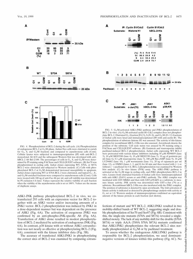
Cyclin B1-Cdc2 is not a BCL-2 kinase. These findings sug-gest that endogenous BCL-2 is phosphorylated by a kinaseactivated at G2/M. An obvious candidate would be Cdc2, al-though the BCL-2 phosphorylation sites S70 and S87 onlyweakly match the phosphorylation consensus site for Cdc2substrates (S/T-P-X-R.S/T-P) (40). To test this possibility,cellular lysates from elutriation-enriched G1, S, or G2/M frac-tions of Jurkat cells were immunoprecipitated with anti-cyclinB1 Ab, and an in vitro kinase assay was performed by usingrecombinant BCL-2-His or histone H1 as the substrate. Cdc2,as expected, was activated at G2/M and phosphorylated itsclassic substrate histone H1 but did not phosphorylate BCL-2(Fig. 5A). This indicates Cdc2 is not the responsible kinaseeven though it has been shown to be activated by paclitaxel andparticipates in the apoptosis of breast cancer cells (68).
To gain information about the BCL-2 kinase, we tested aseries of kinase inhibitors to determine if they affected BCL-2phosphorylation. Jurkat-BCL-2 cells were pretreated with var-ious kinase inhibitors and subsequently exposed to paclitaxel(Fig. 5B). Staurosporine (a broad-spectrum Ser/Thr kinaseinhibitor) and genistein (a Tyr kinase inhibitor) each markedlyinhibited paclitaxel-induced BCL-2 phosphorylation. In con-trast, phosphatidylinositol 3-kinase inhibitors (wortmannin andLY 294002), a MEK inhibitor (PD98059), p38 inhibitors(SB203580 and SB202190), a p70 S6 kinase inhibitor (rapamy-cin), and a PKA inhibitor (Rp-cAMP) did not substantiallyimpair BCL-2 phosphorylation. Thus, this inhibitor study im-plicates both protein tyrosine kinase(s) and Ser/Thr kinase(s)in paclitaxel-induced BCL-2 phosphorylation.
ASK1 and JNK1 are activated at G2/M, and JNK1 phos-phorylates BCL-2. BCL-2 phosphorylation sites conform to the
VOL. 19, 1999 PHOSPHORYLATION AND INACTIVATION OF BCL-2 8473
Dow
nloa
ded
from
http
s://j
ourn
als.
asm
.org
/jour
nal/m
cb o
n 30
Jan
uary
202
2 by
126
.130
.153
.41.
consensus motif for substrates of MAP kinase and JNK/SAPK,yet the lack of response to kinase inhibitors (PD98059,SB203580, and SB202190) suggests that ERKs and p38 are notlikely to be the BCL-2 kinase. Paclitaxel has been shown toresult in JNK/SAPK activation (64). Its candidacy was sup-ported by an immune complex kinase assay performed withendogenous JNK immunoprecipitated from Jurkat cellstreated with anisomycin (10 mg/ml) or UV (300 J/m2) for 30min. The immunoprecipitated, activated JNK phosphorylatedrecombinant BCL-2-His as well as its standard substrate GST–c-Jun (data not shown). Since BCL-2 was phosphorylated atG2/M in normally cycling cells, we next tested whether JNKwas activated at G2/M and capable of phosphorylating BCL-2.JNK was immunoprecipitated from the cell cycle-enriched
fractions of elutriated Jurkat cells and subjected to in vitrokinase assays. JNK from the G2/M fraction was threefold moreactive than at G1, as assayed with c-Jun as a substrate, and alsoproved more potent for phosphorylating recombinant BCL-2.
ASK1 is a member of the MAP3K (MAP kinase kinasekinase) subfamily which activates the JNK pathway and isrequired for tumor necrosis factor- and Fas-Daxx-induced ap-optosis (6, 25). ASK1 has also been noted to be activated bymicrotubule-damaging agents (64). Consequently, we exam-ined ASK1 activity within the cell cycle fractions of Jurkat cells.ASK1 activity was approximately sixfold higher at G2/M thanat the G1 phase (Fig. 5C).
An ASK1/MKK7/JNK1 pathway is responsible for the phos-phorylation of BCL-2 in vivo. To determine whether the
FIG. 3. Substitution of phosphorylation sites in BCL-2 further enhances antiapoptotic activity. (A) Jurkat clones and WEHI-231 clones stably expressing WT oralanine-substituted phosphorylation sites of BCL-2, consisting of Ser70 (S70A), Ser87 (S87A), or all three phosphorylation sites, including Thr69 plus the two serines(AA/A), or an empty vector (Neo) were generated. Cells were treated with 1 mM paclitaxel for Jurkat clones or 1 mM vincristine for WEHI-231 clones and in vivolabeled with 32P-orthophosphate. BCL-2 was immunoprecipitated, separated by SDS-PAGE, and transferred to a membrane. After autoradiography, the samemembranes were used for immunoblotting. Arrowheads denote phosphorylated BCL-2, and dashes denote nonphosphorylated BCL-2. The residual-shifted band onWestern analysis of Jurkat cells bearing AA/A or Neo represents the endogenous hBCL-2 of Jurkat cells. (B) BCL-2 expression levels in Jurkat and WEHI-231 clones,determined by Western blot analysis of lysates from equivalent cells of various Jurkat or WEHI-231 clones with anti-hBCL-2 Ab 6C8 and anti-b-actin Ab. (C to E)Cell viability assays of Jurkat clones (C and D) or WEHI-231 clones (E). Jurkat clones expressing comparable WT, S70A, S87A, AA/A BCL-2, or Neo control vectorwere stimulated with various doses of paclitaxel for 24 h (C) or anti-Fas antibody (100 ng/ml) (D), while WEHI-231 clones were treated with anti-IgM Ab (BET-2supernatant at 1:100 dilution) (E). Viability was determined by PI exclusion using flow cytometry. The results represent the means of triplicate assays. Anotherindependent set of clones expressing matched levels of WT or mutant BCL-2 showed comparable results.
8474 YAMAMOTO ET AL. MOL. CELL. BIOL.
Dow
nloa
ded
from
http
s://j
ourn
als.
asm
.org
/jour
nal/m
cb o
n 30
Jan
uary
202
2 by
126
.130
.153
.41.
ASK1-JNK pathway phosphorylated BCL-2 in vivo, we co-transfected 293 cells with an expression vector for BCL-2 to-gether with an ASK1 vector and/or increasing amounts of aJNK1 vector. BCL-2 phosphorylation was induced by JNK1 ina dose-dependent manner but was dependent on the presenceof ASK1 (Fig. 6A). The activation of exogenous JNK1 wasconfirmed by an anti-phospho-JNK-specific Ab (Fig. 6A).Transfection of ASK1 alone resulted in modest phosphoryla-tion of BCL-2 mediated by activation of endogenous JNK (Fig.6A). In contrast, p38 kinase with or without ASK1 cotransfec-tion was not nearly as effective at phosphorylating BCL-2 (Fig.6A), consistent with the kinase inhibitor data (Fig. 5B).
The accuracy of transfected ASK1/JNK1 in phosphorylatingthe correct sites of BCL-2 was examined by comparing cotrans-
fections of mutant and WT BCL-2. ASK1/JNK1 resulted in twomobility-shifted bands of WT BCL-2, suggesting single and dou-ble phosphorylation of Ser70 and Ser87 (Fig. 6B). Consistent withthis, the single-site mutants (S70A and S87A) revealed a single-shifted moiety. The lack of any mobility shift for the double (S70AS87A) or triple AA/A (T69A S70A S87A) mutants confirmedthat ASK1/JNK1 phosphorylates the relevant BCL-2 sites nor-mally phosphorylated at G2/M or by paclitaxel treatment.
To assess whether the endogenous ASK1/JNK1 pathway isresponsible for BCL-2 phosphorylation, we used dominant-negative versions of kinases within this pathway (Fig. 6C). No
FIG. 4. Phosphorylation of BCL-2 during the cell cycle. (A) Phosphorylationof endogenous BCL-2 at G2/M phase. Jurkat-Neo cells were elutriated to enrichfor G1, S, and G2/M fractions and compared to asynchronous cells (Cont).Cellular lysates were subjected to immunoprecipitation (IP) with anti-BCL-2monoclonal Ab 6C8 and the subsequent Western blot was developed with anti-hBCL-2 Ab Bcl-2/100. The percentages of cells in G1, S, and G2/M were deter-mined by PI staining using FACScan and CELLQUEST software. (B) Ser70 wasphosphorylated in cycling cells. Jurkat clones expressing WT, S70A, or S87ABCL-2 were elutriated and subjected to Western analysis. (C) Cells with phos-phorylated BCL-2 at G2/M demonstrated increased susceptibility to apoptosis.Jurkat clones expressing WT or S70A BCL-2 were elutriated, and typical G1-, S-,and G2/M-enriched fractions were compared to asynchronous cells (Cont). Cellswere treated with 100 ng of anti-Fas Ab per ml, and cell viability was determinedby PI exclusion 6 h later. Values represent the relative viability of each fractionwhen the viability of the asynchronous cells is set at 100%. Values are the meansof duplicate assays.
FIG. 5. G2/M-activated ASK1-JNK1 pathway and JNK1 phosphorylation ofBCL-2 in vitro. (A) G2/M-activated cyclin B1-Cdc2 complex does not phosphor-ylate BCL-2. Elutriated G1 (fraction [Fr] 3), S (Fr 8), and G2/M (Fr 11) fractionsof Jurkat cells were lysed and immunoprecipitated (IP) with anti-cyclin B1. Thephosphorylation of substrate histone H1 was assessed. The activity of this kinasecomplex for recombinant hBCL-2-His was also assessed. Arrowheads denote theposition of the substrate. Cell cycle status was assayed by PI staining using aFACScan and CELLQUEST software. (B) Genistein and staurosporine inhibitpaclitaxel-induced BCL-2 phosphorylation. Jurkat cells expressing WT BCL-2were pretreated with various kinase inhibitors including 50 mM PD98059 (lane3), 10 mM SB203580 (lane 4), 10 mM SB202190 (lane 5), 10 mg of genistein perml (lane 6), 0.1 mM staurosporine (lane 7), 100 mM Rp-cAMP (lane 9), 10 mMLY294002 (lane 10), 1 mM wortmannin (lane 11), 20 ng of rapamycin per ml(lane 12), or DMSO (lanes 1, 2, and 8) for 60 min and then treated with (1) orwithout (2) paclitaxel for 6 h. BCL-2 phosphorylation was examined by Westernblot analysis. (C) In vitro kinase (IVK) assay. The ASK1-JNK1 pathway isactivated at the G2/M stage in cycling cells, and JNK1 phosphorylates BCL-2 invitro. Lysates from elutriated fractions of Jurkat cells were immunoprecipitatedwith anti-ASK1 (DAV) serum or anti-JNK1 antibody. The ASK1 complex wasincubated with GST-MKK6 and then with GST-p38gKN as a substrate to mea-sure kinase activity. JNK activity was determined with GST–c-Jun (79) as asubstrate. Recombinant hBCL-2-His was also incubated with the JNK1 complex.The position of substrates is denoted by open arrowheads. The fold activation ofkinase activity is indicated as detected by phosphorimage analysis with activity atG1 set at 1.0. Western analysis of immunoprecipitates confirmed an equivalentamount of the kinase protein in each fraction (not shown).
VOL. 19, 1999 PHOSPHORYLATION AND INACTIVATION OF BCL-2 8475
Dow
nloa
ded
from
http
s://j
ourn
als.
asm
.org
/jour
nal/m
cb o
n 30
Jan
uary
202
2 by
126
.130
.153
.41.
single dominant-negative kinase expression vector was capableof inhibiting paclitaxel-induced phosphorylation of BCL-2.However, inhibition of multiple steps in the pathway by co-transfection of dnASK1, dnMKK7, and dnJNK1 togethermarkedly inhibited BCL-2 phosphorylation. Of note, dnSEK1was not as effective as dnMKK7 (Fig. 6C). These data supportan ASK1/MKK7/JNK1 axis in the phosphorylation of BCL-2.
DISCUSSION
The phosphorylation of BCL-2 is a further example of howposttranslational modifications can interconvert active to inac-tive conformations of BCL-2 family molecules. However, un-certainty existed in the literature as to whether this phosphor-ylation activates (36) or inactivates (21) the antiapoptoticfunction of BCL-2. Consequently, we elected to first performdetailed 2D mapping and sequence identification of the in vivophosphorylation sites on BCL-2 followed by their individualand collective substitution. Ser70, Ser87, and Thr69 proved tobe phosphorylated, and all are located within the unstructuredloop region between the a1 and a2 helices of BCL-2. The threemobility-shifted bands correspond to one, two, or three sites ofphosphorylation, respectively. The number of phosphorylatedsites appears to depend on the intensity of kinase activation.During normal cell cycle progression, Ser70 is the principalphosphorylation site. Haldar et al. (19) and Srivastava et al.(55) also noted Ser70 as the major site of phosphorylation inresponse to microtubule-damaging agents. We have also notedthat following paclitaxel, Ser70 is phosphorylated before Ser87(not shown) although this sequence of events is not obligate, asSer87 is phosphorylated in an S70A mutant. The single S70Aand S87A mutants and the triple AA/A mutant all demonstrateaugmented antideath activity due to microtubule damage bypaclitaxel or vincristine or to the physiologic anti-IgM or anti-Fas Ab signals. This evidence that BCL-2 phosphorylation isinactivating is in keeping with the observation that phosphor-ylated BCL-2 is less likely to heterodimerize with BAX mole-cules (47, 55). This is consistent with the initial proposal thatthe phosphorylation of BCL-2 would be inactivating (20). Ourfindings disagree with observations on a different system inwhich an S70A BCL-2 mutant displays less protection in NSF/N1.H7 cells, an IL-3-dependent cell line (27), although we
FIG. 6. ASK1-JNK1 pathway phosphorylates BCL-2 in vivo. (A) Dose-de-pendent phosphorylation of BCL-2 by JNK1 in vivo. 293 cells were transfectedwith 20 ng of hBCL-2 or control vector together with indicated amounts ofHA-ASK1, HA-JNK1, and/or HA-p38, as shown. The total amount of trans-fected DNA was kept constant by adding a compensating amount of emptyvector pcDNA3. After 24 h, cells were lysed and the phosphorylation of BCL-2,expression level of each kinase, and activation of JNK and p38 were determinedby Western analysis. Open arrowheads denote the activated endogenous JNKs(p46 and p54). (B) Substrate specificity of ASK1-JNK1 pathway in vivo. WT orphosphorylation site mutant BCL-2 expression vectors (20 ng) were cotrans-fected with ASK1 (500 ng) and JNK (750 ng) or pcDNA3 into 293 cells. Lysatesgenerated 24 h after transfection were assessed for BCL-2 phosphorylation byWestern analysis. (C) Inhibition of BCL-2 phosphorylation by dnASK1,dnMKK7, and dnJNK1. WT BCL-2 (20 ng) was cotransfected with dnASK1 (500ng), dnSEK1 (500 ng), dnMKK7 (500 ng), and dnJNK1 (750 ng), as indicated,into 293 cells. The total amount of DNA transfected was normalized by addingpcDNA3. After 16 h, paclitaxel was added to a final concentration of 1 mM andcells were incubated an additional 8 h. BCL-2 phosphorylation and the expres-sion levels of the dominant-negative (dn) kinases were determined by immuno-blotting.
FIG. 7. Schematic representation of the ASK1/MKK7/JNK1 pathway inBCL-2 phosphorylation. ASK1, a MAP3K, is activated by extracellular andintracellular stimuli to induce JNK pathway activation. JNK phosphorylatesBCL-2, inactivating its antiapoptotic function. TNFR1, tumor necrosis factorreceptor 1.
8476 YAMAMOTO ET AL. MOL. CELL. BIOL.
Dow
nloa
ded
from
http
s://j
ourn
als.
asm
.org
/jour
nal/m
cb o
n 30
Jan
uary
202
2 by
126
.130
.153
.41.
found that the S70A mutant was capable of protecting anotherIL-3-dependent line, FL5.12 (44a). Of note, all BCL-2 mutantsstudied here (S70A, S87A, and the triple AA/A) demonstrateroughly comparable protection, which suggests that the threesites might act in concert to regulate BCL-2. Of interest, theprotection offered by these mutants, including BCL-2 AA/A,which eliminates all phosphorylation, was not as complete asthat reported for mutants lacking the unstructured loop (5, 54).While this might suggest modifications to the loop beyondphosphorylation, it is also conceivable that the loop deletionmore effectively locks BCL-2 in an active conformationwhereas phosphorylation only influences an equilibrium be-tween active and inactive conformers.
The phosphorylation of BCL-2 at the G2/M phase of normallycycling cells indicates that the phosphorylation of BCL-2 is anormal physiologic process rather than exclusively a response tomicrotubule damage. The capacity of drugs with multiple actions,including nocodazole, taxanes and vinca alkaloids, to all result inBCL-2 phosphorylation suggests that arrest at G2/M might be thecommon event. What would be the purpose of inactivating BCL-2at G2/M? Prior evidence of an interrelationship between cell cycleprogression and BCL-2 exists. In IL-3-dependent cells, the phos-phorylation of BCL-2 was temporally correlated with entry intothe cell cycle (47). Bcl-2-deficient T cells demonstrated acceler-ated cell cycle entry but at the risk of increased apoptosis. Incontrast, overexpression of BCL-2 delays entry into S phase andin T cells apparently at the restriction point, reflected by dimin-ished IL-2 production upon activation (31, 37, 45, 61). If BCL-2had a similar arresting affect at G2/M, phosphorylation might berequired for its release. Alternatively, evidence presented heresupports another model: that cells are more susceptible to a deathsignal during G2/M and that characteristic can be attributed tophosphorylation of BCL-2. Thus, a rationale for lowering thethreshold for apoptosis at G2/M would be to ensure the elimina-tion of cells with aberrations of chromosomal segregation. Insupport of this model, overexpression of BCL-XL has been notedto result in increased tetraploidization (39).
Our finding indicated that the same ASK1/JNK pathway isresponsible for the normal cell cycle-related and paclitaxel-induced phosphorylation of BCL-2 (Fig. 7). This suggests thatthe phosphorylation of BCL-2 in response to the microtubule-damaging drugs could simply be an exaggerated normal pro-cess secondary to their G2/M arrest. Alternatively, a moreintimate relationship could exist in that the same agents (tax-anes, vinca alkaloids, nocodazole, and colchicine) that result inthe phosphorylation of BCL-2 also activate the ASK1/JNKkinase pathway (64). In our examination, no single dominantnegative kinase in this pathway was capable of blocking pacli-taxel-induced phosphorylation of BCL-2, consistent with otherobservations (63). However, combined dominant negatives foreach step of the pathway (dnASK1/dnMKK7/dnJNK1) inter-fered quite effectively with BCL-2 phosphorylation. Consistentwith this, the combined overexpression of WT ASK1/JNK1 wascapable of phosphorylating BCL-2 in vivo. The JNK family hasbeen reported to mediate both proapoptotic and antiapoptoticresponses, perhaps reflecting cell type specificity (26). This issupported by Jnk1/Jnk2 double-deficient embryos, which re-vealed region-specific apoptosis with reduced death in thehindbrain but increased apoptosis in the forebrain (29). Nei-ther SEK1 nor p38 had obvious effects despite the fact that p38is implicated as a component of the mitotic spindle checkpoint(59). Recently, activated JNK1/2 has been shown to colocalizewith a MAP3K, MLK (mixed lineage) Ser/Thr kinase, to punc-tate structures along microtubules. Of potential interest, MLKinteracts with the KIF3 family of kinesin motor proteins (42). Infurther support of a direct relationship between microtubules and
the cell death pathway, another BCL-2 family member BIM bindsto the LC8 dynein light-chain protein (48). In this context, or-ganelles including mitochondria are transported along microtu-bules by means of microtubule-associated motor proteins (23).BCL-2 localizes to mitochondria, endoplasmic reticulum, and nu-clear envelope, and transportation along microtubules arrangesthese organelles on a metaphase plate to allow equal segregationof them to daughter cells at mitosis. Thus, BCL-2 associated withthese organelles could be placed in apposition with activated JNKpathway kinases found at microtubules. Thus, drugs which causemicrotubule dysfunction would be activating the same MAP3K/JNK pathway, prompting the phosphorylation of a normal sub-strate(s) including BCL-2 (Fig. 7).
ACKNOWLEDGMENTS
We are grateful to R. J. Davis, S. Gutkind, G. L. Johnson, J. Kyri-akis, E. Nishida, and H. Saito for providing the expression vectorsdescribed above and to Steven F. Dowdy, David S. Pellman, MarkDalton, and Zoltan Oltvai for technical advice and scientific discus-sion. We also thank Deborah S. Maher for secretarial assistance.
K.Y. was a fellow of the National Cancer Institute (U.S.A.)-Japa-nese Foundation for Cancer Research Research Training Program andis supported in part by the Uhehara Memorial Foundation ResearchFellowship.
REFERENCES
1. Bakhshi, A., J. P. Jensen, P. Goldman, J. J. Wright, O. W. McBride, A. L.Epstein, and S. J. Korsmeyer. 1985. Cloning the chromosomal breakpoint oft(14;18) human lymphomas: clustering around JH on chromosome 14 andnear a transcriptional unit on 18. Cell 41:899–906.
2. Blagosklonny, M. V., P. Giannakakou, W. S. el-Deiry, D. G. Kingston, P. I.Higgs, L. Neckers, and T. Fojo. 1997. Raf-1/bcl-2 phosphorylation: a stepfrom microtubule damage to cell death. Cancer Res. 57:130–135.
3. Blume-Jensen, P., R. Janknecht, and T. Hunter. 1998. The kit receptorpromotes cell survival via activation of PI 3-kinase and subsequent Akt-mediated phosphorylation of Bad on Ser136. Curr. Biol. 8:779–782.
4. Boyle, W. J., P. van der Geer, and T. Hunter. 1991. Phosphopeptide mappingand phosphoamino acid analysis by two-dimensional separation on thin-layercellulose plates. Methods Enzymol. 201:110–149.
5. Chang, B. S., A. J. Minn, S. W. Muchmore, S. W. Fesik, and C. B. Thompson.1997. Identification of a novel regulatory domain in Bcl-X(L) and Bcl-2.EMBO J. 16:968–977.
6. Chang, H. Y., H. Nishitoh, X. Yang, H. Ichijo, and D. Baltimore. 1998.Activation of apoptosis signal-regulating kinase 1 (ASK1) by the adapterprotein Daxx. Science 281:1860–1863.
7. Chou, J. J., H. Li, G. S. Salvesen, J. Yuan, and G. Wagner. 1999. Solutionstructure of BID, an intracellular amplifier of apoptotic signaling. Cell 96:615–624.
8. Cleary, M. L., and J. Sklar. 1985. Nucleotide sequence of a t(14;18) chro-mosomal breakpoint in follicular lymphoma and demonstration of a break-point-cluster region near a transcriptionally active locus on chromosome 18.Proc. Natl. Acad. Sci. USA 82:7439–7443.
9. Coso, O. A., M. Chiariello, J. C. Yu, H. Teramoto, P. Crespo, N. Xu, T. Miki,and J. S. Gutkind. 1995. The small GTP-binding proteins Rac1 and Cdc42regulate the activity of the JNK/SAPK signaling pathway. Cell 81:1137–1146.
10. Cryns, V., and J. Yuan. 1998. Proteases to die for. Genes Dev. 12:1551–1570.11. Datta, S. R., H. Dudek, X. Tao, S. Masters, H. Fu, Y. Gotoh, and M. E.
Greenberg. 1997. Akt phosphorylation of BAD couples survival signals to thecell-intrinsic death machinery. Cell 91:231–241.
12. del Peso, L., M. Gonzalez-Garcia, C. Page, R. Herrera, and G. Nunez. 1997.Interleukin-3-induced phosphorylation of BAD through the protein kinaseAkt. Science 278:687–689.
13. Derijard, B., J. Raingeaud, T. Barrett, I. H. Wu, J. Han, R. J. Ulevitch, andR. J. Davis. 1995. Independent human MAP-kinase signal transduction path-ways defined by MEK and MKK isoforms. Science 267:682–685.
14. Ellis, R. E., J. Y. Yuan, and H. R. Horvitz. 1991. Mechanisms and functionsof cell death. Annu. Rev. Cell Biol. 7:663–698.
15. Fang, G., B. S. Chang, C. N. Kim, C. Perkins, C. B. Thompson, and K. N.Bhalla. 1998. “Loop” domain is necessary for taxol-induced mobility shiftand phosphorylation of Bcl-2 as well as for inhibiting taxol-induced cytosolicaccumulation of cytochrome c and apoptosis. Cancer Res. 58:3202–3208.
16. Green, D. R., and J. C. Reed. 1998. Mitochondria and apoptosis. Science281:1309–1312.
17. Gross, A., J. Jockel, M. C. Wei, and S. J. Korsmeyer. 1998. Enforced dimer-ization of BAX results in its translocation, mitochondrial dysfunction andapoptosis. EMBO J. 17:3878–3885.
VOL. 19, 1999 PHOSPHORYLATION AND INACTIVATION OF BCL-2 8477
Dow
nloa
ded
from
http
s://j
ourn
als.
asm
.org
/jour
nal/m
cb o
n 30
Jan
uary
202
2 by
126
.130
.153
.41.
18. Gross, A., X. M. Yin, K. Wang, M. C. Wei, J. Jockel, C. Milliman, H.Erdjument-Bromage, P. Tempst, and S. J. Korsmeyer. 1999. Caspasecleaved BID targets mitochondria and is required for cytochrome c release,while BCL-XL prevents this release but not tumor necrosis factor-R1/Fasdeath. J. Biol. Chem. 274:1156–1163.
19. Haldar, S., A. Basu, and C. M. Croce. 1998. Serine-70 is one of the criticalsites for drug-induced Bcl2 phosphorylation in cancer cells. Cancer Res.58:1609–1615.
20. Haldar, S., J. Chintapalli, and C. M. Croce. 1996. Taxol induces bcl-2 phos-phorylation and death of prostate cancer cells. Cancer Res. 56:1253–1255.
21. Haldar, S., N. Jena, and C. M. Croce. 1995. Inactivation of Bcl-2 by phos-phorylation. Proc. Natl. Acad. Sci. USA 92:4507–4511.
22. Harada, H., B. Becknell, M. Wilm, M. Mann, L. J. Huang, S. S. Taylor, J. D.Scott, and S. J. Korsmeyer. 1999. Phosphorylation and inactivation of BADby mitochondria-anchored protein kinase A. Mol. Cell 3:413–422.
23. Hirokawa, N. 1998. Kinesin and dynein superfamily proteins and the mech-anism of organelle transport. Science 279:519–526.
24. Hsu, Y. T., K. G. Wolter, and R. J. Youle. 1997. Cytosol-to-membraneredistribution of Bax and Bcl-X(L) during apoptosis. Proc. Natl. Acad. Sci.USA 94:3668–3672.
25. Ichijo, H., E. Nishida, K. Irie, P. ten Dijke, M. Saitoh, T. Moriguchi, M.Takagi, K. Matsumoto, K. Miyazono, and Y. Gotoh. 1997. Induction ofapoptosis by ASK1, a mammalian MAPKKK that activates SAPK/JNK andp38 signaling pathways. Science 275:90–94.
26. Ip, Y. T., and R. J. Davis. 1998. Signal transduction by the c-Jun N-terminalkinase (JNK)—from inflammation to development. Curr. Opin. Cell Biol.10:205–219.
27. Ito, T., X. Deng, B. Carr, and W. S. May. 1997. Bcl-2 phosphorylationrequired for anti-apoptosis function. J. Biol. Chem. 272:11671–11673.
28. Johnson, N. L., A. M. Gardner, K. M. Diener, C. A. Lange-Carter, J. Gleavy,M. B. Jarpe, A. Minden, M. Karin, L. I. Zon, and G. L. Johnson. 1996. Signaltransduction pathways regulated by mitogen-activated/extracellular responsekinase kinase kinase induce cell death. J. Biol. Chem. 271:3229–3237.
29. Kuan, C. Y., D. D. Yang, D. R. Samanta Roy, R. J. Davis, P. Rakic, and R. A.Flavell. 1999. The Jnk1 and Jnk2 protein kinases are required for regionalspecific apoptosis during early brain development. Neuron 22:667–676.
30. Li, H., H. Zhu, C. J. Xu, and J. Yuan. 1998. Cleavage of BID by caspase 8mediates the mitochondrial damage in the Fas pathway of apoptosis. Cell94:491–501.
31. Linette, G. P., Y. Li, K. Roth, and S. J. Korsmeyer. 1996. Cross talk betweencell death and cell cycle progression: BCL-2 regulates NFAT-mediated ac-tivation. Proc. Natl. Acad. Sci. USA 93:9545–9552.
32. Ling, Y. H., C. Tornos, and R. Perez-Soler. 1998. Phosphorylation of Bcl-2 isa marker of M phase events and not a determinant of apoptosis. J. Biol.Chem. 273:18984–18991.
33. Lissy, N. A., L. F. Van Dyk, M. Becker-Hapak, A. Vocero-Akbani, J. H.Mendler, and S. F. Dowdy. 1998. TCR antigen-induced cell death occursfrom a late G1 phase cell cycle check point. Immunity 8:57–65.
34. Luo, X., I. Budihardjo, H. Zou, C. Slaughter, and X. Wang. 1998. Bid, a Bcl2interacting protein, mediates cytochrome c release from mitochondria inresponse to activation of cell surface death receptors. Cell 94:481–490.
35. Maundrell, K., B. Antonsson, E. Magnenat, M. Camps, M. Muda, C.Chabert, C. Gillieron, U. Boschert, E. Vial-Knecht, J. C. Martinou, and S.Arkinstall. 1997. Bcl-2 undergoes phosphorylation by c-Jun N-terminal ki-nase/stress-activated protein kinases in the presence of the constitutivelyactive GTP-binding protein Rac1. J. Biol. Chem. 272:25238–25242.
36. May, W. S., P. G. Tyler, T. Ito, D. K. Armstrong, K. A. Qatsha, and N. E.Davidson. 1994. Interleukin-3 and bryostatin-1 mediate hyperphosphoryla-tion of BCL2 alpha in association with suppression of apoptosis. J. Biol.Chem. 269:26865–26870.
37. Mazel, S., D. Burtrum, and H. T. Petrie. 1996. Regulation of cell divisioncycle progression by bcl-2 expression: a potential mechanism for inhibition ofprogrammed cell death. J. Exp. Med. 183:2219–2226.
38. McDonnell, J. M., D. Fushman, C. L. Milliman, S. J. Korsmeyer, and D.Cowburn. 1999. Solution structure of the proapoptotic molecule BID: astructural basis for apoptotic agonists and antagonists. Cell 96:625–634.
39. Minn, A. J., L. H. Boise, and C. B. Thompson. 1996. Expression of Bcl-xLand loss of p53 can cooperate to overcome a cell cycle checkpoint induced bymitotic spindle damage. Genes Dev. 10:2621–2631.
40. Moreno, S., and P. Nurse. 1990. Substrates for p34cdc2: in vivo veritas? Cell61:549–551.
41. Moriguchi, T., F. Toyoshima, N. Masuyama, H. Hanafusa, Y. Gotoh, and E.Nishida. 1997. A novel SAPK/JNK kinase, MKK7, stimulated by TNFalphaand cellular stresses. EMBO J. 16:7045–7053.
42. Nagata, K., A. Puls, C. Futter, P. Aspenstrom, E. Schaefer, T. Nakata, N.Hirokawa, and A. Hall. 1998. The MAP kinase kinase kinase MLK2 co-localizes with activated JNK along microtubules and associates with kinesinsuperfamily motor KIF3. EMBO J. 17:149–158.
43. Nishitoh, H., M. Saitoh, Y. Mochida, K. Takeda, H. Nakano, M. Rothe, K.Miyazono, and H. Ichijo. 1998. ASK1 is essential for JNK/SAPK activationby TRAF2. Mol. Cell 2:389–395.
44. Oltvai, Z. N., and S. J. Korsmeyer. 1994. Checkpoints of dueling dimers foildeath wishes. Cell 79:189–192.
44a.Oltvai, Z. N., and S. J. Korsmeyer. Unpublished observation.45. O’Reilly, L. A., D. C. Huang, and A. Strasser. 1996. The cell death inhibitor
Bcl-2 and its homologues influence control of cell cycle entry. EMBO J.15:6979–6990.
46. Ottilie, S., J. L. Diaz, W. Horne, J. Chang, Y. Wang, G. Wilson, S. Chang, S.Weeks, L. C. Fritz, and T. Oltersdorf. 1997. Dimerization properties ofhuman BAD. Identification of a BH-3 domain and analysis of its binding tomutant BCL-2 and BCL-XL proteins. J. Biol. Chem. 272:30866–30872.
47. Poommipanit, P., B. Chen, and Z. N. Oltvai. 1999. Interleukin-3 induces thephosphorylation of a distinct fraction of bcl-2. J. Biol. Chem. 274:1033–1039.
48. Puthalakath, H., D. C. Huang, L. A. O’Reilly, S. M. King, and A. Strasser.1999. The proapoptotic activity of the Bcl-2 family member Bim is regulatedby interaction with the dynein motor complex. Mol. Cell 3:287–296.
49. Raff, M. C. 1992. Social controls on cell survival and cell death. Nature356:397–400.
50. Rana, A., K. Gallo, P. Godowski, S. Hirai, S. Ohno, L. Zon, J. M. Kyriakis,and J. Avruch. 1996. The mixed lineage kinase SPRK phosphorylates andactivates the stress-activated protein kinase activator, SEK-1. J. Biol. Chem.271:19025–19028.
51. Rodi, D. J., R. W. Janes, H. J. Sanganee, R. A. Holton, B. A. Wallace, and L.Makowski. 1999. Screening of a library of phage-displayed peptides identifieshuman bcl-2 as a taxol-binding protein. J. Mol. Biol. 285:197–203.
52. Ruvolo, P. P., X. Deng, B. K. Carr, and W. S. May. 1998. A functional rolefor mitochondrial protein kinase Calpha in Bcl2 phosphorylation and sup-pression of apoptosis. J. Biol. Chem. 273:25436–25442.
53. Scatena, C. D., Z. A. Stewart, D. Mays, L. J. Tang, C. J. Keefer, S. D. Leach,and J. A. Pietenpol. 1998. Mitotic phosphorylation of Bcl-2 during normalcell cycle progression and Taxol-induced growth arrest. J. Biol. Chem. 273:30777–30784.
54. Srivastava, R. K., Q. S. Mi, J. M. Hardwick, and D. L. Longo. 1999. Deletionof the loop region of Bcl-2 completely blocks paclitaxel-induced apoptosis.Proc. Natl. Acad. Sci. USA 96:3775–3780.
55. Srivastava, R. K., A. R. Srivastava, S. J. Korsmeyer, M. Nesterova, Y. S.Cho-Chung, and D. L. Longo. 1998. Involvement of microtubules in theregulation of Bcl2 phosphorylation and apoptosis through cyclic AMP-de-pendent protein kinase. Mol. Cell. Biol. 18:3509–3517.
56. Steller, H. 1995. Mechanisms and genes of cellular suicide. Science 267:1445–1449.
57. Sullivan, S., and T. W. Wong. 1991. A manual sequencing method foridentification of phosphorylated amino acids in phosphopeptides. Anal. Bio-chem. 197:65–68.
58. Takekawa, M., and H. Saito. 1998. A family of stress-inducible GADD45-like proteins mediate activation of the stress-responsive MTK1/MEKK4MAPKKK. Cell 95:521–530.
59. Takenaka, K., T. Moriguchi, and E. Nishida. 1998. Activation of the proteinkinase p38 in the spindle assembly checkpoint and mitotic arrest. Science280:599–602.
60. Tsujimoto, Y., J. Cossman, E. Jaffe, and C. M. Croce. 1985. Involvement ofthe bcl-2 gene in human follicular lymphoma. Science 228:1440–1443.
61. Vairo, G., K. M. Innes, and J. M. Adams. 1996. Bcl-2 has a cell cycleinhibitory function separable from its enhancement of cell survival. Onco-gene 13:1511–1519.
62. Vaux, D. L., and S. J. Korsmeyer. 1999. Cell death in development. Cell96:245–254.
63. Wang, T. H., D. M. Popp, H. S. Wang, M. Saitoh, J. G. Mural, D. C. Henley,H. Ichijo, and J. Wimalasena. 1999. Microtubule dysfunction induced bypaclitaxel initiates apoptosis through both c-Jun N-terminal kinase (JNK)-dependent and -independent pathways in ovarian cancer cells. J. Biol. Chem.274:8208–8216.
64. Wang, T. H., H. S. Wang, H. Ichijo, P. Giannakakou, J. S. Foster, T. Fojo,and J. Wimalasena. 1998. Microtubule-interfering agents activate c-Jun N-terminal kinase/stress-activated protein kinase through both Ras and apo-ptosis signal-regulating kinase pathways. J. Biol. Chem. 273:4928–4936.
65. White, E. 1996. Life, death, and the pursuit of apoptosis. Genes Dev. 10:1–15.
66. Wolter, K. G., Y. T. Hsu, C. L. Smith, A. Nechushtan, X. G. Xi, and R. J.Youle. 1997. Movement of Bax from the cytosol to mitochondria duringapoptosis. J. Cell Biol. 139:1281–1292.
67. Yin, X. M., Z. N. Oltvai, and S. J. Korsmeyer. 1994. BH1 and BH2 domainsof Bcl-2 are required for inhibition of apoptosis and heterodimerization withBax. Nature 369:321–323.
68. Yu, D., T. Jing, B. Liu, J. Yao, M. Tan, T. J. McDonnell, and M. C. Hung.1998. Overexpression of ErbB2 blocks Taxol-induced apoptosis by upregu-lation of p21Cip1, which inhibits p34Cdc2 kinase. Mol. Cell 2:581–591.
69. Zha, J., H. Harada, K. Osipov, J. Jockel, G. Waksman, and S. J. Korsmeyer.1997. BH3 domain of BAD is required for heterodimerization with BCL-XLand pro-apoptotic activity. J. Biol. Chem. 272:24101–24104.
70. Zha, J., H. Harada, E. Yang, J. Jockel, and S. J. Korsmeyer. 1996. Serinephosphorylation of death agonist BAD in response to survival factor resultsin binding to 14-3-3 not BCL-X(L). Cell 87:619–628.