The BD OneFlow™ ALOT (Acute Leukemia Orientation Tube) is intended for flow-cytometric immunophenotyping of aberrant immature populations of hematopoietic cells (lymphoid and non-lymphoid lineage) in bone marrow and peripheral blood as an aid in the diagnosis of acute lymphoblastic leukemia and non-lymphoid acute leukemia. The BD OneFlow ALOT is designed for use with a suitably equipped BD flow cytometer and software designated for in vitro diagnostic use.
2. SUMMARY AND EXPLANATION
Acute leukemias are a heterogeneous group of diseases characterized as having a clonal (neoplastic) population of immature hematopoietic cells in the peripheral blood (PB) or bone marrow (BM).1 There are two major classes of acute leukemias: lymphoid precursor leukemias and acute myeloid leukemias (AML). The lymphoid precursor leukemias are divided into B-cell and T-cell precursor lymphoblastic leukemias (BCP-ALL and T-ALL, respectively). In addition, a small number of neoplasms do not fit into any of these categories because they either show no clear expression of markers indicative of a particular lineage, or they express markers specific to more than one lineage. They include acute undifferentiated leukemia (AUL) and mixed phenotype acute leukemia (MPAL).
The EuroFlow™* Consortium designed multicolor antibody panels to fully characterize the cell populations in a patient specimen using immunophenotypic markers that are indicative of normal and abnormal cells.1 In addition to the optimized multicolor antibody
* The EuroFlow trademark and logo and the EuroFlow™ antibody panels are property of the EuroFlow Consortium and cannot be reproduced or published without prior written permission from the EuroFlow coordinator (www.euroflow.org).
panels, the EuroFlow protocol comprises standardized procedures for cytometer setup, determination of assay settings, sample preparation, staining, and acquisition, and data analysis.2
The single-tube screening panels and multi-tube classification panels fit into the EuroFlow diagnostic algorithm for the identification and classification of hematological disorders. Each tube contains a set of backbone markers and a set of classification markers.1 Backbone markers are shared across a particular set of panels and are used to normalize the samples so that data files can be combined and analyzed as a single large data file. They are markers that identify distinct cell populations in a particular cell lineage. Classification markers have been selected for their diagnostic utility in discriminating between cell types within a given lineage and in classifying the abnormal cell type in the sample.
3. PRINCIPLES OF THE PROCEDURE
Multiparameter flow cytometry is a sensitive and rapid tool for the qualitative and quantitative characterization of cell populations in a specimen. Cells are incubated with fluorochrome-conjugated antibodies which bind to their target molecules. The stained cells can then be analyzed on a single-cell basis. Multiparameter analysis of the data is used to identify the cell populations in the patient specimen and can lead to the identification of an aberrant clonal cell population.
The number of parameters used in flow cytometric immunophenotyping of hematological disorders has increased in recent years. BD OneFlow ALOT contains a panel of fluorochrome-conjugated antibodies that identify immature populations of hematopoietic cells in the B-lymphoid, T-lymphoid, and non-lymphoid lineages. Analysis of the dot plots allows for the identification of clonally expanded populations of immature hematopoietic precursor cells.
7
4. REAGENT COMPOSITION
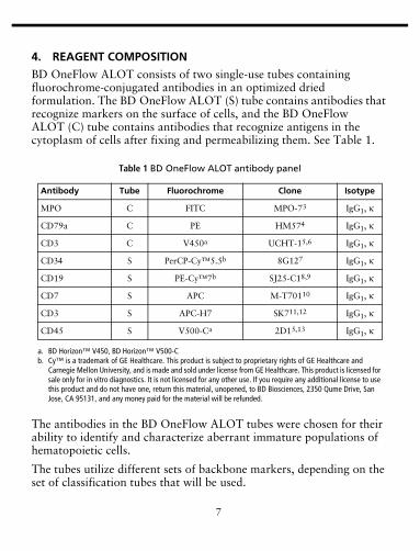
BD OneFlow ALOT consists of two single-use tubes containing fluorochrome-conjugated antibodies in an optimized dried formulation. The BD OneFlow ALOT (S) tube contains antibodies that recognize markers on the surface of cells, and the BD OneFlow ALOT (C) tube contains antibodies that recognize antigens in the cytoplasm of cells after fixing and permeabilizing them. See Table 1.
The antibodies in the BD OneFlow ALOT tubes were chosen for their ability to identify and characterize aberrant immature populations of hematopoietic cells.
The tubes utilize different sets of backbone markers, depending on the set of classification tubes that will be used.
Table 1 BD OneFlow ALOT antibody panel
Antibody Tube Fluorochrome Clone Isotype
MPO C FITC MPO-73 IgG1, κ
CD79a C PE HM574 IgG1, κ
CD3 C V450a
a. BD Horizon™ V450, BD Horizon™ V500-C
UCHT-15,6 IgG1, κ
CD34 S PerCP-Cy™5.5b
b. Cy™ is a trademark of GE Healthcare. This product is subject to proprietary rights of GE Healthcare and Carnegie Mellon University, and is made and sold under license from GE Healthcare. This product is licensed for sale only for in vitro diagnostics. It is not licensed for any other use. If you require any additional license to use this product and do not have one, return this material, unopened, to BD Biosciences, 2350 Qume Drive, San Jose, CA 95131, and any money paid for the material will be refunded.
8G127 IgG1, κ
CD19 S PE-Cy™7b SJ25-C18,9 IgG1, κ
CD7 S APC M-T70110 IgG1, κ
CD3 S APC-H7 SK711,12 IgG1, κ
CD45 S V500-Ca 2D15,13 IgG1, κ
8
For the BCP-ALL panel, the backbone markers in BD OneFlow ALOT are CD45, CD34, and CD19.
For the T-ALL panel, the backbone markers in BD OneFlow ALOT are CD45, cytoplasmic CD3 (cyCD3), and CD3.
For the AML panels, the backbone markers in BD OneFlow ALOT are CD45 and CD34.
CD34 and negative or dim expression of CD45 (CD45neg/dim) are markers for immature cells.
Cytoplasmic myeloperoxidase (cyMPO) is a myeloid lineage marker.
cyCD3 and CD7 are T-cell lineage markers. CD3 is used as a maturity marker for T cells.
CD19 and cytoplasmic CD79a (cyCD79a) are B-cell lineage markers.
Refer to the article describing the EuroFlow antibody panels1 for a full description of the utility of the antibodies chosen for the BD OneFlow ALOT tube.
The BD OneFlow ALOT (S) tube contains 0.7816% 2-methyl-4-isothiazolin-3-one (CAS number 2682-20-4) and 0.1369% sodium azide (CAS number 26628-22-8). The BD OneFlow ALOT (C) tube contains 0.5322% 2-methyl-4-isothiazolin-3-one (CAS number 2682-20-4) and 0.3446% sodium azide (CAS number 26628-22-8). The reagents are classified as hazardous according to the Globally Harmonized System of Classification and Labelling of Chemicals (GHS).
9
5. STORAGE AND HANDLING
Store tubes at 2°C–27°C in the foil pouch. Do not freeze the reagent or expose it to direct light at any time during storage or incubation with cells. The dried fluorochrome-conjugated antibodies are stable until the expiration date shown on the pouch and tube labels when stored as directed. Do not use after the expiration date. Once the pouch is opened, the dried fluorochrome-conjugated antibodies are stable for one month when stored as directed.
CAUTION Ensure that the pouch is completely resealed after removing a tube. The reagent is very sensitive to moisture. Do not remove the desiccant from the reagent pouch.
Warning
H317 May cause an allergic skin reaction.H412 Harmful to aquatic life with long lasting effects.
P261 Avoid breathing dust/fume/gas/mist/vapors/spray.P280 Wear protective gloves/protective clothing/eye protection/face protection.P273 Avoid release to the environment.P302+P352 IF ON SKIN: Wash with plenty of water.P333+P313 If skin irritation or rash occurs: Get medical advice/attention.P362+P364 Take off contaminated clothing and wash it before reuse.P501 Dispose of contents/container to an appropriate treatment and disposal facility in accordance with applicable laws and regulations, and product characteristics at time of disposal.EUH210 Safety Data Sheet available on request.Contains 2-methyl-4-isothiazolin-3-one: May produce an allergic reaction.
10
6. REAGENTS AND MATERIALS
Reagents Provided
BD OneFlow ALOT is provided as single-use tubes in foil pouches. Each kit contains four pouches:
• two pouches, each containing five tubes of BD OneFlow ALOT (S)• two pouches, each containing five tubes of BD OneFlow ALOT (C)
Reagents or Materials Required but Not Provided
• Templates installer CD for BD OneFlow™ Assays (Catalog No. 659305)
The OneFlow ALOT template is provided on an installer CD. The template contains two global worksheets: the BD OneFlow ALOT Acquisition worksheet and the BD OneFlow ALOT Analysis worksheet. Unless you already have the current OneFlow ALOT template, you will have to order the installer CD the first time you order BD OneFlow ALOT. The installer CD also contains the OneFlow Setup template and templates for other BD OneFlow™ reagents.
The Instrument Setup Guide for BD OneFlow™ Assays and the BD OneFlow™ Application Guide for Acute Leukemias are provided on separate CDs along with the installer CD. The Application Guides for BD OneFlow™ Assays CD also contains application guides for other BD OneFlow reagents.
BD OneFlow ALOT is for use on a BD FACSCanto™ II flow cytometer with a 3-laser, 8-color, 4-2H-2V BD default optical configuration (4-2H-2V), running BD FACSDiva™ software v8.0.1 or later.
8. SPECIMENS
BD OneFlow ALOT can be used for immunophenotyping by flow cytometry of PB or BM aspirates collected in EDTA or heparin (for example, in BD Vacutainer® tubes). Each type of specimen can have different storage conditions and limitations that should be considered prior to collection and analysis.14,15,16
Specimens should be processed immediately after collection. If a longer period of time is desired, each laboratory should validate that specimens processed and stored according to their procedures produce equivalent results to specimens processed immediately after collection. PB17,18,19 or BM19,20 specimens collected in anticoagulants may be stored at room temperature for up to 24 hours before testing.
Specimens with large numbers of nonviable cells can give erroneous results due to selective loss of populations and to increased nonspecific binding of antibodies to nonviable cells. Viability of specimens should be assessed and a cutoff value established. A cutoff value of at least 80% viable cells has been suggested.14
† FIX & PERM® is a registered trademark of Nordic-MUbio BV.
12
Specimens should be acquired immediately after staining. If a longer period of time is desired, each laboratory should validate that stained specimens acquired after being held under their storage conditions produce equivalent results to specimens acquired immediately after staining. Protect stained specimens from light until they are acquired.
WARNING All biological specimens and materials coming in contact with them are considered biohazards. Handle as if capable of transmitting infection21,22 and dispose of with proper precautions in accordance with federal, state, and local regulations. Never pipette by mouth. Wear suitable protective clothing, eyewear, and gloves.
Avoid using potentially compromised specimens, including clotted or lipemic specimens.
9. PROCEDURE
Installing the OneFlow ALOT Template
The OneFlow ALOT template has to be installed before you run the assay for the first time. Additional templates can be installed at the same time, as needed. If you will analyze the FCS files on a different workstation from the one used to acquire the samples, ensure that you install the templates on both workstations.
NOTE When you select a template to install, it will always overwrite any template with the same name that was previously installed on the system. If you do not want an existing template on your computer to be overwritten, do not select that template from the installer during the installation process.
1. Insert the installer CD and click the installer icon.
NOTE If the installer does not start automatically, access it through the CD drive and open it.
2. Follow the instructions in the dialog.
13
The installer will copy and paste the templates in the folder D:\BDExport\Templates\Panel\BD Panels.
NOTE If your system has only one drive, the templates will be installed in C:\BDExport\Templates\Panel\BD Panels.
After installation is complete, a dialog opens, summarizing which templates have been successfully copied into the folder.
3. Click OK to close the dialog.
4. The installer ReadMe file opens. Click the close box when you have finished reading it.
5. Eject the installer CD.
Setting Up the Cytometer
1. Use BD FACSDiva CS&T IVD beads and BD FACSDiva software v8.0.1 or later to define the baseline of the cytometer and to run a daily performance check of the cytometer. See the BD FACSDiva™ CS&T IVD Beads IFU and the Instrument Setup Guide for BD OneFlow™ Assays for more information.
2. Use BD OneFlow Setup beads, the OneFlow Setup template, lysed washed blood, and BD FACSDiva software v8.0.1 or later to set photomultiplier tube (PMT) and scatter voltages monthly. See the BD OneFlow™ Setup Beads IFU and the Instrument Setup Guide for BD OneFlow™ Assays for more information.
3. Use BD FC Beads and BD FACSDiva software v8.0.1 or later to set fluorescence compensation monthly. See the BD™ FC Beads 8-color kit for BD OneFlow™ Assays IFU and the Instrument Setup Guide for BD OneFlow™ Assays for more information.
Staining the Specimen
NOTE Before staining the specimen, confirm that the cytometer has been properly set up. We recommend that you confirm that the PMT voltages (PMTVs) are still within their daily target ranges. See the
14
chapter for daily setup in the Instrument Setup Guide for BD OneFlow™ Assays for more information.
1. If the pouches are stored refrigerated, allow them to reach room temperature before opening them.
NOTE The reagent is very sensitive to moisture. To avoid condensation, open the pouches only if they are at room temperature.
2. For each patient specimen, remove a BD OneFlow ALOT (S) tube from its pouch. Do not remove the BD OneFlow ALOT (C) tube from its pouch at this time.
3. Place the tubes in a rack, protected from light.
4. Immediately reseal the pouch with any unused tubes.
NOTE Ensure that the pouch is completely resealed after removing a tube. The reagent is very sensitive to moisture. Do not remove the desiccant from the reagent pouch.
The reagent is very sensitive to light. Start staining the specimen immediately.
5. Write the patient ID on the tube label within the area provided.
NOTE Write the current date on the pouch label when it is first opened. Use the tubes from that pouch within one month before opening the next pouch.
6. Invert the specimen in the collection tube 10 times to mix well.
7. Add 50 µL of wash buffer and 50 µL of unwashed specimen to the BD OneFlow ALOT (S) tube.
15
Staining from 3 × 10⁴ to 2 × 10⁶ white blood cells gives equivalent results.
NOTE Make sure that all of the specimen is in contact with the reagent at the bottom of the tube.
NOTE Do not wipe the outside of the tube with ethanol or isopropanol because the ink on the printed label can run.
8. Vortex vigorously 3–5 seconds to mix well.
9. Incubate for 30 minutes at 20°C–25°C, protected from light.
10. Add 1.5 mL of wash buffer. Vortex vigorously 3–5 seconds to mix well.
11. Add an additional 1.5 mL of wash buffer. Vortex gently to mix.
12. Centrifuge at 540g for 5 minutes at 20°C–25°C.
13. Remove the supernatant without disturbing the cell pellet, leaving approximately 50 µL of residual liquid in the tube.
14. Vortex vigorously until the cell pellet is completely resuspended.
15. Add 100 µL of FIX & PERM Reagent A (fixation solution) to the tube. Vortex vigorously 3–5 seconds to mix well.
16. Incubate for 15 minutes at 20°C–25°C, protected from light.
17. Add 1.5 mL of wash buffer. Vortex vigorously 3–5 seconds to mix well.
18. Add an additional 1.5 mL of wash buffer. Vortex gently to mix.
19. Centrifuge at 540g for 5 minutes at 20°C–25°C.
20. Remove the supernatant without disturbing the cell pellet, leaving approximately 50 µL of residual liquid in the tube.
16
21. Vortex vigorously until the cell pellet is completely resuspended.
NOTE If you are unable to obtain a single-cell suspension, see Troubleshooting.
22. Measure the volume in each tube using a pipet and add wash buffer to give a final volume of 100 µL in each tube. Vortex 3–5 seconds to mix well.
NOTE It is important to have a final volume of 100 µL in each tube so that all of the cells will be completely permeabilized in steps 25–28.
23. Remove the appropriate number of BD OneFlow ALOT (C) tubes from the pouch and reseal the pouch immediately.
NOTE Ensure that the pouch is completely resealed after removing a tube. The reagent is very sensitive to moisture. Do not remove the desiccant from the reagent pouch.
24. Write the patient ID on the BD OneFlow ALOT (C) tube label within the area provided.
NOTE Write the current date on the pouch label when it is first opened. Use the tubes from that pouch within one month before opening the next one.
25. Add 100 µL of FIX & PERM Reagent B (permeabilization solution) to the BD OneFlow ALOT (C) tube.
26. Transfer 100 µL of the sample from the BD OneFlow ALOT (S) tube to the corresponding BD OneFlow ALOT (C) tube.
NOTE Make sure that the patient ID numbers on the two tubes are the same.
27. Vortex the BD OneFlow ALOT (C) tube vigorously 3–5 seconds to mix well.
28. Incubate for 15 minutes at 20°C–25°C, protected from light.
17
29. Add 1.5 mL of wash buffer. Vortex vigorously 3–5 seconds to mix well.
30. Add an additional 1.5 mL of wash buffer. Vortex gently to mix.
31. Centrifuge at 540g for 5 minutes at 20°C–25°C.
32. Remove the supernatant without disturbing the cell pellet, leaving approximately 50 µL of residual liquid in the tube.
33. Add 200 µL of wash buffer to the tube. Vortex vigorously 3–5 seconds to completely resuspend the cell pellet.
NOTE Specimens should be acquired immediately after staining. If a longer period of time is desired, each laboratory should validate that stained specimens acquired after being held under their storage conditions produce equivalent results to specimens acquired immediately after staining. Protect stained specimens from light until they are acquired.
Setting Up the Experiment
1. From the menu bar, select Edit > User Preferences, then navigate to the FCS tab, and select Export FCS after recording, to automatically export the FCS files after acquisition. Click OK.
2. Confirm that the cytometer is in the 4-2H-2V BD default configuration.
3. From the menu bar, select Experiment > New Experiment > Blank Experiment. Click OK.
NOTE You can also create an experiment directly from the Browser using the Experiment icon.
4. If prompted by the CST Mismatch dialog, select Use CST Settings.
5. Rename the experiment according to your laboratory practice.
18
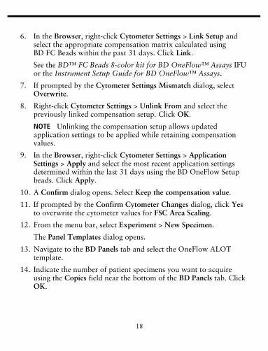
6. In the Browser, right-click Cytometer Settings > Link Setup and select the appropriate compensation matrix calculated using BD FC Beads within the past 31 days. Click Link.
See the BD™ FC Beads 8-color kit for BD OneFlow™ Assays IFU or the Instrument Setup Guide for BD OneFlow™ Assays.
7. If prompted by the Cytometer Settings Mismatch dialog, select Overwrite.
8. Right-click Cytometer Settings > Unlink From and select the previously linked compensation setup. Click OK.
NOTE Unlinking the compensation setup allows updated application settings to be applied while retaining compensation values.
9. In the Browser, right-click Cytometer Settings > Application Settings > Apply and select the most recent application settings determined within the last 31 days using the BD OneFlow Setup beads. Click Apply.
10. A Confirm dialog opens. Select Keep the compensation value.
11. If prompted by the Confirm Cytometer Changes dialog, click Yes to overwrite the cytometer values for FSC Area Scaling.
12. From the menu bar, select Experiment > New Specimen.
The Panel Templates dialog opens.
13. Navigate to the BD Panels tab and select the OneFlow ALOT template.
14. Indicate the number of patient specimens you want to acquire using the Copies field near the bottom of the BD Panels tab. Click OK.
19
15. Rename each specimen, for example, with the appropriate patient ID in front of the specimen name.
NOTE If you have to re-run a particular patient sample, set the current tube pointer to the tube you wish to re-run. Click Next Tube in the Acquisition Dashboard to create another tube for that patient. Do not select Experiment > New Tube from the menu bar or use the New Tube icon from the Browser menu bar to create the additional tube to be acquired because the labels and barcode fields will not be populated.
NOTE If you want to acquire additional patient samples in the experiment, repeat steps 12–15 to add new specimens. Two Confirm dialogs will open asking if you want to create another ALOT acquisition worksheet or another ALOT analysis worksheet. Click Cancel in each dialog.
16. From the menu bar, select Experiment > Experiment Layout and navigate to the Keywords tab.
17. Highlight the Product ID keyword for the appropriate tube, and scan the barcode on the BD OneFlow ALOT (C) tube label.
NOTE If you cannot scan the barcode on the tube label, see Troubleshooting.
18. Manually add the appropriate information to the remaining keywords, as needed.
19. Click OK to close the Experiment Layout.
Acquiring the Stained Sample
1. In the Browser, expand the appropriate specimen and set the current tube pointer to that tube.
2. Select the BD OneFlow ALOT Acquisition worksheet tab.
3. Vortex the stained tube 3–5 seconds at low speed.
20
4. Install the tube on the cytometer. Adjust the flow rate to Medium in the Acquisition Dashboard. Click Acquire Data.
5. Verify that the population is on scale and adjust the gate in the first plot of the ALOT acquisition worksheet to exclude debris, if needed.
6. Click Record Data in the Acquisition Dashboard to collect total events.
NOTE The template automatically collects 100,000 total events. Use the menu in the Acquisition Dashboard to select a different number of events to acquire, if needed. Collecting total events from 3 × 10⁴ to 2 × 10⁶ stained cells gives equivalent results.
7. Inspect the dot plots on the ALOT acquisition worksheet, and adjust the gates as needed.
Some of the dot plots might look different from those in other experiments. The initial FSC-A vs SSC-A dot plot to identify cells and eliminate debris may appear compressed. This is a consequence of the target values used to create the application settings. The values are specified by the EuroFlow Consortium.
NOTE Enlarge the dot plots while adjusting the gates so you can more readily see the populations of interest.
The FSC-A vs SSC-A dot plot is used to identify cells.
The CD45 V500-A vs SSC-A dot plot from the Cells population is used to identify leukocytes.
The CD45 V500-A vs SSC-A dot plot from the Leukocytes population is used to identify the CD45neg/dim population.
The remaining dot plots do not contain gates and are included to assess staining of the CD45neg/dim population with all of the
21
antibodies, therefore serving as an internal quality control for the tube.
NOTE See the BD OneFlow™ Application Guide for Acute Leukemias for examples of the dot plots showing populations of normal cells in the ALOT acquisition worksheet.
8. Continue until all of the tubes have been acquired.
9. From the menu bar, select File > Export > Experiments, and select the Directory Export option. Click OK.
Analyzing the Data Using BD FACSDiva Software
1. From the menu bar, select File > Import > Experiments.
2. Select the experiment that you want to analyze. Click Import.
The experiment with the associated acquisition and analysis worksheets opens.
3. Select the BD OneFlow ALOT Analysis worksheet tab.
4. Inspect the dot plots on page 1 of the ALOT analysis worksheet, and adjust the gates as needed.
Some of the dot plots might look different from those in other experiments. The initial FSC-A vs SSC-A dot plot to identify cells and eliminate debris may appear compressed. This is a consequence of the target values used to create the application settings. The values are specified by the EuroFlow Consortium.
NOTE Enlarge the dot plots while adjusting the gates so you can more readily see the populations of interest.
The first three dot plots on page 1 of the ALOT analysis worksheet identify cells, FSC singlets, and SSC singlets. Debris and doublets are excluded by adjusting the gates.
Examine the Leukocytes population in the CD45 V500-A vs SSC-A dot plot from the SSC Singlets gate.
22
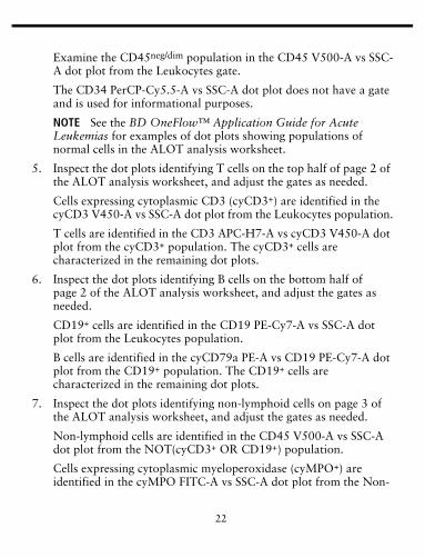
Examine the CD45neg/dim population in the CD45 V500-A vs SSC-A dot plot from the Leukocytes gate.
The CD34 PerCP-Cy5.5-A vs SSC-A dot plot does not have a gate and is used for informational purposes.
NOTE See the BD OneFlow™ Application Guide for Acute Leukemias for examples of dot plots showing populations of normal cells in the ALOT analysis worksheet.
5. Inspect the dot plots identifying T cells on the top half of page 2 of the ALOT analysis worksheet, and adjust the gates as needed.
Cells expressing cytoplasmic CD3 (cyCD3+) are identified in the cyCD3 V450-A vs SSC-A dot plot from the Leukocytes population.
T cells are identified in the CD3 APC-H7-A vs cyCD3 V450-A dot plot from the cyCD3+ population. The cyCD3+ cells are characterized in the remaining dot plots.
6. Inspect the dot plots identifying B cells on the bottom half of page 2 of the ALOT analysis worksheet, and adjust the gates as needed.
CD19+ cells are identified in the CD19 PE-Cy7-A vs SSC-A dot plot from the Leukocytes population.
B cells are identified in the cyCD79a PE-A vs CD19 PE-Cy7-A dot plot from the CD19+ population. The CD19+ cells are characterized in the remaining dot plots.
7. Inspect the dot plots identifying non-lymphoid cells on page 3 of the ALOT analysis worksheet, and adjust the gates as needed.
Non-lymphoid cells are identified in the CD45 V500-A vs SSC-A dot plot from the NOT(cyCD3+ OR CD19+) population.
Cells expressing cytoplasmic myeloperoxidase (cyMPO+) are identified in the cyMPO FITC-A vs SSC-A dot plot from the Non-
23
Lymphoid population. The non-lymphoid cells are characterized in the remaining dot plots.
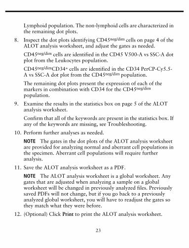
8. Inspect the dot plots identifying CD45neg/dim cells on page 4 of the ALOT analysis worksheet, and adjust the gates as needed.
CD45neg/dim cells are identified in the CD45 V500-A vs SSC-A dot plot from the Leukocytes population.
CD45neg/dimCD34+ cells are identified in the CD34 PerCP-Cy5.5-A vs SSC-A dot plot from the CD45neg/dim population.
The remaining dot plots present the expression of each of the markers in combination with CD34 for the CD45neg/dim population.
9. Examine the results in the statistics box on page 5 of the ALOT analysis worksheet.
Confirm that all of the keywords are present in the statistics box. If any of the keywords are missing, see Troubleshooting.
10. Perform further analyses as needed.
NOTE The gates in the dot plots of the ALOT analysis worksheet are provided for analyzing normal and aberrant cell populations in the specimen. Aberrant cell populations will require further analysis.
11. Save the ALOT analysis worksheet as a PDF.
NOTE The ALOT analysis worksheet is a global worksheet. Any gates that are adjusted when analyzing a sample on a global worksheet will be changed in previously analyzed files. Previously saved PDFs will not change, but if you go back to a previously analyzed global worksheet, you will have to readjust the gates so they match what they were before.
12. (Optional) Click Print to print the ALOT analysis worksheet.
24
13. Analyze the next sample.
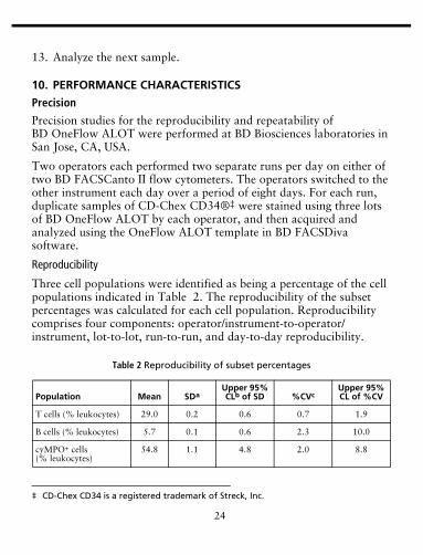
10. PERFORMANCE CHARACTERISTICS
Precision
Precision studies for the reproducibility and repeatability of BD OneFlow ALOT were performed at BD Biosciences laboratories in San Jose, CA, USA.
Two operators each performed two separate runs per day on either of two BD FACSCanto II flow cytometers. The operators switched to the other instrument each day over a period of eight days. For each run, duplicate samples of CD-Chex CD34®‡ were stained using three lots of BD OneFlow ALOT by each operator, and then acquired and analyzed using the OneFlow ALOT template in BD FACSDiva software.
Reproducibility
Three cell populations were identified as being a percentage of the cell populations indicated in Table 2. The reproducibility of the subset percentages was calculated for each cell population. Reproducibility comprises four components: operator/instrument-to-operator/instrument, lot-to-lot, run-to-run, and day-to-day reproducibility.
‡ CD-Chex CD34 is a registered trademark of Streck, Inc.
Table 2 Reproducibility of subset percentages
Population Mean SDaUpper 95% CLb of SD %CVc
Upper 95% CL of %CV
T cells (% leukocytes) 29.0 0.2 0.6 0.7 1.9
B cells (% leukocytes) 5.7 0.1 0.6 2.3 10.0
cyMPO+ cells (% leukocytes)
54.8 1.1 4.8 2.0 8.8
25
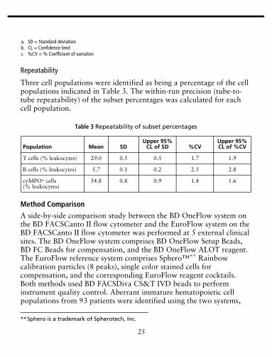
Repeatability
Three cell populations were identified as being a percentage of the cell populations indicated in Table 3. The within-run precision (tube-to-tube repeatability) of the subset percentages was calculated for each cell population.
Method Comparison
A side-by-side comparison study between the BD OneFlow system on the BD FACSCanto II flow cytometer and the EuroFlow system on the BD FACSCanto II flow cytometer was performed at 5 external clinical sites. The BD OneFlow system comprises BD OneFlow Setup Beads, BD FC Beads for compensation, and the BD OneFlow ALOT reagent. The EuroFlow reference system comprises Sphero™** Rainbow calibration particles (8 peaks), single color stained cells for compensation, and the corresponding EuroFlow reagent cocktails. Both methods used BD FACSDiva CS&T IVD beads to perform instrument quality control. Aberrant immature hematopoietic cell populations from 93 patients were identified using the two systems,
a. SD = Standard deviationb. CL = Confidence limitc. %CV = % Coefficient of variation
Table 3 Repeatability of subset percentages
Population Mean SDUpper 95%
CL of SD %CVUpper 95% CL of %CV
T cells (% leukocytes) 29.0 0.5 0.5 1.7 1.9
B cells (% leukocytes) 5.7 0.1 0.2 2.5 2.8
cyMPO+ cells (% leukocytes)
54.8 0.8 0.9 1.4 1.6
**Sphero is a trademark of Spherotech, Inc.
26
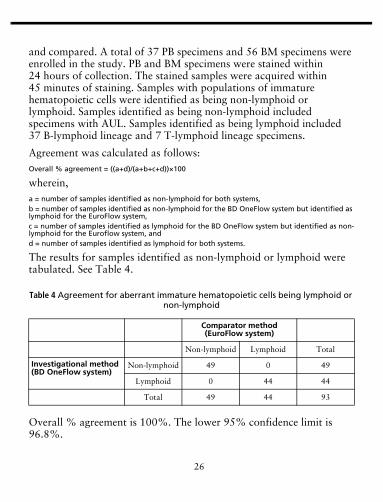
and compared. A total of 37 PB specimens and 56 BM specimens were enrolled in the study. PB and BM specimens were stained within 24 hours of collection. The stained samples were acquired within 45 minutes of staining. Samples with populations of immature hematopoietic cells were identified as being non-lymphoid or lymphoid. Samples identified as being non-lymphoid included specimens with AUL. Samples identified as being lymphoid included 37 B-lymphoid lineage and 7 T-lymphoid lineage specimens.
Agreement was calculated as follows:Overall % agreement = ((a+d)/(a+b+c+d))×100
wherein,a = number of samples identified as non-lymphoid for both systems,b = number of samples identified as non-lymphoid for the BD OneFlow system but identified as lymphoid for the EuroFlow system,c = number of samples identified as lymphoid for the BD OneFlow system but identified as non-lymphoid for the Euroflow system, andd = number of samples identified as lymphoid for both systems.
The results for samples identified as non-lymphoid or lymphoid were tabulated. See Table 4.
Overall % agreement is 100%. The lower 95% confidence limit is 96.8%.
Table 4 Agreement for aberrant immature hematopoietic cells being lymphoid or non-lymphoid
Comparator method (EuroFlow system)
Non-lymphoid Lymphoid Total
Investigational method (BD OneFlow system)
Non-lymphoid 49 0 49
Lymphoid 0 44 44
Total 49 44 93
27
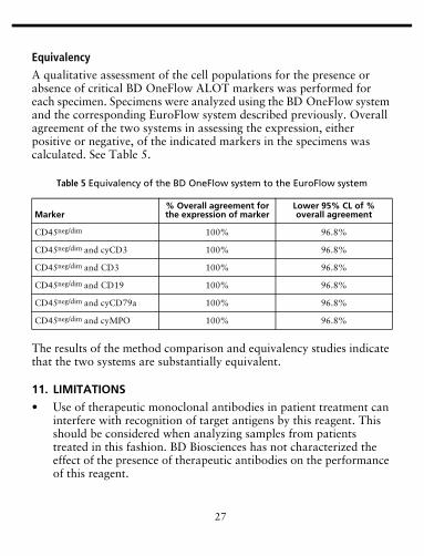
Equivalency
A qualitative assessment of the cell populations for the presence or absence of critical BD OneFlow ALOT markers was performed for each specimen. Specimens were analyzed using the BD OneFlow system and the corresponding EuroFlow system described previously. Overall agreement of the two systems in assessing the expression, either positive or negative, of the indicated markers in the specimens was calculated. See Table 5.
The results of the method comparison and equivalency studies indicate that the two systems are substantially equivalent.
11. LIMITATIONS
• Use of therapeutic monoclonal antibodies in patient treatment can interfere with recognition of target antigens by this reagent. This should be considered when analyzing samples from patients treated in this fashion. BD Biosciences has not characterized the effect of the presence of therapeutic antibodies on the performance of this reagent.
Table 5 Equivalency of the BD OneFlow system to the EuroFlow system
Marker% Overall agreement for the expression of marker
Lower 95% CL of % overall agreement
CD45neg/dim 100% 96.8%
CD45neg/dim and cyCD3 100% 96.8%
CD45neg/dim and CD3 100% 96.8%
CD45neg/dim and CD19 100% 96.8%
CD45neg/dim and cyCD79a 100% 96.8%
CD45neg/dim and cyMPO 100% 96.8%
28
• Use of this reagent for diagnostic evaluation of hematologic disorders should be performed in the context of a thorough immunophenotypic analysis including other relevant markers, for example, markers present in the EuroFlow classification panels, and other diagnostic tests.
• Use of BD OneFlow ALOT requires experience with leukemia and lymphoma immunophenotyping and classification.
• BD OneFlow ALOT has not been tested on specimens from patients with minimal residual disease (MRD).
• Avoid using potentially compromised specimens, including clotted or lipemic specimens.
WARRANTY
Unless otherwise indicated in any applicable BD general conditions of sale for non-US customers, the following warranty applies to the purchase of these products.
THE PRODUCTS SOLD HEREUNDER ARE WARRANTED ONLY TO CONFORM TO THE QUANTITY AND CONTENTS STATED ON THE LABEL OR IN THE PRODUCT LABELING AT THE TIME OF DELIVERY TO THE CUSTOMER. BD DISCLAIMS HEREBY ALL OTHER WARRANTIES, EXPRESSED OR IMPLIED, INCLUDING WARRANTIES OF MERCHANTABILITY AND FITNESS FOR ANY PARTICULAR PURPOSE AND NONINFRINGEMENT. BD’S SOLE LIABILITY IS LIMITED TO EITHER REPLACEMENT OF THE PRODUCTS OR REFUND OF THE PURCHASE PRICE. BD IS NOT LIABLE FOR PROPERTY DAMAGE OR ANY INCIDENTAL OR CONSEQUENTIAL DAMAGES, INCLUDING PERSONAL INJURY, OR ECONOMIC LOSS, CAUSED BY THE PRODUCT.
29
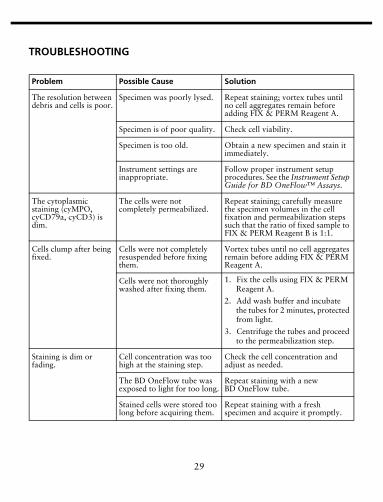
TROUBLESHOOTING
Problem Possible Cause Solution
The resolution between debris and cells is poor.
Specimen was poorly lysed. Repeat staining; vortex tubes until no cell aggregates remain before adding FIX & PERM Reagent A.
Specimen is of poor quality. Check cell viability.
Specimen is too old. Obtain a new specimen and stain it immediately.
Instrument settings are inappropriate.
Follow proper instrument setup procedures. See the Instrument Setup Guide for BD OneFlow™ Assays.
The cytoplasmic staining (cyMPO, cyCD79a, cyCD3) is dim.
The cells were not completely permeabilized.
Repeat staining; carefully measure the specimen volumes in the cell fixation and permeabilization steps such that the ratio of fixed sample to FIX & PERM Reagent B is 1:1.
Cells clump after being fixed.
Cells were not completely resuspended before fixing them.
Vortex tubes until no cell aggregates remain before adding FIX & PERM Reagent A.
Cells were not thoroughly washed after fixing them.
1. Fix the cells using FIX & PERM Reagent A.
2. Add wash buffer and incubate the tubes for 2 minutes, protected from light.
3. Centrifuge the tubes and proceed to the permeabilization step.
Staining is dim or fading.
Cell concentration was too high at the staining step.
Check the cell concentration and adjust as needed.
The BD OneFlow tube was exposed to light for too long.
Repeat staining with a new BD OneFlow tube.
Stained cells were stored too long before acquiring them.
Repeat staining with a fresh specimen and acquire it promptly.
30
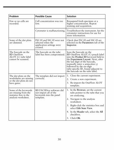
Few or no cells are recorded.
Cell concentration was too low.
Resuspend fresh specimen at a higher concentration. Repeat staining and acquisition.
Cytometer is malfunctioning. Troubleshoot the instrument. See the cytometer instructions for use for more information.
Some of the dot plots are dimmed.
FSC-H and SSC-H were not selected when the application settings were created.
Check that FSC-H and SSC-H are selected on the Parameters tab of the Inspector.
The barcode on the BD OneFlow ALOT (C) tube label cannot be scanned.
The barcode on the tube label has been compromised.
Scan the barcode on the BD OneFlow ALOT (C) pouch label into the Product ID keyword field in the Experiment Layout. Next, after the last digit of the barcode, manually enter a semicolon (;) followed by the six-digit tube-specific ID, found adjacent to the barcode on the tube label.
The dot plots on the worksheets are missing or the dot plots do not have gates.
The template did not import correctly.
1. Close the current experiment.
2. Create a new experiment.
3. Re-import the OneFlow ALOT template.
Some of the keywords are missing from the statistics box in the analysis worksheet.
BD FACSDiva software did not import all of the keywords into the panel template.
1. In the Browser, set the current tube pointer to the tube that you are analyzing.
2. Navigate to the analysis worksheet.
3. Right-click the statistics box and select Edit Stats View.
4. In the Header tab, select the All checkbox.
5. Click OK.
Problem Possible Cause Solution
31
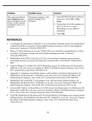
REFERENCES1 van Dongen JJ, Lhermitte L, Böttcher S, et al. EuroFlow antibody panels for standardized
n-dimensional flow cytometric immunophenotyping of normal, reactive and malignant leukocytes. Leukemia. 2012;26:1908-1975.
2 Kalina T, Flores-Montero J, van der Velden VH, et al. EuroFlow standardization of flow cytometer instrument settings and immunophenotyping protocols. Leukemia. 2012;26:1986-2010.
3 Pulford KA, Erber WN, Crick JA, et al. Use of monoclonal antibody against human neutrophil elastase in normal and leukaemic myeloid cells. J Clin Pathol. 1988;41:853-860.
4 Engel P, Wagner N, Tedder TF. CD79 Workshop report. In: Schlossman SF, Boumsell L, Gilks W, et al, eds. Leucocyte Typing V: White Cell Differentiation Antigens. New York, NY: Oxford University Press; 1995:667-670.
5 Appendix C. Summary of antibody names, code numbers, and donor laboratories. In: McMichael AJ, Beverley PC, Cobbold S, et al, eds. Leucocyte Typing III: White Cell Differentiation Antigens. New York, NY: Oxford University Press; 1987:988-993.
6 McMichael AJ, Gotch FM. T-cell antigens: new and previously defined clusters. In: McMichael AJ, Beverley PCL, Cobbold S, et al, eds. Leucocyte Typing III: White Cell Differentiation Antigens. New York, NY: Oxford University Press; 1987:31-62.
7 Greaves MF, Titley I, Colman SM, et al. CD34 cluster workshop report. In: Schlossman SF, Boumsell L, Gilks W, et al, eds. Leucocyte Typing V: White Cell Differentiation Antigens. Vol 1. New York, NY: Oxford University Press; 1995:840-846.
8 Ling NR, Maclennan ICM, Mason DY. B-cell and plasma cell antigens: new and previously defined clusters. In: McMichael AJ, Beverley PC, Cobbold S, et al, eds. Leucocyte Typing III: White Cell Differentiation Antigens. New York, NY: Oxford University Press; 1987:302-335.
The statement For in vitro diagnostic use does not appear in the footer of the analysis worksheet when it is printed.
The paper margins in the printer settings were changed.
1. From the BD FACSDiva software menu bar, select File > Page Setup.
2. Ensure that all of the margins are set to 2.54 cm or 1 inch, depending on your default standards.
3. Click OK.
Problem Possible Cause Solution
32
9 Nadler LM. B Cell/Leukemia Panel Workshop: Summary and Comments. In: Reinherz EL, Haynes BF, Nadler LM, Bernstein ID, eds. Leukocyte Typing II: Human B Lymphocytes. Vol 2. New York, NY: Springer-Verlag; 1986:3-43.
10 Reiter C. Cluster report: CD7. In: Knapp W, Dörken B, Gilks WR, et al, eds. Leucocyte Typing IV: White Cell Differentiation Antigens. New York, NY: Oxford University Press; 1989:341-342.
11 Haynes BF. Summary of T-cell studies performed during the Second International Workshop and Conference on Human Leukocyte Differentiation Antigens. In: Reinherz EL, Haynes BF, Nadler LM, Bernstein ID, eds. Leukocyte Typing II: Human T Lymphocytes. Vol 1. New York, NY: Springer-Verlag; 1986:3-30.
12 Knowles RW. Immunochemical analysis of the T-cell–specific antigens. In: Reinherz EL, Haynes BF, Nadler LM, Bernstein ID, eds. Leukocyte Typing II: Human T Lymphocytes. Vol 1. New York, NY: Springer-Verlag; 1986:259-288.
13 Cobbold SP, Hale G, Waldmann H. Non-lineage, LFA-1 family, and leucocyte common antigens: new and previously defined clusters. In: McMichael AJ, Beverley PC, Cobbold S, et al, eds. Leucocyte Typing III: White Cell Differentiation Antigens. New York, NY: Oxford University Press; 1987:788-803.
14 Rothe G, Schmitz G. Consensus protocol for the flow cytometric immunophenotyping of hematopoietic malignancies. Leukemia. 1996;10:877-895.
15 Stelzer GT, Marti G, Hurley A, McCoy P, Lovett EJ, Schwartz A. US-Canadian consensus recommendations on the immunophenotypic analysis of hematologic neoplasia by flow cytometry: standardization and validation of laboratory procedures. Cytometry. 1997;30:214-230.
16 Davis BH, Dasgupta A, Kussick S, Han JY, Estrellado A; on behalf of ICSH/ICCS working group. Validation of cell-based fluorescence assays: practice guidelines from the ICSH and ICCS - Part II - Preanalytical issues. Cytometry Part B. 2013;84B:286-290.
17 Nicholson JKA, Green TA. Selection of anticoagulants for lymphocyte immunophenotyping: effect of specimen age on results. J Immunol Methods. 1993;165:31-35.
18 Paxton H, Bendele T. Effect of time, temperature, and anticoagulant on flow cytometry and hematological values. Ann NY Acad Sci. 1993;677:440-443.
19 Stetler-Stevenson M, Ahmad E, Barnett D et al. Clinical Flow Cytometric Analysis of Neoplastic Hematolymphoid Cells; Approved Guideline—Second Edition. Wayne, PA: Clinical and Laboratory Standards Institute; 2005. CLSI document H43-A2.
20 Stetler-Stevenson M, Greig B, Yuan C. Flow cytometric specimen collection, processing, and reporting. In: Kottke-Marchant K, Davis BH, eds. Laboratory Hematology Practice. First Edition. Hoboken, NJ; Wiley-Blackwell Inc.; 2012:105-114.
21 Protection of Laboratory Workers from Occupationally Acquired Infections; Approved Guideline—Fourth Edition. Wayne, PA: Clinical and Laboratory Standards Institute; 2014. CLSI document M29-A4.
33
22 Centers for Disease Control. Perspectives in disease prevention and health promotion update: universal precautions for prevention of transmission of human immunodeficiency virus, hepatitis B virus, and other bloodborne pathogens in health-care settings. MMWR. 1988;37:377-388.


![l>lf·· E ·B; -I,:,C-·-1·1V · cat. no.i bd lj.657 bd lj.6]5 bd 4630 bd 4·627 bd 4628 bd 4886 bd 4546 bd 4·545 bd 4544 bd 4542 bd lj,588 bd lj.593 bd 0102 bd 4636 bd 4632 bd](https://static.documents.pub/doc/80x56/5f7c69bb7d840d18665ab1e6/llf-e-b-ic-11v-cat-noi-bd-lj657-bd-lj65-bd-4630-bd-4627-bd-4628-bd.jpg)


