Inhibition of Protein Kinase M� Disrupts the Stable SpatialDischarge of Hippocampal Place Cells in a FamiliarEnvironment
Jeremy M. Barry,1,2 Bruno Rivard,1,2 Steven E. Fox,1,2 Andre A. Fenton,1,2,3 Todd C. Sacktor,1,2 and Robert U. Muller1,2
1Department of Physiology and Pharmacology, State University of New York, Downstate Medical Center, Brooklyn, New York 11203, 2The Robert F.Furchgott Center for Neural & Behavioral Science, State University of New York, Downstate Medical Center, Brooklyn, New York 11203, and 3Center forNeural Science, New York University, New York, New York 10003
It is widely held that spatial computations in the rodent hippocampus require the location-specific discharge of place cells that togetherform a stable cognitive map used to solve and perform spatial tasks. It is not known, however, if map stability requires persistenthippocampal synaptic strength changes that are vulnerable to blockade of protein kinase M� (PKM�) phosphorylation activity, a manip-ulation that reverses hippocampal LTP and disrupts multiple forms of long-term memory. Here we report that acute intrahippocampalinhibition of PKM� disrupts place cell activity in a familiar environment, where the map is expected to be stable. After this disruption,new, stable spatial firing patterns can later form, but the new and original maps are unrelated even though the rat is exposed to a constantenvironment. We therefore propose that the previously demonstrated erasure of stored spatial memory and the disruption of place cellfiring are parallel effects of PKM� blockade. We similarly propose that the known sparing of new spatial memory formation depends onthe sparing of new map formation. On these bases, we argue that the loss of the map used to perform a practiced spatial task leads tobehavioral performance deficits, and that synaptic plasticity maintained by PKM�, which stabilizes the map, is essential for the properexpression of spatial memory.
IntroductionThe ensemble location-specific discharge of hippocampal placecells is believed to reflect the operation of a spatial map of theenvironment that enables rodents to solve complex spatial prob-lems (O’Keefe and Nadel, 1978); interference with location-specific firing, however induced, leads to parallel performancedeficits in spatial tasks (McHugh et al., 1996; Barnes et al., 1997;Cho et al., 1998; Kentros et al., 1998, 2004; Ekstrom et al., 2001;Liu et al., 2003; Nakazawa et al., 2004). It is also agreed that themap is environment-specific; when the same cells are recorded insufficiently different circumstances, their individual firing fieldsappear unrelated. Moreover, because a particular map is reacti-vated whenever the animal returns to a particular environment, itis inferred that map stability requires the presence of a memorymechanism (Muller et al., 1987).
In support of this inference, it is known that geneticallyaltering biochemical pathways required for normal hip-
pocampal LTP produces unstable place cells (McHugh et al.,1996; Rotenberg et al., 1996, 2000; Cho et al., 1998; Yan et al.,2002; Taverna et al., 2005). Antagonizing NMDAR transmis-sion does not affect established place cells or remapping in anovel environment but prevents stabilization of the new placecells (Kentros et al., 1998). Furthermore, inducing LTP causespartial remapping of place cell firing fields (Dragoi et al.,2003). Thus, there is clear evidence that NMDAR-dependentLTP plays a role in place cell activity.
Recent evidence also suggests that crucial aspects of memorydepend on the functional state of the atypical PKC isoform pro-tein kinase M� (PKM�) (Sacktor, 2011). Thus, reducing PKM�activity with the pseudosubstrate zeta inhibitory peptide (ZIP) orother agents erases both LTP induced by high-frequency stimu-lation and information acquired during experience (Pastalkovaet al., 2006; Li et al., 2010; Madronal et al., 2010; Migues et al.,2010; Sacktor, 2011). PKM� inhibition does not affect baselinetransmission so that normal signaling is possible (Ling et al.,2002; Serrano et al., 2005; Pastalkova et al., 2006; Madronal et al.,2010). Furthermore, this inhibition appears to erase establishedmemories without damaging the ability to later acquire newmemories (Pastalkova et al., 2006; Shema et al., 2007, 2011;Madronal et al., 2010; Migues et al., 2010; von Kraus et al., 2010;Sacktor, 2011). Specifically, blocking hippocampal PKM� activ-ity has the stated effects on synaptic activity and erases severalspatial memory forms dependent on normal hippocampal func-tion (Pastalkova et al., 2006; Serrano et al., 2008). We thereforeinfer that prior LTP-like synaptic plasticity within the hippocampus
Received Jan. 22, 2012; revised July 26, 2012; accepted July 29, 2012.Author contributions: J.M.B., S.E.F., A.A.F., and R.U.M. designed research; J.M.B and R.U.M. performed research;
J.M.B., B.R., and R.U.M. contributed unpublished reagents/analytic tools; J.M.B., S.E.F., A.A.F., and R.U.M. analyzeddata; J.M.B., A.A.F., T.C.S., and R.U.M. wrote the paper.
This work was funded by National Institutes of Health Grants MH057068, MH053576 and DA034970 to T.C.S. andNS20686 to R.U.M. We thank Dr. John Kubie for discussions concerning experimental design.
The authors declare no competing financial interests.Correspondence should be addressed to Dr. Robert U. Muller, Department of Physiology and Pharmacology,
Downstate Medical Center, 450 Clarkson Ave., Brooklyn, NY 11203. E-mail: [email protected]:10.1523/JNEUROSCI.0319-12.2012
The Journal of Neuroscience, October 3, 2012 • 32(40):13753–13762 • 13753
maintains organized place cell firing fieldsand predict that hippocampal ZIP injec-tions should modify or abolish place cell fir-ing fields in a familiar environment butleave intact the ability to subsequently gen-erate new, stable fields; our goal is to testthese predictions.
Materials and MethodsTrainingThe subjects were eight male Long–Evans rats(300 –350 g) handled in accordance with theUnited States Public Health Service guidelinesfor treatment of laboratory animals. They wereindividually housed using a 12 h light/dark cy-cle. The first 5 training days consisted of (1)daily handling, (2) food deprivation reducingweight to 85% ad libitum, and (3) 20 min/d of‘‘pellet chasing’’ learning to forage for 25 mg ofsugar pellets scattered �2 per minute into anarena. The arena was a white 76 � 76 cm box 66cm high with a 45 cm wide, 60 cm high black-and-white striped card on one wall. The boxwas centered in a 2.5 � 2.5 m black soundproofroom on a renewable piece of gray paper. Cen-tered 2 m overhead was a black disk with fourlights along the edge separated by 90°, a videocamera in the middle, and a pellet feeder near the middle.
SurgeryThe rat was anesthetized (Nembutal, 50 mg/kg i.p.) and placed in astereotaxic frame. The skull was exposed and four screws inserted, twojust anterior to the left and right edges of bregma, and two above left andright cerebella. Grounding was via the right cerebellum screw. Using thecoordinates of Paxinos and Watson (2007), 0.8 mm holes were drilled at�3.5 mm anteroposterior and �3.7 mm mediolateral above the left andright dorsal hippocampuses. These holes allowed 22 gauge injectionguides to reach 1.8 mm dorsoventral (DV). The guides were kept patentwith 30 gauge wires that reached 3.6 mm DV. A 2.6 mm hole 0.5 mmmedial to the left guide hole was made for an implant, which was loweredso its tetrode tips were 2.0 mm below the brain surface. The skull holeswere protected with sterile Vaseline. The implant was fixed via the skullscrews with Grip Cement. The wound was sutured and topical antibioticapplied. The interval between surgery and cell screening was 1 week.
RecordingsRecordings were made with an eight tetrode implant (Fig. 1). Each 25 �mnichrome wire was gold plated, lowering its resistance to �100 k�. Te-trode signals were amplified �1 at the rat’s head and led via a cable to acommutator. One tetrode was left in neocortex as a reference. Signalswere bandpass filtered 300 – 6000 Hz and digitized at 30 kHz with a 12 bitA/D converter. A spike event was stored whenever the voltage on a te-trode wire exceeded an 85–100 �V threshold.
Rat trackingTwo red LEDs 3 mm apart were attached to the headstage. Video fromthe overhead camera was led to a frame grabber (DT3120, Data Transla-tion) and digitized at 30 Hz. Each frame was scanned for the LEDs whoselocations were stored as an XY time series at 3.0 � 3.0 cm resolution.
Cell screeningCell-screening sessions lasted 10 – 45 min. If there were �10 wellisolated cells, inactive tetrodes were advanced �60 �m and the ratspent at least 2 h in its cage. Once �10 place cells were isolated, a 16min preinjection, baseline session was recorded. As shown below andin Results, we could identify the same cells in the preinjection sessionand in all postinjection sessions during the main experimental day. Insome cases, we could still identify these cells on subsequent days; inothers, the tetrodes had apparently moved so that waveform match-
ing was not possible. In these cases, additional recordings were madeon the new waveforms, without moving the tetrodes.
InjectionsThree types of injections were made, two as controls and the third to testthe effects of ZIP on place cell activity in a familiar environment, as acell-level equivalent to the ability of this inhibitor of PKM� to reversebehavioral memory. Muscimol was used to directly demonstrate that asubstance with known pharmacological actions could produce predict-able effects after injections near single-cell recording sites; we show inResults that muscimol has the expected effect of reducing or silencing thedischarge of identified place cells. Saline injections enabled us to showthat muscimol and ZIP each have major but distinct effects on place cellactivity.
Muscimol. The GABAA receptor agonist muscimol (5-aminomethyl-3-hydroxyisoxazole; Sigma-Aldrich) was infused into the left hippocam-pus (n � 3) ipsilateral to the implant; the concentration was 0.5 �g/�l inPBS, pH 7.4. The injection (1.0 �l at 0.33 �l/min) was through a 30 gaugetube in the guide. The tube was withdrawn 3 min later. After 5 min, therat was returned to the box for 45– 60 min of recording. Recordings thenext day tested for recovery from muscimol.
Saline. Tris-buffered saline, pH 7.2, was injected (1.0 �l at 0.33 �l/min) bilaterally into the dorsal hippocampuses (n � 4). The injectionswere separated by �5 min; the first was contralateral to the tetrodes.Next, the rat was returned to its home cage for 2 h, after which three 16min recording sessions were done, separated by 8 min. After 2 morehours in its cage, the rat underwent two more sessions, separated by 8min.
ZIP. Bilateral myristolated ZIP injections (10 nmol/�l, pH 7.2, in Tris-buffered saline, Anaspec) were made (n � 4) using the same protocol asthat for saline injections.
Waveform clustering and stabilityWaveform clustering was done with Off-line Sorter (Plexon). The crite-ria used to establish clusters in the preinjection (baseline) session wereapplied to all subsequent sessions recorded from the same animal suchthat an identified single unit was recognized across multiple sessions if itappeared on the same tetrode and had the same cluster boundaries thatdistinguished it as a single unit. Only unitary waveforms that appearedconstant across sessions were studied. The across-session stability of thiswaveform subset was quantified as described below. The clusters werefurther culled by inspecting a subset of 30 superimposed waveforms
Figure 1. The recording plus injection implant. a, The entire implant. Visible below are the tetrodes, injection guides (blue), andcannula (yellow). b, Enlargement of the tip. c, Bottom of the tip showing the relation of the cannula to the near (N) and far (F)tetrodes.
13754 • J. Neurosci., October 3, 2012 • 32(40):13753–13762 Barry et al. • Place Cell Instability Caused by PKM� Blockade
randomly selected from each of the sessions recorded for a given rat. Ifthe candidate cross-session matches were not agreed to by three indepen-dent observers, the cluster was dropped from further analysis.
We used a correlational method to show that cross-session matchesmade in this way are overwhelmingly better than matches between ran-domly paired clusters. For a given session pair, we randomly chose, foreach session, n � 100 waveforms from the putative cluster and computedn 2 Pearson product–moment correlations; the estimated waveform sim-ilarity, S, for this single cluster was the averaged z-transform of the n 2
values. S averaged over all clusters for the session pair measures relativewaveform stability. For the same session pair, we repeated this calcula-tion, but now each cluster was matched against a different, randomlyselected (without replacement) cluster yielding a second stability esti-mate R. Random selection was done by pairing only with a cluster fromthe same tetrode, pairing with any other cluster recorded from the samerat, and pairing with another cluster recorded from any rat. Because theresults were substantially the same for each method, we report only thelast one.
For each injected substance, we compared S to R for two key sessionpairs. To show that the injection did not disturb the ability to matchwaveforms, we used the baseline session (Pre) and the first postinjectionsession (P1) and compared SPre/P1 to RPre/P1. To show longer term wave-form stability, we used P1 and the last postinjection session (P5) andcompared SP1/P5 to RP1/P5. The results, summarized in Table 1, demon-strate that matching using the clustering template is much more reliablethan random cluster matching.
Next, we did an ANOVA on SPre/P1 values to determine whether wave-forms are altered less by saline. For the main effect of substance, F(2,157) �4.49; p � 0.013. Tukey’s post hoc tests revealed that SPre/P1 is lower formuscimol ( p � 0.030) and ZIP ( p � 0.036) than for saline, but muscimoland ZIP do not differ from each other ( p � 0.962). We conclude that ourcorrelation method is sensitive enough to detect drug-induced waveformchanges compared with our estimate of variation caused by injectingvehicle. We further conclude that such drug-induced changes are insuf-ficient to cause the profound effects of ZIP on spatial firing becausesimilar-sized waveform effects produced by muscimol cause a totallydifferent pattern of effects, which is properly explained by its reversibleactivation of GABAA receptors.
Data analysisRecordings were from dorsal CA1. Cells were pyramidal cells or in-terneurons (Ranck, 1973; Fox and Ranck, 1975, 1981), but only pyrami-dal cells were analyzed.
The spatial resolution for analysis was 3.0 � 3.0 cm. Sixty-four bysixty-four firing rate arrays were constructed using standard methods(Muller et al., 1987). Rate arrays were used to (1) make colored maps tovisualize positional firing distributions, (2) quantify positional distribu-tions, and (3) compare the similarity of the firing distribution of cells insession pairs and in session halves.
In firing rate maps, the rate was zero in yellow pixels. Increasing ratesare in the following order: orange, red, green, blue, and purple. Unvisitedpixels are white. Positional firing distribution properties measures werebased mainly on the idea of a firing field. The definition of a field is aregion of �9 contiguous pixels with firing rate �0.1 spikes/s. We calcu-lated the overall rate, field rate, coherence, and field areas. The overallrate was the number of spikes fired by a cell divided by session time. Thefield rate was the number of spikes in the field divided by time in the field.
To determine coherence, we used nearest-neighbor 2-D rate autocorre-lation (Muller and Kubie, 1989). Parallel lists were constructed for thefiring rate in each pixel and the average in the eight nearest neighbors.Next, the product–moment correlation, r, between the two lists was cal-culated. Coherence is the z-transform of r: z � 0.5 ln [(1 r)/(1 � r)].Pyramidal cells with coherence �0.3 are taken by trained observers to beplace cells. The field area was the fractional apparatus area occupied bythe firing field.
A pyramidal cell was a place cell if it met the following criteria: (1) 0.1spikes/s � overall rate � 5.0 spikes/s; field rate � 1.0 spikes/s; (2) coher-ence � 0.3; (3) field area � 0.6 apparatus area.
We also calculated similarity. Positional firing stability was estimatedfrom the similarity of a cell’s spatial firing pattern in two time intervals.Similarity is the Fisher z-transform of the product–moment correlationbetween the firing rates in corresponding pixels for two intervals. Wecalculated similarities between session pairs and between first and secondsession halves.
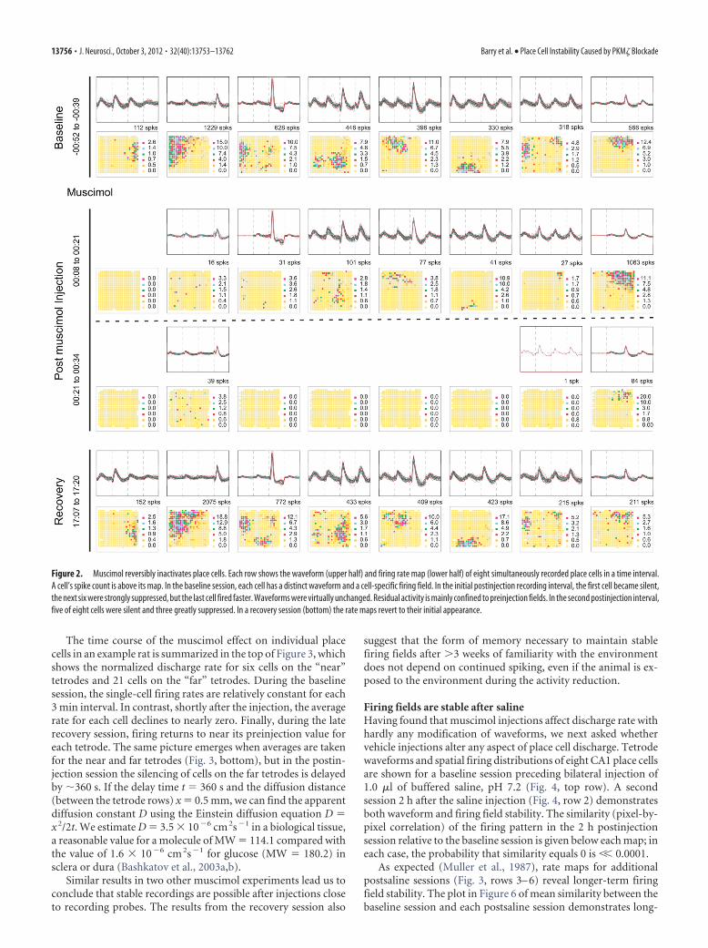
ResultsMuscimol reduces place cell firingTo examine the predictions stated in the Introduction, we firstshowed that volumetric injections of aqueous solutions can bemade in the near vicinity of recording tetrodes in freely movingrats without disturbing the ability to resolve individual wave-forms (Hafting et al., 2008). Specifically, we found that muscimolinjected ipsilateral to the tetrodes reduces or silences spiking bygating of GABAA receptors with only small changes of waveformconfiguration (see Materials and Methods). Implants of the de-sign shown in Figure 1 were placed into the dorsal hippocampusof three rats so that the injection cannula was situated 3.5 mmbelow bregma. The individually movable tetrodes were graduallylowered until spikes from at least 10 well isolated CA1 pyramidalcells were observed as the rat foraged for 25 mg of food pelletsscattered into a familiar 75 cm square chamber. A preinjectionbaseline session was followed �1 h later by injecting 1.0 �l con-taining 4.4 nmol of muscimol. Recordings were made soon afterthe injection (�10 min) to detect the drug effect and again muchlater (�10 h) to look for recovery.
As seen in Figure 2, muscimol had the expected effect on cellactivity. The properties of 8 CA1 pyramidal cells are shown in twoways for four different time intervals during the experiment. Eachtime interval is displayed as a row consisting of two panels, anelectrical recording above that shows identified waveforms for acell, and a firing rate map below that shows the spatial dischargedistribution of the corresponding cell. In the baseline session(row 1), it is seen that each cell has a distinct waveform “signa-ture” because of the disposition of the four tetrode wires relativeto the cell. Moreover, each cell has a spatially restricted “firingfield” that qualifies it as a place cell (see Materials and Methods).During the first half of the 26 min postinjection session (row 2),the place cell in the first column ceased to discharge and six ofseven of the others fired much more slowly. As expected, musci-mol decreased most firing rates but did not strongly alter wave-forms. Interestingly, the residual firing was mainly confined topreinjection firing fields, suggesting that location-specificity doesnot depend entirely on the precise rate or timing of spikes. Themuscimol effect was even greater during the second half of thepostinjection session (row 3). Now five of eight cells were com-pletely silent and the discharge of the remaining three wasstrongly reduced although waveform constancy was hardlychanged. The fixed relationship between the target cells and therecording tetrodes is corroborated by the recovery session (row4), when the spatial firing patterns return to their initial statesafter a delay of �17 h postinjection.
Table 1. t Values and probabilities for comparing waveform similarities aftermatching across two sessions according to the waveform template from thebaseline session and according to random assignment of other clusters
Substance Session pair t df Probability
Saline RPre/P1 vs SPre/P1 12.6 122 7.9E-24RP1/P5 vs SP1/P55 9.4 120 4.6E-16
Muscimol RPre/P1 vs SPre/P1 7.1 80 4.5E-10RP1/P5 vs SP1/P55 6.8 116 4.8E-10
ZIP RPre/P1 vs SPre/P1 7.0 108 2.3E-10RP1/P5 vs SP1/P55 7.7 80 3.1E-11
Barry et al. • Place Cell Instability Caused by PKM� Blockade J. Neurosci., October 3, 2012 • 32(40):13753–13762 • 13755
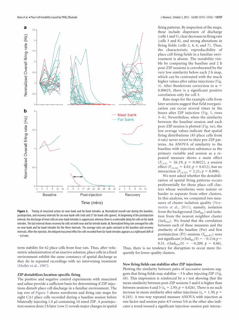
The time course of the muscimol effect on individual placecells in an example rat is summarized in the top of Figure 3, whichshows the normalized discharge rate for six cells on the “near”tetrodes and 21 cells on the “far” tetrodes. During the baselinesession, the single-cell firing rates are relatively constant for each3 min interval. In contrast, shortly after the injection, the averagerate for each cell declines to nearly zero. Finally, during the laterecovery session, firing returns to near its preinjection value foreach tetrode. The same picture emerges when averages are takenfor the near and far tetrodes (Fig. 3, bottom), but in the postin-jection session the silencing of cells on the far tetrodes is delayedby �360 s. If the delay time t � 360 s and the diffusion distance(between the tetrode rows) x � 0.5 mm, we can find the apparentdiffusion constant D using the Einstein diffusion equation D �x 2/2t. We estimate D � 3.5 � 10�6 cm 2s�1 in a biological tissue,a reasonable value for a molecule of MW � 114.1 compared withthe value of 1.6 � 10�6 cm 2s�1 for glucose (MW � 180.2) insclera or dura (Bashkatov et al., 2003a,b).
Similar results in two other muscimol experiments lead us toconclude that stable recordings are possible after injections closeto recording probes. The results from the recovery session also
suggest that the form of memory necessary to maintain stablefiring fields after �3 weeks of familiarity with the environmentdoes not depend on continued spiking, even if the animal is ex-posed to the environment during the activity reduction.
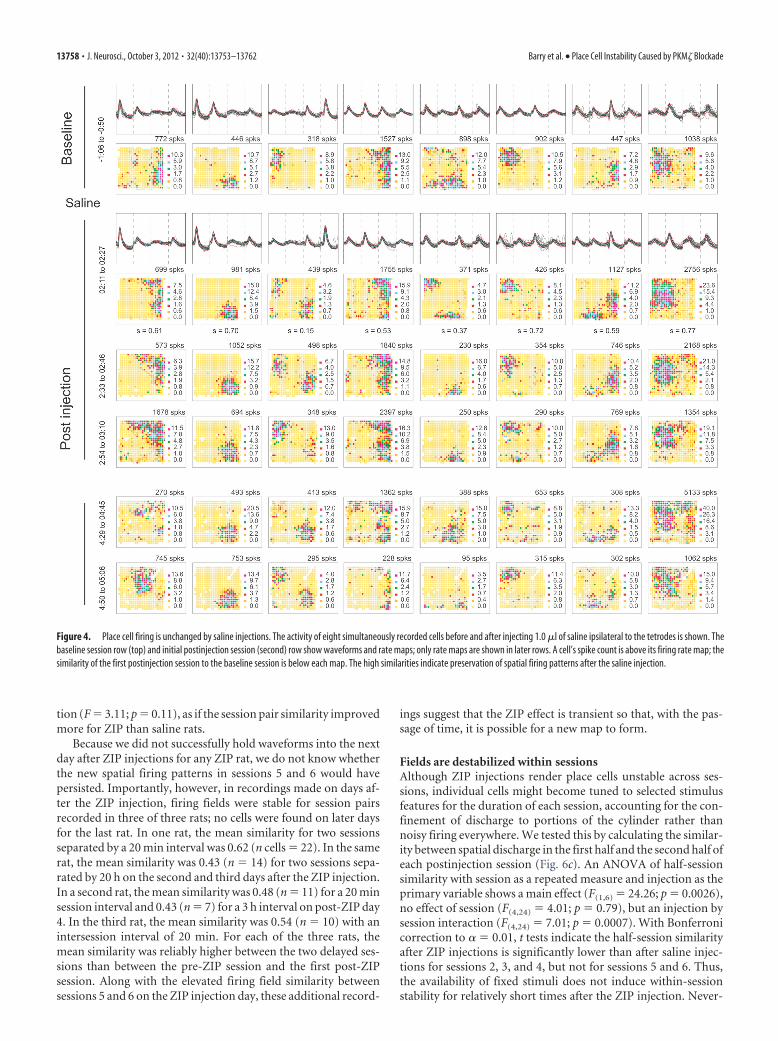
Firing fields are stable after salineHaving found that muscimol injections affect discharge rate withhardly any modification of waveforms, we next asked whethervehicle injections alter any aspect of place cell discharge. Tetrodewaveforms and spatial firing distributions of eight CA1 place cellsare shown for a baseline session preceding bilateral injection of1.0 �l of buffered saline, pH 7.2 (Fig. 4, top row). A secondsession 2 h after the saline injection (Fig. 4, row 2) demonstratesboth waveform and firing field stability. The similarity (pixel-by-pixel correlation) of the firing pattern in the 2 h postinjectionsession relative to the baseline session is given below each map; ineach case, the probability that similarity equals 0 is �� 0.0001.
As expected (Muller et al., 1987), rate maps for additionalpostsaline sessions (Fig. 3, rows 3– 6) reveal longer-term firingfield stability. The plot in Figure 6 of mean similarity between thebaseline session and each postsaline session demonstrates long-
Figure 2. Muscimol reversibly inactivates place cells. Each row shows the waveform (upper half) and firing rate map (lower half) of eight simultaneously recorded place cells in a time interval.A cell’s spike count is above its map. In the baseline session, each cell has a distinct waveform and a cell-specific firing field. In the initial postinjection recording interval, the first cell became silent,the next six were strongly suppressed, but the last cell fired faster. Waveforms were virtually unchanged. Residual activity is mainly confined to preinjection fields. In the second postinjection interval,five of eight cells were silent and three greatly suppressed. In a recovery session (bottom) the rate maps revert to their initial appearance.
13756 • J. Neurosci., October 3, 2012 • 32(40):13753–13762 Barry et al. • Place Cell Instability Caused by PKM� Blockade
term stability for 62 place cells from four rats. Thus, after volu-metric administration of an inactive solution, place cells in a fixedenvironment exhibit the same constancy of spatial discharge asthey do in repeated recordings with no intervening treatment(Muller et al., 1987).
ZIP destabilizes location-specific firingThe positive and negative control experiments with muscimoland saline provide a sufficient basis for determining if ZIP injec-tions disturb place cell discharge in a familiar environment. Thetop row of Figure 5 shows waveforms and firing rate maps foreight CA1 place cells recorded during a baseline session beforebilaterally injecting 1.0 �l containing 10 nmol ZIP. A postinjec-tion session done 2 h later (row 2) reveals major changes in spatial
firing patterns. By inspection of the maps,these include dispersion of discharge(cells 1 and 5), clear decreases in firing rate(cells 3 and 8), and strong alterations infiring fields (cells 2, 4, 6, and 7). Thus,the characteristic reproducibility ofplace cell firing fields in a familiar envi-ronment is absent. The instability visi-ble by comparing the baseline and 2 hpost-ZIP sessions is corroborated by thevery low similarity below each 2 h map,which can be contrasted with the muchhigher values after saline injections (Fig.4). After Bonferroni correction to � �0.00625, there is a significant positivecorrelation only for cell 3.
Rate maps for the example cells fromlater sessions suggest that field reorgani-zation can occur several times in thehours after ZIP injection (Fig. 5, rows3– 6). Nevertheless, when the similaritybetween the baseline session and eachpost-ZIP session is plotted (Fig. 6a), thelow average values indicate that spatialfiring distributions (83 place cells from4 rats) never revert to their pre-ZIP pat-terns. An ANOVA of similarity to thebaseline with injection substance as theprimary variable and session as a re-peated measure shows a main effect(F(1,6) � 26.19; p � 0.0022), a sessioneffect (F(4,24) � 4.01; p � 0.012), but nointeraction (F(4,24) � 2.21; p � 0.098).
We next asked whether the destabili-zation of spatial firing patterns occurspreferentially for those place cell clus-ters whose waveforms were noisier orharder to separate from other clusters.In this analysis, we computed two mea-sures of cluster isolation quality (Ney-motin et al., 2011), namely, isolationfrom the background (IsolBG) and isola-tion from the nearest neighbor cluster(IsolNN). We found that the correlationbetween each of these measures and thesimilarity of the baseline (Pre) and firstpostinjection (P1) sessions (Spre/P1) werenot significant [r(IsolBG/S) � �0.114; p �0.31. r(IsolNN/S) � �0.209; p � 0.06].
Thus, there is no tendency for disruption to occur more fre-quently for lower-quality clusters.
New firing fields can stabilize after ZIP injectionsPlotting the similarity between pairs of successive sessions sug-gests that firing fields may stabilize �5 h after injecting ZIP (Fig.6b). This impression is reinforced by a t test showing that themean similarity between post-ZIP sessions 5 and 6 is higher thanbetween sessions 4 and 5 (t6 � 2.95; p � 0.026). There is no suchincrease in mean similarity after saline injections (t6 � 1.30; p �0.245). A two-way repeated measure ANOVA with injection asone factor and session pairs 4/5 versus 5/6 as the other also indi-cates a trend toward a significant injection–session pair interac-
Figure 3. Timing of muscimol action on near-bank and far-bank tetrodes. a, Normalized overall rate during the baseline,postinjection, and recovery intervals for six near-bank cells (red) and 21 far-bank cells (green). At beginning of the postinjectioninterval, the discharge of most cells on near-bank tetrodes is suppressed, whereas there is a noticeable delay for cells on far-banktetrodes. The last interval shows recovery for cells on both near and far tetrode banks. b, Normalized firing rate averaged for cellson near-bank and far-bank tetrodes for the three intervals. The average rates are quite constant in the baseline and recoveryintervals. After the injection, the delayed muscimol effect for cells recorded from far-bank tetrodes appears as a rightward shift of�6.0 min.
Barry et al. • Place Cell Instability Caused by PKM� Blockade J. Neurosci., October 3, 2012 • 32(40):13753–13762 • 13757
tion (F � 3.11; p � 0.11), as if the session pair similarity improvedmore for ZIP than saline rats.
Because we did not successfully hold waveforms into the nextday after ZIP injections for any ZIP rat, we do not know whetherthe new spatial firing patterns in sessions 5 and 6 would havepersisted. Importantly, however, in recordings made on days af-ter the ZIP injection, firing fields were stable for session pairsrecorded in three of three rats; no cells were found on later daysfor the last rat. In one rat, the mean similarity for two sessionsseparated by a 20 min interval was 0.62 (n cells � 22). In the samerat, the mean similarity was 0.43 (n � 14) for two sessions sepa-rated by 20 h on the second and third days after the ZIP injection.In a second rat, the mean similarity was 0.48 (n � 11) for a 20 minsession interval and 0.43 (n � 7) for a 3 h interval on post-ZIP day4. In the third rat, the mean similarity was 0.54 (n � 10) with anintersession interval of 20 min. For each of the three rats, themean similarity was reliably higher between the two delayed ses-sions than between the pre-ZIP session and the first post-ZIPsession. Along with the elevated firing field similarity betweensessions 5 and 6 on the ZIP injection day, these additional record-
ings suggest that the ZIP effect is transient so that, with the pas-sage of time, it is possible for a new map to form.
Fields are destabilized within sessionsAlthough ZIP injections render place cells unstable across ses-sions, individual cells might become tuned to selected stimulusfeatures for the duration of each session, accounting for the con-finement of discharge to portions of the cylinder rather thannoisy firing everywhere. We tested this by calculating the similar-ity between spatial discharge in the first half and the second half ofeach postinjection session (Fig. 6c). An ANOVA of half-sessionsimilarity with session as a repeated measure and injection as theprimary variable shows a main effect (F(1,6) � 24.26; p � 0.0026),no effect of session (F(4,24) � 4.01; p � 0.79), but an injection bysession interaction (F(4,24) � 7.01; p � 0.0007). With Bonferronicorrection to � � 0.01, t tests indicate the half-session similarityafter ZIP injections is significantly lower than after saline injec-tions for sessions 2, 3, and 4, but not for sessions 5 and 6. Thus,the availability of fixed stimuli does not induce within-sessionstability for relatively short times after the ZIP injection. Never-
Figure 4. Place cell firing is unchanged by saline injections. The activity of eight simultaneously recorded cells before and after injecting 1.0 �l of saline ipsilateral to the tetrodes is shown. Thebaseline session row (top) and initial postinjection session (second) row show waveforms and rate maps; only rate maps are shown in later rows. A cell’s spike count is above its firing rate map; thesimilarity of the first postinjection session to the baseline session is below each map. The high similarities indicate preservation of spatial firing patterns after the saline injection.
13758 • J. Neurosci., October 3, 2012 • 32(40):13753–13762 Barry et al. • Place Cell Instability Caused by PKM� Blockade
theless, at longer times, within-session stability tends to increasein parallel with between-session stability.
DiscussionTo ask how blocking PKM� activity affects place cells, we designedand used an implant to inject aqueous solutions �1.5–2.0 mm fromtetrodes in the CA1 pyramidal cell layer. We first saw that injectingthe GABAA agonist muscimol gradually reduced neuronal spikingactivity without strongly altering waveforms or the spatial location ofresidual activity. We also showed that the muscimol effect delaybetween the near and far tetrodes could be ascribed to diffusion of aspecies with the molecular weight of muscimol. The recovery offiring rate in unchanged firing fields �20 h after muscimol injectionalso implies the method is sound, as do the near invariance of wave-form and location-specific firing after saline injections. Interestingly,waveform shape, while largely preserved after all three injectiontypes, was detectably more altered by muscimol and ZIP than saline,which is as expected if muscimol and ZIP (but not saline) affect
synaptic conductances and, therefore, extracellular current flowduring spikes.
Although we did not test spatial performance in this study,our central finding is that bilateral hippocampal ZIP injec-tions profoundly alter the location-specific firing of CA1 placecells, providing an account of why similar injections erasehippocampal-dependent spatial memory. In this view, theZIP-induced reorganization of spatial firing patterns explainthe rat’s inability to avoid a specific region of space, to efficientlygo to a hidden goal in the Morris swimming task, or to visit onlybaited arms of a radial maze (Pastalkova et al., 2006; Serrano etal., 2008). The irreversible memory loss has a parallel with thepersistently reduced similarity between the pre-ZIP and post-ZIPspatial firing patterns; the original firing field set does not return.Based on cognitive map theory (O’Keefe and Nadel, 1978), weargue that such memory losses occur not because the rat hasforgotten the task itself, but because of disruptions of map-likerepresentations of familiar environments maintained by the rat’s
Figure 5. ZIP injections disrupt place cells. The activity of eight simultaneously recorded cells before and after bilateral injections of 1.0 �l ZIP. The baseline (top) row and initial postinjection(second) row show each cell’s waveform and rate map; only rate maps are shown in later rows. Spike counts are above each map. The similarity (s) of the first postinjection session to the baselinesession is below each map. The low similarities indicate that the spatial firing patterns have been drastically modified by the ZIP injection. Spatial firing patterns show little constancy betweenconsecutive sessions except for the last pair (rows 5 and 6).
Barry et al. • Place Cell Instability Caused by PKM� Blockade J. Neurosci., October 3, 2012 • 32(40):13753–13762 • 13759
hippocampal place cells; the rat cannot ac-curately use environmental features be-cause it is lost in effectively unexploredspace (Pastalkova et al., 2006; Serrano etal., 2008). Note, however, that rules forthe task performance may have been dis-rupted. Also, we have to consider that,while the map appears to have beenerased, it may still exist in a latent formthat can no longer be retrieved.
A hallmark of memory deficits follow-ing ZIP injections is that the rat can beretrained in the same task; performance islost because the memory is erased and notbecause the storage mechanism is irre-versibly damaged (Pastalkova et al., 2006;Shema et al., 2007). In parallel with thiskey observation, we see that new firingfields for some continuously recorded pyramidal cells begin tostabilize with the passage of sufficient time after ZIP injection.Specifically, similarity values for place cells in session pairs begin-ning 5 h after ZIP injections approach the levels seen after salineinjections; within-session stability also improves around thesame time. In addition, when new sets of waveforms are recordedlong after a ZIP injection, their spatial firing patterns are stablebetween sessions, as expected if the storage machinery is intact.Thus, the ability of rats to relearn a spatial task after ZIP erasuredoes not require transfer of function to alternative neural ma-chinery. Instead, the rats use the very same network componentsoriginally involved.
The observed formation of new, stable firing fields 5 h afterinjection provides an estimate of the duration of action of ZIP. Iffuture place cell recordings reveal that fields present at 5 h remainstable overnight, the prediction is that retraining in a behavioraltask will be effective after 5 h.
Our data are inconsistent with the idea that place cell activity istriggered by a fixed set of converging input cells with constantsynaptic input strengths, including grid cells (Fyhn et al., 2004;Hafting et al., 2005), ventral medial entorhinal place-like cells(Quirk et al., 1992), or some unknown cell type, as is proposed byseveral theories (McNaughton et al., 2006; Solstad et al., 2006). Ifsuch feed-forward models were correct, the spatial firing patternof each cell would return to its original state after any transientdisruption caused by ZIP. We find, however, that the new patternis as different from the original as that formed after remapping.This suggests that the spatial firing patterns of place cells areinstead constructed from a process in which place cell activity isfed back to the entorhinal cells. Our observations also suggest thatplace cell firing fields may also arise from a random combinationof inputs selected by an activity-dependent process, likely an en-dogenous LTP-like synaptic plasticity maintained by PKM� ac-tivity (Pastalkova et al., 2006; Sacktor, 2011).
If the reorganization of firing fields after ZIP injections are ascomplete as after remapping (Muller and Kubie, 1987; Lever etal., 2002), how do the two phenomena differ? We suggest thereare two essential distinctions. First, ZIP induces firing patternchanges in a constant, familiar environment, whereas remappingoccurs when the surroundings are sufficiently modified. Remap-ping, or at least partial remapping, can occur in a familiar envi-ronment, but this requires prolonged exposure (Bostock et al.,1991; Lever et al., 2002), changes in the task (Markus et al., 1995),or changes of behavioral contingencies (Moita et al., 2004). In ouropinion, ZIP-induced reorganizations most closely resemble the
spontaneous remappings reported in mice without reinforce-ment to explore their environment (Kentros et al., 2004) and insenescent rats paralleled by transient performance deficits(Barnes et al., 1997). The ZIP effect differs from these observa-tions as well as other multistable map-switching phenomena thathave been observed (Kelemen and Fenton, 2010) and is predictedby models in which the hippocampus can toggle between tran-siently stable map-like collections of place cell firing patterns (forreview, see Redish, 1999; Touretzky and Muller, 2006). The keydifference is that after ZIP the firing fields do not jump from onestable across-cell state to another but instead continuously driftacross and within post-ZIP sessions 1–5.
A second difference between ZIP effects and remapping is thecharacteristic time course of subsequent stabilization; in remap-ping between a familiar and a novel environment, the new firingpatterns are stable after a single exposure to the novel environ-ment (Kentros et al., 1998), whereas it takes hours for place cellactivity to become constant after ZIP. It is interesting that block-ade of NMDA receptors during the initial exposure to the novelenvironment does not interfere with remapping nor with short-term (1–2 h) stability (Kentros et al., 1998; Ekstrom et al., 2001).The implication is that the immediate firing pattern changes thatoccur during NMDAR antagonism are supported by an alterna-tive form of plasticity. Thus, NMDAR-based plasticity is requiredfor stabilization (consolidation) of firing fields but not their ini-tial formation. On the other hand, as our results suggest, oncefields are stabilized by NMDAR-initiated processes, they are vul-nerable to interference with the AMPA receptor-based machin-ery that maintains the plastic modifications (Sacktor, 2011).
The consequences of ZIP injections may also be differentiatedfrom “overdispersion,” another form of firing field instability inwhich place cells discharge much more rapidly or slowly thanexpected from their time-averaged rates on individual passes ofthe rat through fields (Fenton and Muller, 1998; Olypher et al.,2002). The instability of overdispersion occurs on an attention-like timescale of �1 s and is associated with normal field stabilitywithin a recording session and between sessions (Jackson andRedish, 2007; Fenton et al., 2010). In contrast, ZIP injectionsdecreased field stability between the first and second halves ofearly sessions and between successive early sessions. Indeed, theZIP-induced instability precludes the overdispersion calculationsfor measuring the firing variability on individual passes of the ratthrough a firing field because the calculations assume within-session field stability (Fenton and Muller, 1998).
Figure 6. Plots of average similarity comparing the effects of saline (black) and ZIP (red) injections. a, Similarity of spatial firingpatterns in the postinjection session sequence to the firing pattern in the baseline (preinjection) session. The average similarity ofpostinjection sessions to the baseline session is consistently lower after ZIP injections. b, Similarity of spatial firing patterns insuccessive sessions. Similarity is high before and after the saline injection and stays high. Similarity is low after the ZIP injection andremains low until session pair S5S6. c, Half-session similarity after saline and ZIP. Half-session similarity after ZIP injections is lowerfor sessions 2, 3, and 4, but not for sessions 5 and 6.
13760 • J. Neurosci., October 3, 2012 • 32(40):13753–13762 Barry et al. • Place Cell Instability Caused by PKM� Blockade
The lack of within-session stability after ZIP is interesting be-cause it suggests that the stimuli to which place cells are tunedfluctuate at a relatively rapid rate, on the order of minutes. Be-cause tetanus-induced, PKM�-mediated LTP maintenance is ini-tiated by NMDA receptor activation (Sacktor, 2011), at firstglance these ZIP-induced, within-session firing field fluctuationsseem to contradict the idea of a non-NMDAR-based plasticitymechanism that accounts for normal field formation and the 1–2h-long interval of stability during NMDA receptor blockade(Kentros et al., 1998). We speculate, however, that an essentialconsequence of either NMDAR or non-NMDAR synaptic poten-tiation is insertion by PKM� of additional AMPA receptors atappropriate postsynaptic sites. We argue, in other words, that ZIPinjections reveal a final common pathway for synaptic modifica-tion at CA1 and likely other contacts in the hippocampal forma-tion. At the same time, however, it seems there is always a form ofshort time-scale tuning because the temporal variations do notlead to noisy, environment-wide discharge. It is as if the processof place cell formation is always being initiated but cannot go tocompletion; incipient firing fields may decay almost as quickly asthey coalesce.
ReferencesBarnes CA, Suster MS, Shen J, McNaughton BL (1997) Multistability of
cognitive maps in the hippocampus of old rats. Nature 388:272–275.CrossRef Medline
Bashkatov AN, Genina EA, Sinichkin IuP, Kochubeı VI, Lakodina NA,Tuchin VV (2003a) Determination of glucose diffusion coefficient inthe human eye sclera (in Russian). Biofizika 48:309 –313. Medline
Bashkatov AN, Genina EA, Sinichkin YP, Kochubey VI, Lakodina NA,Tuchin VV (2003b) Glucose and mannitol diffusion in human dura ma-ter. Biophys J 85:3310 –3318. CrossRef Medline
Bostock E, Muller RU, Kubie JL (1991) Experience-dependent modifica-tions of hippocampal place cell firing. Hippocampus 1:193–205. CrossRefMedline
Cho YH, Giese KP, Tanila H, Silva AJ, Eichenbaum H (1998) Abnormalhippocampal spatial representations in alphaCaMKIIT286A andCREBalphaDelta- mice. Science 279:867– 869. CrossRef Medline
Dragoi G, Harris KD, Buzsaki G (2003) Place representation within hip-pocampal networks is modified by long-term potentiation. Neuron 39:843– 853. CrossRef Medline
Fenton AA, Muller RU (1998) Place cell discharge is extremely variable dur-ing individual passes of the rat through the firing field. Proc Natl Acad SciU S A 95:3182–3187. CrossRef Medline
Fenton AA, Lytton WW, Barry JM, Lenck-Santini PP, Zinyuk LE, Kubík S,Bures J, Poucet B, Muller RU, Olypher AV (2010) Attention-like mod-ulation of hippocampus place cell discharge. J Neurosci 30:4613– 4625.CrossRef Medline
Fox SE, Ranck JB Jr (1975) Localization and anatomical identification oftheta and complex spike cells in dorsal hippocampal formation of rats.Exp Neurol 49:299 –313. CrossRef Medline
Fox SE, Ranck JB Jr (1981) Electrophysiological characteristics of hip-pocampal complex-spike cells and theta cells. Exp Brain Res 41:399 – 410.Medline
Fyhn M, Molden S, Witter MP, Moser EI, Moser MB (2004) Spatial repre-sentation in the entorhinal cortex. Science 305:1258 –1264. CrossRefMedline
Hafting T, Fyhn M, Molden S, Moser MB, Moser EI (2005) Microstructureof a spatial map in the entorhinal cortex. Nature 436:801– 806. CrossRefMedline
Hafting T, Fyhn M, Bonnevie T, Moser MB, Moser EI (2008)Hippocampus-independent phase precession in entorhinal grid cells. Na-ture 453:1248 –1252. CrossRef Medline
Jackson J, Redish AD (2007) Network dynamics of hippocampal cell-assemblies resemble multiple spatial maps within single tasks. Hippocam-pus 17:1209 –1229. CrossRef Medline
Kelemen E, Fenton AA (2010) Dynamic grouping of hippocampal neuralactivity during cognitive control of two spatial frames. PLoS Biol8:e1000403. CrossRef Medline
Kentros CG, Agnihotri NT, Streater S, Hawkins RD, Kandel ER (2004) In-creased attention to spatial context increases both place field stability andspatial memory. Neuron 42:283–295. CrossRef Medline
Kentros C, Hargreaves E, Hawkins RD, Kandel ER, Shapiro M, Muller RV(1998) Abolition of long-term stability of new hippocampal place cellmaps by NMDA receptor blockade. Science 280:2121–2126. CrossRefMedline
Lever C, Wills T, Cacucci F, Burgess N, O’Keefe J (2002) Long-term plastic-ity in hippocampal place-cell representation of environmental geometry.Nature 416:90 –94. CrossRef Medline
Li XY, Ko HG, Chen T, Descalzi G, Koga K, Wang H, Kim SS, Shang Y, KwakC, Park SW, Shim J, Lee K, Collingridge GL, Kaang BK, Zhuo M (2010)Alleviating neuropathic pain hypersensitivity by inhibiting PKMzeta inthe anterior cingulate cortex. Science 330:1400 –1404. CrossRef Medline
Ling DS, Benardo LS, Serrano PA, Blace N, Kelly MT, Crary JF, Sacktor TC(2002) Protein kinase Mzeta is necessary and sufficient for LTP mainte-nance. Nat Neurosci 5:295–296. CrossRef Medline
Liu X, Muller RU, Huang LT, Kubie JL, Rotenberg A, Rivard B, Cilio MR,Holmes GL (2003) Seizure-induced changes in place cell physiology: re-lationship to spatial memory. J Neurosci 23:11505–11515. Medline
Madronal N, Gruart A, Sacktor TC, Delgado-García JM (2010) PKMzetainhibition reverses learning-induced increases in hippocampal synapticstrength and memory during trace eyeblink conditioning. PLoS One5:e10400. CrossRef Medline
Markus EJ, Qin YL, Leonard B, Skaggs WE, McNaughton BL, Barnes CA(1995) Interactions between location and task affect the spatial and di-rectional firing of hippocampal neurons. J Neurosci 15:7079 –7094.Medline
McHugh TJ, Blum KI, Tsien JZ, Tonegawa S, Wilson MA (1996) Impairedhippocampal representation of space in CA1-specific NMDAR1 knock-out mice. Cell 87:1339 –1349. CrossRef Medline
McNaughton BL, Battaglia FP, Jensen O, Moser EI, Moser MB (2006) Pathintegration and the neural basis of the “cognitive map”. Nat Rev Neurosci7:663– 678. CrossRef Medline
Migues PV, Hardt O, Wu DC, Gamache K, Sacktor TC, Wang YT, Nader K(2010) PKMzeta maintains memories by regulating GluR2-dependentAMPA receptor trafficking. Nat Neurosci 13:630 – 634. CrossRef Medline
Moita MA, Rosis S, Zhou Y, LeDoux JE, Blair HT (2004) Putting fear in itsplace: remapping of hippocampal place cells during fear conditioning.J Neurosci 24:7015–7023. CrossRef Medline
Muller RU, Kubie JL (1987) The effects of changes in the environment onthe spatial firing of hippocampal complex-spike cells. J Neurosci 7:1951–1968. Medline
Muller RU, Kubie JL (1989) The firing of hippocampal place cells predictsthe future position of freely moving rats. J Neurosci 9:4101– 4110.Medline
Muller RU, Kubie JL, Ranck JB Jr (1987) Spatial firing patterns of hip-pocampal complex-spike cells in a fixed environment. J Neurosci 7:1935–1950. Medline
Nakazawa K, McHugh TJ, Wilson MA, Tonegawa S (2004) NMDA recep-tors, place cells and hippocampal spatial memory. Nat Rev Neurosci5:361–372. CrossRef Medline
Neymotin SA, Lytton WW, Olypher AV, Fenton AA (2011) Measuring thequality of neuronal identification in ensemble recordings. J Neurosci 31:16398 –16409. CrossRef Medline
O’Keefe J, Nadel L (1978) The hippocampus as a cognitive map. Oxford:Clarendon.
Olypher AV, Lansky P, Fenton AA (2002) Properties of the extra-positionalsignal in hippocampal place cell discharge derived from the overdisper-sion in location-specific firing. Neuroscience 111:553–566. CrossRefMedline
Pastalkova E, Serrano P, Pinkhasova D, Wallace E, Fenton AA, Sacktor TC(2006) Storage of spatial information by the maintenance mechanism ofLTP. Science 313:1141–1144. CrossRef Medline
Paxinos G, Watson C (2007) The rat brain in stereotaxic coordinates, 6thedition. Amsterdam: Academic/Elsevier.
Quirk GJ, Muller RU, Kubie JL, Ranck JB Jr (1992) The positional firingproperties of medial entorhinal neurons: description and comparisonwith hippocampal place cells. J Neurosci 12:1945–1963. Medline
Barry et al. • Place Cell Instability Caused by PKM� Blockade J. Neurosci., October 3, 2012 • 32(40):13753–13762 • 13761
Ranck JB Jr (1973) Studies on single neurons in dorsal hippocampal forma-tion and septum in unrestrained rats. I. Behavioral correlates and firingrepertoires. Exp Neurol 41:461–531. Medline
Redish AD (1999) Beyond the cognitive map: from place cells to episodicmemory. Cambridge, MA: MIT.
Rotenberg A, Mayford M, Hawkins RD, Kandel ER, Muller RU (1996) Miceexpressing activated CaMKII lack low frequency LTP and do not formstable place cells in the CA1 region of the hippocampus. Cell 87:1351–1361. CrossRef Medline
Rotenberg A, Abel T, Hawkins RD, Kandel ER, Muller RU (2000) Parallelinstabilities of long-term potentiation, place cells, and learning caused bydecreased protein kinase A activity. J Neurosci 20:8096 – 8102. Medline
Sacktor TC (2011) How does PKMzeta maintain long-term memory? NatRev Neurosci 12:9 –15. CrossRef Medline
Serrano P, Yao Y, Sacktor TC (2005) Persistent phosphorylation by proteinkinase Mzeta maintains late-phase long-term potentiation. J Neurosci25:1979 –1984. CrossRef Medline
Shema R, Sacktor TC, Dudai Y (2007) Rapid erasure of long-term memoryassociations in the cortex by an inhibitor of PKM zeta. Science 317:951–953. CrossRef Medline
Shema R, Haramati S, Ron S, Hazvi S, Chen A, Sacktor TC, Dudai Y (2011)Enhancement of consolidated long-term memory by overexpression ofprotein kinase Mzeta in the neocortex. Science 331:1207–1210. CrossRefMedline
Solstad T, Moser EI, Einevoll GT (2006) From grid cells to place cells: amathematical model. Hippocampus 16:1026 –1031. CrossRef Medline
Taverna FA, Georgiou J, McDonald RJ, Hong NS, Kraev A, Salter MW,Takeshima H, Muller RU, Roder JC (2005) Defective place cell activityin nociceptin receptor knockout mice with elevated NMDA receptor-dependent long-term potentiation. J Physiol 565:579 –591. CrossRefMedline
Touretzky D, Muller RU (2006) Place field dissociation and multiple mapsin hippocampus. Neurocomputing 69:1260 –1263. CrossRef
von Kraus LM, Sacktor TC, Francis JT (2010) Erasing sensorimotor mem-ories via PKMzeta inhibition. PLoS One 5:e11125. CrossRef Medline
Yan J, Zhang Y, Jia Z, Taverna FA, McDonald RJ, Muller RU, Roder JC(2002) Place-cell impairment in glutamate receptor 2 mutant mice.J Neurosci 22:RC204. Medline
13762 • J. Neurosci., October 3, 2012 • 32(40):13753–13762 Barry et al. • Place Cell Instability Caused by PKM� Blockade


![Dimensions: [mm] Recommended Hole Pattern: [mm] Electrical ... · WCAP-FTBE Film Capacitors MPSP075103KA00DCPP45004 ORDER CODE 890493422002CS SIZE REVISION STATUS DATE (YYYY-MM-DD)](https://static.documents.pub/doc/80x56/5e20dcef4e03f05b1d7a1268/dimensions-mm-recommended-hole-pattern-mm-electrical-wcap-ftbe-film-capacitors.jpg)


