Behavioural Brain Research 227 (2012) 330–339 Contents lists available at ScienceDirect Behavioural Brain Research journal homepage: www.elsevier.com/locate/bbr Research report Complementary activation of hippocampal–cortical subregions and immature neurons following chronic training in single and multiple context versions of the water maze Jason S. Snyder ∗ , Meredith A. Clifford, Sarah I. Jeurling, Heather A. Cameron Unit on Neuroplasticity, National Institute of Mental Health, National Institutes of Health, Building 35/3C911, MSC3718, Bethesda, MD 20892, USA article info Article history: Received 18 December 2010 Received in revised form 31 May 2011 Accepted 20 June 2011 Available online 28 June 2011 Keywords: Adult neurogenesis Hippocampus Neocortex Spatial learning Memory Immediate-early gene abstract Neurobiological studies of memory typically involve single learning sessions that last minutes or days. In natural settings, however, animals are constantly learning. Here we investigated how several weeks of spatial water maze training influences subsequent activation of neocortical and hippocampal subregions, including adult-born neurons. Mice were either trained in a single context or in a variant of the task in which the spatial cues and platform location changed every 3 days, requiring constant new learning. On the final day, half of the mice in each training group were tested in a novel context and the other half were tested in their previous, familiar context. Two hours later mice were perfused to measure subregion-specific expression of the immediate-early gene zif268, a marker of neuronal activation. None of the training paradigms affected the magnitude of adult neurogenesis. However, different neuronal populations were activated depending on prior training history, final context novelty, or a combination of these 2 factors. The anterior cingulate cortex was more activated by novel context exposure, regardless of the type of prior training. The suprapyramidal blade of the dentate gyrus and region CA3 showed greater activation in mice trained in multiple contexts, primarily after exposure to a familiar context. In immature granule neurons, multiple context training enhanced activation regardless of final context novelty. CA1 showed no significant changes in zif268 expression across any training condition. In naïve control mice, training on the final day increased zif268 expression in CA3, CA1 and the anterior cingulate cortex, but not the dentate gyrus, relative to mice that remained in their cages (transport controls). Unexpectedly, immature granule cells showed a decrease in zif268 expression in naïve learners relative to transport controls. These findings suggest novel and complementary roles for hippocampal, neocortical, and immature neuronal populations in learning and memory. Published by Elsevier B.V. 1. Introduction Spatially guided behavior is associated with distinct patterns of activation across hippocampal and neocortical subregions that can be observed through electrophysiological recordings and immediate-early gene (IEG) expression [1–7]. Within the dentate gyrus (DG) spatial exploration and spatial learning also induce expression of IEGs in adult-generated neurons [8–13]. In most of these studies, neuronal activation has been measured following acute experiences in a single environment, but it is known that prior experience can shape activity. For example, hippocampal Arc expression is increased when the escape platform is moved to a novel location in the water maze [14], anterior cingulate Fos expres- sion is dependent on the age of a water maze memory [15], and the number of Fos+ hippocampal neurons increases when familiar ∗ Corresponding author. Tel.: +1 301 451 8281; fax: +1 301 480 4564. E-mail address: [email protected](J.S. Snyder). spatial cues are rearranged in a radial maze task [16]. Addition- ally, mature adult-born neurons are more likely to be active during water maze retention tests if they were sufficiently integrated at the time of original training [10], and new neurons are particularly activated by water maze re-training at remote time-points after a previous training session [9]. While it is known that neuronal activ- ity is modulated by both previous and current spatial experiences, it remains unclear how neuronal populations are recruited during spatial learning situations in animals that are highly familiar with a context or highly experienced at learning new spatial contexts. Such regular learning is common in natural populations. To investigate the effects of extended prior experience on neu- ronal activation we trained mice for several weeks in a standard spatial water maze or a variant where the spatial context changed every 3 days. We chose the water maze because it is highly hippocampal-dependent [17–19], it induces IEG expression in the hippocampus and neocortex [10,11,14,15], and behavioral perfor- mance is readily quantifiable, in contrast to exploration paradigms where learning cannot be measured. On the final day of training 0166-4328/$ – see front matter. Published by Elsevier B.V. doi:10.1016/j.bbr.2011.06.025
Transcript
R
Cnw
JU
a
ARRAA
KAHNSMI
1
ocigetapenst
0d
Behavioural Brain Research 227 (2012) 330–339
Contents lists available at ScienceDirect
Behavioural Brain Research
journa l homepage: www.e lsev ier .com/ locate /bbr
esearch report
omplementary activation of hippocampal–cortical subregions and immatureeurons following chronic training in single and multiple context versions of theater maze
ason S. Snyder ∗, Meredith A. Clifford, Sarah I. Jeurling, Heather A. Cameronnit on Neuroplasticity, National Institute of Mental Health, National Institutes of Health, Building 35/3C911, MSC3718, Bethesda, MD 20892, USA
r t i c l e i n f o
rticle history:eceived 18 December 2010eceived in revised form 31 May 2011ccepted 20 June 2011vailable online 28 June 2011
Neurobiological studies of memory typically involve single learning sessions that last minutes or days. Innatural settings, however, animals are constantly learning. Here we investigated how several weeks ofspatial water maze training influences subsequent activation of neocortical and hippocampal subregions,including adult-born neurons. Mice were either trained in a single context or in a variant of the task inwhich the spatial cues and platform location changed every 3 days, requiring constant new learning.On the final day, half of the mice in each training group were tested in a novel context and the otherhalf were tested in their previous, familiar context. Two hours later mice were perfused to measuresubregion-specific expression of the immediate-early gene zif268, a marker of neuronal activation. Noneof the training paradigms affected the magnitude of adult neurogenesis. However, different neuronalpopulations were activated depending on prior training history, final context novelty, or a combinationof these 2 factors. The anterior cingulate cortex was more activated by novel context exposure, regardlessof the type of prior training. The suprapyramidal blade of the dentate gyrus and region CA3 showedgreater activation in mice trained in multiple contexts, primarily after exposure to a familiar context.In immature granule neurons, multiple context training enhanced activation regardless of final context
novelty. CA1 showed no significant changes in zif268 expression across any training condition. In naïvecontrol mice, training on the final day increased zif268 expression in CA3, CA1 and the anterior cingulatecortex, but not the dentate gyrus, relative to mice that remained in their cages (transport controls).Unexpectedly, immature granule cells showed a decrease in zif268 expression in naïve learners relative totransport controls. These findings suggest novel and complementary roles for hippocampal, neocortical,
opul
and immature neuronal p
. Introduction
Spatially guided behavior is associated with distinct patternsf activation across hippocampal and neocortical subregions thatan be observed through electrophysiological recordings andmmediate-early gene (IEG) expression [1–7]. Within the dentateyrus (DG) spatial exploration and spatial learning also inducexpression of IEGs in adult-generated neurons [8–13]. In most ofhese studies, neuronal activation has been measured followingcute experiences in a single environment, but it is known thatrior experience can shape activity. For example, hippocampal Arcxpression is increased when the escape platform is moved to a
ovel location in the water maze [14], anterior cingulate Fos expres-ion is dependent on the age of a water maze memory [15], andhe number of Fos+ hippocampal neurons increases when familiar
166-4328/$ – see front matter. Published by Elsevier B.V.oi:10.1016/j.bbr.2011.06.025
ations in learning and memory.Published by Elsevier B.V.
spatial cues are rearranged in a radial maze task [16]. Addition-ally, mature adult-born neurons are more likely to be active duringwater maze retention tests if they were sufficiently integrated atthe time of original training [10], and new neurons are particularlyactivated by water maze re-training at remote time-points after aprevious training session [9]. While it is known that neuronal activ-ity is modulated by both previous and current spatial experiences,it remains unclear how neuronal populations are recruited duringspatial learning situations in animals that are highly familiar witha context or highly experienced at learning new spatial contexts.Such regular learning is common in natural populations.
To investigate the effects of extended prior experience on neu-ronal activation we trained mice for several weeks in a standardspatial water maze or a variant where the spatial context changedevery 3 days. We chose the water maze because it is highly
hippocampal-dependent [17–19], it induces IEG expression in thehippocampus and neocortex [10,11,14,15], and behavioral perfor-mance is readily quantifiable, in contrast to exploration paradigmswhere learning cannot be measured. On the final day of training
e trained mice in a novel or familiar environment and measuredxpression of the activity-dependent IEG zif268 [20], which haseen used as a reliable marker of activation of adult-born neu-ons [8,9,12,21] and of hippocampal and cortical brain regions6,7,22,23]. Neuronal activation induced by water maze trainingas examined in adult-born granule neurons, the granule cell pop-lation as a whole, and CA3 and CA1 pyramidal cells. In addition, wexamined zif268 expression in the anterior cingulate cortex (ACC),ecause this region has been shown to be an important site for
ong-term storage of spatial memory in other tasks [6,7,15,24]. Con-rary to our expectations, we found no effects of long-term water
aze training on new neuron survival but did find enhanced activ-ty in selective neuronal populations that differed according to priorraining condition (single vs. multiple contexts), final context expo-ure (familiar vs. novel), or a combination of these two factors.ollectively, these results point to new roles for adult-born neu-ons and spatial memory networks that have not been addressedith acute behavioral testing procedures.
. Methods
.1. Animals and general procedures
A total of 40 male C57Bl/6 (National Cancer Institute Animal Production Area,rederick, MD) mice were used in this study. Mice arrived at 8 weeks of age and werellowed 1 week to acclimate to the vivarium prior to any experimentation. Mice wereoused 4/cage with ad libitum availability of food and water and 12:12 h light:darkchedule with lights on at 6:00 a.m. Each mouse was given a single injection of BrdU200 mg/kg, I.P. in 0.9% NaCl with 0.007 N NaOH) 31 days prior to sacrifice.
.2. Behavioral procedures
All mice (including controls) were handled during the week prior to testing toamiliarize them with the experimenter and minimize stress associated with water
aze training. Mice were trained for several weeks in a spatial water maze. On train-ng days, all mice (including controls) were brought to the testing area 30 min prioro testing. The tails of all mice (including controls) were numbered with a marker toid animal identification during training. On the day prior to spatial training, miceere given 2 water maze trials with the curtains drawn (i.e., minimal cues) and thelatform in the center of the pool, cued by an object hanging overhead. For spa-ial training, mice were placed in the N, S, E, or W points of the water maze in aystematically random and counterbalanced fashion. Mice were given 90 s to find a0 cm platform submerged 1 cm below the surface of the water, which was madepaque by the addition of white nontoxic paint. If they did not find the platformuring this time, they were guided there by the experimenter. Mice were trainedith 4 trials each day with 2–4 min inter-trial intervals. Training occurred either
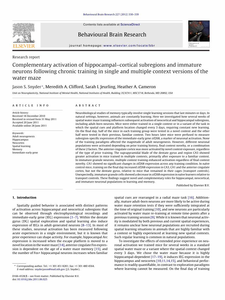
n a single context throughout the entire experiment or in contexts that changedvery 3 days (n = 16 for each; see Fig. 1a for complete breakdown of groups tested).n the standard context, used for single context training, there were prominent cuesn 3 walls and a curtain behind which the experimenter remained during the trial.or multiple context training, distinct contexts were created using a variety of cur-ains and cues (flags, 3 dimensional objects such as buckets and tables) positioned inarious arrangements. Inevitably, some cues (primarily static features of the room)ere used in multiple contexts, but efforts were made to make the contexts as dis-
inct as possible. The platform location remained constant within each context butoved when contexts changed (Fig. 2h). The single and multiple context-trainedice were further subdivided into groups that were only trained after BrdU injection
nd groups that were trained both before and after BrdU injection. For the post-BrdUrained groups, mice were trained for 24 days beginning 7 days after BrdU injection.or the pre/post-BrdU trained groups, mice were similarly trained for 24 days butlso received 9 additional days of training ending 2 days prior to BrdU injection.aïve cage control mice received BrdU but underwent no water maze training onays 0–41.
On the final day of the experiment (day 42), mice in each group were split intoubgroups and either tested in the previous, familiar (F) context or a novel (N) con-ext to examine the role of context novelty on zif268 expression. Naïve control miceither remained in their cage (cageC, i.e., transport controls) or were tested in a singlelock of spatial water maze trials for the first time (cageN). Mice in all other groupsere tested with a single block of spatial water maze trials in either the same famil-
ar context as on the prior day (singleF and multiF) or in a completely novel contextsingleN and multiN). The final testing context was the same for all mice except the
ingleN group. Thus, most mice experienced the same context on the final day, buthe degree of novelty associated with it varied across groups. The block of 4 trialsasted approximately 20 min, after which mice remained undisturbed in their homeage in the testing area until 2 h after the 1st trial, at which time mice were rapidlyuthanized.
Research 227 (2012) 330–339 331
2.3. Histological methods
Mice were anesthetized with isoflurane and perfused with 4% paraformalde-hyde. Brains were post-fixed for 24 h in 4% paraformaldehyde, transferred to a 20%sucrose solution for at least 24 h, and then sectioned coronally at 40 �m on a freez-ing sliding microtome. Fluorescent immunostaining was performed as previouslydescribed [25], on sections containing dorsal hippocampus. To measure experience-dependent activity in hippocampal and cortical subregions, zif268 immunostainingwith chromogenic detection was performed. Sections were incubated for 3 days,free floating, in PBS containing 0.5% tween20, 3% goat serum and rabbit anti-zif268antibody (1:1000; Santa Cruz Biotechnology, sc-189). Sections were then washedand incubated with a biotinylated goat anti-rabbit secondary antibody (1:250; Jack-son Immunolabs) for 60 min, detected using Vector SG peroxidase substrate (Vectorlabs), cleared, and coverslipped under Permount. To assess activity in young neu-rons, combined fluorescent immunostaining against zif268 and doublecortin (DCX)was performed as previously described [25]. Briefly, free-floating sections wereincubated simultaneously in rabbit anti-zif268 (1:1000; Santa Cruz Biotechnology,sc-110), and goat anti-DCX (1:200; Santa Cruz Biotechnology, sc-8066), followedby visualization with Alexa conjugated secondary antibodies (Invitrogen). For com-bined BrdU and DCX immunostaining, free floating sections were treated with 2 NHCl and then incubated simultaneously with rat anti-BrdU (1:2500; Accurate Anti-bodies, OBT0030) and anti-DCX antibodies (as above). For BrdU+ cell counting,immunostaining with BrdU visualization was performed as previously described[25]. Briefly, slide-mounted sections were pre-treated with heat, trypsin, and HClto denature DNA, incubated with mouse anti-BrdU (1:100), and visualized withavidin–biotin–HRP (Vector Labs) followed by DAB.
2.3.1. Histological data analysisAnalysis of immunoperoxidase-stained zif268 expression in the DG, CA3 and
CA1 was performed on 2 sections spaced 240 �m apart containing dorsal hip-pocampus (between −1.8 and −2.4 mm relative to Bregma). Zif268 expression inthe anterior cingulate cortex (Cg1 and Cg2, according to Ref. [26]) was examinedin 4 sections, each spaced 120 �m apart, between 0 mm and 1.0 mm relative toBregma. Images of the hippocampus and cortex were acquired with a 4× objec-tive, using Stereoinvestigator 9.0 software (Microbrightfield). To ensure consistentlevels of brightness and quality, all sections were imaged during the same sessionand with the same exposure and gain settings. Images were analyzed using ImageJsoftware (http://rsbweb.nih.gov/ij/). Boundaries were traced and the percentage ofarea within each brain subregion that exhibited above-threshold zif268 stainingintensity was calculated, as an estimate of the proportion of cells in the subregionshowing activation. The same thresholds were applied across all sections, but due tothe greater cell packing in CA1 and greater staining intensity in the anterior cingu-late cortex, the threshold for these regions was 22% lower than that used for the DGand CA3, in order to avoid saturation/ceiling effects that could obscure differencesbetween groups.
To assess the behavioral activation of adult-born neurons, DCX+ neurons wereexhaustively examined for co-labeling with zif268 in one hemisphere of a singledorsal hippocampal section. The mean number of DCX+ cells examined per mousewas 374, spanning both the infrapyramidal and suprapyramidal blades. DCX+ cellswere initially inspected under epifluorescence using a 60× oil-immersion objective,and putative double-labeling was confirmed via confocal microscopy as previouslydescribed [25]. Specifically, cells with a zif268 intensity of 1.4× background wereconsidered positive. Zif268 expression was also observed in BrdU+ cells but dueto the small number of BrdU-labeled cells these cells were not examined further.Additionally, the total number of DCX+ cells was divided by the sampled volumeof the granule cell layer to obtain a density measure for this marker of immatureneurons. To determine the proportion of BrdU+ cells expressing DCX, all BrdU+ cellsin both hemispheres of a single dorsal hippocampal section (mean = 34 per mouse)were examined for co-expression of DCX via confocal microscopy. For the survivalanalysis, DAB-labeled BrdU+ cells were counted in a 1:12 series through the entireDG, and the total count was multiplied by 12 to provide a stereological estimate ofthe total number of BrdU+ cells in the DG.
2.4. Statistical analysis
Comparisons between groups were performed by ANOVA with Bonferroni posthoc tests as required.
3. Results
3.1. Behavioral performance in single- and multiple contextvariants of the water maze
Comparison of Pre/Post-BrdU and Post-BrdU trained mice in
the single context condition by 2-way repeated measures ANOVAshowed a significant main effect of training day (F23,322 = 8.5,P < 0.0001) and a significant interaction (F23,322 = 5.2, P < 0.0001) butno main effect of prior training (F1,322 = 1.1, P > 0.3). Post hoc tests
332 J.S. Snyder et al. / Behavioural Brain Research 227 (2012) 330–339
Fig. 1. Experimental design. (a) Timeline. All mice received BrdU injections on day 11 and were perfused on day 42. Cage control mice were brought to the testing room withthe rest of the mice but received no other treatment through day 41. Mice in other groups were trained in a spatial water maze task, either in a single spatial context, shown inblack, or in multiple contexts, represented by other colors. Some mice received water maze training only after BrdU administration (Post-BrdU), while others (Pre/Post-BrdU)received water maze training both before and after BrdU administration. On the final day of the experiment (day 42), mice were subjected to a single block of 4 water mazetrials in either a familiar or novel context (or no training for some control mice) and were perfused 2 h after the initial training trial. (b) Panoramic images of 4 contexts. Thecontext shown on top is the standard context, used to train mice in the single context condition and to test most of the groups on the final day. The other 3 contexts are the2nd, 5th, and 8th contexts used for Pre/Post-BrdU training of mice in the multiple context condition. (For interpretation of the references to color in this figure legend, thereader is referred to the web version of the article.)
J.S. Snyder et al. / Behavioural Brain Research 227 (2012) 330–339 333
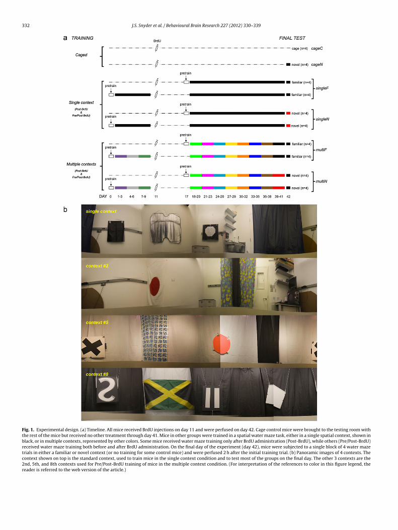
Fig. 2. Behavioral performance in single- and multiple-context water mazes. (a) Latency to find the hidden platform decreased across in the single-context training condition.(b) Proximity, i.e., mean within-trial distance, to the platform location decreased over days of training for mice in the single-context training condition. Here, and in (d) Post-BrdU and Pre/Post-BrdU groups are pooled for clarity. Therefore, days 1–9 reflect behavioral performances for only the Pre/Post-BrdU group and days 18–41 reflect behavioralperformances for both groups. (c) Latency to find the hidden platform for the multiple-context groups. (d) Proximity scores for the multiple-context groups. (e) Averagedlatencies for the 3 days of training in each context for mice in the multiple-context condition. (f) Averaged proximity scores for the 3 days of training in each context formice in the multiple-context condition. (g) Proximity scores for the final 4 days of the experiment (days 39–42). Days 39–41 show performance at the end of training andd t (singg latform(
sttittcddmtPeFtP
ewotfaAPt
ay 42 shows performance as mice are tested further in either the previous contexroup of mice did not receive training but was only tested on day 42 (cageN). (h) Psequentially ordered) that used a given platform location.
howed that Pre/Post-BrdU trained mice were significantly fastero locate the hidden platform on days 18, 19 and 21, as expected dueo prior training on days 1–9 (post hoc all P < 0.01). To confirm thatmproved escape latencies reflect spatial learning we also analyzedhe average proximity to the escape platform over days [27]. As withhe latency measure, proximity to the platform increased signifi-antly over days (i.e., average within-trial distance to the platformecreased; Fig. 2b, ANOVAs for both groups both P < 0.001). Overays 18–41, there was no main effect of prior training on perfor-ance; however, there was a prior training × day interaction such
hat mice in the Pre/Post-BrdU group outperformed mice in theost-BrdU group on day 19 (2-way repeated measures ANOVA,ffect of training day F23,322 = 8.5, P < 0.0001; effect of prior training1,322 = 0.00003, P > 0.9; interaction F23,322 = 3.3, P < 0.0001). Sincehere was no main effect of prior training, the Pre/Post-BrdU andost-BrdU groups are pooled in Fig. 2b.
Mice in the multiple-context condition showed improvedscape latencies and spatial proximity to the platform across daysithin each spatial context, i.e., performance typically improved
ver 3 days and then was impaired as mice were challengedo learn a new spatial context (Fig. 2c and d). To measure per-ormance, latency and proximity scores across contexts were
veraged for each mouse for each day. Two-way repeated measuresNOVAs showed no differences between mice in the Post-BrdU andre/Post-BrdU multiple context training groups (F1,28 ≤ 1.0, P > 0.3)herefore the 2 groups were pooled. Mice trained in multiple con-
leF, multiF) or a novel context (singleN, multiN) to induce zif268 expression. Onelocations for the multiple-context enriched mice. Numbers indicate the contexts
texts showed reduced latency over days 1–3 (F2,30 = 29.7, P < 0.0001;Fig. 2e), and post hoc comparisons indicated significant differencesin latency between days 1 and 2 and between days 1 and 3 (P < 0.001for both). Proximity to the platform location changed over days(F2,30 = 82.6, P < 0.0001; Fig. 2f), with post hoc tests revealing signif-icant reductions in the proximity score each day (all comparisonsP < 0.001). To investigate the possibility that mice formed an allo-centric representation of the room that carried over across contexts,we compared the latencies to 20 cm annuli surrounding the pre-vious versus current platform locations on the first trial in a novelcontext. On average, 42% ± 12% (SEM) swam to the current platformlocation first, which was not different from chance (one sample t-test, T6 = 0.7, P = 0.5), suggesting poor performance was due to thealtered context and not simply because the platform had moved toa new location as in spatial match to place testing [28].
On the final day (day 42), mice in each group were eitherexposed to the same context seen over the last 3 or more daysor to a novel context (see Fig. 1 schematic). Proximity scores forthe final 4 days are shown in Fig. 2g. Collectively, performance ofmice in novel contexts was impaired relative to those in famil-iar contexts on the final day, as expected (2-way ANOVA; maineffect of final context novelty, F1,28 = 13, P = 0.001; effect of prior
training condition, F1,28 = 0.6, P = 0.5; interaction, F1,28 = 0.5, P = 0.5).To investigate how performance changed from day 41 to day 42when the final novel/familiar context was introduced, a 2-way(group × day) repeated measures ANOVA was performed. There
334 J.S. Snyder et al. / Behavioural Brain Research 227 (2012) 330–339
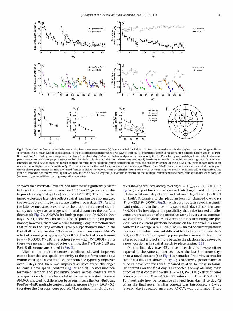
Fig. 3. Adult neurogenesis following spatial water maze training. (a) Stereological counts of 31-day-old BrdU+ cells showed no significant training effects. Inset shows ar ntater as notb
wp4mtutrd
3r
nmwittfsaNvDtn
3sc
BooAiomPzcmot(iw(
epresentative BrdU+ cell in the DG. (b) The density of DCX+ cells in the dorsal deepresentative BrdU+/DCX+ cell. (c) The proportion of BrdU+ cells expressing DCX wars 10 �m.
as a significant group × day interaction (F3,28 = 6, P < 0.01), andost hoc tests revealed that mice tested in a novel context on day2 performed significantly worse than on day 41 (singleN P < 0.01,ultiN P = 0.05). In contrast, mice in the multiple context condition
hat were trained in a familiar context on day 42 (multiF) contin-ed to improve their performance relative to day 41 (P < 0.05). Micerained in the same context through the entire experiment (singleF)eached asymptotic performance many days earlier, and were notifferent on day 41 compared to day 42 (P = 0.2).
.2. Water maze training did not affect neuronal survival or theate of expression of the immature marker doublecortin
Long-term exposure to enriched housing conditions and run-ing wheels enhance survival of young granule neurons in adultice [25,29]. We hypothesized that long-term exposure to spatialater maze training, a strongly hippocampal-dependent behav-
or, would also enhance new neuron survival in these mice. Theotal number of BrdU+ cells was estimated for each training condi-ion and subjected to a 1-way ANOVA. Mice exposed to novel andamiliar contexts on the final day were grouped, since this expo-ure was after the normal time period of cell death [30–32] and inll likelihood too close to the time of perfusion to have an effect.o significant group differences were found in the number of sur-iving BrdU+ cells (Fig. 3a; F4,31 = 0.8, P = 0.5) or in the density ofCX+ cells (Fig. 3b; 1-way ANOVA, F4,35 = 0.5, P = 0.7). Additionally,
he proportion of BrdU+ cells expressing DCX across groups wasot different (Fig. 3c; F4,34 = 0.7, P = 0.6).
.3. Distinct subregion-specific patterns of activation followingpatial water maze training and exposure to familiar versus novelontexts
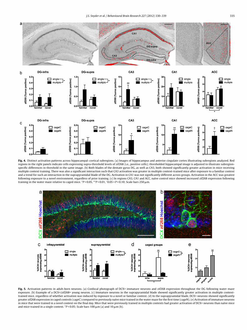
Because behavior was not different in the mice trained post-rdU and pre/post-BrdU, these groups were pooled for analysesf zif268 expression on the final day. Within each hippocampalr cortical subregion, zif268 expression was analyzed using 2-wayNOVAs with training (single or multiple context) and final test-
ng context (familiar or novel) as factors (Fig. 4). In both bladesf the DG, more cells expressed zif268 in multiple context-trainedice than in single context-trained mice (for both blades, F1,24 > 5,< 0.05). In the suprapyramidal blade there was a trend for moreif268 in multiple context-trained mice exposed to the familiarontext on day 42 (interaction F1,25 = 2.7, P = 0.11; post hoc vs.ultiN P = 0.08, post hoc vs. singleF P < 0.01). A similar pattern
f activation was also observed in CA3, where multiple context-rained mice had more zif268 expression than single-trained mice
main effect of training: F1,25 = 7, P < 0.05), and there was a signif-cant training × final test context interaction (F1,25 = 5.5, P < 0.05)
ith the greatest zif268 expression in mice in the multiF conditionpost hoc, P < 0.05 vs. multiN and P < 0.01 vs. singleF). In contrast
gyrus was not different between training groups. Inset shows confocal image of adifferent between groups. Bars represent mean ± SEM, n = 7–8 in each group. Scale
to the DG and CA3, zif268 expression in CA1 showed no significantdifferences across trained groups. Lastly, the ACC showed signifi-cantly enhanced activation in response to novel context exposure,regardless of prior training status (main effect of final test context,F1,05 = 5.3, P < 0.05).
Zif268 expression was also examined in control groups, whichreceived no prior training and were exposed to the spatial watermaze for the first time on day 42 (cageN) or remained in their cages(cageC) just prior to perfusion (Fig. 4c). As expected based on stud-ies of naïve rodents learning a spatial water maze [14,33], cageNmice showed enhanced zif268 expression relative to cageC mice inregions CA3, CA1 and ACC (main effect of brain region, main effectof experience and interaction all P < 0.01; post hocs – CA3 P < 0.05,CA1 P < 0.001, ACC P = 0.05; Fig. 4c). In contrast, zif268 levels did notdiffer significantly between these two groups in the DG (post hocboth blades P > 0.05).
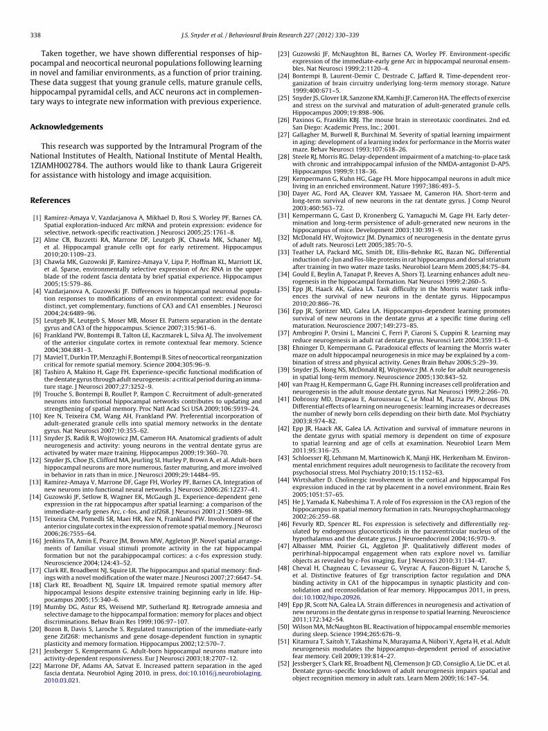
3.4. Multiple context training enhances activation of adult-bornneurons
Zif268 expression was quantified in adult-born granule cells inthe DG identified via fluorescent immunostaining for DCX (Fig. 5).Activation was measured as the proportion of DCX+ neuronsco-expressing zif268. In the infrapyramidal blade, DCX+/zif268+expression showed no main effect of training or of final test con-text and no interaction between these two factors (all P > 0.1). Inthe suprapyramidal blade, however, there was a significant effectof training, with a greater proportion of DCX+ neurons expressingzif268 in mice subjected to multiple context training compared tothe single context training condition (F1,22 = 10.0, P < 0.01; Fig. 5c).There was no effect of final context on DCX+/zif268+ expression(F1,22 < 0.1, P = 0.9).
Zif268 expression in DCX+ cells was assessed in control mice,which remained in their home cage until the final day. Using 2-wayANOVA with blade (supra- or infrapyramidal) and training (noveltraining or no training) as factors, significant main effects of bladeand training and a significant interaction (all F > 5, P < 0.05) werefound (Fig. 5d). Zif268 expression in DCX+ neurons was greater inthe suprapyramidal blade than in the infrapyramidal blade, as seenin the overall granule cell population (Fig. 4c). But in contrast towhat was seen in mature neurons in all examined subregions, DCX+cells in the suprapyramidal blade of the cageC group, had signifi-cantly greater zif268 expression compared to those in the cageNgroup (P < 0.01). DCX+/zif268+ expression was also significantlygreater in the suprapyramidal blade than in the infrapyramidalblade of cageC mice (P < 0.01).
To further examine activation of immature neurons as a func-
tion of prior training experience, we compared all groups thatwere trained in a novel context on the final day. One-way ANOVAshowed that mice in the multiN condition had greater suprapyrami-dal DCX+/zif268+ expression than both singleN cageN mice, which
J.S. Snyder et al. / Behavioural Brain Research 227 (2012) 330–339 335
Fig. 4. Distinct activation patterns across hippocampal–cortical subregions. (a) Images of hippocampus and anterior cingulate cortex illustrating subregions analyzed. Redregions in the right panels indicate cells expressing supra-threshold levels of zif268 (i.e., positive cells); thresholded hippocampal image is adjusted to illustrate subregion-specific differences in threshold in the same image. (b) Both blades of the dentate gyrus DG, as well as CA3, both showed significantly greater activation in mice receivingmultiple context training. There was also a significant interaction such that CA3 activation was greater in multiple context-trained mice after exposure to a familiar contextand a trend for such an interaction in the suprapyramidal blade of the DG. Activation in CA1 was not significantly different across groups. Activation in the ACC was greatestfollowing exposure to a novel environment, regardless of prior training. (c) In regions CA3, CA1 and ACC, naïve control mice showed increased zif268 expression followingtraining in the water maze relative to caged mice. *P < 0.05, **P < 0.01, †0.05 < P < 0.10; Scale bars 250 �m.
Fig. 5. Activation patterns in adult-born neurons. (a) Confocal photograph of DCX+ immature neurons and zif268 expression throughout the DG following water mazeexposure. (b) Example of a DCX+/zif268+ young neuron. (c) Immature neurons in the suprapyramidal blade showed significantly greater activation in multiple context-trained mice, regardless of whether activation was induced by exposure to a novel or familiar context. (d) In the suprapyramidal blade, DCX+ neurons showed significantlygreater zif268 expression in caged controls (cageC) compared to previously naïve mice trained in the water maze for the first time (cageN). (e) Activation of immature neuronsin mice that were trained in a novel context on the final day. Mice that were previously trained in multiple contexts had greater activation of DCX+ neurons than naïve miceand mice trained in a single context. *P < 0.05; Scale bars 100 �m (a) and 10 �m (b).
3 Brain
wpg
4
itamcnvwsomaitttwedttfiTnwrcra
4
dwdiceanavlpumtlss[cssnte
36 J.S. Snyder et al. / Behavioural
ere not significantly different from each other (F2,16 = 6.1, P < 0.01;ost hoc multiN vs. singleN and cageN both P < 0.05, post hoc sin-leN vs. cageN P > 0.2; Fig. 5e).
. Discussion
In the current study we used chronic standard water maze train-ng or a novel variant where contextual cues regularly changedo examine (1) how prolonged spatial learning affects survival ofdult-born neurons and (2) how prior spatial learning experiencesodulate activity during exploration of novel and familiar spatial
ontexts. We found no effect of chronic spatial learning on neweuron survival. However, after chronic training in static or varyingersions of the spatial water maze, distinct patterns of activationere revealed across neuronal populations. Specifically, the ACC
howed enhanced activation following exposure to a novel contextn the final test day (day 42), regardless of prior training (single vs.ultiple). In contrast, the DG (supra) and CA3 were preferentially
ctivated in multiple context-trained mice but only when testedn a familiar context. CA1 appeared to have a similar pattern forhe means across groups, but zif268 expression showed no statis-ically significant differences. Lastly, immature granule neurons inhe suprapyramidal blade showed enhanced activation in mice thatere repeatedly trained in different spatial contexts, with no pref-
rential activation in novel versus familiar contexts on the finalay. In addition to chronically trained mice, control groups wereransported to the testing room in their home cages throughouthe training period and either remained in their home cages on thenal test day or were trained in the water maze for the first time.raining increased zif268 expression in CA3, CA1 and the ACC butot in the DG. We unexpectedly found that young granule cellsere actually inhibited during initial exposure to the water maze
elative to their very high rate of activation in the cage controlondition. Collectively, these results suggest novel complementaryoles for hippocampal and neocortical regions in spatial learningnd memory.
.1. Survival of adult-born hippocampal neurons
Experience-dependent effects on neuronal survival have beenemonstrated with environmental enrichment [29] and spatialater maze learning [34], with the direction of change varyingepending on factors such as task difficulty and cell age [35,36]. It
s somewhat surprising, therefore, that the current study found nohange in young granule cell survival or DCX+ cell density, since ourxperimental design contained features of both of these paradigmsnd involved training mice during a prolonged period when neweurons die and could be potentially rescued [30–32]. However,lthough several studies have found changes in new neuron sur-ival in rats [34,37] (but see Ref. [39]), the water maze may haveess of an effect on new neuron survival in mice [38,40]. Anotherossibility is that survival was increased when the labeled gran-le cells were very young and was subsequently decreased as theyatured, as previously described in rats [41,42]. The sum of these
wo changes could result in a lack of net effect on survival with pro-onged learning. A previous study has suggested that the stress ofpatial water maze training has a negative effect on young neuronurvival, since decreased survival was eliminated with pretraining38], but a similar effect of pretraining was not observed in theurrent study. Enhanced survival of new neurons is consistentlyeen in mice with environmental enrichment [8,29,43]. There are
everal possible reasons why long-term water maze training didot produce the survival effect seen with enrichment, includinghe length of daily exposure (brief versus continuous), sex differ-nces (males versus females, which have been used in virtually all
Research 227 (2012) 330–339
mouse enrichment studies), or differences in stress levels producedby water maze learning and environmental enrichment.
4.2. Activation in naïve mice
Previous studies have shown that training naïve animals in thewater maze increases IEG expression relative to cage controls inCA1 and CA3 [14,33] and that non-naïve behaving animals typicallyhave greater IEG expression in the hippocampus and neocortex rel-ative to cage controls [1,15,23,44,45]. Our data showing increasedzif268 expression in CA3, CA1 and the ACC relative to cage controlsare consistent with these findings. The DG, however, did not showincreased activation following water maze exposure in naïve micerelative to cage controls, in apparent contrast with several studiesreporting IEG increases in behaving animals relative to cage con-trols [1,3,10] and two studies showing increased Fos and Arc innaïve water maze-trained rats relative to cage controls [11,14]. It isunlikely that lack of expression reflects a problem with immunos-taining or with the behavioral manipulations because zif268 levelswere increased other brain regions (CA3, CA1) in the same sec-tions. Currently, little is known about the function of the DG andthe factors that activate populations of DG neurons, but stress candecrease IEG expression relative to cage controls [46], and DG IEGexpression can be reduced by novelty [47]. Species differencesand/or distinct functions of zif268 [48] versus Fos and Arc may alsoexplain differential expression of these IEGs in the current study ascompared to previous studies. Furthermore, electrophysiologicaland IEG data indicate that the same DG neurons are often reacti-vated by different experiences, including sleep [1,2,5]. Thus, the DGof naïve trained mice, relative to cage controls, was either (1) notsignificantly activated, (2) did not undergo zif268-dependent formsof plasticity with 2 h post-training, or (3) did not activate additionalpopulations of neurons during water maze acquisition.
Unlike the other neurons examined, young adult-born gran-ule cells in the current study showed reduced zif268 expressionin naïve water maze-trained naïve mice relative to the cage con-trol condition. Other studies have reported a lack of increase inzif268 protein in young neurons following behavioral stimulation[8,49], but this is the first study to our knowledge to find a sig-nificant reduction in IEG expression in new neurons with novelexperience. This decrease could reflect inhibition of young granulecells in stressful conditions, since mice that were familiar with thewater maze (single/multiple context-trained mice) had numbersof DCX+/zif268+ neurons that were intermediate between thosein the highly stressful first exposure and (presumably stress-free)cage controls. Alternatively, it is possible that relatively high lev-els of zif268 in young neurons in cage controls reflect sleep-relatedconsolidation activity [1,50], since undisturbed mice spend muchof their time sleeping during the light phase, and adult-born neu-rons are required for consolidation and long-term spatial memory[39,51,52]. A previous study [53] has found increased zif268 acti-vation following REM sleep in the DG of rats exposed to a novelenvironment; cage control mice in the current study were brieflyhandled upon transport to the testing room and then slept formuch of the subsequent ∼3 h prior to perfusion. These experiencesand/or social interactions that occurred as a result of transport andhandling could have led to increased zif268 expression in new neu-rons during subsequent sleep bouts. It will be useful to determinespecific conditions under which resting animals show neuronalactivation and expression of various IEGs. Although stress and/orsleep effects could potentially explain the inhibition of zif268 inyoung neurons induced by water maze training in naïve mice, nei-
ther of these factors seems likely to play a role in the changesobserved within the groups of chronically trained mice, which wereall equally awake and probably all mildly stressed during the train-ing period.
Brain
4t
dicacfi
nbtcimhiecbhpasfen
ruDIrac
4c
ptgt(ttfzmstctfpsmtcibi
J.S. Snyder et al. / Behavioural
.3. Response to novel conditions in the ACC of chronicallyrained mice
Chronically trained mice were highly familiar with the taskemands, unlike naïve mice, which had experiences (swimming
n a spatial water maze in a novel room or remaining in the homeage) that differed in spatial layout and procedural aspects as wells novelty. Thus, differences in activation patterns across chroni-ally trained mice can be more easily linked to training history ornal context novelty than those in the naïve mice.
Only neurons in the ACC showed a selective response to theovel context condition; pyramidal neurons in CA3 and CA1 andoth the overall population of DG neurons as well as the imma-ure subset showed no increase in response to the novel spatialontext. The ACC response to novelty is unexpected in light of stud-es showing increased ACC activation following retrieval of remote
emory relative to recent memory [6,7,15]. Although we did notave test groups that were directly comparable to previous stud-
es, one might have expected that mice in the singleF group, whichxperienced a context that was well-trained and presumably well-onsolidated, would have shown the highest levels of activation,ut this was not the case. Our finding that mice in the singleF groupad less ACC activation than mice in the singleN group and com-arable activation to mice in the cageN group, despite retrievingmemory that was as old (24/41 days) as that in typical rodent
tudies of remote memory, suggests that a key factor requiredor activation of the ACC during remote memory retrieval is anxtended training–testing interval during which the animals areot exposed to the testing environment.
In contrast to the ACC response, the DG showed identicalesponses in the novel and familiar spatial conditions. This wasnexpected since lesion data specifically points to a role for theG in detecting spatial novelty [54,55]. This disconnect between
EG activation and lesion-induced deficits may reflect differentequirements for detecting spatial rearrangement of objects withincontext, as in the earlier studies, and learning an entirely new
ontext, as in the current study.
.4. Effects of prior training on hippocampal activation inhronically trained mice
Unlike ACC activation, hippocampal activation was related torior training, and in some cases there was an interaction betweenhe training type and novelty. For example, mice in the multiFroup had elevated zif268 expression in CA3, and there was a trendoward this same effect in the suprapyramidal blade of the DGand perhaps CA1). This activation pattern could reflect differen-ial learning during the final training episode, since these werehe only mice that showed improvement from the prior day’s per-ormance levels. The other groups, which showed lower levels ofif268 expression, were at different stages of behavioral perfor-ance. Mice trained in a novel context on the final day (multiN,
ingleN) had just begun to form a memory of the spatial con-ext and platform location, as evidenced by impaired performanceompared to the prior day. At the other extreme, mice trained inhe same context throughout the entire experiment (singleF) per-ormed as well as mice in the multiF group on the final day, but theirerformance had plateaued approximately 2 weeks earlier and theyhowed no evidence of continued learning. Increased zif268 in theultiF mice on the final day may therefore reflect activation related
o mastery of the current context and/or early stages of memory
onsolidation. This interpretation fits with previous data show-ng mossy fiber sprouting and, presumably, increased connectivityetween the DG and CA3 after 4–5 days of spatial water maze train-
ng [56]. An unresolved question is whether increased activation on
Research 227 (2012) 330–339 337
the 4th day of training in a given context is dependent on extensiveprior training.
The parallels between zif268 activity and behavior on the finalday do not hold as well for infrapyramidal DG granule cells andimmature granule neurons in the suprapyramidal blade. Neuronalactivation in these populations was determined by prior experi-ence rather than current stimuli; these neurons were more activein mice trained in multiple contexts than mice trained in a singlecontext and were completely unaffected by the novelty/familiarityof the final context. It is interesting to note that the mature gran-ule cells of the suprapyramidal blade showed an activity patternthat resembled that of CA3 and CA1 pyramidal cells, while the pat-tern in the infrapyramidal blade was very different, resemblingonly the immature granule cells. Previous studies have found thatexperiences such as undirected exploration or spatial water mazetraining rapidly increase expression of immediate-early genes c-Fos and Arc in mature granule cells within the suprapyramidal butnot infrapyramidal blade [1,3,11,57]. This difference may be drivenby differences in GABAergic inhibition to the two blades [58]. Thecurrent study found that although activation is always lower inthe infrapyramidal blade than in the suprapyramidal blade, maturegranule cells in the infrapyramidal blade can be rapidly activatedby spatial water maze experience if the animals have undergoneprior training. The increased activation in the multiple exposureanimals, which are still learning on the final day of training, alongwith previous findings that the infrapyramidal blade is activated6–8 h after exposure to a novel environment [1] seems consistentwith the possibility that the infrapyramidal blade is involved inmemory consolidation.
An interesting question is how prior experience affects imma-ture neurons that are not even born when training begins. Granulecells in mice can be activated shortly after they are 2 weeks old,and some retain doublecortin until they are greater than 4 weeks ofage [12]. Thus, it is possible that zif268 expression in doublecortin-positive neurons reflects participation in memory formation onlyduring the final 1–2 weeks of training and subsequent retrievalof those memories on the final day. A second possibility is thatprior memory of the training affects subsequent perception, strat-egy, or other aspects of behavior and it is this upstream difference,rather than a change in the young neurons themselves, that affectsrecruitment of immature neurons.
There are several possible consequences of increased activa-tion of young granule neurons. Previous studies have found thata small proportion of granule neurons, often the same popula-tion, are active in different contexts [2,3,5,22]. Growth in the activepopulation seen here could reflect the recruitment of additionalpopulations of neurons in order to better pattern-separate relatedcontexts, perhaps partly via enhanced integration of immature neu-rons by water maze training [59,60]. Alternatively, it has beenproposed that a developmental window of increased activity mayallow immature neurons to bind together temporally related expe-riences [61]. A twist on this idea is the possibility that increasedactivation of (infrapyramidal and) immature neurons during multi-ple context learning serves to relate similar experiences, perhaps byrecruiting additional neurons that were recently activated by simi-lar contexts. Repeated context learning may therefore promote theformation of a schematic understanding of the related spatial watermaze contexts, akin to that which is formed during the systemsconsolidation of memory [62,63]. Such a role would be consistentwith IEG imaging studies and neurogenesis ablation studies thathave implicated young neurons [9,39,51] and the infrapyramidalblade [1] in memory consolidation and the processing of remote
memory. In any case, the activity and/or plasticity in these granuleneurons is unusual in its dependence on the prior experience andcould be thought of as an instance of metaplasticity at a cellularlevel.
3 Brain
piTht
A
N1f
R
[
[
[
[
[
[
[
[
[
[
[
[
[
[
[
[
[
[
[
[
[
[
[
[
[
[
[
[
[
[
[
[
[
[
[
[
[
[
[
[
[
[
38 J.S. Snyder et al. / Behavioural
Taken together, we have shown differential responses of hip-ocampal and neocortical neuronal populations following learning
n novel and familiar environments, as a function of prior training.hese data suggest that young granule cells, mature granule cells,ippocampal pyramidal cells, and ACC neurons act in complemen-ary ways to integrate new information with previous experience.
cknowledgements
This research was supported by the Intramural Program of theational Institutes of Health, National Institute of Mental Health,ZIAMH002784. The authors would like to thank Laura Grigereitor assistance with histology and image acquisition.
eferences
[1] Ramirez-Amaya V, Vazdarjanova A, Mikhael D, Rosi S, Worley PF, Barnes CA.Spatial exploration-induced Arc mRNA and protein expression: evidence forselective, network-specific reactivation. J Neurosci 2005;25:1761–8.
[3] Chawla MK, Guzowski JF, Ramirez-Amaya V, Lipa P, Hoffman KL, Marriott LK,et al. Sparse, environmentally selective expression of Arc RNA in the upperblade of the rodent fascia dentata by brief spatial experience. Hippocampus2005;15:579–86.
[4] Vazdarjanova A, Guzowski JF. Differences in hippocampal neuronal popula-tion responses to modifications of an environmental context: evidence fordistinct, yet complementary, functions of CA3 and CA1 ensembles. J Neurosci2004;24:6489–96.
[5] Leutgeb JK, Leutgeb S, Moser MB, Moser EI. Pattern separation in the dentategyrus and CA3 of the hippocampus. Science 2007;315:961–6.
[6] Frankland PW, Bontempi B, Talton LE, Kaczmarek L, Silva AJ. The involvementof the anterior cingulate cortex in remote contextual fear memory. Science2004;304:881–3.
[7] Maviel T, Durkin TP, Menzaghi F, Bontempi B. Sites of neocortical reorganizationcritical for remote spatial memory. Science 2004;305:96–9.
[8] Tashiro A, Makino H, Gage FH. Experience-specific functional modification ofthe dentate gyrus through adult neurogenesis: a critical period during an imma-ture stage. J Neurosci 2007;27:3252–9.
[9] Trouche S, Bontempi B, Roullet P, Rampon C. Recruitment of adult-generatedneurons into functional hippocampal networks contributes to updating andstrengthening of spatial memory. Proc Natl Acad Sci USA 2009;106:5919–24.
10] Kee N, Teixeira CM, Wang AH, Frankland PW. Preferential incorporation ofadult-generated granule cells into spatial memory networks in the dentategyrus. Nat Neurosci 2007;10:355–62.
11] Snyder JS, Radik R, Wojtowicz JM, Cameron HA. Anatomical gradients of adultneurogenesis and activity: young neurons in the ventral dentate gyrus areactivated by water maze training. Hippocampus 2009;19:360–70.
12] Snyder JS, Choe JS, Clifford MA, Jeurling SI, Hurley P, Brown A, et al. Adult-bornhippocampal neurons are more numerous, faster maturing, and more involvedin behavior in rats than in mice. J Neurosci 2009;29:14484–95.
14] Guzowski JF, Setlow B, Wagner EK, McGaugh JL. Experience-dependent geneexpression in the rat hippocampus after spatial learning: a comparison of theimmediate-early genes Arc, c-fos, and zif268. J Neurosci 2001;21:5089–98.
15] Teixeira CM, Pomedli SR, Maei HR, Kee N, Frankland PW. Involvement of theanterior cingulate cortex in the expression of remote spatial memory. J Neurosci2006;26:7555–64.
16] Jenkins TA, Amin E, Pearce JM, Brown MW, Aggleton JP. Novel spatial arrange-ments of familiar visual stimuli promote activity in the rat hippocampalformation but not the parahippocampal cortices: a c-fos expression study.Neuroscience 2004;124:43–52.
17] Clark RE, Broadbent NJ, Squire LR. The hippocampus and spatial memory: find-ings with a novel modification of the water maze. J Neurosci 2007;27:6647–54.
18] Clark RE, Broadbent NJ, Squire LR. Impaired remote spatial memory afterhippocampal lesions despite extensive training beginning early in life. Hip-pocampus 2005;15:340–6.
19] Mumby DG, Astur RS, Weisend MP, Sutherland RJ. Retrograde amnesia andselective damage to the hippocampal formation: memory for places and objectdiscriminations. Behav Brain Res 1999;106:97–107.
20] Bozon B, Davis S, Laroche S. Regulated transcription of the immediate-earlygene Zif268: mechanisms and gene dosage-dependent function in synapticplasticity and memory formation. Hippocampus 2002;12:570–7.
22] Marrone DF, Adams AA, Satvat E. Increased pattern separation in the agedfascia dentata. Neurobiol Aging 2010, in press, doi:10.1016/j.neurobiolaging.2010.03.021.
[
Research 227 (2012) 330–339
23] Guzowski JF, McNaughton BL, Barnes CA, Worley PF. Environment-specificexpression of the immediate-early gene Arc in hippocampal neuronal ensem-bles. Nat Neurosci 1999;2:1120–4.
24] Bontempi B, Laurent-Demir C, Destrade C, Jaffard R. Time-dependent reor-ganization of brain circuitry underlying long-term memory storage. Nature1999;400:671–5.
25] Snyder JS, Glover LR, Sanzone KM, Kamhi JF, Cameron HA. The effects of exerciseand stress on the survival and maturation of adult-generated granule cells.Hippocampus 2009;19:898–906.
26] Paxinos G, Franklin KBJ. The mouse brain in stereotaxic coordinates. 2nd ed.San Diego: Academic Press, Inc.; 2001.
27] Gallagher M, Burwell R, Burchinal M. Severity of spatial learning impairmentin aging: development of a learning index for performance in the Morris watermaze. Behav Neurosci 1993;107:618–26.
28] Steele RJ, Morris RG. Delay-dependent impairment of a matching-to-place taskwith chronic and intrahippocampal infusion of the NMDA-antagonist D-AP5.Hippocampus 1999;9:118–36.
29] Kempermann G, Kuhn HG, Gage FH. More hippocampal neurons in adult miceliving in an enriched environment. Nature 1997;386:493–5.
30] Dayer AG, Ford AA, Cleaver KM, Yassaee M, Cameron HA. Short-term andlong-term survival of new neurons in the rat dentate gyrus. J Comp Neurol2003;460:563–72.
31] Kempermann G, Gast D, Kronenberg G, Yamaguchi M, Gage FH. Early deter-mination and long-term persistence of adult-generated new neurons in thehippocampus of mice. Development 2003;130:391–9.
32] McDonald HY, Wojtowicz JM. Dynamics of neurogenesis in the dentate gyrusof adult rats. Neurosci Lett 2005;385:70–5.
33] Teather LA, Packard MG, Smith DE, Ellis-Behnke RG, Bazan NG. Differentialinduction of c-Jun and Fos-like proteins in rat hippocampus and dorsal striatumafter training in two water maze tasks. Neurobiol Learn Mem 2005;84:75–84.
34] Gould E, Beylin A, Tanapat P, Reeves A, Shors TJ. Learning enhances adult neu-rogenesis in the hippocampal formation. Nat Neurosci 1999;2:260–5.
35] Epp JR, Haack AK, Galea LA. Task difficulty in the Morris water task influ-ences the survival of new neurons in the dentate gyrus. Hippocampus2010;20:866–76.
36] Epp JR, Spritzer MD, Galea LA. Hippocampus-dependent learning promotessurvival of new neurons in the dentate gyrus at a specific time during cellmaturation. Neuroscience 2007;149:273–85.
37] Ambrogini P, Orsini L, Mancini C, Ferri P, Ciaroni S, Cuppini R. Learning mayreduce neurogenesis in adult rat dentate gyrus. Neurosci Lett 2004;359:13–6.
38] Ehninger D, Kempermann G. Paradoxical effects of learning the Morris watermaze on adult hippocampal neurogenesis in mice may be explained by a com-bination of stress and physical activity. Genes Brain Behav 2006;5:29–39.
39] Snyder JS, Hong NS, McDonald RJ, Wojtowicz JM. A role for adult neurogenesisin spatial long-term memory. Neuroscience 2005;130:843–52.
40] van Praag H, Kempermann G, Gage FH. Running increases cell proliferation andneurogenesis in the adult mouse dentate gyrus. Nat Neurosci 1999;2:266–70.
41] Dobrossy MD, Drapeau E, Aurousseau C, Le Moal M, Piazza PV, Abrous DN.Differential effects of learning on neurogenesis: learning increases or decreasesthe number of newly born cells depending on their birth date. Mol Psychiatry2003;8:974–82.
42] Epp JR, Haack AK, Galea LA. Activation and survival of immature neurons inthe dentate gyrus with spatial memory is dependent on time of exposureto spatial learning and age of cells at examination. Neurobiol Learn Mem2011;95:316–25.
43] Schloesser RJ, Lehmann M, Martinowich K, Manji HK, Herkenham M. Environ-mental enrichment requires adult neurogenesis to facilitate the recovery frompsychosocial stress. Mol Psychiatry 2010;15:1152–63.
44] Wirtshafter D. Cholinergic involvement in the cortical and hippocampal Fosexpression induced in the rat by placement in a novel environment. Brain Res2005;1051:57–65.
45] He J, Yamada K, Nabeshima T. A role of Fos expression in the CA3 region of thehippocampus in spatial memory formation in rats. Neuropsychopharmacology2002;26:259–68.
46] Fevurly RD, Spencer RL. Fos expression is selectively and differentially reg-ulated by endogenous glucocorticoids in the paraventricular nucleus of thehypothalamus and the dentate gyrus. J Neuroendocrinol 2004;16:970–9.
47] Albasser MM, Poirier GL, Aggleton JP. Qualitatively different modes ofperirhinal-hippocampal engagement when rats explore novel vs. familiarobjects as revealed by c-Fos imaging. Eur J Neurosci 2010;31:134–47.
48] Cheval H, Chagneau C, Levasseur G, Veyrac A, Faucon-Biguet N, Laroche S,et al. Distinctive features of Egr transcription factor regulation and DNAbinding activity in CA1 of the hippocampus in synaptic plasticity and con-solidation and reconsolidation of fear memory. Hippocampus 2011, in press,doi:10.1002/hipo.20926.
49] Epp JR, Scott NA, Galea LA. Strain differences in neurogenesis and activation ofnew neurons in the dentate gyrus in response to spatial learning. Neuroscience2011;172:342–54.
50] Wilson MA, McNaughton BL. Reactivation of hippocampal ensemble memoriesduring sleep. Science 1994;265:676–9.
51] Kitamura T, Saitoh Y, Takashima N, Murayama A, Niibori Y, Ageta H, et al. Adult
neurogenesis modulates the hippocampus-dependent period of associativefear memory. Cell 2009;139:814–27.
52] Jessberger S, Clark RE, Broadbent NJ, Clemenson Jr GD, Consiglio A, Lie DC, et al.Dentate gyrus-specific knockdown of adult neurogenesis impairs spatial andobject recognition memory in adult rats. Learn Mem 2009;16:147–54.
[63] Winocur G, Moscovitch M, Bontempi B. Memory formation and long-term
J.S. Snyder et al. / Behavioural
53] Ribeiro S, Goyal V, Mello CV, Pavlides C. Brain gene expression during REM sleepdepends on prior waking experience. Learn Mem 1999;6:500–8.
54] Hunsaker MR, Rosenberg JS, Kesner RP. The role of the dentate gyrus, CA3a,b,and CA3c for detecting spatial and environmental novelty. Hippocampus2008;18:1064–73.
55] Lee I, Hunsaker MR, Kesner RP. The role of hippocampal subregions in detectingspatial novelty. Behav Neurosci 2005;119:145–53.
56] Ramirez-Amaya V, Balderas I, Sandoval J, Escobar ML, Bermudez-Rattoni F. Spa-tial long-term memory is related to mossy fiber synaptogenesis. J Neurosci2001;21:7340–8.
Septo-temporal gradients of neurogenesis and activity in 13-month-old rats.Neurobiol Aging 2011;32:1149–56.
58] Scharfman HE, Sollas AL, Smith KL, Jackson MB, Goodman JH. Structural andfunctional asymmetry in the normal and epileptic rat dentate gyrus. J CompNeurol 2002;454:424–39.
Research 227 (2012) 330–339 339
59] Ambrogini P, Cuppini R, Lattanzi D, Ciuffoli S, Frontini A, Fanelli M. Synapto-genesis in adult-generated hippocampal granule cells is affected by behavioralexperiences. Hippocampus 2009;20:799–810.
60] Tronel S, Fabre A, Charrier V, Oliet SH, Gage FH, Abrous DN. Spatial learningsculpts the dendritic arbor of adult-born hippocampal neurons. Proc Natl AcadSci USA 2010;107:7963–8.
62] Tse D, Langston RF, Kakeyama M, Bethus I, Spooner PA, Wood ER, et al. Schemas
retention in humans and animals: convergence towards a transforma-tion account of hippocampal-neocortical interactions. Neuropsychologia2010;48:2339–56.