Page 1
Betanodavirus Infections of Finfish
Australia and New Zealand Standard Diagnostic Procedure, 2014 Page 1 of 25
Betanodavirus Infections of Finfish
NJG Moody
MStJ Crane
AAHL Fish Diseases Laboratory
CSIRO Australian Animal Health Laboratory
Private Bag 24
Geelong VIC 3220
Australia
[email protected]
AAHL Fish Diseases Laboratory
CSIRO Australian Animal Health Laboratory
Private Bag 24
Geelong VIC 3220
Australia
[email protected]
Part 1 - Diagnostic Overview
Summary
Viral nervous necrosis (VNN), syn. viral encephalopathy and retinopathy (VER), is a
serious viral disease of finfish caused by a Betanodavirus, syn. nervous necrosis virus
(NNV). Disease has been reported in over 40 fish species from tropical to temperate
climates in most continents around the world, and most reports of disease have been
associated with species in aquaculture facilities. Clinical disease is most commonly
observed in larval and juvenile finfish, and this is the case in Australia, although reports of
clinical disease in adult fish are increasing. Mortality rates of up to 100% are most
commonly seen in larval fish and mortality rates tend to decrease as the age of the infected
fish increases. Fish surviving infection can become sub-clinical carriers. The most
common mode of transmission appears to be vertical, from sub-clinically infected
broodstock to progeny during spawning, but horizontal transmission can occur.
Betanodavirus infection has a significant economic, social and environmental impact in
Australia through direct losses, inhibition of trade, restriction on locations suitable for
aquaculture expansion and suspension of fish restocking programs due to concerns on the
impact of the virus to native fish species due to translocation of infected stock. Tests have
been developed to detect the infection in fish and for health certification purposes. As
serology testing for the detection of antibodies in finfish is not routinely practiced in
diagnostic laboratories, all tests are based on direct detection of the virus. Virus isolation
is used to detect infectious virus in fresh unfixed material. Real-time reverse-transcriptase
polymerase chain reaction (RT-qPCR) and nested reverse-transcriptase polymerase chain
reaction (RT-nPCR) tests are used to detect viral nucleic acid sequences in fresh and/or
fixed material, and histological examination is used for disease diagnosis in formalin-fixed
material. Immunological tests (immunocytochemical test (ICCT), or indirect
immunofluorescent antibody test (IFAT)), RT-qPCR or RT-nPCR are used to confirm the
identity of viruses isolated in cell culture as Betanodavirus, with sequencing of amplicons
produced by conventional RT-PCR used for confirmation of strain identity. The
immunohistochemical test (IHCT) or IFAT are used to confirm or exclude nodavirus from
fixed histological tissue sections where the nature of the lesions may be ambiguous.
Page 2
Betanodavirus Infections of Finfish
Australia and New Zealand Standard Diagnostic Procedure, 2014 Page 2 of 25
Aetiology
The virus causing VNN is a member of the genus Betanodavirus of the family
Nodaviridae.1 Virions are non-enveloped and icosahedral in shape with a diameter of
approximately 25 to 30 nm and contain two segments (RNA1 and RNA2) of positive-sense
single-stranded RNA (ssRNA) with the RNA2 segment containing the sequence for the
viral coat protein.2 The RNA2 segment is highly conserved among isolates and is the target
for the detection of viral RNA by PCR methods. The viral coat protein is the target for
immunological methods. There are four recognised genotypes: striped jack nervous
necrosis virus (SJNNV), tiger puffer nervous necrosis virus (TPNNV), barfin flounder
nervous necrosis virus (BFNNV) and red-spotted grouper nervous necrosis virus
(RGNNV).3 A possible fifth genotype, turbot betanodavirus (TNV), has been proposed.
4
All Australian Betanodavirus isolates have been identified as members of the RGNNV
genogroup, where they occur in two distinct clusters (RGNNV 1a and RGNNV 1c).5
Clinical Signs
Clinical signs are most commonly observed in larvae and fry and are due to damage to the
nervous tissue of the spinal cord, brain and retina caused by the viral infection. Typically,
affected fish display abnormal behaviour, including spiral swimming and rapid
uncoordinated darting movement, with mass mortalities occurring over a short period of
time.6, 7
Changes in surface pigmentation (usually darkening of the skin of diseased fish),
cessation of feeding, abnormal inflation of the swim bladder and increased susceptibility to
cannibalism may also be observed.
Epidemiology
VNN is an acute infectious disease of primarily larvae and fry of a range of finfish species
cultured in seawater. Mortalities of up to 100% are most common in larvae with
susceptibility generally decreasing as the age of the fish increases. However, significant
mortalities of some fish species at harvest size8, 9
and of some species cultured in
freshwater have been reported.10
As other finfish species are evaluated for aquaculture
potential, the range of species found to be susceptible to infection is likely to increase.
Nodaviruses have been detected in juvenile fish surviving experimental and natural
infection7, 11-13
and while the duration of viral persistence is unknown, virus has been
detected in halibut and Australian bass surviving acute infection for at least 12 months
after the initial disease outbreak.11, 14
Virus has also been detected in healthy adult fish of
known susceptible species,8, 11-13, 15
and from healthy fish of species in which disease has
not been observed previously.16, 17
Exposure to the virus, or recombinant coat protein of the
virus, can induce a protective neutralising antibody response in susceptible fish,18
although
duration of this response is unknown. Antibodies have been detected by enzyme-linked
immunosorbent assay (ELISA) in broodstock.19, 20
As ELISA-positive fish have still
produced VNN virus-positive offspring,21
the relationship between antibody status and
presence of infectious virus within the fish remains to be clarified. The most common
mode of transmission is thought to be vertical from infected broodstock to progeny;6, 22
however, horizontal transmission can occur.6, 23
Occurrence and Distribution
The disease has been described in more than 40 fish species from all continents. In
Australia, Betanodavirus has been detected in Australian bass (Macquaria
Page 3
Betanodavirus Infections of Finfish
Australia and New Zealand Standard Diagnostic Procedure, 2014 Page 3 of 25
novemaculeata), barramundi (Lates calcarifer), barramundi cod (Cromileptes altilevis),
goldspotted rockcod (Epinephelus coioides), flowery cod (Epinephelus fuscoguttatus) and
striped trumpeter (Latris lineata) from marine aquaculture facilities in New South Wales,
the Northern Territory, Queensland, South Australia and Tasmania and from sleepy cod
(Oxyeleotris lineolatus) from a freshwater aquaculture facility in Queensland.5 The
Australian isolates have been identified as members of the RGNNV genotype, where they
occur in two distinct clusters (RGNNV 1a and RGNNV 1c).5 The RGNNV 1a group
contains isolates from Queensland, the Northern Territory and Tasmania and the RGNNV
1c group contains isolates from New South Wales and South Australia. As the effect of the
isolates on other native finfish species is unknown, strict controls are in place to reduce the
risk of translocation of virus with commercial stock or stock used for restocking programs,
and to reduce the risk of escape of virus from aquaculture facilities into the environment.
Exclusion of the virus from aquaculture premises, good hygiene in these premises and
reduced stocking densities have contributed to decreasing the incidence of VNN outbreaks.
Screening by nested RT-PCR and use of only NNV-negative broodstock has also reduced
the occurrence of disease in larvae.21, 24, 25
Betanodavirus infection has not been reported in
New Zealand.
Pathology
Gross lesions are uncommon, but over-inflation of the swim bladder in infected sevenband
grouper8 and red drum
26 has been reported. VNN is characterised pathologically by
vacuolating encephalopathy and retinopathy.27
Lesions are usually less severe in older fish
than in juvenile fish and, depending on the age of the fish, severity of the vacuolation can
range from one or two affected cells to necrosis of entire regions of the brain and retina. In
barramundi, vacuolation occurs more often in the optic tectum and cerebellum than in the
telencephalon and medulla oblongata. Focal pyknosis and karyorrhexis of the neural cells,
granularity of the neuropil and accumulation of eosinophilic material in macrophages may
also be observed.27
Detailed descriptions of microscopic changes in infected fish species of
Australian origin have been published.6, 7, 27
Diagnostic Tests
Clinical signs in larvae, fry and juvenile finfish are indicative of nodavirus infection, but
definitive diagnosis requires observation of vacuoles in nervous tissue in histological
sections combined with detection of the viral antigen in fresh or fixed tissue, detection of
viral genome by molecular tests or virus isolation followed by identification of
Betanodavirus by immunological or molecular methods.
Tests on fixed material
Diagnosis can be made on the observation of vacuoles in the nervous tissue of the spinal
cord, brain and retina fixed in formalin or Bouin’s fixative, processed and stained with
haematoxylin and eosin (H&E), using light microscopy. If clinical signs have been
observed, especially in larvae or fry, then observation of vacuoles in nervous tissue is
sufficient for diagnosis. However, if there is any doubt, confirmation of the presence of
virus in the tissue sections by transmission electron microscopy (TEM),
immunohistochemistry test (IHCT) or indirect immunofluorescent antibody test (IFAT) is
required. While both the IHCT and IFAT perform equally well when lesions are observed
in H&E-stained sections by light microscopy, the IHCT is more sensitive at detecting
nodavirus in sub-clinically infected fish than the IFAT, which in turn is more sensitive than
histology alone.13
Comparisons of TEM with the IHCT and IFAT are not available;
Page 4
Betanodavirus Infections of Finfish
Australia and New Zealand Standard Diagnostic Procedure, 2014 Page 4 of 25
however, as TEM requires specialised equipment and expertise that is not always readily
available, the IHCT is recommended for routine confirmation of infection in fixed
material.
The real-time reverse-transcriptase polymerase chain reaction (RT-qPCR) is the most
appropriate test for molecular detection of viral RNA in tissue fixed in 80-95% analytical
grade ethanol or RNAlater™ as this assay is 10-1000-fold more sensitive than the nested
reverse-transcriptase polymerase chain reaction (RT-nPCR), depending on the genotypes
being tested.11
Confirmation of RT-qPCR results by conventional RT-nPCR and
sequencing of representative amplicons should be undertaken to confirm the positive
result. This is especially important when no clinical signs have been observed or the
disease is suspected in a new species or in a new geographical location.
Tests on unfixed material
Detection of NNV in unfixed (fresh) nervous tissue is undertaken by RT-qPCR11
or RT-
nPCR,5, 13, 28
or virus isolation in cultures of susceptible cell lines.22
Virus isolation can be
undertaken in cultures of either the snakehead (SSN-1)29
or snakehead clone (E-11)30
cell
lines with confirmation of NNV isolation in cell culture undertaken by RT-qPCR, RT-
nPCR, ICCT or IFAT.
Testing of broodstock reproductive or spawning material (unfertilised eggs, fertilised eggs
or milt) and blood can be undertaken by RT-qPCR or RT-nPCR, although optimal sample
type, sample collection method, sample preparation and testing protocols are still to be
determined. While positive results of valid tests can be accepted, negative results may be
false-negative results.
Summary for recommended tests for surveillance and diagnostic testing
The methods currently available for targeted surveillance and diagnosis of VER/VNN are
listed in Table 1. The assigned designations in the table are based on those of the OIE
manual,22
published research and the authors’ experience.
Table 1 Methods currently used for surveillance and diagnostic testing for Betanodavirus
Method Targeted
surveillance
Presumptive
Diagnosis
Confirmatory
diagnosis
Histology d c d
Histology followed by IHCT or IFAT c a a
Transmission EM d c b
Virus isolation c b b
Virus isolation followed by IHCT or IFAT or
RT-nPCR and sequencing b b a
RT-nPCR b a c
RT-qPCR a a b
RT-nPCR followed by sequencing b a a
The designations used in the table, according to the OIE Manual,22
indicate: a = the
recommended method for reasons of availability, utility, and diagnostic specificity and
Page 5
Betanodavirus Infections of Finfish
Australia and New Zealand Standard Diagnostic Procedure, 2014 Page 5 of 25
sensitivity; b = standard method with good diagnostic sensitivity and specificity, but cost
and availability limit its application; c = the method has application in some situations but
other factors severely limit its application; d = the method is not recommended for this
purpose.
These are somewhat subjective as suitability involves issues of reliability, sensitivity,
specificity and utility. Although not all of the tests listed as category a or b have undergone
formal standardisation and validation, their routine nature and the fact that they have been
used widely without dubious results, makes them acceptable.22
Safety and Biosecurity Requirements
Betanodavirus is not known or suspected to cause human infection. Further, while this
virus is widespread in the Australian marine environment, laboratory containment is
required due to the potential for different genetic variants to be introduced into naïve
environments. Laboratory studies involving the handling of infectious material or
amplification of virus should preferably be conducted under PC2 or similar containment
conditions as described in AS/NZ 2243.3:2010. 31
Part 2 - Test Methods
The target audience of this ANZSDP is staff working in laboratories performing routine
diagnostic testing within an accredited quality system. As such, competencies relating to
health and safety requirements for reagents and equipment used, and technical skills
required to perform the assays and interpret the results, have been assumed
Storage of tissue specimens
Samples for histology, including immunodiagnostic testing, or TEM testing should be
placed in the appropriate fixative (10% formalin or Bouin’s) immediately after euthanasia
of the fish and processed using standard procedures.
Tissue fixed in 80-95% analytical grade ethanol for PCR tests can be transported and
stored at ambient temperature, and tissue fixed in RNAlater™ (or equivalent) for PCR tests
can be transported at ambient temperature and stored according to the manufacturer’s
recommendations.
Unfixed samples should be held at temperatures less than 10C at all times and transported
with wet ice to the laboratory within 24 hours. If samples cannot be transported within this
time they should be frozen at -70C or lower, until transport to the laboratory with dry ice.
Samples can be stored at -70C or lower for at least two years before loss of integrity is
observed.
Immunohistochemistry test (IHCT) for detection of Betanodavirus in fixed material
Principle of the Test
This test uses polyclonal antibodies raised in sheep against the recombinant coat protein
(rCP) of a barramundi (BNNV) or sleepy cod Betanodavirus (SCNNV) isolate and an anti-
sheep immunoglobulin G (IgG)-horseradish peroxidase conjugated secondary antibody, to
localise viral coat protein in histological sections containing neural tissue of finfish. The
test is used to confirm or exclude Betanodavirus as the agent causing lesions observed in
Page 6
Betanodavirus Infections of Finfish
Australia and New Zealand Standard Diagnostic Procedure, 2014 Page 6 of 25
H&E-stained sections, or to diagnose Betanodavirus infection in tissue sections in the
absence of histology expertise.
Reagents and Materials
Polyclonal antibody. The primary sheep anti-BNNV rCP or sheep anti-SCNNV rCP
polyclonal antibody is available from the suppliers described in Part 3.
Peroxidase-conjugated secondary antibody. The assay described here was validated using
rabbit anti-sheep IgG [H+L] horseradish peroxidise (HRP) (Cat No. 313-35-003, Jackson
ImmunoResearch, USA). Any commercial anti-sheep IgG [H+L] HRP conjugate can be
used. New batches should be tested using positive and negative control slides. Other
reagents are:
Tris buffered saline (TBS; 20 mM Tris, 500 mM NaCl)
Tris 9.68 g
NaCl 116.9 g
Deionised water 4 L
0.1% trypsin in TBS
Trypsin (1:250) 0.2 g
TBS 4 L
3% H2O2 in methanol
H2O2 6.0 mL
Methanol 200 mL
5% bovine serum albumin (BSA) in TBS
BSA 2.5 g
TBS 50 mL
2.5% BSA in TBS
BSA 1.25 g
TBS 50 mL
Metal Enhanced DAB Substrate Kit (Pierce).
Any commercially-available HRP substrate can be used. New batches should be tested
using positive and negative control slides.
Deionised water. Any source of deionised water is suitable.
Mayer’s haematoxylin
Aluminium ammonium sulphate 10 g
Deionised water 200 mL
Haematoxylin 0.2 g
Sodium iodate 0.04 g
Citric acid 0.2 g
Chloral hydrate 10 g
Dissolve the aluminium sulphate in the distilled water using a magnetic stirrer and large
stir bar. Do not heat. When completely dissolved, add the haematoxylin. Once the
haematoxylin is completely dissolved, add in the following order: sodium iodate, citric
acid and chloral hydrate. Ensure that all chemicals are completely dissolved.
Page 7
Betanodavirus Infections of Finfish
Australia and New Zealand Standard Diagnostic Procedure, 2014 Page 7 of 25
Lithium carbonate
Lithium carbonate 2.8 g
Deionised water 200 mL
Mounting medium: Any standard aqueous mounting medium is suitable.
Deparaffinised, rehydrated tissue sections mounted on positively charged glass histology
slides.
Hydrophobic marker.
Humidified 37C chamber.
Compound light microscope.
Test Procedure
Circle the tissue sections with a hydrophobic marker. Tissue sections should not be
allowed to dry at any stage. Add 1 mL 0.1% (w/v) trypsin to each tissue section and
incubate at 37C for 30 minutes in a humidified chamber. Wash three times with TBS.
Block endogenous peroxidase by immersing the tissue sections in 3% (v/v) H2O2 in
methanol at room temperature for 20 minutes. Wash three times with TBS. Block non-
specific binding sites by incubating each tissue section with 1 mL 5% (w/v) BSA in TBS in
a humidified chamber at 37C for 20 minutes. Wash three times with TBS. Add 1 mL
sheep anti-NNV rCP polyclonal antibody, diluted 1/1000 in 2.5% BSA in TBS, to each
tissue section and incubate in a humidified chamber at 37C for 60 minutes. Wash three
times with TBS. Add 1 mL of rabbit anti-sheep IgG [H+L] HRP conjugate, diluted 1/1000
in 2.5% BSA in TBS, to each tissue section and incubate in a humidified chamber at 37C
for 60 minutes. Wash three times with TBS. Prepare the Metal Enhanced DAB Substrate
Kit according to the manufacturer’s instructions. Add 1 mL to each tissue section and
incubate for 10 minutes at room temperature. Stop development by immersing the slides in
deionised water. Counterstain tissue sections with Mayer’s haematoxylin for 60 seconds,
rinse in tap water for 60 seconds, blue in lithium carbonate for 60 seconds and rinse in tap
water for 60 seconds. Mount tissue sections under a coverslip using an aqueous mounting
medium and examine with a compound microscope.
Positive and negative control slides must be included in every test. Ideally, a positive slide
showing a low level of infection should also be included. For the test to be valid, dark
brown or black staining of neuronal cells must be observed in the neural tissue of the spinal
cord, brain and/or retina of the positive control slides (Figure 1). No specific staining
should be seen in the negative control slide. Some non-specific staining may be seen in the
stomach.
Interpretation of Results
Any positive staining indicates the presence of Betanodavirus coat protein and the fish is
considered to be undergoing an active infection. When a single fish from a larger group is
positive in an IHCT, it is a strong indication of the presence of the virus and additional
sampling and/or testing should be considered.
Page 8
Betanodavirus Infections of Finfish
Australia and New Zealand Standard Diagnostic Procedure, 2014 Page 8 of 25
Figure 1 An example of a positive NNV IHCT in the brain of a heavily infected
barramundi, indicated by the dark brown/black staining.
Indirect Fluorescent Antibody Test (IFAT) for detection of Betanodavirus in fixed
material
Principle of the Test
This test uses polyclonal antibodies raised in sheep against the recombinant coat protein of
a barramundi or sleepy cod Betanodavirus isolate and an anti-sheep IgG fluorescein-
conjugated secondary antibody, to localise viral coat protein in histological sections
containing neural tissue of finfish. The test is used to confirm or exclude Betanodavirus as
the agent causing lesions observed in H&E-stained sections, or to diagnose Betanodavirus
infection in tissue sections in the absence of histology expertise.
Reagents and Materials
Polyclonal antibody. The primary sheep anti-BNNV rCP or sheep anti-SCNNV rCP
polyclonal antibody is available from the suppliers, as described in Part 3 (contact details
for relevant suppliers are listed in Part 4).
Fluorescent conjugated secondary antibody. The assay described here was validated using
rabbit anti-sheep IgG [H+L] Cy2 (No longer available, Jackson ImmunoResearch, USA). Any
commercially-available anti-sheep IgG [H+L] fluorescent conjugate can be used. Cyanine
2 (Cy2) or equivalent is more intense and less prone to photo-bleaching than fluorescein
isothiocyanate (FITC). New batches should be tested using positive and negative control
slides.
Page 9
Betanodavirus Infections of Finfish
Australia and New Zealand Standard Diagnostic Procedure, 2014 Page 9 of 25
Tris buffered saline (TBS; 20 mM Tris, 500 mM NaCl)
Tris 9.68 g
NaCl 116.9 g
Deionised water 4 L
0.1% trypsin in TBS
Trypsin (1:250) 0.2 g
TBS 4 L
5% BSA in TBS
BSA 2.5 g
TBS 50 mL
2.5% BSA in TBS
BSA 1.25 g
TBS 50 mL
Aqueous mounting medium.
Deparaffinised, rehydrated tissue sections mounted on positively charged glass histology
slides.
Hydrophobic marker.
Humidified 37C chamber.
Fluorescence microscope.
Test Procedure
Circle the tissue sections with a hydrophobic marker. Tissue sections should not be
allowed to dry at any stage. Block non-specific binding sites by incubating each tissue
section with 1 mL 5% BSA in TBS in a humidified chamber at 37C for 60 minutes. Wash
three times with TBS. Add 1 mL sheep anti-NNV rCP polyclonal antibody, diluted 1/1000
in 2.5% BSA in TBS, to each tissue section and incubate in a humidified chamber at 37C
for 60 minutes. Wash three times with TBS. Add 1 mL of rabbit anti-sheep IgG [H+L]-
Cy2
conjugate, diluted 1/1000 in 2.5% BSA in TBS, to each tissue section and incubate
in a humidified chamber at 37C for 60 minutes. Wash three times with TBS. Mount the
tissue sections under a coverslip using an aqueous mounting medium and examine with a
fluorescence microscope.
Positive and negative control slides must be included every time the test is performed.
Ideally, a positive slide showing a low level of infection should also be included. For the
test to be valid, bright green fluorescent staining of neuronal cells must be observed in the
neural tissue of the spinal cord, brain and/or retina of the positive control slides (Figure 2).
No specific staining should be seen in the negative control slide.
Interpretation of Results
Any positive staining indicates the presence of the Betanodavirus coat protein and the fish
is considered to be undergoing an active infection. When a single fish from a larger group
is positive in an IFAT, it is a strong indication of the presence of the virus and additional
sampling and/or testing should be considered.
Page 10
Betanodavirus Infections of Finfish
Australia and New Zealand Standard Diagnostic Procedure, 2014 Page 10 of 25
Figure 2 An example of a positive NNV IFAT in the retina of a heavily infected
barramundi, indicated by the bright green staining.
Real-time RT-PCR (RT-qPCR)
Principle of the Test
RT-qPCR is used to amplify a specific sequence from an RNA target. Firstly, reverse
transcription (RT) converts the specific target sequence of viral RNA into complementary
DNA (cDNA). Incorporation of a sequence-specific probe with a fluorescent dye at the 5’
end and a quencher at the 3’ end increases the specificity of the assay, as this probe must
also bind to the target sequence with the primers during the annealing stage. As the PCR
amplifies the specific sequence from this cDNA to produce multiple copies, the 5’ to 3’
exonuclease activity of the Taq polymerase releases the fluorophore from the bound probe.
As the effect of the quencher has been eliminated, the fluorescence emission is detected in
real time. As the amount of target sequence is increased by the PCR, the amount of
fluorescence increases as the PCR continues. The primers, probe and cycling conditions
used in these procedures are fully described.11
The protocol described here is the
modification validated for use through equivalence testing at the Commonwealth Scientific
Industrial Research Organisation (CSIRO) Australian Animal Health Laboratory (AAHL)
Fish Diseases Laboratory (AFDL).
Page 11
Betanodavirus Infections of Finfish
Australia and New Zealand Standard Diagnostic Procedure, 2014 Page 11 of 25
Table 2 Primer and probe sequences used in the NNV RT-qPCR test
Primer Sequence
QR2T-F 5’- CTT CCT GCC TGA TCC AAC TG -3’
QR2T-R 5’- GTT CTG CTT TCC CAC CAT TTG -3’
Probe
QR2Tprobe 5’- 6FAM CAA CGA CTG CAC CAC GAG TTG TAMRA -3’
Due to the highly sensitive nature of PCR tests, highly developed technical skills as well as
quality control procedures and separate work areas for the different components of the
PCR test are required to avoid cross-contamination and production of false-positive results.
Reagents and Materials
Transportation Medium (TM). Hank’s balanced salt solution (Life Technologies)
containing 200 IU/mL penicillin, 200 µg/mL streptomycin sulphate and 2% (v/v) foetal
bovine serum. Any transportation medium can be used after validation for use.
Fixatives. Ethanol (80-95% analytical grade) or RNAlater
QIAamp Viral RNA Mini Kit (Qiagen) or MagMAX™-96 Viral RNA Isolation Kit (Life
Technologies). Both kits have been validated for use by AFDL for RNA and DNA
extractions. Alternative kits should be validated before use.
AgPath-ID One-Step RT-PCR Kit (Life Technologies). The AgPath-ID One-Step RT-PCR
Kit is specifically mentioned as this reagent is routinely used for RT-qPCR assays by
AFDL. The QuantiTect Probe RT-PCR Kit (Qiagen) has also been evaluated during
equivalence testing. Alternative kits should be validated for use.
Primers and probe (Table 2)
RNase-free deionised water
Real-time PCR instrument (Life Technologies 7500 Fast). Any real-time PCR instrument
can be used after validation.
Test Procedure
Whole larvae/fry or organ samples are homogenised in buffer (e.g. TM or homogenisation
buffers provided with extraction kits). The tissue sample required is dependent on the size
of the fish (Table 3) with a 1:10 (w/v) tissue:TM ratio required for subsequent analysis.
Homogenisation can be achieved using a mortar and pestle, bead beater11
or stomacher bag
and 2 lb hammer.13
Table 3 Tissue samples required
Fish size (length) Tissue sample
<1 cm Whole fish
1–5 cm Whole head
5–8 cm Trimmed head
>8 cm Dissected brain and eye
For smaller sample volumes, tissue is homogenised in a mortar and pestle. The
homogenate is clarified by centrifugation at 3000 g for 15 minutes at 4°C. An aliquot of
the clarified supernatant is used for RNA extraction.
Page 12
Betanodavirus Infections of Finfish
Australia and New Zealand Standard Diagnostic Procedure, 2014 Page 12 of 25
Samples obtained from broodstock (eggs and sperm) are homogenised by bead-beating or
drawing the sample repeatedly back and forth through an 18 gauge needle in
homogenization buffer, until the viscosity is reduced and an even homogenate is produced.
Preparation of broodstock spawning material has not been optimized or validated for
testing by RT-qPCR.
Blood samples are lysed by addition of an equal volume of sterile deionised water and
incubated at 4C for 60 minutes. Cellular debris is removed by centrifugation at 10,000 g
at 5C for 10 minutes and the supernatant used for RNA extraction.
Cell-free supernatants from cell cultures can be used directly for RNA extraction.
RNA is extracted according to the manufacturer’s instructions. Two µL of template is
added to replicate wells of Master Mix containing 12.5 L 2× AgPath-ID One-step RT-
PCR Buffer, 1 L 25× RT-PCR Enzyme Mix, 0.9 M each primer, 0.25 M probe and
water to 23 L. Negative (e.g. no template control (NTC), extraction negative control),
positive and internal controls must be included with each test run. Reverse transcription
and PCR amplification are conducted in a thermal cycler programmed as follows: 1 cycle
of 50C for 30 minutes, 1 cycle of 95C for 10 minutes, 45 cycles of 95C for 15 seconds
and 60C for 60 seconds. Results are recorded and analysed using the software provided by
the manufacturer of the real-time PCR instrument.
For the assay and test results to be accepted the following criteria must be fulfilled: The
NTC and/or negative sample control must have no evidence of typical amplification
curves. Each positive control must yield a typical amplification curve and mean CT values
within the acceptable range according to quality control data accumulated by the National
Association of Testing Authorities Australia (NATA)-accredited diagnostic laboratory.
Test samples with typical amplification curves are considered positive. At the AFDL any
sample producing a typical amplification curve is considered positive and additional
verification techniques are undertaken. These include conventional RT-nPCR and
sequencing of amplicons. This is an acceptable alternative to using a CT cut-off to
determine positive or negative status of a test sample32
. Each laboratory will need to
determine their own appropriate cut-off values according to results of testing during
implementation of the assays.
Interpretation of Results
A positive RT-qPCR result is indicative of the presence of Betanodavirus RNA in the
sample. However, a positive RT-qPCR does not indicate whether the sample contains
infectious virus. Follow-up testing with conventional RT-nPCR and sequencing of
amplicons should be undertaken where samples are test-positive from facilities, species, or
geographical locations in which Betanodavirus infections have not been reported
previously. This is especially important when no other diagnostic test has been used. A
negative result from finfish tissue is indicative of the absence of nodavirus RNA in the
sample.
Nested RT-PCR (RT-nPCR)
Principle of the Test
RT-nPCR is used to amplify a specific sequence from an RNA target. Firstly, reverse
transcription (RT) converts the specific target on the viral RNA into cDNA. Then the
primary (first-step) PCR amplifies the specific sequence from this cDNA to produce
multiple copies. To achieve even greater sensitivity a second, or ‘nested’, PCR, which
Page 13
Betanodavirus Infections of Finfish
Australia and New Zealand Standard Diagnostic Procedure, 2014 Page 13 of 25
targets a specific DNA sequence within the primary PCR amplicon, is used. The NNV RT-
nPCR test is based on the R3-F2 primers (Table 4) and RT-PCR cycling conditions to
amplify a 426bp primary sequence and the nested primers NR’3-NF’2 (Table 4) and nested
PCR cycling conditions to amplify a 294bp secondary sequence from the T4 region of the
Betanodavirus coat protein gene. The primers and cycling conditions used in these
procedures,33, 34
with modifications, are fully described.5, 13
One significant modification
exists for testing of blood, where BSA is added at a concentration of 1 g/L to the
primary PCR. StrataScript RT was originally used for the optimisation and validation
work for the NNV RT-nPCR. However, this reagent is no longer available and the one-step
RT-nPCR procedure described below is in use at the AFDL after comparative testing.
Table 4 Primers used in the NNV RT-nPCR test
Primer Sequence
R3: 5’- CGA GTC AAC ACG GGT GAA GA -3’
F2: 5’- CGT GTC AGT CAT GTG TCG CT -3’
NR'3: 5’- GGA TTT GAC GGG GCT GCT CA -3’
NF'2: 5’- GTT CCC TGT ACA ACG ATT CC -3’
Due to the highly sensitive nature of the PCR, highly developed technical skills as well as
quality control procedures and separate work areas for the different components of the
PCR test are essential to avoid contamination and production of false positive results.
Reagents and Materials
Transport Medium (TM). Hank’s balanced salt solution (Life Technologies) containing
200 IU/mL penicillin, 200 µg/mL streptomycin sulphate and 2% (v/v) foetal bovine serum.
Fixatives. Ethanol (80-95% analytical grade) or RNAlater
QIAamp Viral RNA Mini Kit (Qiagen, USA) or MagMAX™-96 Viral RNA Isolation Kit
(Life Technologies, USA). Both kits have been validated for use by AFDL for RNA and
DNA extractions. Alternative kits should be validated for use.
SuperScript III One Step RT-PCR with Platinum Taq (Life Technologies). Any RT could
be used; however, not all enzymes perform to the same standard, and comparative testing
should be undertaken if reagents are changed.
Primers (see Table 4).
RNase-free deionised water.
HotStarTaq™ Master Mix Kit (QIAGEN). Any Taq could be used; however, not all
enzymes perform to the same standard,13
and comparative testing should be undertaken if
reagents are changed. The HotStarTaq™ Master Mix Kit is specifically mentioned as this
reagent was used for the optimisation and validation work for the NNV RT-nPCR and the
hot start format reduces the risk of reagent degradation due to temperature fluctuations, and
of contamination due to a reduction in the number of components the operator must add.
10 mg/mL BSA, for testing blood.
DNA ladder and loading dye. Any commercially available DNA ladder that contains bands
which enable easy confirmation of the size of the amplicons (~430bp and 295bp) can be
used.
Agarose.
Dye (e.g. SYBRSafe gel stain).
Page 14
Betanodavirus Infections of Finfish
Australia and New Zealand Standard Diagnostic Procedure, 2014 Page 14 of 25
Thermal Cycler (Eppendorf MasterCycler).
Gel electrophoresis system.
Gel documentation system.
Test Procedure
Whole larvae/fry or organ samples are homogenised in buffer (e.g. TM or homogenisation
buffers provided with extraction kits). The tissue sample required is dependent on the size
of the fish (Table 5) with a 1:10 (w/v) tissue:TM ratio required for subsequent analysis.
Homogenisation can be achieved using a mortar and pestle, bead beater11
or stomacher bag
and 2 lb hammer.13
Table 5 Tissue samples required
Fish size (length) Tissue sample
<1 cm Whole fish
1 – 5 cm Whole head
5 – 8 cm Trimmed head
>8 cm Dissected brain and eye
For smaller sample volumes, tissue is homogenised in a mortar and pestle. The
homogenate is clarified by centrifugation at 3000 g for 15 minutes at 4°C. An aliquot of
the clarified supernatant is used for RNA extraction.
Samples obtained from broodstock (eggs and sperm) are homogenised by bead-beating or
drawing the sample repeatedly back and forth through an 18 gauge needle in
homogenization buffer, until the viscosity is reduced and an even homogenate is produced.
Preparation of broodstock spawning material has not been optimized or validated for
testing by RT-nPCR.
Blood samples are lysed by addition of an equal volume of sterile deionised water and
incubated at 4C for 60 minutes. Cellular debris is removed by centrifugation at 10,000 g
at 5C for 10 minutes and the supernatant used for RNA extraction.
Cell-free supernatants from cell cultures can be used directly for RNA extraction.
RNA is extracted according to the manufacturer’s instructions.
RT-PCR is performed in a single step 25 L reaction mix, containing 2 L RNA sample, 2
x Reaction Mix, 0.18 M of each primer (F2 and R3), 1 µL SuperScript III/Platinum Taq
Mix and DEPC-treated water.
RT-PCR amplification is carried out in a thermal cycler programmed as follows: 50C for
30 minutes 94C for 2 minutes, then 35 cycles of 95C for 45 seconds, 50C for 45
seconds and 68C for 45 seconds with a final extension at 68C for 7 minutes.
Nested PCR amplification is carried out in a 25 L reaction mix, containing 2 L of the
primary PCR reaction, 0.36 M of each primer (NR’3 and NF’2), 12.5 L HotStarTaq™
Master Mix and deionised water. Reactions are conducted in a thermal cycler programmed
as follows: 95C for 15 minutes, then 30 cycles of 94C for 45 seconds, 50C for 45
seconds and 72C for 45 seconds with a final extension at 72C for 7 minutes.
Reaction products are analysed after electrophoresis through a 1.5% to 2% agarose gel
(using standard procedures) and amplicons are visualised with SYBRSafe gel stain
according to the manufacturer’s instructions.
Page 15
Betanodavirus Infections of Finfish
Australia and New Zealand Standard Diagnostic Procedure, 2014 Page 15 of 25
The NTC and/or negative sample control must have no evidence of specific amplicons. A
positive reaction for the primary RT-PCR results in the production of a discrete 426bp
amplicon and for the nested RT-PCR the production of a discrete 294bp amplicon (Figure
3a). Amplicons should be sequenced for confirmation of the presence of nodavirus.
Some non-specific banding is observed after primary RT-PCR testing of broodstock
spawning fluids and blood but this is not seen after nested RT-PCR testing and does not
affect the quality of the test.
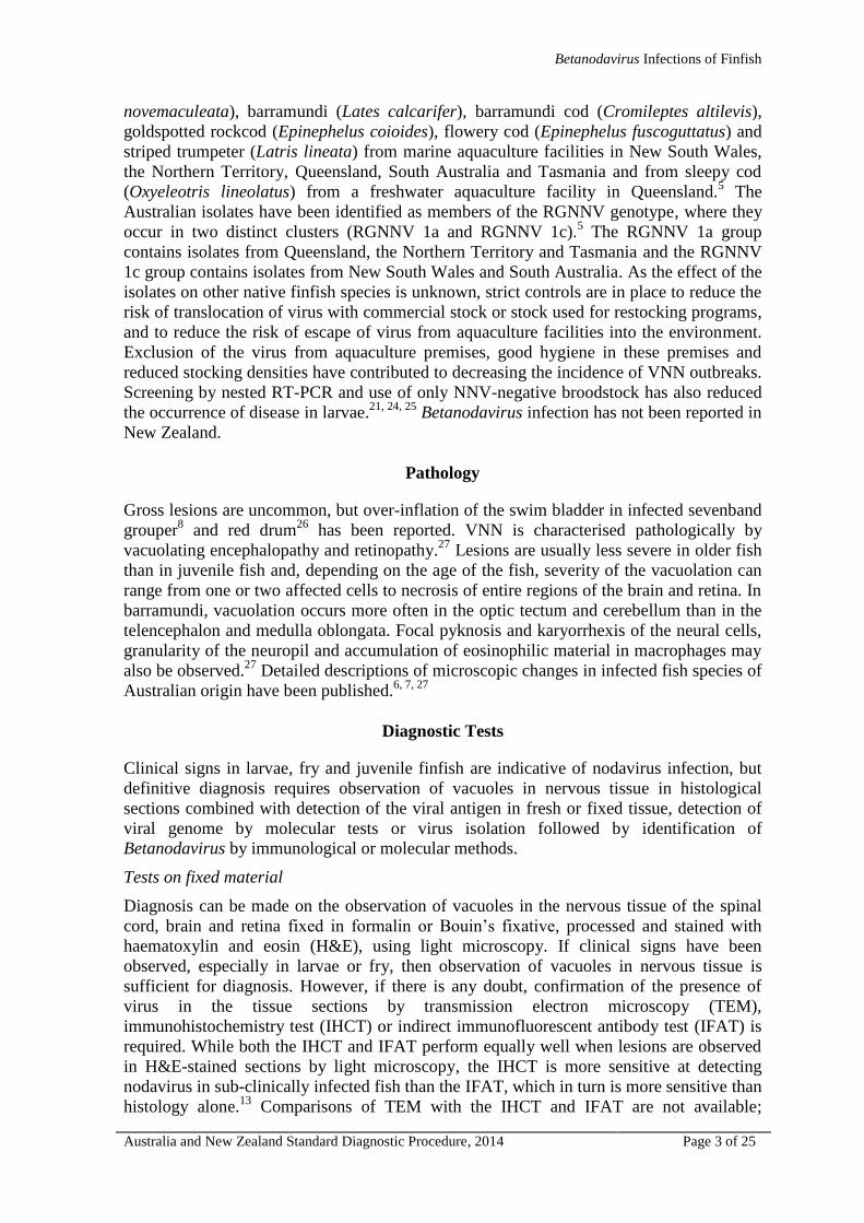
A non-specific band of approximately 900bp is sometimes observed when testing
barramundi and Australian bass samples infected with RGNNV 1c isolates (Figure 3b).
Interpretation of Results
Positive RT-nPCR results from finfish tissue are indicative of the presence of
Betanodavirus RNA in the sample. However, a positive RT-nPCR does not indicate
whether the sample contains infectious virus. Amplicons from positive RT-nPCR results
from facilities, species or geographical locations where Betanodavirus infections have not
been reported previously should be sequenced and the sequence compared with known
Betanodavirus sequences to confirm the result. This is especially important when no other
diagnostic test has been used. A negative result from finfish tissue is indicative of the
absence of Betanodavirus RNA in the sample. When testing broodstock, blood or
spawning material, a positive RT-nPCR is indicative of the presence of Betanodavirus
RNA in the sample. However, a positive RT-nPCR does not indicate whether the sample
contains infectious virus. A negative result when testing broodstock blood or spawning
material is indicative of the absence of nodavirus RNA in the sample. The role of
Betanodavirus infections in broodstock, in particular barramundi, is not well understood
and a negative RT-nPCR result may be a false-negative result.
Figure 3a An example of positive RT-nPCR results for Australian RGNNV 1a isolates.
Upper gel: Primary RT-PCR – positive amplicon of 426bp (arrowed). Lower gel: RT-
nPCR – positive amplicon of 294bp (arrowed).
Page 16
Betanodavirus Infections of Finfish
Australia and New Zealand Standard Diagnostic Procedure, 2014 Page 16 of 25
Figure 3b An example of positive RT-nPCR results for Australian RGNNV 1c isolates
obtained from barramundi. RT-nPCR – positive amplicon of 294bp (white arrow) and
non-specific amplicon of ~900bp (red arrow). M: molecular weight marker, N: no
template control, P: positive control
Virus Isolation
Principle of the Test
Virus isolation in monolayer cultures of susceptible cell lines is the “gold standard” for
determining the presence of infectious virus. For tissue samples submitted in appropriate
condition (unfixed, stored under correct temperature and transportation conditions),
processed correctly for virus isolation at a NATA-accredited diagnostic laboratory and
inoculated onto cultures of susceptible cell lines, then incubated under appropriate
conditions, the development of viral cytopathic effect (CPE) indicates the presence of
infectious virus.
Reagents and Materials
SSN-1 and/or E11 cell line(s) (American Type Culture Collection or European Collection
of Animal Cell Cultures).
Growth medium: Leibovitz’s L-15 medium supplemented with 10% (v/v) foetal bovine
serum, 2 mM glutamine, 100 IU/mL penicillin and 100 µg/mL streptomycin sulphate.
Maintenance medium: Leibovitz’s L-15 medium supplemented with 2% (v/v) foetal bovine
serum, 2 mM glutamine, 100 IU/mL penicillin and 100 µg/mL streptomycin sulphate.
Equipment required for the establishment and maintenance of a fish cell culture laboratory
has been discussed previously.35
Test Procedure
Whole larvae/fry or organ samples are homogenised in TM and should be kept chilled (4°C
to 10°C) during processing. Sample preparation and virus isolation are ideally undertaken
on the day of sample receipt. If this is not possible, samples should be stored at -80°C. All
manipulations are undertaken in a Class II Biological Safety Cabinet using aseptic
technique and sterile equipment and reagents. The sample/tissue required is dependent on
the size of the fish (Table 6).
Table 6 Tissue samples required
Fish size (length) Tissue sample
<1 cm Whole fish
1–5 cm Whole head
5–8 cm Trimmed head
>8 cm Dissected brain and eye
M ----------------------------------------------- Test Samples ------------------------------------------------------ N P
Page 17
Betanodavirus Infections of Finfish
Australia and New Zealand Standard Diagnostic Procedure, 2014 Page 17 of 25
Prepare two sets of sterile centrifuge tubes labelled with the submission identification (ID)
number, sample number and dilution. One set is used for the 1/10 sample dilution and the
other set for the 1/100 sample dilution. Add 4.5 mL TM to each 1/100 tube. A clarified
1:10 (w/v) tissue suspension in TM is required after homogenisation. Therefore, weigh a
sample container with a tissue sample and subtract the weight of an empty sample
container to obtain an estimate of the weight of the tissue sample. Operating within a Class
II Biological Safety Cabinet and using 10 mL sterile pipettes, dispense 4.5 mL TM into
each 1/100 tube. Place all tubes in a test tube rack in an ice slurry. Homogenise the tissue
sample and resuspend in extra TM to achieve a 1:10 (w/v) tissue homogenate. Transfer to
the tubes labelled 1/10 and clarify the homogenate by centrifugation at 3000 g for 15
minutes at 4°C. Pipette 0.5 mL of the supernatant from each 1/10 tissue sample dilution
into the corresponding 1/100 test tubes containing 4.5 mL TM.
The procedure described is for virus isolation in cultures of the SSN-1 or E11 cell line
established in 24-well tissue culture plates. If different culture vessels are used (e.g. 96-
well plates, 25 cm² tissue culture flasks), volumes are adjusted proportionally. Cells are
seeded at a density of 1.7 105 cells/mL with 1.5 mL of cell suspension (in L-15 medium
complete with 10% FBS and antibiotics) and incubated at 25°C overnight. Cultures should
be less than 24 hours old when inoculated with the diagnostic sample. On the day of
sample inoculation, examine cell cultures to be used by inverted light microscopy. Ensure
that they are approximately 70% to 80% confluent, free from overt microbial
contamination, and that mitotic figures are visible (that is, the cultures contain actively
dividing cells). Any problems should be noted and, if necessary, fresh cultures prepared for
use on the next day. Discard the cell culture medium from the SSN-1 or E-11 cultures.
Inoculate duplicate cultures with 150 µL of each sample dilution (1/10 and 1/100). One set
(column) of four well-cultures on each 24-well tissue culture plate should be used as
negative controls, which are inoculated with 150 µL TM only. Incubate the cultures at
25°C for one hour to allow adsorption of any virus present. Following adsorption, add 1.5
mL maintenance medium to each culture (negative controls first) yielding final sample
dilutions of 1/100 and 1/1000. Place culture plates in a 25°C incubator. On the day
following inoculation, and every 1-3 days thereafter, examine the cultures by inverted light
microscopy for any microbial contamination, tissue sample (non-specific) cytotoxicity and
viral cytopathic effect (CPE).
Subculturing
At 7-12 days after inoculation, test cultures not showing CPE should be passaged. For each
tissue sample, the contents (medium and cells scraped from the substrate) of each of the
four replicate cultures, irrespective of dilution, are pooled into sterile centrifuge tubes. If
tissue sample cytotoxicity or bacterial/fungal contamination has been observed during the
initial culture period for each pool, the pooled contents should be filtered (0.45 μm) into
the sterile centrifuge tube. Alternatively, samples can be centrifuged at 10 000 g for 10
minutes at 5°C. Each pooled sample is diluted (1/10) by pipetting 0.5 mL of the
supernatant into a test tube containing 4.5 mL TM. Without decanting the cell culture
medium, inoculate duplicate fresh cell cultures in 24-well culture plates, prepared as
described above, with 150 µL of the pooled undiluted supernatant and a further duplicate
set of wells with the 150 µL of the 1/10 dilution of the pooled supernatant. Place culture
plates in a 25°C incubator. Observe these cultures and record observations. Irrespective of
the time at which the passage occurred, cell cultures should be observed for at least 21
days for completion of the assay. The test is valid if the negative control cultures retain
normal cellular morphology for the full period of incubation in the absence of bacterial
Page 18
Betanodavirus Infections of Finfish
Australia and New Zealand Standard Diagnostic Procedure, 2014 Page 18 of 25
contamination. To ensure cell lines, used on a routine basis, are susceptible to the viruses
of concern, titrations of a positive control viral stock on each of the cell lines should be
carried out on a regular basis (e.g. 6 monthly).
Interpretation of Results
The test sample is negative if the inoculated cell cultures retain normal cellular
morphology similar to the negative control cultures and the cellular monolayer retains
normal integrity. If virus isolation is the only test performed, confirmation by ICCT, IFAT
or RT-qPCR should be undertaken on a sample of the cell culture to avoid false-negative
results. If any of the cell cultures inoculated with test samples demonstrate any
abnormalities, such as increased intracellular vacuolation, or monolayer disruption (Figure
4), further investigation is required, such as examination by electron microscopy, further
sub-culturing or confirmation of the presence of Betanodavirus by molecular methods as
described above, or by ICCT or IFAT (see below).
A
Page 19
Betanodavirus Infections of Finfish
Australia and New Zealand Standard Diagnostic Procedure, 2014 Page 19 of 25
Figure 4 Examples of an uninfected SSN-1 cell culture, 4 days post-seeding (A) and
of a BNNV-infected SSN-1 cell culture, 4 days post-seeding, 3 days post-inoculation (B)
Immunocytochemistry Test (ICCT) and Indirect Immunofluorescent Antibody
Test (IFAT) of cell cultures
Principle of the Test
This test uses polyclonal antibodies raised in sheep against the recombinant coat protein of
a barramundi or sleepy cod Betanodavirus isolate and an anti-sheep IgG horseradish
peroxidise or Cy2™-conjugated secondary antibody, to localise viral coat protein in fixed
cell cultures used in an attempt to isolate NNV from fish tissues. The test is used to
confirm or exclude Betanodavirus as the agent causing observed CPE or to exclude
Betanodavirus infection in cell cultures not showing CPE.
Reagents and Materials
As previously stated.
Test Procedure
Fixation
Drain culture medium from the cell cultures. Dispose of the supernatant according to
standard procedures. Rinse the cell cultures gently in TBS. Fix the cell cultures in 50%
acetone/50% methanol. Add 1 mL per well for 24-well plates (or 3 mL per 25 cm² flask).
Incubate for 5 minutes at room temperature, with gentle agitation. Remove fixative and
rinse fixed cultures with TBS.
B
Page 20
Betanodavirus Infections of Finfish
Australia and New Zealand Standard Diagnostic Procedure, 2014 Page 20 of 25
ICCT
Block endogenous peroxidase by adding 3% H2O2 in methanol. Add 1 mL per well for 24-
well plates (or 3 mL per 25 cm² flask). Incubate for 20 minutes at room temperature. Rinse
three times with TBS and block non-specific binding sites with 5% BSA in TBS by adding
1 mL per well for 24-well plates (or 3 mL per 25 cm² flask) and incubate for 30 minutes at
37C. Rinse each well three times with TBS. Immediately before use dilute sheep anti-
NNV rCP polyclonal antibody (1 Ab) 1/1000 in 2.5% BSA in TBS and add to the cell
culture: add 500 L to each well for 24-well plates (or 3 mL per 25 cm² flask). Incubate at
37C for 60 minutes. Rinse each well three times with TBS. Immediately before use dilute
rabbit anti-sheep IgG [H+L] HRP conjugate 1/1000 in 2.5% BSA in TBS and add to the
cell culture: Add 500 L to each well for 24-well plates (or 3 mL per 25 cm² flask).
Incubate at 37C for 60 minutes. Prepare the HRP Chromogen, according to the
manufacturer’s instructions and add 1 mL per well for 24-well plates (or 3 mL per 25 cm2
flask). Stop development after 10 minutes by aspirating the HRP Chromogen and replacing
with Milli-Q water. Counterstain in Mayer’s haematoxylin for 60 seconds, rinse in Milli-Q
water for 60 seconds and ‘blue’ with lithium carbonate for 60 seconds By adding 1 mL per
well for 24-well plates (or 3 mL per 25 cm² flask). Rinse in Milli-Q water, rinse each well
three times with TBS and observe under an inverted light microscope.
IFAT
Block non-specific binding sites by adding 1 mL 5% (w/v) BSA in TBS to each well and
incubate for 30 minutes at 37C. Rinse each well three times with TBS. Immediately
before use dilute sheep anti-NNV rCP polyclonal antibody (1 Ab) 1/1000 in 2.5% BSA
(w/v in TBS) and add to the cell culture. Use 500 L to each well for 24-well plates (or 3
mL per 25 cm2 flask). Incubate at 37C for 60 minutes. Rinse each well three times with
TBS. Immediately before use dilute rabbit anti-sheep IgG [H+L] Cy2™ conjugate 1/1000
in 2.5% BSA (w/v in TBS) and add to the cell culture. Add 500 L to each well for 24-
well plates (or 3 mL per 25 cm2 flask). Incubate at 37C for 60 minutes. Turn on the
mercury lamp of a fluorescence microscope 20 minutes prior to use. Wash each well three
times with TBS and observe under the fluorescence microscope.
Quality Control Aspects
Positive and negative control cell cultures must be included in every test. Ideally, a positive
cell culture exhibiting a low level of infection should also be included. For the ICCT to be
valid, dark brown or black staining cytoplasm of cells must be observed in the positive
control cultures (Figure 5a). For the IFAT to be valid, bright green fluorescence in the
cytoplasm must be observed in the positive control cultures (Figure 5b). No specific
staining should be seen in the negative control cultures.
Interpretation of Results
Any positive staining indicates the presence of the Betanodavirus coat protein and the
culture is considered to be positive for Betanodavirus infection.
Page 21
Betanodavirus Infections of Finfish
Australia and New Zealand Standard Diagnostic Procedure, 2014 Page 21 of 25
Figure 5 Examples of an ICCT positive BNNV-infected SSN-1 cell culture (A) and
of an IFAT positive BNNV-infected SSN-1 cell culture (B)
A
B
Page 22
Betanodavirus Infections of Finfish
Australia and New Zealand Standard Diagnostic Procedure, 2014 Page 22 of 25
References
1. Thiéry R, Johnson KL, Nakai T et al. Nodaviridae. In: King AMQ, Lefkowitz E, Adams MJ,
Carstens EB, editors. Virus taxonomy: classification and nomenclature of Viruses Ninth report of the
International Committee on Taxonomy of Viruses. Elsevier, 2012:1061-1067.
2. Mori KI, Nakai T, Muroga K et al. Properties of a new virus belonging to Nodaviridae found in
larval striped jack (Pseudocaranx dentex) with nervous necrosis. Virology 1992;187:368-371.
3. Nishizawa T, Furuhashi M, Nagai T et al. Genomic classification of fish nodaviruses by molecular
phylogenetic analysis of the coat protein gene. Applied and Environmental Microbiology 1997;63:1633-
1636.
4. Johansen R, Sommerset I, Torud B et al. Characterization of nodavirus and viral encephalopathy
and retinopathy in farmed turbot, Scophthalmus maximus (L.). Journal of Fish Diseases 2004;27:591-601.
5. Moody NJG, Horwood PF, Reynolds A et al. Phylogenetic analysis of betanodavirus isolates from
Australian finfish. Diseases of Aquatic Organisms 2009;87:151-160.
6. Munday BL, Kwang J, Moody N. Betanodavirus infections of teleost fish: a review. Journal of Fish
Diseases 2002;25:127-142.
7. Anderson IG, Moody NJ. The effect of barramundi nodavirus on important freshwater fishes.
Fisheries Research and Development Corporation Project 1999/205 Final Report 2004.
8. Fukuda Y, Nguyen HD, Furuhashi M et al. Mass mortality of cultured sevenband grouper,
Epinephelus septemfasciatus, associated with viral nervous necrosis. Fish Pathology 1996;31:165-170.
9. LeBreton A, Grisez L, Sweetman J et al. Viral nervous necrosis (VNN) associated with mass
mortalities in cage-reared sea bass, Dicentrarchus labrax (L). Journal of Fish Diseases 1997;20:145-151.
10. Athanassopoulou F, Billinis C, Prapas T. Important disease conditions of newly cultured species in
intensive freshwater farms in Greece: first incidence of nodavirus infection in Acipenser sp. Diseases of
Aquatic Organisms 2004;60:247-252.
11. Hick P, Whittington RJ. Optimisation and validation of a real-time reverse transcriptase-polymerase
chain reaction assay for detection of betanodavirus. Journal of Virological Methods 2010;163:368-377.
12. Johansen R, Amundsen M, Dannevig BH et al. Acute and persistent experimental nodavirus
infection in spotted wolffish Anarhichas minor. Diseases of Aquatic Organisms 2003;57:35-41.
13. Moody NJ, Horwood PF, McHardy S. Development of diagnostic tests for the detection of
nodavirus. Fisheries Research and Development Corporation Project 2001/626 Final Report 2004.
14. Johansen R, Grove S, Svendsen AK et al. A sequential study of pathological findings in Atlantic
halibut, Hippoglossus hippoglossus (L.), throughout one year after an acute outbreak of viral encephalopathy
and retinopathy. Journal of Fish Diseases 2004;27:327-341.
15. Anderson IG, Oakey HJ. The production of nodavirus-free fish fry and the nodaviruses natural
distribution. Fisheries Research and Development Corporation Project 2002/043 Final Report 2008.
16. Castric J, Thiery R, Jeffroy J et al. Sea bream Sparus aurata, an asymptomatic contagious fish host
for nodavirus. Diseases of Aquatic Organisms 2001;47:33-38.
17. Gomez DK, Sato J, Mushiake K et al. PCR-based detection of betanodaviruses from cultured and
wild marine fish with no clinical signs. Journal of Fish Diseases 2004;27:603-608.
18. Tanaka S, Mori K, Arimoto M et al. Protective immunity of sevenband grouper, Epinephelus
septemfasciatus Thunberg, against experimental viral nervous necrosis. Journal of Fish Diseases 2001;24:15-
22.
19. Breuil G, Romestand B. A rapid ELISA method for detecting specific antibody level against
nodavirus in the serum of the sea bass, Dicentrachus labrax (L.): application to the screening of spawners in
a sea bass hatchery. Journal of Fish Diseases 1999;22:45-52.
20. Whittington RJ. Aquatic Animal Health Subprogram: tools for investigation of the nodavirus carrier
state in marine, euryhaline and freshwater fish and control of NNV through integrated management.
Fisheries Research and Development Corporation Project 2008/041 Final Report 2012.
21. Watanabe K, Nishizawa T, Yoshimizu M. Selection of brood stock candidates of barfin flounder
using an ELISA system with recombinant protein of barfin flounder nervous necrosis virus. Diseases of
Aquatic Organisms 2000;41:219-223.
22. OIE. Chapter 2.3.11 Viral encephalopathy and retinopathy, Manual of Diagnostic Tests for Aquatic
Animals, The World Organisation for Animal Health (OIE).
http://www.oie.int/fileadmin/Home/eng/Health_standards/aahm/current/2.3.11_VER.pdf. 2013. Retrieved
11/11/13
23. Hick P, Schipp G, Bosmans J et al. Recurrent outbreaks of viral nervous necrosis in intensively
cultured barramundi (Lates calcarifer) due to horizontal transmission of betanodavirus and recommendations
for disease control. Aquaculture 2011;319:41-52.
Page 23
Betanodavirus Infections of Finfish
Australia and New Zealand Standard Diagnostic Procedure, 2014 Page 23 of 25
24. Mori K, Mushiake K, Arimoto M. Control measures for viral nervous necrosis in striped jack. Fish
Pathology 1998;33:443-444.
25. Mushiake K, Nishizawa T, Nakai T et al. Control of VNN in striped jack: selection of spawners
based on the detection of SJNNV gene by polymerase chain reaction (PCR). Fish Pathology 1994;29:177-
182.
26. Oh MJ, Jung SJ, Kim SR et al. A fish nodavirus associated with mass mortality in hatchery-reared
red drum, Sciaenops ocellatus. Aquaculture 2002;211:1-7.
27. Munday BL, Langdon JS, Hyatt AD et al. Mass mortality associated with viral-induced vacuolating
encephalopathy and retinopathy of larval and juvenile barramundi, Lates calcarifer Bloch. Aquaculture
1992;103:197-211.
28. Crane MSJ, McColl KA, Davies KR et al. Evaluation of the draft Australian and New Zealand
Standard Diagnostic Procedure (ANZSDP) for PCR-detection of betanodaviruses. Fish Pathology
2007;42:173-179.
29. Frerichs GN, Morgan D, Hart D et al. Spontaneously productive C-type retrovirus infection of fish
cell-lines. Journal of General Virology 1991;72:2537-2539.
30. Iwamoto T, Nakai T, Mori K et al. Cloning of the fish cell line SSN-1 for piscine nodaviruses.
Diseases of Aquatic Organisms 2000;43:81-89.
31. Standards Australia. Safety in laboratories. Part 3: Microbiological safety and containment. AS/NZ
2243.3.2010. Standards Australia, Sydney, NSW, 2010.
http://infostore.saiglobal.com/store/Details.aspx?ProductID=1430097
32. Caraguel CGB, Stryn H, Gagné N et al. Selection of a cutoff for real-time plymerase chain reaction
results to fit a diagnostic purpose: analytical and epidemiologic approaches. Journal of Veterinary Diagnostic
Investigation 2011;23:2-15.
33. Nishizawa T, Mori K, Nakai T et al. Polymerase chain reaction (PCR) amplification of RNA of
striped jack nervous necrosis virus (SJNNV). Diseases of Aquatic Organisms 1994;18:103-107.
34. Thiery R, Raymond JC, Castric J. Natural outbreak of viral encephalopathy and retinopathy in
juvenile sea bass, Dicentrarchus labrax: study by nested reverse transcriptase-polymerase chain reaction.
Virus Research 1999;63:11-17.
35. Crane MSJ, Williams LM. Viruses of salmonids: Virus isolation in fish cell lines, Australian and
New Zealand Standard Diagnostic Procedure, Sub-Committee on Animal Health Laboratory Standards
http://www.scahls.org.au/__data/assets/pdf_file/0008/1522691/Virus_Isolation.pdf. 2008. Retrieved 11/11/13
Part 3 - Reagents and Kits
Betanodavirus-specific polyclonal antibody
Polyclonal antibodies, raised in sheep against the recombinant coat proteins from
barramundi and sleepy cod Betanodavirus isolates, are available from the Queensland
Government Department of Agriculture, Fisheries and Forestry, Biosecurity Sciences
Laboratory (BSL), Brisbane, Queensland.
Betanodavirus-infected finfish for positive controls for IHCT
Tissue from Betanodavirus-infected finfish on histological slides is available from the
Queensland Government Department of Agriculture, Fisheries and Forestry Biosecurity
Sciences Laboratory (BSL), Brisbane, Queensland.
Betanodavirus-infected finfish homogenate and Betanodavirus-infected SSN-1 cell
culture supernatant for positive controls for RT-qPCR and RT-nPCR
Homogenates from Betanodavirus infected finfish and Betanodavirus-infected cell culture
supernatant are available from the authors.
Page 24
Betanodavirus Infections of Finfish
Australia and New Zealand Standard Diagnostic Procedure, 2014 Page 24 of 25
Suppliers of Reagents and Kits
In Australia:
AAHL Fish Diseases Laboratory
CSIRO Australian Animal Health Laboratory
5 Portarlington Rd
East Geelong VIC 3219
Ph: + 61 3 5227 5000
Fax: +61 3 5227 5555
Email: [email protected]
Biosecurity Sciences Laboratory (BSL)
Health and Food Science Precinct
Block 12, 39 Kessels Road,
Coopers Plains QLD 4108
Ph: +61 7 3276 6062
Fax +61 7 3216 6620
Email: [email protected]
Jackson Immunoresearch
http://www.jacksonimmuno.com/home.asp
Abacus ALS Australia
12 Mowbray Tce
East Brisbane QLD 4169
Email: [email protected]
Tel: 1800 222 287
Fax: 1800 287 222
Pierce
www.piercenet.com
Thermo Fisher Scientific
www.thermofisher.com.au
5 Caribbean Drive
P.O. Box 9092
Scoresby VIC 3179
Email: [email protected]
Tel: 1300 735 292
Qiagen
www.qiagen.com
PO Box 169
Chadstone Centre VIC 3148
Email: [email protected]
Ph: 1800 243 066
Fax: 03-9840-9888
Life Technologies
http://www.lifetechnologies.com/au/en/home.html
Life Technologies Australia Pty Ltd.
30-32 Compark Circuit
Mulgrave VIC 3170
Email: [email protected]
Tel: 1800 636 327
Fax: 1800 143 363
Page 25
Betanodavirus Infections of Finfish
Australia and New Zealand Standard Diagnostic Procedure, 2014 Page 25 of 25
In New Zealand:
Jackson Immunoresearch
http://www.jacksonimmuno.com/home.asp
Abacus ALS New Zealand
PO Box 97-923
South Auckland Mail Center
Auckland
E-Mail: [email protected]
Tel:0800 222 170
Fax: 0800 222 180
Pierce
www.piercenet.com
Thermo Fisher Scientific
http://www.thermofisher.co.nz/
5 Caribbean Drive
P.O. Box 9092
Scoresby VIC 3179
Email: [email protected]
Tel: 1300 735 292
Qiagen
www.qiagen.com
Bio-Strategy Ltd
www.bio-strategy.com
PO Box 303 385
Auckland 0751
E-mail: [email protected]
Tel: 0800 34 24 66
Life Technologies
www.Life Technologies.com.au
Life Technologies New Zealand Limited
18-24 Botha Road
Penrose
Auckland 1006
Email: [email protected]
Tel: 0800 636 327