72
Big Group Discussion Group C3B Malig-Marayag Carbuncle with Ipsilateral Facial Cellulitis in a Diabetic
| Date post: | 01-Jan-2016 |
| Category: |
Documents |
| Upload: | eugenia-porter |
| View: | 220 times |
| Download: | 0 times |

Big Group DiscussionGroup C3B
Malig-Marayag
Carbuncle with Ipsilateral Facial Cellulitis in a Diabetic
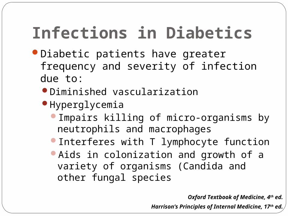
Infections in DiabeticsDiabetic patients have greater frequency
and severity of infection due to:Diminished vascularizationHyperglycemia
Impairs killing of micro-organisms by neutrophils and macrophages
Interferes with T lymphocyte functionAids in colonization and growth of a variety
of organisms (Candida and other fungal species
Harrison’s Principles of Internal Medicine, 17th ed.
Oxford Textbook of Medicine, 4th ed.
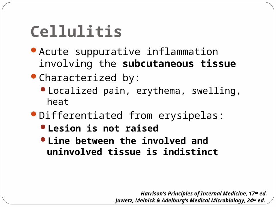
CellulitisAcute suppurative inflammation involving
the subcutaneous tissueCharacterized by:
Localized pain, erythema, swelling, heatDifferentiated from erysipelas:
Lesion is not raisedLine between the involved and
uninvolved tissue is indistinct
Harrison’s Principles of Internal Medicine, 17th ed.Jawetz, Melnick & Adelburg’s Medical Microbiology, 24th ed.

CellulitisMild local erythema and tenderness
Rapidly becomes intense and spreadsArea becomes infiltrated and pits on pressureCentral part may become nodular and
develop a vesicle that ruptures and discharges pus and necrotic material
MalaiseFever and chills
Andrews’ Diseases of the Skin: Clinical Dermatology, 10 th ed.

CellulitisMost commonly caused by indigenous flora
Staphylococcus aureus – usually associated with an abscess, folliculitis, or foreign body
Streptococcus pyogenes – spreads more rapidly; associated with fever and lymphangitis
Bacteria may gain access to the epidermis through:Cracks in the skin, abrasions, cuts, burns,
insect bites, surgical incisions, intravenous catheters
Harrison’s Principles of Internal Medicine, 17th ed.
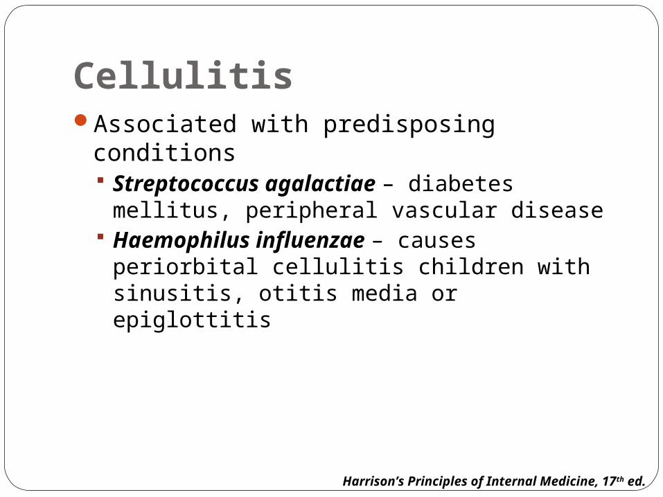
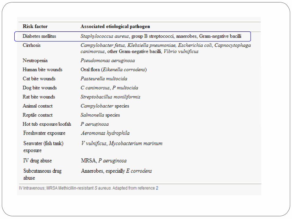
CellulitisAssociated with predisposing conditions
Streptococcus agalactiae – diabetes mellitus, peripheral vascular disease
Haemophilus influenzae – causes periorbital cellulitis children with sinusitis, otitis media or epiglottitis
Harrison’s Principles of Internal Medicine, 17th ed.
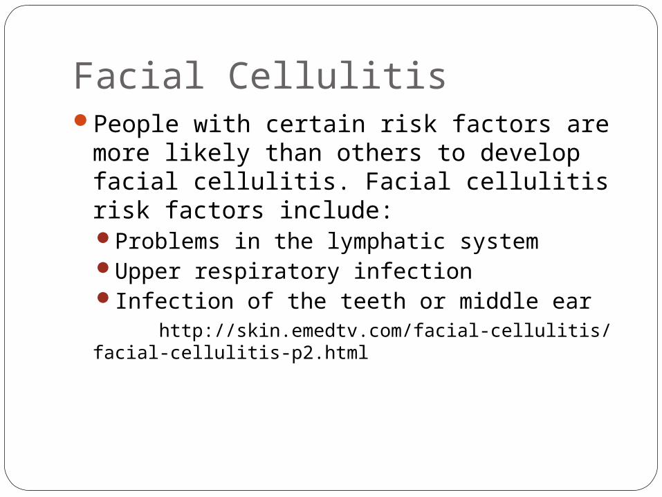
Facial CellulitisPeople with certain risk factors are more
likely than others to develop facial cellulitis. Facial cellulitis risk factors include:Problems in the lymphatic system Upper respiratory infection Infection of the teeth or middle ear
http://skin.emedtv.com/facial-cellulitis/facial-cellulitis-p2.html
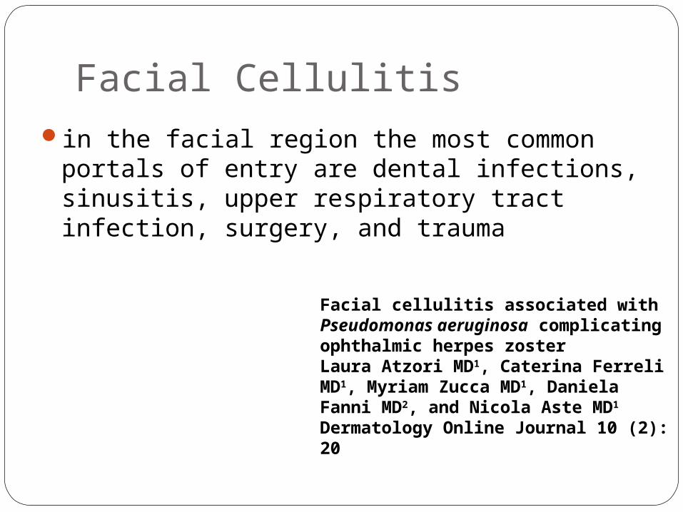
Facial Cellulitisin the facial region the most common portals
of entry are dental infections, sinusitis, upper respiratory tract infection, surgery, and trauma
Facial cellulitis associated with Pseudomonas aeruginosa complicating ophthalmic herpes zoster Laura Atzori MD1, Caterina Ferreli MD1, Myriam Zucca MD1, Daniela Fanni MD2, and Nicola Aste MD1 Dermatology Online Journal 10 (2): 20
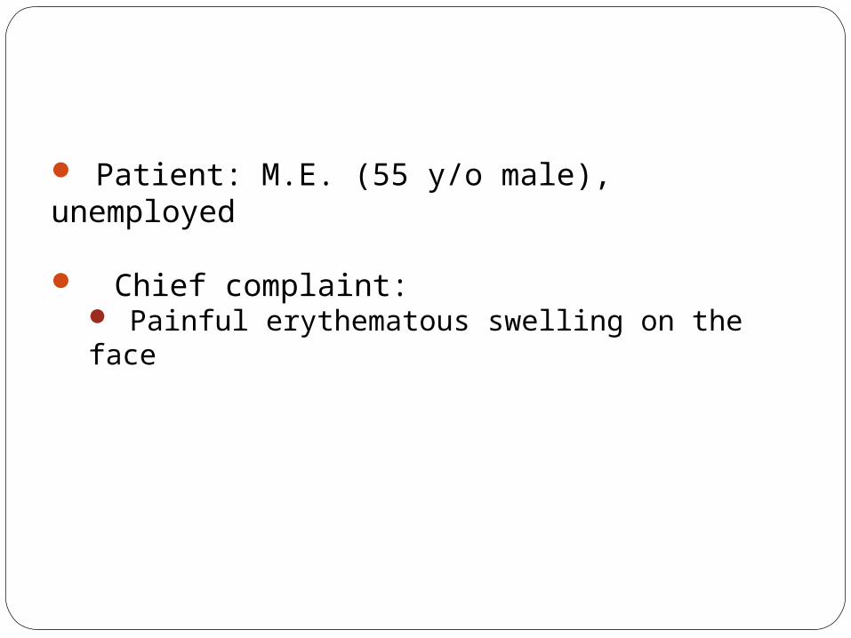
Patient: M.E. (55 y/o male), unemployed
Chief complaint: Painful erythematous swelling on the face

HISTORY OF PRESENT ILLNESS
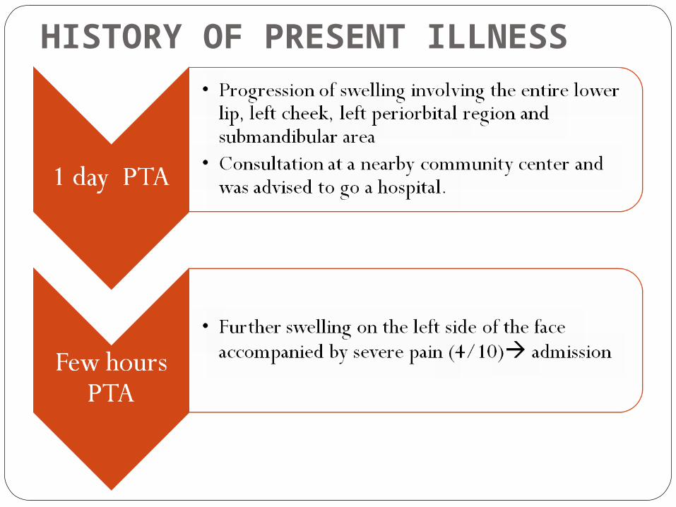
HISTORY OF PRESENT ILLNESS
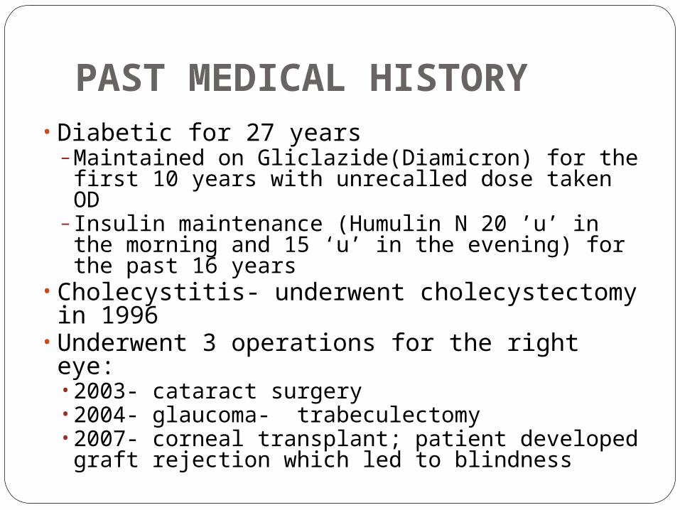
PAST MEDICAL HISTORY• Diabetic for 27 years
– Maintained on Gliclazide(Diamicron) for the first 10 years with unrecalled dose taken OD
– Insulin maintenance (Humulin N 20 ’u’ in the morning and 15 ‘u’ in the evening) for the past 16 years
• Cholecystitis- underwent cholecystectomy in 1996
• Underwent 3 operations for the right eye:• 2003- cataract surgery • 2004- glaucoma- trabeculectomy • 2007- corneal transplant; patient developed graft rejection which led to blindness
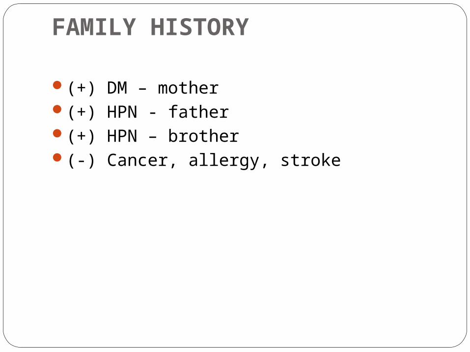
FAMILY HISTORY
(+) DM – mother(+) HPN - father(+) HPN – brother(-) Cancer, allergy, stroke
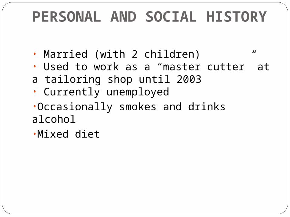
PERSONAL AND SOCIAL HISTORY
• Married (with 2 children)• Used to work as a “master cutter” at a tailoring shop until 2003• Currently unemployed•Occasionally smokes and drinks alcohol•Mixed diet
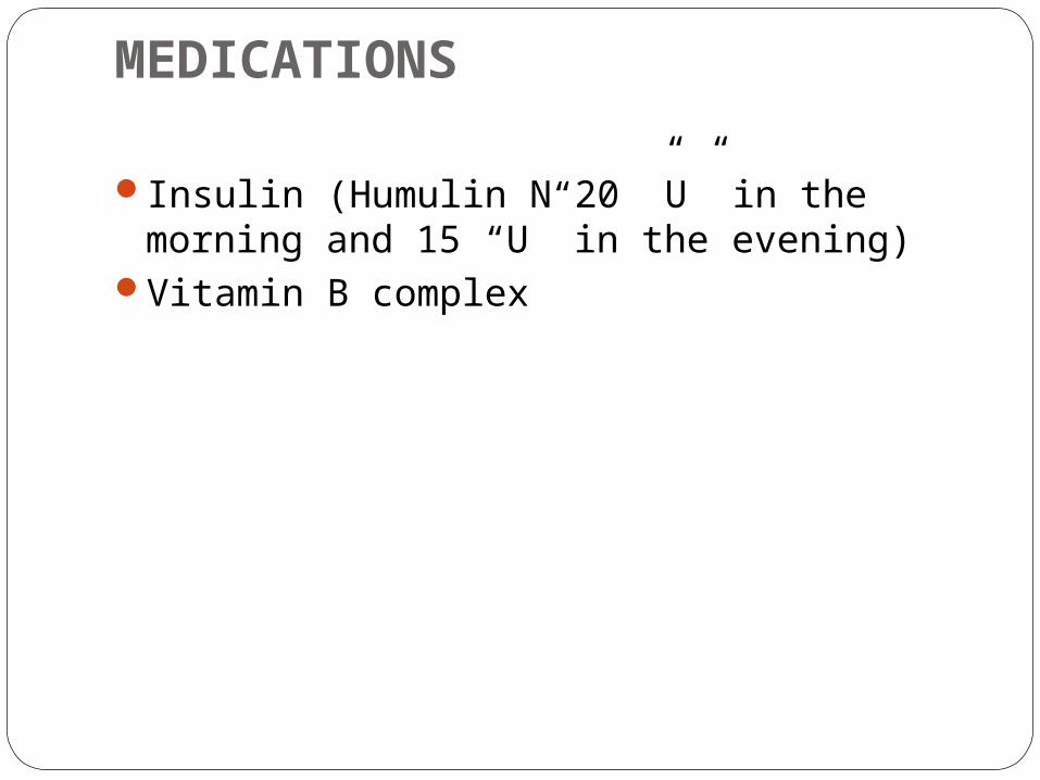
MEDICATIONS
Insulin (Humulin N 20 ”U” in the morning and 15 “U” in the evening)
Vitamin B complex
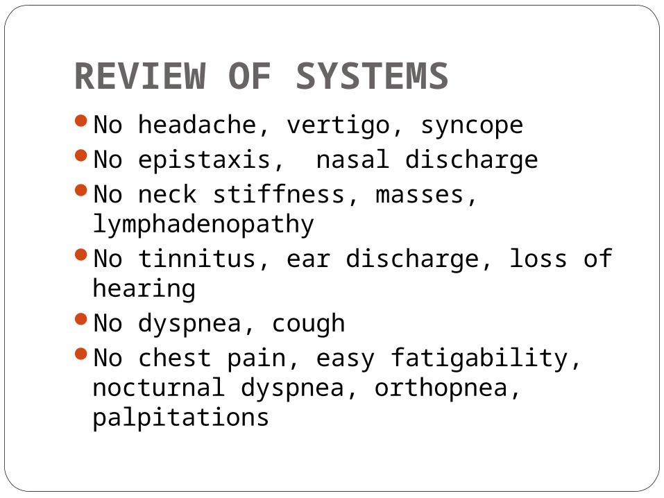
REVIEW OF SYSTEMSNo headache, vertigo, syncopeNo epistaxis, nasal dischargeNo neck stiffness, masses,
lymphadenopathyNo tinnitus, ear discharge, loss of
hearingNo dyspnea, coughNo chest pain, easy fatigability,
nocturnal dyspnea, orthopnea, palpitations
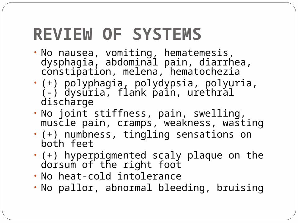
REVIEW OF SYSTEMS• No nausea, vomiting, hematemesis,
dysphagia, abdominal pain, diarrhea, constipation, melena, hematochezia
• (+) polyphagia, polydypsia, polyuria, (-) dysuria, flank pain, urethral discharge
• No joint stiffness, pain, swelling, muscle pain, cramps, weakness, wasting
• (+) numbness, tingling sensations on both feet
• (+) hyperpigmented scaly plaque on the dorsum of the right foot
• No heat-cold intolerance• No pallor, abnormal bleeding, bruising

ON ADMISSION (SEPT 18, 2009)
Conscious, coherent and oriented to time, place and person
Vital Signs: BP: 130/70mmHg PR - 109bpm, RR-20cpm, Temp: 36.7°C
Weight= 66 kg Height= 170cm BMI=23 HEENT: (+) periorbital swelling,
nonhyperemic conjunctivae, anicteric sclerae, cornea opaque, OU, retina pigmented OU, lens cannot be assessed, (+) light perception, OD, (+) hand movement, OS
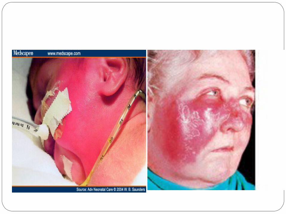
No nasoaural discharge, (+) swelling with violaceous discoloration of the lower lip and surrounding skin topped with multiple erosions, crusts and pus, buccal mucosa cannot be assessed
NECK: supple neck, no masses, (-) palpable cervical LN,right, LN on the left cannot be assessed due to swelling over the submandibular area, (-) neck vein distension
Conscious, coherent and oriented to time, place and person
Vital Signs: BP: 130/70mmHg PR - 109bpm, RR-20cpm, Temp: 36.7°C
Weight= 66 kg Height= 170cm BMI=23 HEENT: (+) periorbital swelling,
nonhyperemic conjunctivae, anicteric sclerae, cornea opaque, OU, retina pigmented OU, lens cannot be assessed, (+) light perception, OD, (+) hand movement, OS
No nasoaural discharge, (+) swelling with violaceous discoloration of the lower lip and surrounding skin topped with multiple erosions, crusts and pus, buccal mucosa cannot be assessed
NECK: supple neck, no masses, (-) palpable cervical LN,right, LN on the left cannot be assessed due to swelling over the submandibular area, (-) neck vein distension
SEPT 19, 2009 Conscious, coherent and oriented to
time, place and person Vital Signs: BP: 120/80mmHg PR -
90bpm, RR-24cpm, Temp: 37.3°C Weight= 66 kg Height= 170cm BMI=23 HEENT: (+) periorbital swelling,
nonhyperemic conjunctivae, anicteric sclerae, cornea opaque OU
No nasoaural discharge (+) swelling with ruptured carbuncle on
the left lower lip sorrounded by cellulitis, buccal mucosa cannot be assessed
NECK: supple neck, no masses, (-) palpable cervical LN,right, LN on the left cannot be assessed due to swelling over the submandibular area, (-) neck vein distension
Conscious, coherent and oriented to time, place and person
Vital Signs: BP: 120/80mmHg PR - 90bpm, RR-24cpm, Temp: 37.3°C
Weight= 66 kg Height= 170cm BMI=23 HEENT: (+) periorbital swelling,
nonhyperemic conjunctivae, anicteric sclerae, cornea opaque OU
No nasoaural discharge (+) swelling with ruptured carbuncle on
the left lower lip sorrounded by cellulitis, buccal mucosa cannot be assessed
NECK: supple neck, no masses, (-) palpable cervical LN,right, LN on the left cannot be assessed due to swelling over the submandibular area, (-) neck vein distension
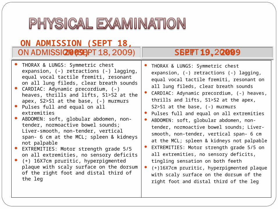
THORAX & LUNGS: Symmetric chest expansion, (-) retractions (-) lagging, equal vocal tactile fremiti, resonant on all lung fileds, clear breath sounds
CARDIAC: Adynamic precordium, (-) heaves, thrills and lifts, S1>S2 at the apex, S2>S1 at the base, (-) murmurs
Pulses full and equal on all extremities ABDOMEN: soft, globular abdomen, non-
tender, normoactive bowel sounds; Liver-smooth, non-tender, vertical span- 6 cm at the MCL; spleen & kidneys not palpable
EXTREMITIES: Motor strength grade 5/5 on all extremities, no sensory deficits
(+) 16X7cm pruritic, hyperpigmented plaque with scaly surface on the dorsum of the right foot and distal third of the leg
THORAX & LUNGS: Symmetric chest expansion,
(-) retractions (-) lagging, equal vocal tactile
fremiti, resonant on all lung fileds, clear breath
sounds CARDIAC: Adynamic precordium, (-) heaves,
thrills and lifts, S1>S2 at the apex, S2>S1 at the
base, (-) murmurs Pulses full and equal on all extremities ABDOMEN: soft, globular abdomen, non-tender,
normoactive bowel sounds; Liver-smooth, non-
tender, vertical span- 6 cm at the MCL; spleen &
kidneys not palpable EXTREMITIES: Motor strength grade 5/5 on all
extremities, no sensory deficits, tingling sensation
on both feeth (+)16X7cm pruritic, hyperpigmented plaque with
scaly surface on the dorsum of the right foot and
distal third of the leg
ON ADMISSION (SEPT 18, 2009) SEPT 19, 2009
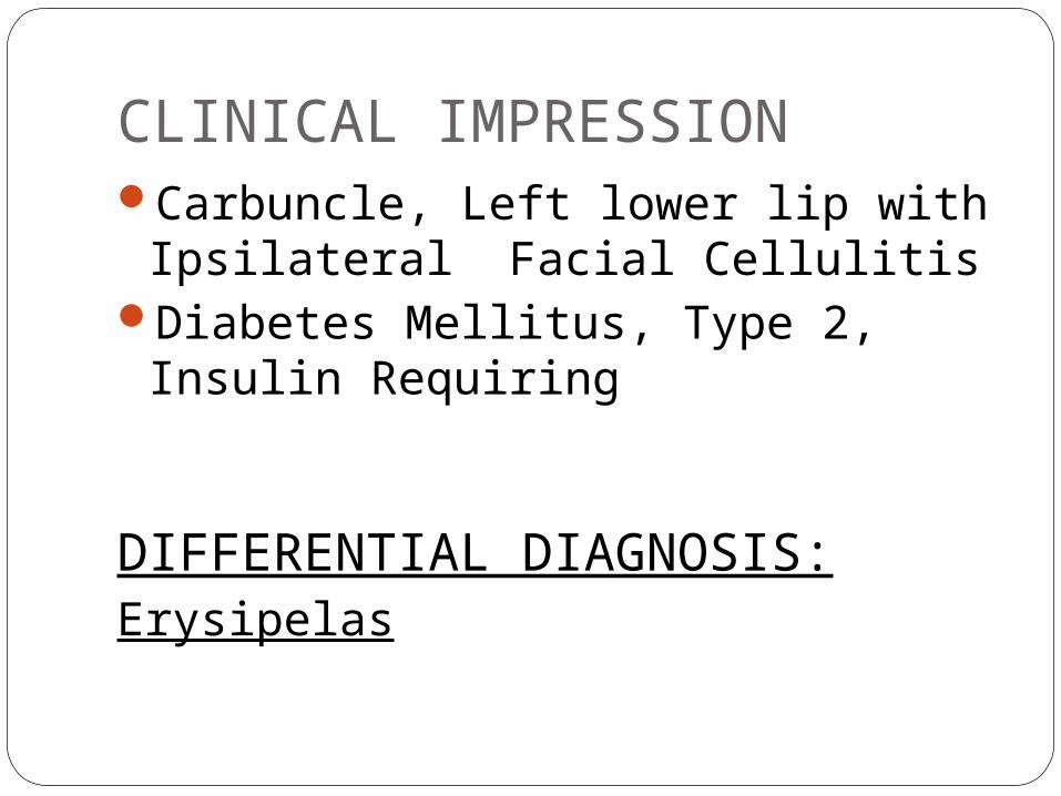
CLINICAL IMPRESSIONCarbuncle, Left lower lip with
Ipsilateral Facial Cellulitis Diabetes Mellitus, Type 2, Insulin
Requiring
DIFFERENTIAL DIAGNOSIS:Erysipelas
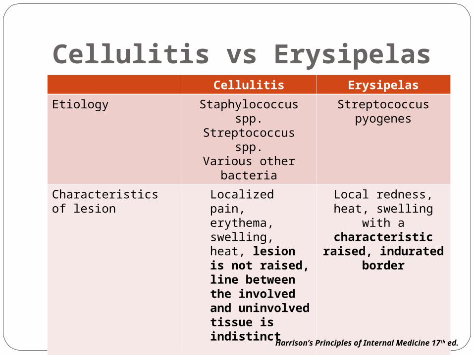
Cellulitis vs ErysipelasCellulitis Erysipelas
Etiology Staphylococcus spp.Streptococcus spp.
Various other bacteria
Streptococcus pyogenes
Characteristics of lesion
Localized pain, erythema, swelling, heat, lesion is not raised, line between the involved and uninvolved tissue is indistinct
Local redness, heat, swelling with a characteristic
raised, indurated border
Accompanying signs and symptoms
Malaise, chills, fever Malaise, chills, high fever, headache,
vomiting, joint painsHarrison’s Principles of Internal Medicine 17th ed.

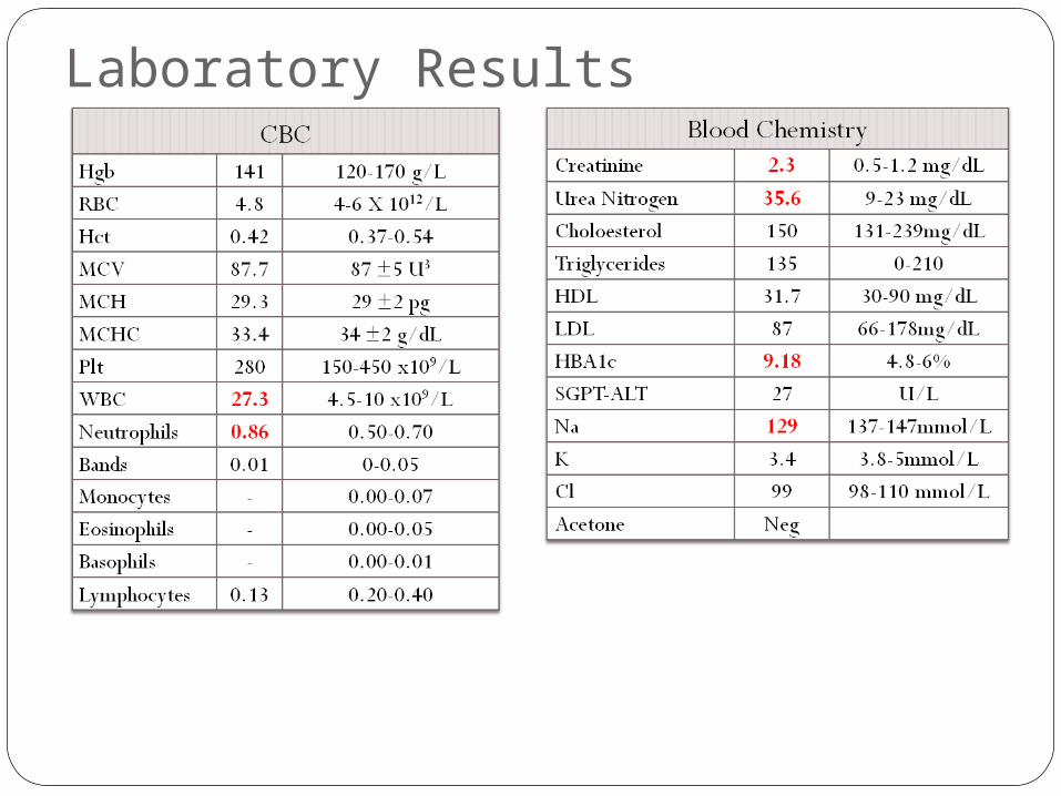
DIAGNOSTIC PLANS:CBCCBGBlood Chemistry
creatinineLipid profileHBA1cSodium Potassium
Arterial Blood Gas

THERAPEUTIC PLANSANTIBIOTIC- what?INSULIN THERAPY ACCORDING TO CBG
RESULTS(SLIDING SCALE)/INSULIN DRIP?DIETARY MANAGEMENT- DIABETIC/RENAL
DIET specific diet: kilocalories/day, protein &
carbohydrate requirementHYDRATION-PLAIN NSS (HOW MUCH
INFUSION/MIN)
Laboratory Results
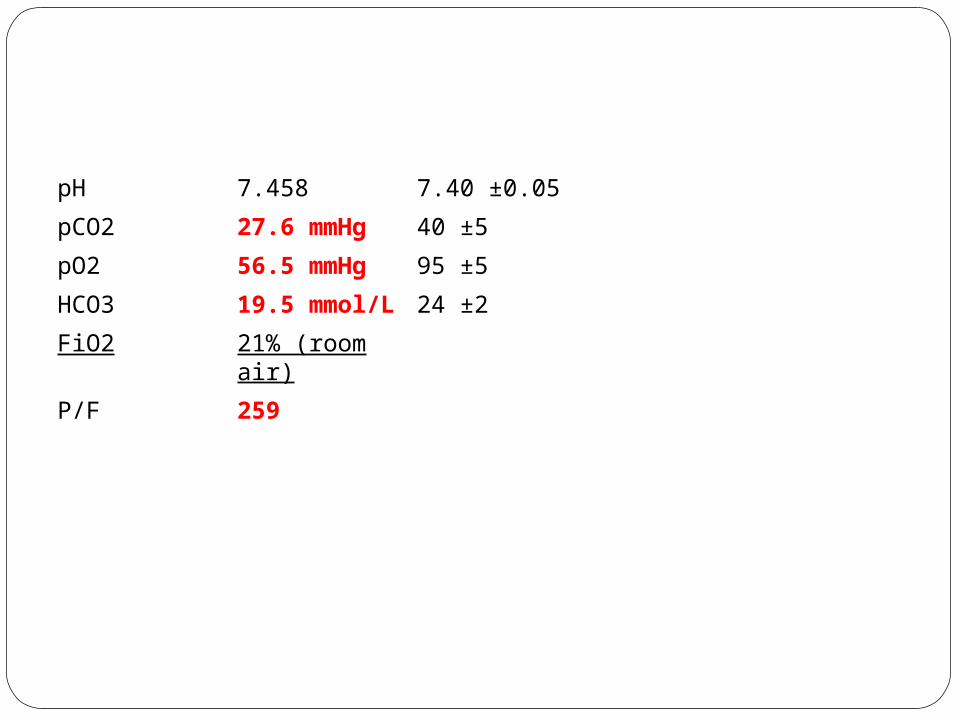
pH 7.458 7.40 ±0.05
pCO2 27.6 mmHg 40 ±5
pO2 56.5 mmHg 95 ±5
HCO3 19.5 mmol/L
24 ±2
FiO2 21% (room air)
P/F 259

DIAGNOSTIC TEST FOR DIABETES MELLITUS
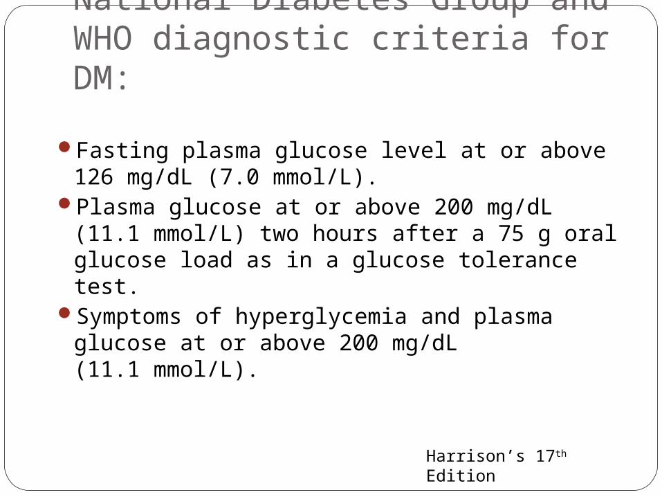
National Diabetes Group and WHO diagnostic criteria for DM:
Fasting plasma glucose level at or above 126 mg/dL (7.0 mmol/L).
Plasma glucose at or above 200 mg/dL (11.1 mmol/L) two hours after a 75 g oral glucose load as in a glucose tolerance test.
Symptoms of hyperglycemia and plasma glucose at or above 200 mg/dL (11.1 mmol/L).
Harrison’s 17th Edition

Fasting blood glucose test • The most common test for diagnosis of
diabetes.
• blood glucose levels are checked after fasting for between 12 and 14 hours.
• Patients with fasting glucose levels from 100 to 125 mg/dL (6.1 and 7.0 mmol/L) are considered to have impaired fasting glucose
• Patients with diabetes may be asked to delay their diabetes medication or insulin dose until the test is completed.
http://ovennewyork.com/diabetes-mellitus-laboratory-tests-or-diagnostic-tests.html

Random blood glucose test blood glucose levels are checked at various
times during the day, and it doesn’t matter when you last ate.
Blood glucose levels tend to stay constant in a person who doesn’t have diabetes.
http://ovennewyork.com/diabetes-mellitus-laboratory-tests-or-diagnostic-tests.html

Oral glucose tolerance test (OGTT) • FBS is obtained before the ingestion of a 50- to
200-g glucose load (usual amount is 75 g),
• blood samples are drawn at ½, 1, 2, and 3 hours (may be 4- or 5-hour sampling).
• Blood samples are checked at regular intervals for two hours.
• Glucose tolerance tests are used when the results of the fasting blood glucose are borderline.
http://ovennewyork.com/diabetes-mellitus-laboratory-tests-or-diagnostic-tests.html

• They are also used to diagnose diabetes in pregnancy (gestational diabetes).
• NORMAL: the results of the glucose tolerance test will show that their blood sugar levels fall within the normal range
• Patients with plasma glucose at or above 140 mg/dL or 7.8 mmol/L, but not over 200, two hours after a 75 g oral glucose load are considered to have impaired glucose tolerance.

Glycated Hemoglobin (Glycohemoglobin, HbA1c) for Diabetes Mellitus
• Measures glycemic control over a 60- to 120-day period by measuring the irreversible reaction of glucose to hemoglobin through freely permeable erythrocytes during their 120-day lifecycle.
• While not used for diagnosis, an elevated level of glucose irreversibly bound to hemoglobin of 6.0% or higher (the 2003 revised U.S. standard) is considered abnormal by most labs
http://ovennewyork.com/diabetes-mellitus-laboratory-tests-or-diagnostic-tests.html
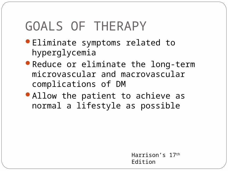
GOALS OF THERAPYEliminate symptoms related to
hyperglycemiaReduce or eliminate the long-term
microvascular and macrovascular complications of DM
Allow the patient to achieve as normal a lifestyle as possible
Harrison’s 17th Edition
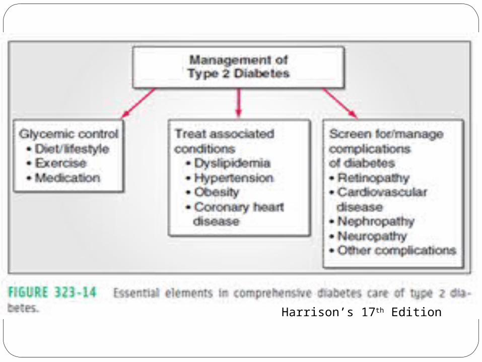
Harrison’s 17th Edition

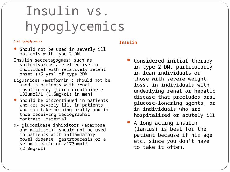
Insulin vs. hypoglycemicsOral hypoglycemics
Should not be used in severly ill patients with type 2 DM
Insulin secretagogues: such as sulfonlyureas are effective in individual with relatively recent onset (<5 yrs) of type 2DM
Biguanides (metformin): should not be used in patients with renal insufficency [serum creatinine > 133umol/L (1.5mg/dL) in men]
Should be discontinued in patients who are severly ill, in patients who can take nothing orally and in thoe receiving radiograohic contrast material
α- glucosidase inhibitors (acarbose and miglitol): should not be used in patients with inflammatory bowel disease, gastroparesis or a serum creatinine >177umol/L (2.0mg/dL)
Insulin
Considered initial therapy in type 2 DM, particularly in lean individuals or those with severe weight loss, in individuals with underlying renal or hepatic disease that precludes oral glucose-lowering agents, or in individuals who are hospitalized or acutely ill
A long acting insulin (lantus) is best for the patient because if his age etc. since you don’t have to take it often.

Patient education about DM, nutrition and exercise• Patient with DM should receive education
about:– Nutrition– Exercise– Care of diabetes during illness– Medications to lower the plasma glucose
• Continuing process with regular visits for reinforcement
• Diabetes self-management education (DSME)
Harrison’s 17th Edition
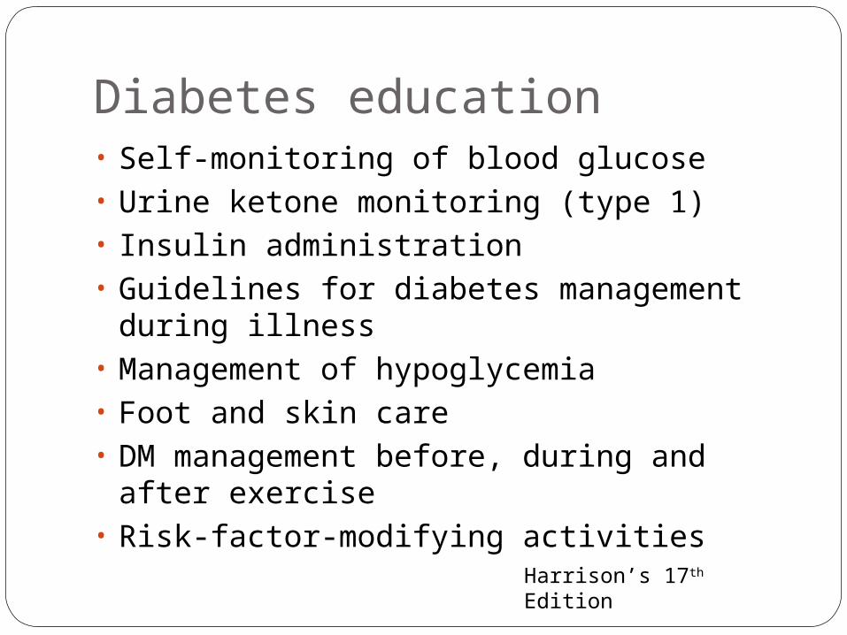
Diabetes education• Self-monitoring of blood glucose• Urine ketone monitoring (type 1)• Insulin administration• Guidelines for diabetes management
during illness• Management of hypoglycemia• Foot and skin care• DM management before, during and after
exercise• Risk-factor-modifying activities
Harrison’s 17th Edition
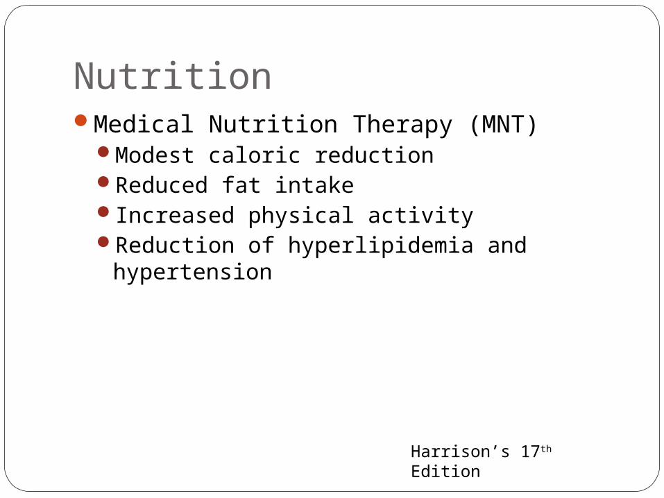
NutritionMedical Nutrition Therapy (MNT)
Modest caloric reductionReduced fat intakeIncreased physical activityReduction of hyperlipidemia and
hypertension
Harrison’s 17th Edition
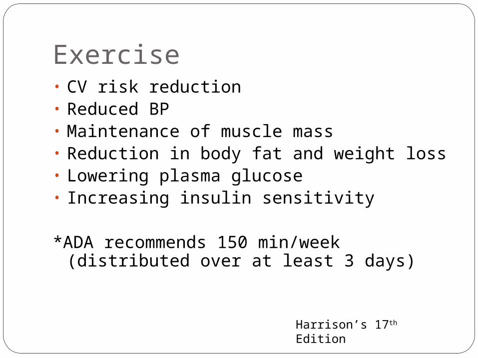
Exercise• CV risk reduction• Reduced BP• Maintenance of muscle mass• Reduction in body fat and weight loss• Lowering plasma glucose• Increasing insulin sensitivity
*ADA recommends 150 min/week (distributed over at least 3 days)
Harrison’s 17th Edition

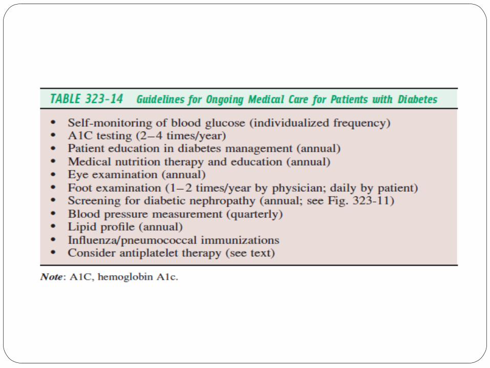
Monitoring the level of glyceminc controlPlasma glucose measurements by the
patient and assessment of long-term control by the physician
Measurement of A1C and review of the patient’s self-measurement of plasma glucose
Harrison’s 17th Edition
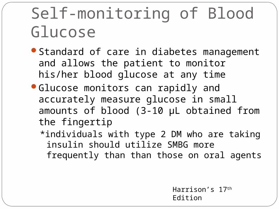
Self-monitoring of Blood GlucoseStandard of care in diabetes management
and allows the patient to monitor his/her blood glucose at any time
Glucose monitors can rapidly and accurately measure glucose in small amounts of blood (3-10 µL obtained from the fingertip*individuals with type 2 DM who are taking
insulin should utilize SMBG more frequently than than those on oral agents
Harrison’s 17th Edition
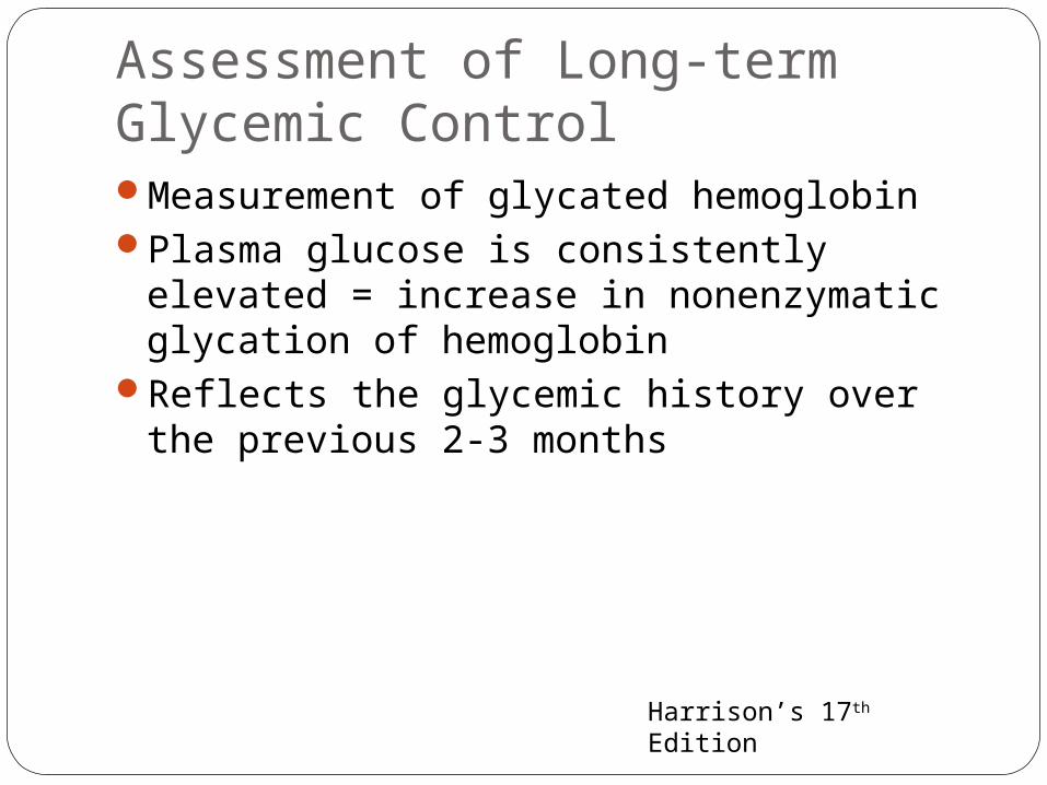
Assessment of Long-term Glycemic ControlMeasurement of glycated hemoglobinPlasma glucose is consistently elevated =
increase in nonenzymatic glycation of hemoglobin
Reflects the glycemic history over the previous 2-3 months
Harrison’s 17th Edition

CELLULITIS
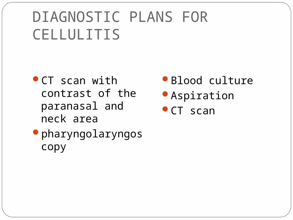
DIAGNOSTIC PLANS FOR CELLULITIS
CT scan with contrast of the paranasal and neck area
pharyngolaryngoscopy
Blood cultureAspirationCT scan

*As clinically indicated; †Ulcerated lesions should be cleaned and debrided before having wound base swabbed; ‡Most useful if vesicle/bullae or fluid abscess present; §Seek out bone trauma and air fluid levels; ¶Indications –neurological deficits, vision nonassessable, proptosis/deteriorating acuity or colour/bilateral edema/ophthalmoplegia, no
improvement after 24 h and swinging pyrexia not resolving within 36 h (for head only); **Only if central nervous system involvement suspected

DiagnosisBased on appearance of the skin and patient
history
Drainage from an abscess or weeping wound associated with cellulitis should be sent for culture and sensitivities.
Material from needle aspiration of inflamed skin or skin biopsy can be cultured in cases of cellulitis without purulence, abscess, or a necrotic
Indications for blood cultures include significant fever and chills, severe immunocompromise, periorbital cellulitis, and cellulitis superimposed on lymphedema.
A polymorphonuclear leukocytosis is often present with cellulitis; a complete blood cell count and differential may help gauge the severity of infection and the hematologic response.

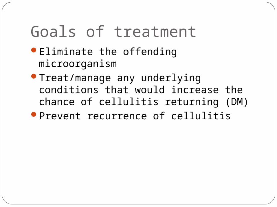
Goals of treatmentEliminate the offending microorganismTreat/manage any underlying conditions
that would increase the chance of cellulitis returning (DM)
Prevent recurrence of cellulitis

oral therapy for mild infections
intravenous therapy for severe infections achievement of high
drug levels with rapid delivery.
Therapeutic approach
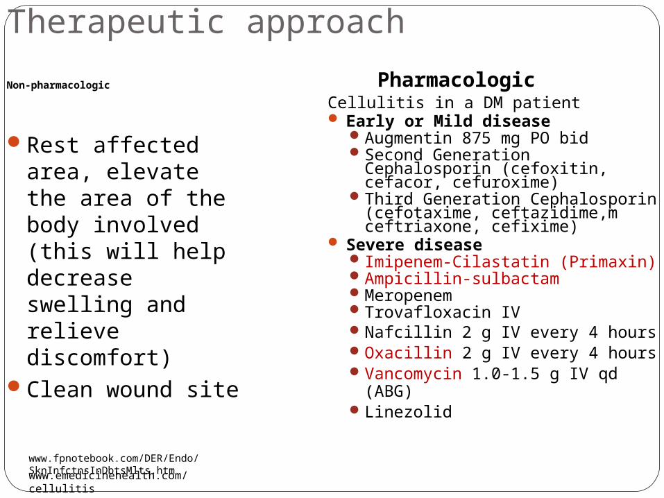
Non-pharmacologic
Rest affected area, elevate the area of the body involved (this will help decrease swelling and relieve discomfort)
Clean wound site
PharmacologicCellulitis in a DM patient Early or Mild disease
Augmentin 875 mg PO bidSecond Generation Cephalosporin
(cefoxitin, cefacor, cefuroxime)Third Generation Cephalosporin
(cefotaxime, ceftazidime,m ceftriaxone, cefixime)
Severe disease Imipenem-Cilastatin (Primaxin)Ampicillin-sulbactamMeropenemTrovafloxacin IV Nafcillin 2 g IV every 4 hours Oxacillin 2 g IV every 4 hours Vancomycin 1.0-1.5 g IV qd (ABG)Linezolid
www.emedicinehealth.com/cellulitis
www.fpnotebook.com/DER/Endo/SknInfctnsInDbtsMlts.htm
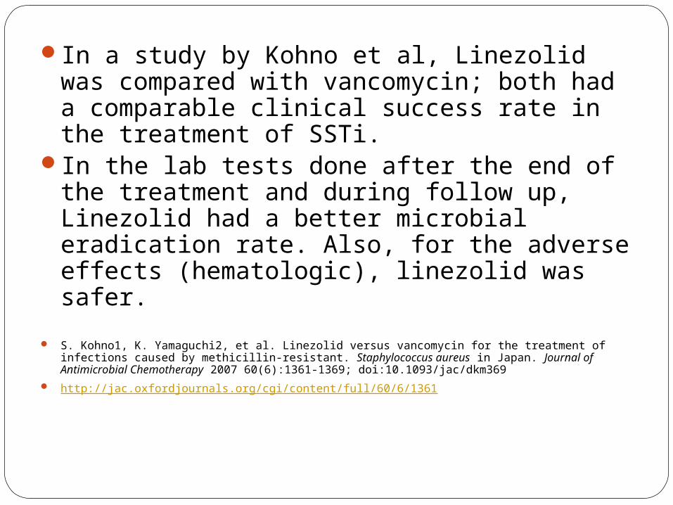
In a study by Kohno et al, Linezolid was compared with vancomycin; both had a comparable clinical success rate in the treatment of SSTi.
In the lab tests done after the end of the treatment and during follow up, Linezolid had a better microbial eradication rate. Also, for the adverse effects (hematologic), linezolid was safer.
S. Kohno1, K. Yamaguchi2, et al. Linezolid versus vancomycin for the treatment of infections caused by methicillin-resistant. Staphylococcus aureus in Japan. Journal of Antimicrobial Chemotherapy 2007 60(6):1361-1369; doi:10.1093/jac/dkm369
http://jac.oxfordjournals.org/cgi/content/full/60/6/1361
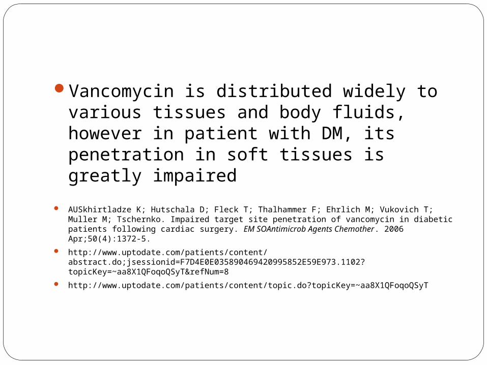
Vancomycin is distributed widely to various tissues and body fluids, however in patient with DM, its penetration in soft tissues is greatly impaired
AUSkhirtladze K; Hutschala D; Fleck T; Thalhammer F; Ehrlich M; Vukovich T; Muller M; Tschernko. Impaired target site penetration of vancomycin in diabetic patients following cardiac surgery. EM SOAntimicrob Agents Chemother. 2006 Apr;50(4):1372-5.
http://www.uptodate.com/patients/content/abstract.do;jsessionid=F7D4E0E035890469420995852E59E973.1102?topicKey=~aa8X1QFoqoQSyT&refNum=8
http://www.uptodate.com/patients/content/topic.do?topicKey=~aa8X1QFoqoQSyT

In a study by Stein et al, Linezolid was effective in the treatment of Staphylococcus aureus in diabetic patients
G E. Stein1,* S Schooley1, et al. Linezolid tissue penetration and serum activity against strains of methicillin-resistant Staphylococcus aureus with reduced vancomycin susceptibility in diabetic patients with foot infections. Journal of Antimicrobial Chemotherapy, doi:10.1093/jac/dkm271
http://jac.oxfordjournals.org/cgi/content/full/dkm271v1
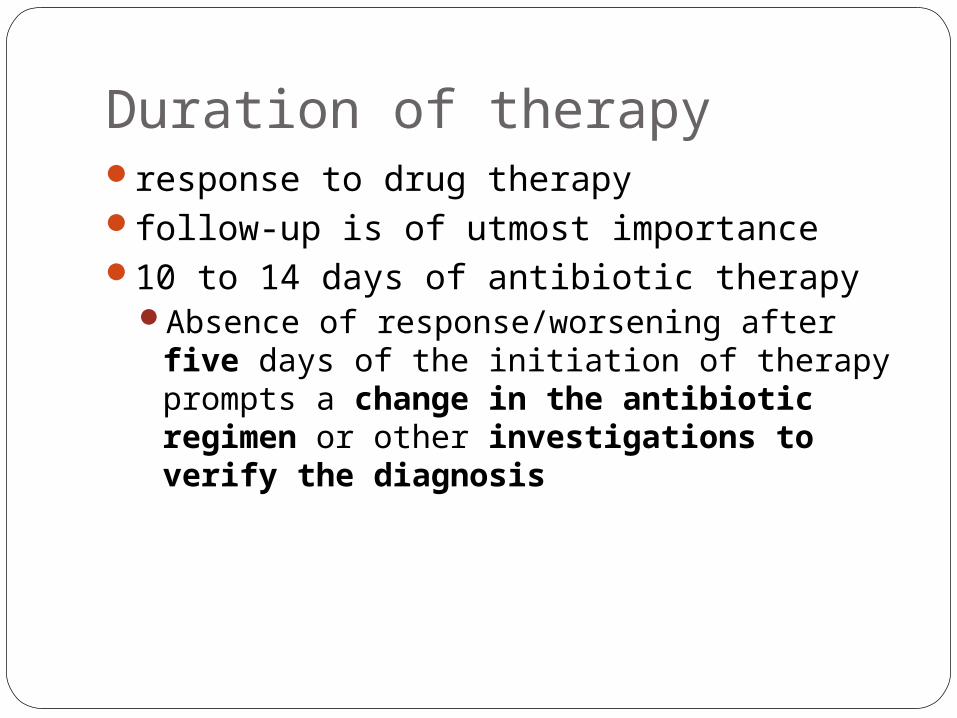
Duration of therapyresponse to drug therapyfollow-up is of utmost importance10 to 14 days of antibiotic therapy
Absence of response/worsening after five days of the initiation of therapy prompts a change in the antibiotic regimen or other investigations to verify the diagnosis

Preventing a recurrence of cellulitisCellulitis tends to recur in people with
certain medical conditions that can lead to skin breakdown, such as edema (fluid buildup), fungal or bacterial infections, diabetes, or peripheral arterial disease. edema, support stockings and good skin hygiene
may reduce or eliminate recurrence of cellulitis.fungal infections, regular use of antifungal
medicines may help reduce recurrent cellulitis. high risk for recurring cellulitis, (when with open
wound or cut) taking preventive antibiotics may help
http://www.everettclinic.com/kbase/topic/mini/tr5105/treatmnt.htm
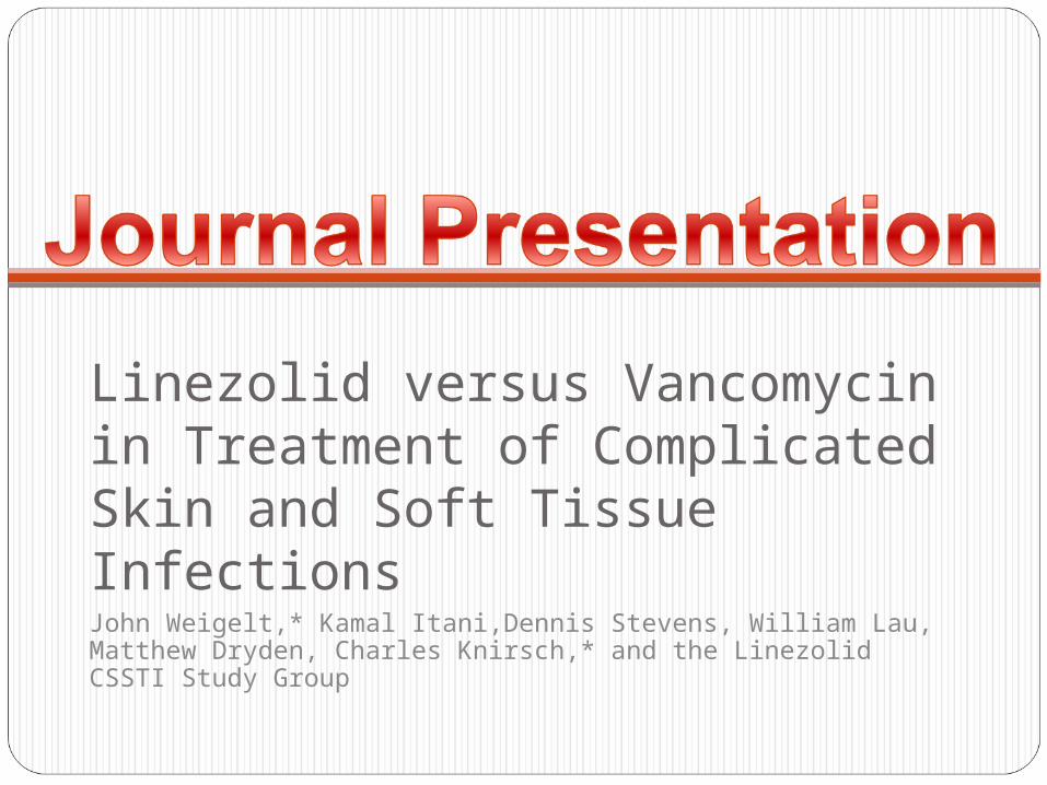
Linezolid versus Vancomycin in Treatment of Complicated Skin and Soft Tissue InfectionsJohn Weigelt,* Kamal Itani,Dennis Stevens, William Lau, Matthew Dryden, Charles Knirsch,* and the Linezolid CSSTI Study Group
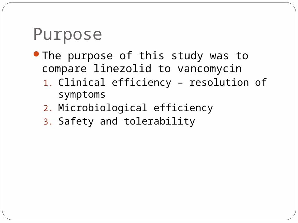
PurposeThe purpose of this study was to compare
linezolid to vancomycin1. Clinical efficiency – resolution of symptoms2. Microbiological efficiency 3. Safety and tolerability

Background of the StudyThis was a randomized, open-label,
comparator-controlled, multicenter, multinational study that included patients with suspected or proven methicillin-resistant Staphylococcus aureus (MRSA) infections that involved substantial areas of skin or deeper soft tissues, such as cellulitis, abscesses, infected ulcers, or burns <10% of total body surface area.

PathogensSENTRY Antimicrobial Surveillance Program:
SSTIs may be caused by a wide range of pathogens Staphylococcus aureus 40% 5% of infections included
Pseudomonas aeruginosa (12%)Escherichia coli (10%)Enterococcus spp. (8%)Klebsiella spp. (6%)Enterobacter spp. (6%)

Materials and MethodStudy period: October 2002-March 2003Inclusions:
Required physical findings included (i) erythema with or without induration, (ii) fluctuation, (iii) heat/localized warmth, (iv) pain/tenderness, and (v) drainage/discharge.
at least one of the following symptoms: (i) fever, (ii) hypothermia, (iii) hypotension, (iv) a white blood cell count of 10,000/mm3, or (v) 15% immature neutrophils regardless of white blood cell count


Statistical AnalysisMicrobiological outcomes were categorized
as success (documented or presumed
eradicationof the pathogen present at baseline)
failure (documented or presumed persistence of pathogen present at baseline)
indeterminate (pathogen data indeterminate),
missing (pathogen data missing).
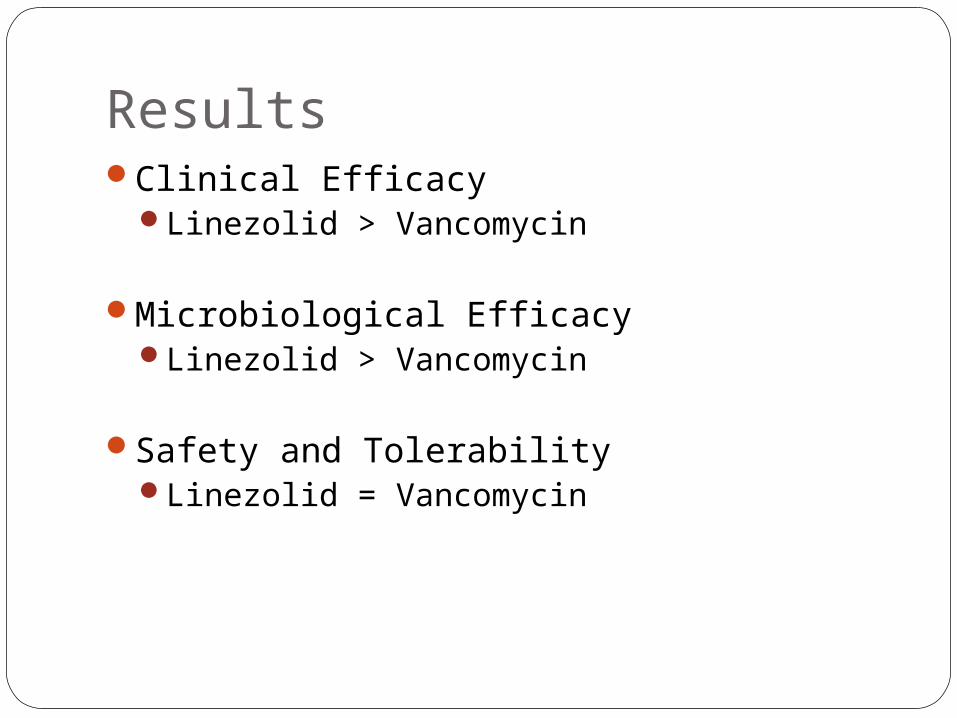
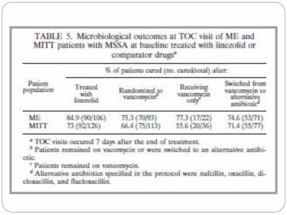
ResultsClinical Efficacy
Linezolid > Vancomycin
Microbiological EfficacyLinezolid > Vancomycin
Safety and TolerabilityLinezolid = Vancomycin

CostLinezolid Vancomycin
IV Zyvox 2mg/mL x 300mLP 3,418.28
Vancocin 500mg P 1,929.00
Suspension
Zyvox 100 mg/5 mL x 150 mL P 14,637.28
NA
Tablet Zyvox 600mgP 2,950.40
NA
Linezolid Vancomycin
600mg IV or PO every 12 hours
IV: 3,418.28 x 2 = P 6,836.56PO: 2,950.40 x 2 = P 5,900.80
1g IV every 12 hours
IV: 1,929.00 x 4 = P 7,716

References Vincent Ki, MD and Coleman Rotstein, MD. Bacterial skin and soft
tissue infections in adults: A review of their epidemiology, pathogenesis, diagnosis, treatment and site of care. Can J Infect Dis Med Microbiol. 2008 March; 19(2): 173-184.
G E. Stein1,* S Schooley1, et al. Linezolid tissue penetration and serum activity against strains of methicillin-resistant Staphylococcus aureus with reduced vancomycin susceptibility in diabetic patients with foot infections. Journal of Antimicrobial Chemotherapy, doi:10.1093/jac/dkm271
AUSkhirtladze K; Hutschala D et al. Impaired target site penetration of vancomycin in diabetic patients following cardiac surgery. EM SOAntimicrob Agents Chemother. 2006 Apr;50(4):1372-5.
S. Kohno1, K. Yamaguchi2, et al. Linezolid versus vancomycin for the treatment of infections caused by methicillin-resistant. Staphylococcus aureus in Japan. Journal of Antimicrobial Chemotherapy 2007 60(6):1361-1369; doi:10.1093/jac/dkm369
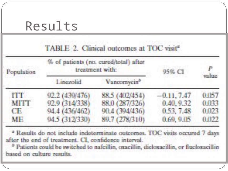
Results