permission from Dove Medical Press Limited, provided the work is properly attributed. Permissions beyond the scope of the License are administered by Dove Medical Press Limited. Information on how to request permission may be found at: http://www.dovepress.com/permissions.php
Clinical, Cosmetic and Investigational Dermatology 2015:8 485–488
Clinical, Cosmetic and Investigational Dermatology Dovepress
submit your manuscript | www.dovepress.com
Dovepress 485
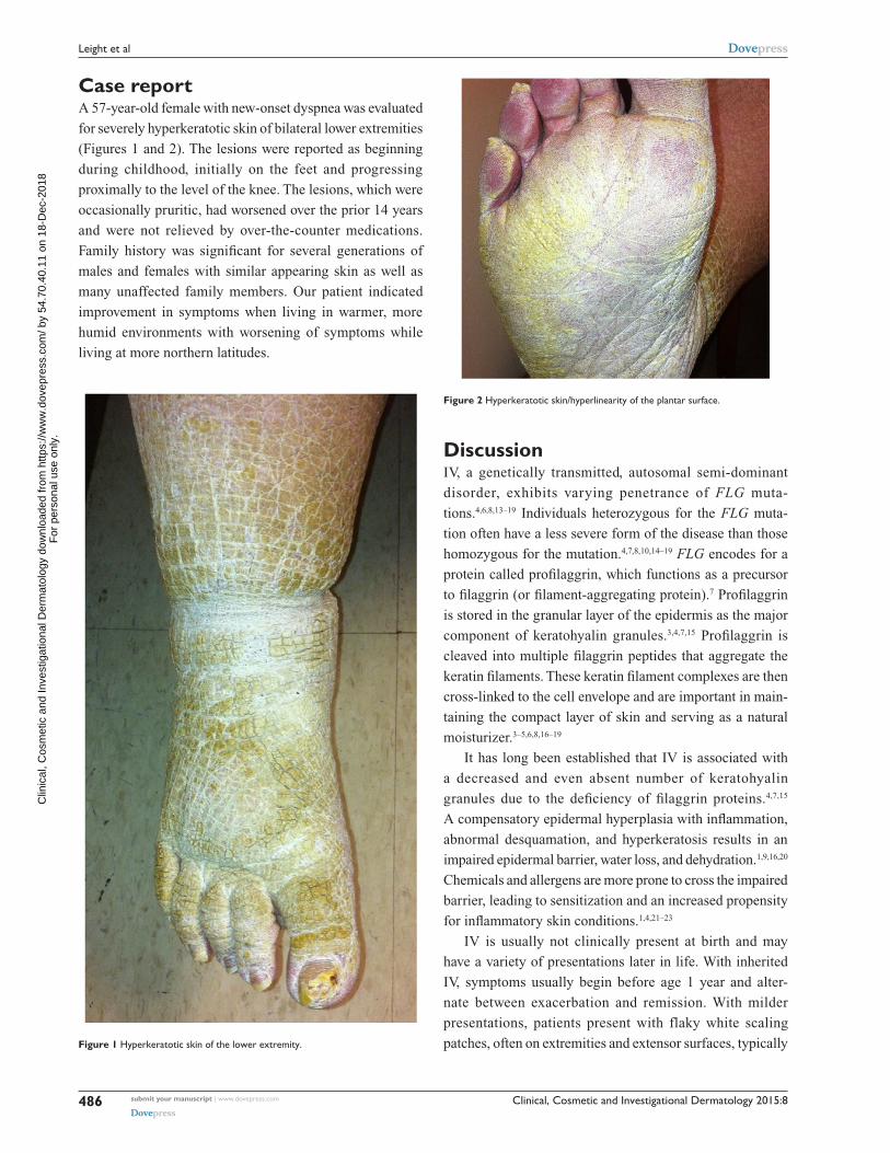
C a s e R e p o Rt
open access to scientific and medical research
open access Full text article
http://dx.doi.org/10.2147/CCID.S89871
Bilateral lower extremity hyperkeratotic plaques: a case report of ichthyosis vulgaris
Hayley LeightZachary Zinnomid JalaliDepartment of Dermatology, West Virginia University, Morgantown, WV, Usa
Clinical, Cosmetic and Investigational Dermatology
Publish your work in this journal
Submit your manuscript here: http://www.dovepress.com/clinical-cosmetic-and-investigational-dermatology-journal
Clinical, Cosmetic and Investigational Dermatology is an interna-tional, peer-reviewed, open access, online journal that focuses on the latest clinical and experimental research in all aspects of skin disease and cosmetic interventions. All areas of dermatology will be covered; contributions will be welcomed from all clinicians and
basic science researchers globally. This journal is indexed on CAS. The manuscript management system is completely online and includes a very quick and fair peer-review system, which is all easy to use. Visit http://www.dovepress.com/testimonials.php to read real quotes from published authors.
Clinical, Cosmetic and Investigational Dermatology 2015:8submit your manuscript | www.dovepress.com
Dovepress
Dovepress
Dovepress
488
Leight et al
for providing medical student opportunity for case study
research. Manuscript editing assistance was provided by
Susan B Leight, EdD, FNP-BC, FAANP, Director, School
of Nursing, West Virginia Wesleyan College. The authors
advise that the West Virginia University Ethics Committee/
Institutional Review Board does not require patient approval
for case reports.
DisclosureThe authors report no conflicts of interest in this work.
PA: Elsevier Saunders; 2012. 2. Styperek AR, Rice ZP, Kamalpour L, et al. Annual direct and indirect
health costs of the congenital ichthyoses. Pediatr Dermatol. 2010; 27(4):325–335.
3. Wolff K, Goldsmith LA, Katz SI, Gilchrest BA, Paller AS, Leffell DJ. Fitzpatrick’s Dermatology in General Medicine. 7th ed. New York, NY: McGraw Hill; 2008.
4. Thyssen JP, Godoy-Gijon E, Elias PM. Ichthyosis vulgaris: the filaggrin mutation disease. Br J Dermatol. 2013;168(6):1155–1166.
5. Li M, Cheng R, Shi M, et al. Analyses of FLG mutation frequency and filaggrin expression in isolated ichthyosis vulgaris (IV) and atopic dermatitis-associated IV. Br J Dermatol. 2013;168(6):1335–1338.
6. Wells RS, Kerr CB. Clinical features of autosomal dominant and sex-linked ichthyosis in an English population. BMJ. 1966;1(5493):947–950.
7. Liu P, Yang Q, Wang X, et al. Identification of a genetic locus for ichthyosis vulgaris on chromosome 10q22.3-q24.2. J Invest Dermatol. 2008;128(6):1418–1422.
8. Sandilands A, Terron-Kwiatkowski A, Hull PR, et al. Comprehensive analysis of the gene encoding filaggrin uncovers prevalent and rare mutations in ichthyosis vulgaris and atopic eczema. Nat Genet. 2007;39(5):650–654.
9. Bellew S, Del Rosso JQ. Overcoming the Barrier Treatment of Ichthyosis: A Combination-therapy Approach. J Clin Aesthetic Dermatol. 2010;3(7):49–53.
10. Perusquίa-Ortiz AM, Oji V, Sauerland MC, et al. Complete filaggrin deficiency in ichthyosis vulgaris is associated with only moderate changes in epidermal permeability barrier function profile. J Eur Acad Dermatol Venereol. 2013;27(12):1552–1558.
11. Dreyfus I, Bourrat E, Maruani A, et al. Factors associated with impaired quality of life in adult patients suffering from ichthyosis. Acta Derm Venereol. 2014;94(3):344–346.
12. Dreyfus I, Pauwels C, Bourrat E, et al. Burden of inherited ichthyosis: a French national survey. Acta Derm Venereol. 2015;95(3):326–328.
13. Esparza-Gordillo J, Matanovic A, Marenholz I, et al. Maternal filaggrin mutations increase the risk of atopic dermatitis in children: an effect independent of mutation inheritance. PLoS Genet. 2015;11(3):1–16.
14. Mendes MS, Aquino TA, de Padua Lima A, Kouzak SS, Takano GH. Mosaic epidermolytic ichthyosis – case report. An Bras Dermatol. 2013;88(6 Suppl 1):116–119
15. Brown SJ, McLean WH. One remarkable molecule: filaggrin. J Invest Dermatol. 2012;132(3 Pt 2):751–762.
16. Akiyama M. FLG mutations in ichthyosis vulgaris and atopic eczema: spectrum of mutations and population genetics. Br J Dermatol. 2010; 162(3):472–477.
17. Zhang X, Liu S, Chen X, et al. Novel and recurrent mutations in the filaggrin gene in Chinese patients with ichthyosis vulgaris. Br J Dermatol. 2010;163(1):63–69.
18. Nomura T, Akiyama M, Sandilands A, et al. Prevalent and rare muta-tions in the gene encoding filaggrin in Japanese patients with ichthyo-sis vulgaris and atopic dermatitis. J Invest Dermatol. 2009;129(5): 1302–1305.
19. Winge MCG, Hoppe T, Berne B, et al. Filaggrin genotype determines functional and molecular alterations in skin of patients with atopic dermatitis and ichthyosis vulgaris. PLoS One. 2011;6(12):e28254.
20. Blanchet-Bardon C, Tadini G, Machado Matos M, Delarue A. Association of glycerol and paraffin in the treatment of ichthyosis in children: an international, multicentric, randomized, controlled, double-blind study. J Eur Acad Dermatol Venereol. 2012;26(8):1014–1019.
21. Fleckman P, Newell BD, van Steensel MA, Yans, AC. Topical treatment of ichthyoses. Dermatol Ther. 2013;26(1):16–25.
22. Bodemer C, Bourrat, E, Mazereeuw-Hautier J, et al. Short- and medium-term efficacy of specific hydrotherapy in inherited ichthyosis. Br J Dermatol. 2011;165(5):1087–1094.
23. Chan A, Godoy-Gijon E, Nuno-Gonzalez A. et al. Cellular basis of secondary infections and impaired desquamation in certain inherited ichthyoses. JAMA Dermatol. 2015;151(3):285–292.
24. Divyashree RA, Naveen KN, Pai VV, Athanikar SB, Gupta G. Cutaneous manifestations of obesity among dermatology patients in a tertiary care center. Indian J Dermatol Venereol Leprol. 2014;80(3):278.
25. Greene AK, Grant FD, Slavin SA. Lower-extremity lymphedema and elevated body-mass index. N Engl J Med. 2012;366(22):2136–2137.
26. Weitman ES, Aschen SZ, Farias-Eisner, et al. Obesity impairs lymphatic fluid transport and dendritic cell migration to lymph nodes. PLoS One. 2013;8(8):e70703.
27. Arngrim N, Simonsen L, Holst JJ, Bülow J. Reduced adipose tissue lymphatic drainage of macromolecules in obese subjects: a possible link between obesity and local tissue inflammation? Int J Obes (Lond). 2013;37(5):748–750.
28. Scheinfeld, NS. Obesity and dermatology. Clin Dermatol. 2004; 22(4):303–309.
30. Lu S, Tran TA, Jones DM, et al. Localized lymphedema (elephantiasis): a case series and review of the literature. J Cutan Pathol. 2009;36(4): 1–20.
31. Kaba H, Bakar Y, Ozdemir Oç, Sertel S. Complex Decongestive Physiotherapy Treats Skin Changes like Hyperkeratosis Caused by Lymphedema. Case Rep Dermatol Med. 2012;2012:416421.
32. Linnitt N. Complex skin changes in chronic oedemas. Br J Community Nurs. 2007;12(4):S10–S15.
33. Nayak S, Acharjya B, Mohanty P. Ichthyosis hystrix. Indian Dermatol Online J. 2013;4(1):47–49.