Bimolecular Fluorescence Complementation (BiFC) Analysis as a Probe of Protein Interactions in Living Cells Tom K. Kerppola Howard Hughes Medical Institute and Department of Biological Chemistry, University of Michigan Medical School, Ann Arbor, Michigan 48109-0650; email: [email protected]Annu. Rev. Biophys. 2008. 37:465–87 First published online as a Review in Advance on March 26, 2008 The Annual Review of Biophysics is online at biophys.annualreviews.org This article’s doi: 10.1146/annurev.biophys.37.032807.125842 Copyright c 2008 by Annual Reviews. All rights reserved 1936-122X/08/0609-0465$20.00 Key Words fluorescence imaging, subcellular localization, visualization of molecular complexes, multicolor BiFC analysis Abstract Protein interactions are a fundamental mechanism for the generation of biological regulatory specificity. The study of protein interactions in living cells is of particular significance because the interactions that occur in a particular cell depend on the full complement of pro- teins present in the cell and the external stimuli that influence the cell. Bimolecular fluorescence complementation (BiFC) analysis en- ables direct visualization of protein interactions in living cells. The BiFC assay is based on the association between two nonfluorescent fragments of a fluorescent protein when they are brought in prox- imity to each other by an interaction between proteins fused to the fragments. Numerous protein interactions have been visualized us- ing the BiFC assay in many different cell types and organisms. The BiFC assay is technically straightforward and can be performed using standard molecular biology and cell culture reagents and a regular fluorescence microscope or flow cytometer. 465 Annu. Rev. Biophys. 2008.37:465-487. Downloaded from arjournals.annualreviews.org by University of Texas - Austin on 06/30/08. For personal use only.
Transcript
ANRV343-BB37-22 ARI 24 April 2008 15:55
Bimolecular FluorescenceComplementation (BiFC)Analysis as a Probeof Protein Interactionsin Living CellsTom K. KerppolaHoward Hughes Medical Institute and Department of Biological Chemistry,University of Michigan Medical School, Ann Arbor, Michigan 48109-0650;email: [email protected]
Annu. Rev. Biophys. 2008. 37:465–87
First published online as a Review in Advance onMarch 26, 2008
The Annual Review of Biophysics is online atbiophys.annualreviews.org
This article’s doi:10.1146/annurev.biophys.37.032807.125842
AbstractProtein interactions are a fundamental mechanism for the generationof biological regulatory specificity. The study of protein interactionsin living cells is of particular significance because the interactionsthat occur in a particular cell depend on the full complement of pro-teins present in the cell and the external stimuli that influence thecell. Bimolecular fluorescence complementation (BiFC) analysis en-ables direct visualization of protein interactions in living cells. TheBiFC assay is based on the association between two nonfluorescentfragments of a fluorescent protein when they are brought in prox-imity to each other by an interaction between proteins fused to thefragments. Numerous protein interactions have been visualized us-ing the BiFC assay in many different cell types and organisms. TheBiFC assay is technically straightforward and can be performed usingstandard molecular biology and cell culture reagents and a regularfluorescence microscope or flow cytometer.
465
Ann
u. R
ev. B
ioph
ys. 2
008.
37:4
65-4
87. D
ownl
oade
d fr
om a
rjou
rnal
s.an
nual
revi
ews.
org
by U
nive
rsity
of
Tex
as -
Aus
tin o
n 06
/30/
08. F
or p
erso
nal u
se o
nly.
ANRV343-BB37-22 ARI 24 April 2008 15:55
Contents
INTRODUCTION. . . . . . . . . . . . . . . . . 466VISUALIZATION OF PROTEIN
INTERACTIONS . . . . . . . . . . . . . . . 467An Abbreviated History of
Complementation Assays . . . . . . 467Comparison of the BiFC Approach
Many proteins have different functions in var-ious cell types and in response to distinctextracellular signals. The effects of the cel-lular environment on protein functions areoften mediated by interactions with differentpartners under different conditions. Proteininteractions also integrate signals from dif-ferent signaling pathways and developmentalprograms and coordinate regulatory mecha-nisms in the cell. Studies of protein interac-
tions in living cells can provide insights intothese functions, as interactions with differentpartners may occur in different cells, at dif-ferent times, and in different subcellular lo-cations. The visualization of interactions inindividual cells also enables analysis of differ-ences among different cells in the population.Studies in intact cells also avoid the possibilityof changes in protein interactions as a resultof cell lysis and mixing of the contents of dif-ferent cellular compartments. Consequently,
466 Kerppola
Ann
u. R
ev. B
ioph
ys. 2
008.
37:4
65-4
87. D
ownl
oade
d fr
om a
rjou
rnal
s.an
nual
revi
ews.
org
by U
nive
rsity
of
Tex
as -
Aus
tin o
n 06
/30/
08. F
or p
erso
nal u
se o
nly.
ANRV343-BB37-22 ARI 24 April 2008 15:55
the direct visualization of protein complexesin living cells provides a valuable complementto other methods for the study of proteininteractions.
VISUALIZATION OF PROTEININTERACTIONS
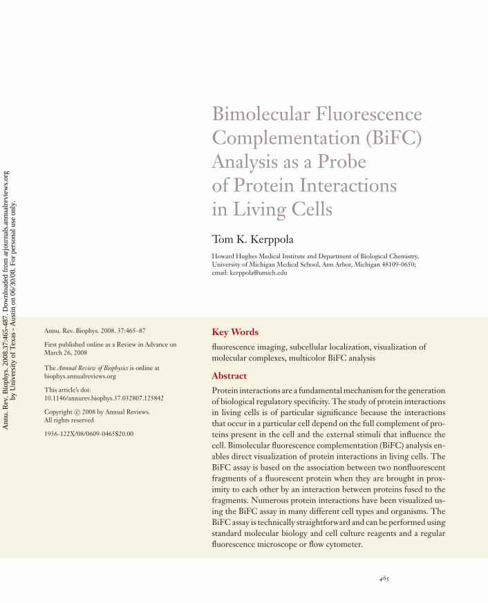
Several methods enable the visualization ofprotein interactions in living cells. Most ofthese methods require either elaborate in-strumentation and complex data processing,or staining with exogenous fluorophores ordyes. The bimolecular fluorescence comple-mentation (BiFC) assay enables simple anddirect visualization of protein interactions inliving cells (44). The BiFC approach is basedon the formation of a fluorescent complexwhen two proteins fused to nonfluorescentfragments of a fluorescent protein interactwith each other (Figure 1). The interactionbetween the fusion proteins facilitates theassociation between the fragments of the fluo-rescent protein. This approach enables visual-ization of the subcellular locations of specificprotein complexes in the normal cellular en-vironment. The BiFC approach can be usedfor the analysis of interactions between manytypes of proteins and does not require infor-mation about the structures of the interactionpartners. It can be performed using a standardepifluorescence microscope and does not re-quire staining of the cells with exogenous fluo-rophores or dyes.
An Abbreviated History ofComplementation Assays
Protein complementation has now been stud-ied for about half a century. Fragments ofmany proteins can associate with each otherto form a functional complex. Complementa-tion between enzyme fragments was originallyobserved by Richards (96) using subtilisin-cleaved bovine pancreatic ribonuclease. Ge-netic complementation between different al-leles of the same gene was characterized byUllmann, Jacob, and Monod (113–115), who
YN
A B
YC
A B
YN-YC
Figure 1Schematic representation of the principle of the BiFC assay. Two non-fluorescent fragments (YN and YC) of the yellow fluorescent protein (YFP)are fused to putative interaction partners (A and B). The association of theinteraction partners allows formation of a bimolecular fluorescent complex.
used β-galactosidase mutants that conferredgrowth on lactose when coexpressed in thesame cell. Subsequently, fragments of manyproteins have been shown to spontaneouslyassociate to form a functional complex.
Of particular significance for the study ofprotein interactions was the demonstrationthat the association between some proteinfragments could be facilitated by fusion of thefragments to specific interaction partners asfirst demonstrated for fragments of ubiquitinin yeast by Johnsson & Varshavsky (50). Sub-sequently, conditional complementation be-tween fragments of β-galactosidase was visu-alized by Blau and coworkers (97) in intactmammalian cells. Conditional complementa-tion by fragments of dihydrofolate reductasewas reported by Michnick and colleagues (87).
Complementation between fragments ofa green fluorescent protein (GFP) variantwas first detected in Escherichia coli by Re-gan and coworkers (33) using fusions to ar-tificial, interacting peptides. Fragments of theyellow fluorescent protein (YFP) were shownto produce fluorescent complexes in mam-malian cells when fused to calmodulin andthe M13 calmodulin binding peptide by theMiyawaki laboratory (74). Conditional com-plementation between fragments of YFP inmammalian cells was demonstrated by Huin my laboratory (44). Fragments of severalother proteins have been used in conditionalcomplementation assays (Table 1) (55). Each
www.annualreviews.org • BiFC Analysis as a Probe of Protein Interactions in Living Cells 467
Ann
u. R
ev. B
ioph
ys. 2
008.
37:4
65-4
87. D
ownl
oade
d fr
om a
rjou
rnal
s.an
nual
revi
ews.
org
by U
nive
rsity
of
Tex
as -
Aus
tin o
n 06
/30/
08. F
or p
erso
nal u
se o
nly.
ANRV343-BB37-22 ARI 24 April 2008 15:55
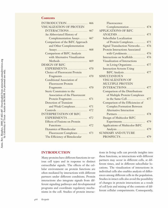
Table 1 Comparison of complementation methods using fragments of different proteins
Reporter ligation Cell population Hours Cultured, implanted cells (79)
β-lactamase CCF2/AMhydrolysis
Cellular Minutes Cultured cells, primaryneurons
(32, 104, 121)
Fireflyluciferase
Luciferin hydrolysis Cell population Hours Cultured, implanted cells (85)
Renillaluciferase
Coelenterazineluminescence
Cell population Minutes–hours Cultured, implanted cells (84)
Gaussialuciferase
Coelenterazineluminescence
Cell population Minutes Cultured cells (94)
TEV protease Coupled reporters Cellular Minutes Cultured cells (120)
aThe spatial and temporal resolution as well as the experimental systems used reflect those reported in publications using these approaches are notintended to represent the limits of performance of these methods.
complementation approach has specific ad-vantages and limitations. This chapter focuseson complementation between fragments offluorescent proteins.
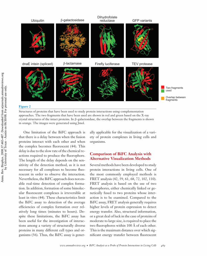
The structures of the complexes formedby complementation have not been deter-mined. However, it is likely that the struc-tures resemble those of the intact proteins be-cause they reproduce many of their functions(Figure 2). Proteins with a variety of struc-tures can be reconstituted from fragments.However, only a few of the peptide bondsin any particular protein can be broken toproduce fragments that can associate to forma functional complex. This limitation mayreflect the folding pathways of the respec-tive proteins. Greater insight into the foldingpathways of complexes formed by the proteinfragments would be valuable for understand-ing the factors that determine which proteinfragments can associate to produce a func-tional complex.
Comparison of the BiFC Approachand Other Complementation Assays
The advantage of the BiFC approach com-pared to other complementation methodsis that the assembled complex has strongintrinsic fluorescence that allows direct vi-sualization of the protein interaction. Theinteraction can therefore be detected with-out exogenous fluorogenic or chromogenicagents, avoiding potential perturbation of thecells by these agents. This also avoids po-tential problems caused by uneven distri-butions of the chromogenic or fluorogenicsubstrates or ligands. Using the BiFC ap-proach, living cells can be observed overtime and the possibility that experimentalmanipulations alter the result can be mini-mized. Moreover, as described below, multi-ple protein interactions can be visualized inparallel using spectrally distinct bimolecularfluorescent complexes.
468 Kerppola
Ann
u. R
ev. B
ioph
ys. 2
008.
37:4
65-4
87. D
ownl
oade
d fr
om a
rjou
rnal
s.an
nual
revi
ews.
org
by U
nive
rsity
of
Tex
as -
Aus
tin o
n 06
/30/
08. F
or p
erso
nal u
se o
nly.
ANRV343-BB37-22 ARI 24 April 2008 15:55
Ubiquitin β-galactosidaseDihydrofolate
reductase GFP variants
dnaE intein (spliced) Firefly luciferaseβ-lactamase TEV protease
Two fragments used
Overlap between fragments
Figure 2Structures of proteins that have been used to study protein interactions using complementationapproaches. The two fragments that have been used are shown in red and green based on the X-raycrystal structures of the intact proteins. In β-galactosidase, the overlap between the fragments is shownin orange. The images were generated using Jmol.
One limitation of the BiFC approach isthat there is a delay between when the fusionproteins interact with each other and whenthe complex becomes fluorescent (44). Thisdelay is due to the slow rate of the chemical re-actions required to produce the fluorophore.The length of the delay depends on the sen-sitivity of the detection method, as it is notnecessary for all complexes to become fluo-rescent in order to observe the interaction.Nevertheless, the BiFC approach does not en-able real-time detection of complex forma-tion. In addition, formation of some bimolec-ular fluorescent complexes is irreversible atleast in vitro (44). These characteristics limitthe BiFC assay to detection of the averageefficiencies of complex formation over rel-atively long times (minutes to hours). De-spite these limitations, the BiFC assay hasbeen useful for the investigation of interac-tions among a variety of structurally diverseproteins in many different cell types and or-ganisms (56). Thus, the BiFC assay is gener-
ally applicable for the visualization of a vari-ety of protein complexes in living cells andorganisms.
Comparison of BiFC Analysis withAlternative Visualization Methods
Several methods have been developed to studyprotein interactions in living cells. One ofthe most commonly employed methods isFRET analysis (42, 59, 61, 68, 72, 102, 110).FRET analysis is based on the use of twofluorophores, either chemically linked or ge-netically fused to two proteins whose inter-action is to be examined. Compared to theBiFC assay, FRET analysis generally requireshigher levels of protein expression to detectenergy transfer. Also, structural information,or a great deal of luck in the case of proteins ofmoderate to large size, is required to place thetwo fluorophores within 100 A of each other.This is the maximum distance over which sig-nificant energy transfer between fluorescent
www.annualreviews.org • BiFC Analysis as a Probe of Protein Interactions in Living Cells 469
Ann
u. R
ev. B
ioph
ys. 2
008.
37:4
65-4
87. D
ownl
oade
d fr
om a
rjou
rnal
s.an
nual
revi
ews.
org
by U
nive
rsity
of
Tex
as -
Aus
tin o
n 06
/30/
08. F
or p
erso
nal u
se o
nly.
ANRV343-BB37-22 ARI 24 April 2008 15:55
proteins can be detected. The fraction of pro-teins that form complexes must also be highenough to produce a sufficient change in thedonor and acceptor fluorescence intensities.To exclude alternative interpretations of theresults, numerous controls must be performedand the fluorescence intensities must be mea-sured with high quantitative accuracy. Despitethese limitations, FRET has been successfullyused for the analysis of many protein inter-actions in living cells. A great advantage ofFRET over BiFC analysis is that the com-plexes are in principle at equilibrium, allowingreal-time detection of complex formation anddissociation.
Several characteristics of the BiFC assaymake it valuable for many studies of pro-tein interactions. First, it enables direct vi-sualization of protein interactions and doesnot depend on detection of secondary effects.Second, the interactions can be visualized inliving cells, eliminating potential artifacts as-sociated with cell lysis or fixation. Third, theproteins are expressed in their normal cellularcontext, ideally at levels comparable to theirendogenous counterparts. Thus, they are pre-dicted to reflect the properties of the corre-sponding native proteins, including the ef-fects of any posttranslational modifications.Fourth, the BiFC assay does not require com-plex formation by a large fraction of the pro-teins but can detect interactions between sub-populations of each protein. Fifth, multicolorBiFC analysis allows simultaneous visualiza-tion of multiple protein complexes in the samecell and enables analysis of the competitionbetween alternative interaction partners forcomplex formation with a shared subunit. Fi-nally, BiFC analysis does not require special-ized equipment, apart from an inverted fluo-rescence microscope equipped with objectivesthat allow imaging of fluorescence in cells.The direct detection of bimolecular complexfluorescence requires no postacquisition im-age processing for interpretation of the data.In sum, BiFC is a powerful tool for cell biol-ogists seeking to understand protein interac-tions in intact cells.
DESIGN OF BiFC EXPERIMENTS
BiFC analysis is based on enhancement of theassociation between fluorescent protein frag-ments by fusion of the fragments to proteinsthat interact with each other. This will onlyoccur under some conditions. Thus, experi-ments that make use of the BiFC assay mustbe designed to take into account parametersthat affect the association of the fluorescentprotein fragments.
Choice of FluorescentProtein Fragments
We have identified several combinations offluorescent protein fragments that can beused for bimolecular fluorescence comple-mentation (44, 45). The combinations of flu-orescent protein fragments recommended forBiFC analysis are listed in Table 2. Formost purposes, fragments of YFP truncatedat residue 155 (YN155, N-terminal residues1–154 and YC155, C-terminal residues 155–238) are recommended, as they produce rela-tively bright fluorescence signals in complexesformed by many interaction partners but pro-duce low fluorescence when fused to proteinsthat do not interact with each other under ap-propriate conditions (see below). Fragmentsof YFP truncated at residue 173 (YN173,N-terminal residues 1–172 and YC173, C-terminal residues 172–238) as well as frag-ments of other fluorescent proteins can alsobe used (45). Fragments of the Venus fluores-cent protein often produce brighter fluores-cence in BiFC analysis (90, 101). However,these fragments can also produce a higherlevel of fluorescence when fused to proteinsthat do not normally interact with each other.Homologous fragments of related fluorescentproteins can also be used in BiFC analysis (48),although their properties have not been char-acterized in similar detail.
Conditional Association ofFluorescent Protein Fragments
The interaction between the proteins fusedto the fluorescent protein fragments must
470 Kerppola
Ann
u. R
ev. B
ioph
ys. 2
008.
37:4
65-4
87. D
ownl
oade
d fr
om a
rjou
rnal
s.an
nual
revi
ews.
org
by U
nive
rsity
of
Tex
as -
Aus
tin o
n 06
/30/
08. F
or p
erso
nal u
se o
nly.
ANRV343-BB37-22 ARI 24 April 2008 15:55
Table 2 Combinations of fluorescent protein fragments recommended for BiFC analysis
Concurrent visualization of Aand B interaction with Z
500/20 nm and 436/10 nm 535/30 nm and 470/30 nm
aYN155 corresponds to residues 1–154 of EYFP. YC155 corresponds to residues 155–238 of EYFP. YN173corresponds to residues 1–172 of EYFP. YC173 corresponds to residues 173–238 of EYFP. CN155 corresponds toresidues 1–154 of ECFP. CC155 corresponds to residues 155–238 of ECFP.
produce a sufficiently large increase in theefficiency of association between the fluores-cent protein fragments to be detectable underthe experimental conditions to be used. Theassociation between the fluorescent proteinfragments is thought to depend on their lo-cal concentrations. Many fluorescent proteinfragments can associate with each other inde-pendently when expressed at sufficiently highconcentrations (13). It is therefore generallyadvantageous to express the fusion proteins atthe lowest levels that permit detection of fluo-rescence complementation. Ideally, the fusionproteins should be expressed at the same levelsas their endogenous counterparts. The ten-dency of the fluorescent protein fragmentsto associate is also often reduced when theyare fused to proteins that do not associatewith each other. It is essential to test the ef-fects of mutations that are predicted to reduceor eliminate the interaction on fluorescencecomplementation.
Steric Constraints to theAssociation of the FluorescentProtein Fragments
The association of the fluorescent proteinfragments does not require that the interac-tion partners position the fragments in thecorrect relative orientation for association.However, the fragments of the fluorescentproteins must have sufficient freedom of mo-
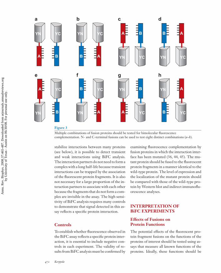
tion in the complex to allow them to col-lide with each other sufficiently frequentlyto facilitate bimolecular fluorescent complexformation. It is generally not possible topredict the arrangement of the fluorescentprotein fragments that will produce maximalsignal. Fusion proteins that produce optimalsignal must therefore be identified by empir-ical testing of several combinations of fusionproteins. For true interaction partners, it isalmost always possible to find a combinationof fusion proteins that produces a detectablesignal. Unless it is known that fusions to oneend of the protein are likely to be nonfunc-tional or that topological constraints are likelyto preclude the association between the frag-ments in some fusions, it is recommended thatfusions to both the N- and C-terminal endsof the proteins under investigation be tested.Schematic diagrams of the different permuta-tions of fusion proteins that can be used forBiFC analysis are shown in Figure 3. Thefluorescent protein fragments should be fusedto the interaction partners using flexible linkersequences to allow maximal mobility of thefragments after complex formation.
Detection of Transientand Weak Complexes
Because BiFC analysis is based on the as-sociation between fluorescent protein frag-ments, and this association is likely to
www.annualreviews.org • BiFC Analysis as a Probe of Protein Interactions in Living Cells 471
Ann
u. R
ev. B
ioph
ys. 2
008.
37:4
65-4
87. D
ownl
oade
d fr
om a
rjou
rnal
s.an
nual
revi
ews.
org
by U
nive
rsity
of
Tex
as -
Aus
tin o
n 06
/30/
08. F
or p
erso
nal u
se o
nly.
ANRV343-BB37-22 ARI 24 April 2008 15:55
YN YC
A B
YN YC
B A YN YC
A B
YN YC
B A
YC
B
YC
A
YN
A
YN
B
a b c d
e f g h
YN
A
YC
B
YN
B
YC
A
Figure 3Multiple combinations of fusion proteins should be tested for bimolecular fluorescencecomplementation. N- and C-terminal fusions can be used to test eight distinct combinations (a–h).
stabilize interactions between many proteins(see below), it is possible to detect transientand weak interactions using BiFC analysis.The interaction partners do not need to form acomplex with a long half-life because transientinteractions can be trapped by the associationof the fluorescent protein fragments. It is alsonot necessary for a large proportion of the in-teraction partners to associate with each otherbecause the fragments that do not form a com-plex are invisible in the assay. The high sensi-tivity of BiFC analysis requires many controlsto demonstrate that signal detected in this as-say reflects a specific protein interaction.
Controls
To establish whether fluorescence observed inthe BiFC assay reflects a specific protein inter-action, it is essential to include negative con-trols in each experiment. The validity of re-sults from BiFC analysis must be confirmed by
examining fluorescence complementation byfusion proteins in which the interaction inter-face has been mutated (36, 44, 45). The mu-tant protein should be fused to the fluorescentprotein fragments in a manner identical to thewild-type protein. The level of expression andthe localization of the mutant protein shouldbe compared with those of the wild-type pro-tein by Western blot and indirect immunoflu-orescence analyses.
INTERPRETATION OFBiFC EXPERIMENTS
Effects of Fusions onProtein Functions
The potential effects of the fluorescent pro-tein fragment fusions on the functions of theproteins of interest should be tested using as-says that measure all known functions of theproteins. Ideally, these functions should be
472 Kerppola
Ann
u. R
ev. B
ioph
ys. 2
008.
37:4
65-4
87. D
ownl
oade
d fr
om a
rjou
rnal
s.an
nual
revi
ews.
org
by U
nive
rsity
of
Tex
as -
Aus
tin o
n 06
/30/
08. F
or p
erso
nal u
se o
nly.
ANRV343-BB37-22 ARI 24 April 2008 15:55
I
+t½< 1 s
t½< 10 s
+ +
II
t½= 60 s
III
t½= 3000 s
IV
Nonfluorescentaggregates
t½~ 60 s t½~ 60 s
t½~ 60 s
V
Figure 4Pathway for formation of bimolecular fluorescent complexes. The pathway for fluorescent complexformation has been deduced based on in vitro studies of the dynamics of bimolecular fluorescencecomplementation using purified proteins (44). See the text for a description of the steps in this pathway.
measured under the same conditions used tovisualize the protein interactions. It is partic-ularly important to examine potential conse-quences of the stabilization of protein interac-tions by association of the fluorescent proteinfragments.
Dynamics of BimolecularFluorescent Complexes
The dynamics of BiFC complexes have beeninvestigated in vitro to elucidate the pathwayfor fluorescent complex formation (Figure 4)(44). The initial association between the fu-sion proteins (Figure 4, complex I) is medi-ated by the interaction partners. Results fromcompetition studies in which proteins lack-ing fluorescent protein fragment fusions wereadded to the mixture of fusion proteins indi-cate that the initial association between the
fusion proteins can be displaced to form com-plexes containing only one fluorescent pro-tein fragment (Figure 4, complexes II). Theefficiency of this competition decreases witha half-time of 1 min after mixing of the fu-sion proteins, suggesting that the complexisomerizes to form an irreversible associationbetween the fusion proteins (Figure 4, com-plex III). The increase in fluorescence displayssigmoidal kinetics, consistent with an initiallag corresponding to the time required forassociation of the fluorescent protein frag-ments, followed by maturation with a half-time of 50 min to produce the fluorescentBiFC complex (Figure 4, complex IV). Thefluorophores of all GFP variants are formedvia autocatalytic reactions after folding ofthe polypeptide, a process known as matura-tion. The rate of maturation of BiFC com-plex fluorescence was equivalent to the rate of
www.annualreviews.org • BiFC Analysis as a Probe of Protein Interactions in Living Cells 473
Ann
u. R
ev. B
ioph
ys. 2
008.
37:4
65-4
87. D
ownl
oade
d fr
om a
rjou
rnal
s.an
nual
revi
ews.
org
by U
nive
rsity
of
Tex
as -
Aus
tin o
n 06
/30/
08. F
or p
erso
nal u
se o
nly.
ANRV343-BB37-22 ARI 24 April 2008 15:55
maturation of the corresponding intact fluo-rescent protein.
The fluorescent protein fragments that donot associate with a complementary fragmentbecome trapped in a form that is not com-petent for subsequent association (Figure 4,complex V). This loss of competence for asso-ciation is likely to be significant for the speci-ficity of BiFC analysis because it results in akinetic barrier to the association of fluores-cent protein fragments that are fused to pro-teins that do not normally interact with eachother. This model based on in vitro studiescan account for many of the results observedin cells, including the requirement for a spe-cific interaction between the proteins fused tothe fluorescent protein fragments for efficientBiFC complex formation.
Fragments of a different GFP derivativeconjugated to nucleic acid interaction part-ners can produce fluorescence with more than100-fold-faster kinetics in vitro (22) than thefusion proteins originally investigated (44).These results may reflect chemical matu-ration of the fluorescent protein fragmentsduring expression or purification, possiblyassisted by the intein cleavage or biotin con-jugation reactions. The fluorescence inten-sity of BiFC complexes produced by thesefragments was reduced under conditions pre-dicted to destabilize the nucleic acid interac-tion (22). These results are consistent withrapid fluorescent complex formation and dis-sociation by the fluorescent protein frag-ments, suggesting that BiFC analysis can beused for nearly real-time visualization of someinteractions under the in vitro conditions usedin these experiments.
The differences in the dynamics of BiFCcomplex fluorescence in these experimentsmay reflect differences in the experimentalconditions, the fluorescent protein fragmentsused, or the interactions studied. Becausethese studies were performed in vitro, a sig-nificant question is whether BiFC complexesexhibit rapid fluorescent complex formationand dissociation in cells. Several studies of in-teractions between various proteins have re-
ported rapid changes in fluorescence intensityobserved in BiFC analysis in response to stim-uli predicted to affect the interactions (38, 66,98). However, it is difficult to exclude the pos-sibility that changes in the cellular environ-ment or variations in protein turnover affectthe fluorescence intensity measured in theseexperiments. Further studies of the dynamicsof BiFC complexes in living cells are neededto address this issue.
The Efficiency of BimolecularFluorescence Complementation
The fluorescence intensity produced bybimolecular fluorescence complementationvaries widely for interactions between dif-ferent partners and for different fusions tothe same partners. The fluorescence intensityproduced by BiFC complexes in living cellsis generally less than 10% of that producedby expression of an intact fluorescent protein.Nevertheless, because autofluorescence in thevisible range is extremely low in most cells, thesignal from bimolecular fluorescence comple-mentation is often orders of magnitude higherthan background fluorescence.
The efficiency of fluorescence comple-mentation is defined as the fluorescence in-tensity produced by bimolecular fluorescentcomplex formation when a specific level of fu-sion proteins is expressed in the cell. The effi-ciencies of bimolecular fluorescence comple-mentation produced by structurally unrelatedproteins cannot be used to determine the effi-ciencies of complex formation because manyfactors unrelated to the efficiency of complexformation influence the efficiency of bimolec-ular fluorescence complementation. Never-theless, in situations in which all these fac-tors are predicted to be identical, such as inthe case of wild-type and mutated interactionspartners, differences in the efficiencies of bi-molecular fluorescence complementation canprovide information about the relative effi-ciencies of complex formation. Thus, the ef-fects of single amino acid substitutions thatdo not alter the level of protein expression or
474 Kerppola
Ann
u. R
ev. B
ioph
ys. 2
008.
37:4
65-4
87. D
ownl
oade
d fr
om a
rjou
rnal
s.an
nual
revi
ews.
org
by U
nive
rsity
of
Tex
as -
Aus
tin o
n 06
/30/
08. F
or p
erso
nal u
se o
nly.
ANRV343-BB37-22 ARI 24 April 2008 15:55
its localization can be examined by comparingthe efficiencies of fluorescence complemen-tation by the wild-type and mutated proteins(44, 45).
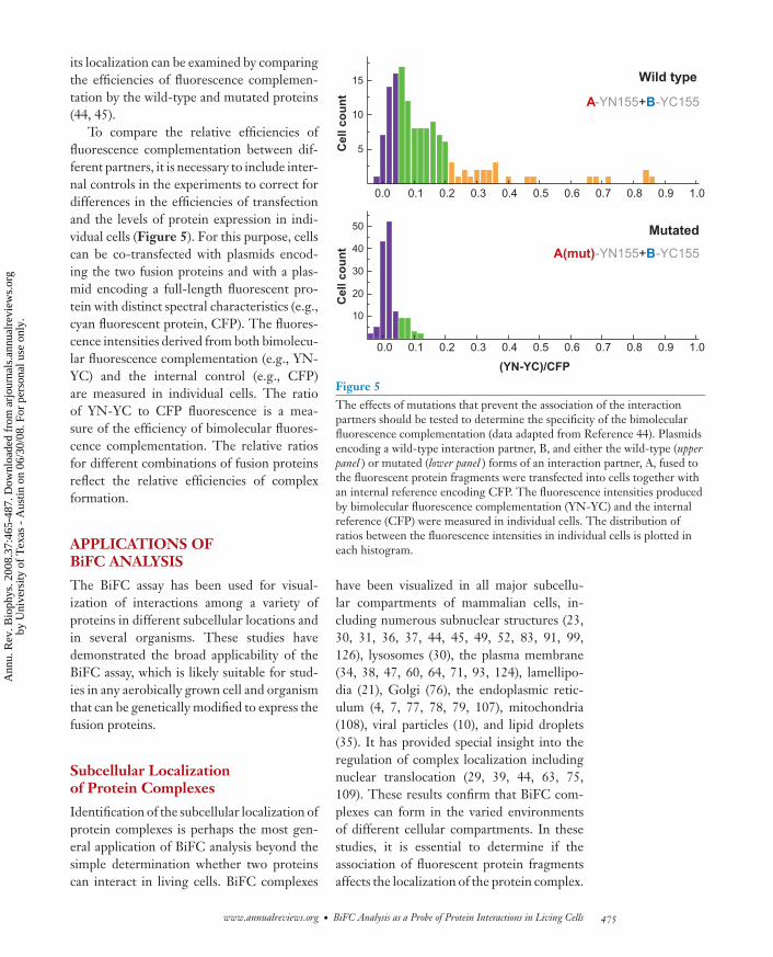
To compare the relative efficiencies offluorescence complementation between dif-ferent partners, it is necessary to include inter-nal controls in the experiments to correct fordifferences in the efficiencies of transfectionand the levels of protein expression in indi-vidual cells (Figure 5). For this purpose, cellscan be co-transfected with plasmids encod-ing the two fusion proteins and with a plas-mid encoding a full-length fluorescent pro-tein with distinct spectral characteristics (e.g.,cyan fluorescent protein, CFP). The fluores-cence intensities derived from both bimolecu-lar fluorescence complementation (e.g., YN-YC) and the internal control (e.g., CFP)are measured in individual cells. The ratioof YN-YC to CFP fluorescence is a mea-sure of the efficiency of bimolecular fluores-cence complementation. The relative ratiosfor different combinations of fusion proteinsreflect the relative efficiencies of complexformation.
APPLICATIONS OFBiFC ANALYSIS
The BiFC assay has been used for visual-ization of interactions among a variety ofproteins in different subcellular locations andin several organisms. These studies havedemonstrated the broad applicability of theBiFC assay, which is likely suitable for stud-ies in any aerobically grown cell and organismthat can be genetically modified to express thefusion proteins.
Subcellular Localizationof Protein Complexes
Identification of the subcellular localization ofprotein complexes is perhaps the most gen-eral application of BiFC analysis beyond thesimple determination whether two proteinscan interact in living cells. BiFC complexes
A(mut)-YN155+B-YC155
A-YN155+B-YC155
10
20
30
40
50
0.0 0.1 0.2 0.3 0.4 0.5 0.6 0.7 0.8 0.9 1.0
0.0 0.1 0.2 0.3 0.4 0.5 0.6 0.7 0.8 0.9 1.0
5
10
15
(YN-YC)/CFP
Cel
l co
un
tC
ell c
ou
nt
Wild type
Mutated
Figure 5The effects of mutations that prevent the association of the interactionpartners should be tested to determine the specificity of the bimolecularfluorescence complementation (data adapted from Reference 44). Plasmidsencoding a wild-type interaction partner, B, and either the wild-type (upperpanel ) or mutated (lower panel ) forms of an interaction partner, A, fused tothe fluorescent protein fragments were transfected into cells together withan internal reference encoding CFP. The fluorescence intensities producedby bimolecular fluorescence complementation (YN-YC) and the internalreference (CFP) were measured in individual cells. The distribution ofratios between the fluorescence intensities in individual cells is plotted ineach histogram.
have been visualized in all major subcellu-lar compartments of mammalian cells, in-cluding numerous subnuclear structures (23,30, 31, 36, 37, 44, 45, 49, 52, 83, 91, 99,126), lysosomes (30), the plasma membrane(34, 38, 47, 60, 64, 71, 93, 124), lamellipo-dia (21), Golgi (76), the endoplasmic retic-ulum (4, 7, 77, 78, 79, 107), mitochondria(108), viral particles (10), and lipid droplets(35). It has provided special insight into theregulation of complex localization includingnuclear translocation (29, 39, 44, 63, 75,109). These results confirm that BiFC com-plexes can form in the varied environmentsof different cellular compartments. In thesestudies, it is essential to determine if theassociation of fluorescent protein fragmentsaffects the localization of the protein complex.
www.annualreviews.org • BiFC Analysis as a Probe of Protein Interactions in Living Cells 475
Ann
u. R
ev. B
ioph
ys. 2
008.
37:4
65-4
87. D
ownl
oade
d fr
om a
rjou
rnal
s.an
nual
revi
ews.
org
by U
nive
rsity
of
Tex
as -
Aus
tin o
n 06
/30/
08. F
or p
erso
nal u
se o
nly.
ANRV343-BB37-22 ARI 24 April 2008 15:55
One strategy to accomplish this is to deter-mine the localization of one interaction part-ner in the presence of an overexpressed part-ner lacking the fluorescent protein fragment(36). In addition to numerous interactions in-volving soluble proteins, BiFC analysis hasalso been used to study interactions involvingintegral membrane proteins (21, 64), demon-strating that the topological constraints ofthese proteins do not prevent the use of BiFCanalysis.
Signal Transduction Networks
Many proteins interact with a large numberof different partners. The sum of these inter-actions produces a complex network of con-nections where signals impinging on a singlenode (protein) can be propagated through-out the network. Visualization of individualinteractions within this network can provideinsight into the relationships between a spe-cific interaction within the network and thesignals that modulate its localization and effi-ciency. Numerous interactions involving bothdiffusible components of such networks (2, 19,20, 28, 41, 46, 95, 98, 118) and membrane re-ceptors (16, 21, 64) have been visualized usingBiFC analysis. Interactions between cytoplas-mic and nuclear signal transduction compo-nents (3, 119) have enabled tracking of sig-nal transduction between different cellularcompartments.
Protein Interactions Associatedwith Cytokinesis
Interactions that occur in a cell cycle–regulated manner are particularly interestingand challenging subjects for imaging studies.This is because the complexes are transient,placing special requirements on the efficiencyand rate of complex detection. Faithful rep-resentation of the cell cycle–regulated forma-tion of these complexes also requires that themethods used for imaging them do not dis-tort the temporal regulation of complex for-
mation and dissociation or degradation. BiFCanalysis has been used to visualize the com-plex formed by Grr1 and Hof1 (8). Grr1 in-teracts with Hof1 specifically in the bud neckbetween the mother and daughter cells duringthe G2-M stage of the cell cycle. This associ-ation results in degradation of Hof1, which isrequired for efficient contraction of the actinring closing the bud neck and cytokinesis. In-teractions between the p0071 catenin familymember with the RhoA small GTPase and theEct guanine nucleotide exchange factor havealso been visualized using BiFC analysis at themidbody, a structure located at the site of cy-tokinesis during telophase in mammalian cells(54). These results demonstrate the detectionof spatially and temporally restricted complexformation by BiFC analysis.
Interactions on Scaffolds
Many proteins can be brought in proximity toeach other by binding to the same interactionpartner that can serve as a scaffold for the as-sembly of multiprotein complexes. Such scaf-folds are not limited to proteins but includenucleic acids, carbohydrates, and other cel-lular macromolecules. Simultaneous bindingby two proteins in the vicinity of each otheron the same scaffold can be detected by BiFCanalysis. This principle has been used to de-tect RNA binding by fusing the fragments ofthe fluorescent protein to two RNA bindingproteins (90). It has also been used to visu-alize RNA export complexes in the nucleusand to measure the turnover rate of such com-plexes (99). By designing fusion proteins thatcan bind to a single type of RNA molecule, thisapproach has been used to track RNA insideliving cells (80). Similar fusion proteins with adesigned binding specificity for DNA displayfluorescence complementation in vitro uponbinding to a specific DNA oligonucleotide(105). One concern regarding this generalstrategy is that the fluorescent complex assem-bled on the scaffold might remain fluorescentfollowing dissociation from the scaffold.
476 Kerppola
Ann
u. R
ev. B
ioph
ys. 2
008.
37:4
65-4
87. D
ownl
oade
d fr
om a
rjou
rnal
s.an
nual
revi
ews.
org
by U
nive
rsity
of
Tex
as -
Aus
tin o
n 06
/30/
08. F
or p
erso
nal u
se o
nly.
ANRV343-BB37-22 ARI 24 April 2008 15:55
Visualization of Interactionsin Living Organisms
The BiFC assay has been applied to stud-ies in a variety of unicellular and multicellu-lar organisms. Many interactions have beenvisualized in E. coli (6, 33, 67, 73), Agrobac-terium tumefaciens (15, 111), and Bacillus sub-tilis (75, 103). Among fungi, BiFC analysis hasbeen extensively used in Saccharomyces cere-visiae (Baker’s yeast) (8, 53, 82), and also inAcremonium chrysogenum (43), Aspergillus nidu-lans (9), and Magnaporthe grisea (125). Amonghigher eukaryotic organisms, BiFC analysishas been used to visualize numerous interac-tions in many plant species (1, 5, 11, 12, 14,17, 18, 24, 25, 40, 51, 57, 58, 62, 65, 69, 70,81, 86, 88, 92, 100, 106, 112, 116, 117, 122,123). Virtually all these studies have been per-formed by transient expression using heterol-ogous expression vectors, suggesting that theexpression of the fusion proteins is unlikelyto reflect their normal tissue-specific patterns.BiFC analysis has also been used to visual-ize interactions between Caenorhabditis elegansproteins (75).
Interaction Screens UsingBiFC Analysis
The BiFC assay can be used as a screeningtool to identify potential interaction partnersas well as modifiers of known interactions(26, 93). The challenge of implementing ascreen for interaction partners is that the lev-els of expression of different fusion proteinsin a library are likely to vary over a largerange and may not reflect the levels of expres-sion of the corresponding endogenous pro-teins. Thus, differences in BiFC signal arelikely affected by a variety of factors unrelatedto the efficiency of the protein interaction.Nevertheless, several novel interaction part-ners have been identified using this strategy(26, 93).
BiFC analysis can also be used to screenfor small-molecule modulators (66). Thereare numerous mechanisms whereby smallmolecules could influence the fluorescence in-
tensity produced in BiFC assays. Neverthe-less, because many of these mechanisms couldalso influence the endogenous proteins, thisprovides a useful strategy for the identificationof small molecules that alter specific proteincomplexes in living cells. Comparison of theeffects of specific small molecules on a panelof BiFC complexes can provide an indicationof the degree of specificity of their effects.
SIMULTANEOUSVISUALIZATION OF MULTIPLEPROTEIN INTERACTIONS
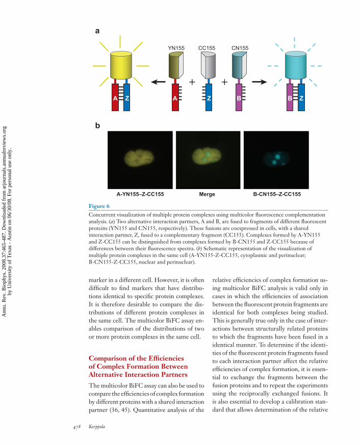
Many proteins have a large number of poten-tial interaction partners. Often these interac-tions are mutually exclusive, such that onlyone protein can interact with a particular pro-tein molecule at any one time. This results incompetition for shared interaction partners incells that express several alternative partners.The multicolor BiFC assay enables visualiza-tion of interactions between multiple combi-nations of proteins in the same cell (45). Thisassay is based on the formation of fluorescentcomplexes with different spectra through theassociation of fragments of different fluores-cent proteins fused to alternative interactionpartners (Figure 6). The multicolor BiFC as-say enables comparison of the subcellular dis-tributions of several protein complexes in thesame cell and allows analysis of the compe-tition between mutually exclusive interactionpartners for binding to a common partner.
Comparison of the Distributionsof Multiple Protein Complexesin the Same Cell
Complexes formed by a protein with differentinteraction partners often have different func-tions. These functional differences can be re-flected in differences between the subcellulardistributions of the protein complexes. Thesubcellular distributions of different proteincomplexes can be compared by identifying amarker that has the same distribution as one orthe other complex and comparing the distri-bution of the second complex with that of the
www.annualreviews.org • BiFC Analysis as a Probe of Protein Interactions in Living Cells 477
Ann
u. R
ev. B
ioph
ys. 2
008.
37:4
65-4
87. D
ownl
oade
d fr
om a
rjou
rnal
s.an
nual
revi
ews.
org
by U
nive
rsity
of
Tex
as -
Aus
tin o
n 06
/30/
08. F
or p
erso
nal u
se o
nly.
ANRV343-BB37-22 ARI 24 April 2008 15:55
A Z
+B
+BA Z Z
a
b
YN155
A-YN155–Z-CC155 B-CN155–Z-CC155
CN155CC155
Merge
Figure 6Concurrent visualization of multiple protein complexes using multicolor fluorescence complementationanalysis. (a) Two alternative interaction partners, A and B, are fused to fragments of different fluorescentproteins (YN155 and CN155, respectively). These fusions are coexpressed in cells, with a sharedinteraction partner, Z, fused to a complementary fragment (CC155). Complexes formed by A-YN155and Z-CC155 can be distinguished from complexes formed by B-CN155 and Z-CC155 because ofdifferences between their fluorescence spectra. (b) Schematic representation of the visualization ofmultiple protein complexes in the same cell (A-YN155-Z-CC155, cytoplasmic and perinuclear;B-CN155-Z-CC155, nuclear and perinuclear).
marker in a different cell. However, it is oftendifficult to find markers that have distribu-tions identical to specific protein complexes.It is therefore desirable to compare the dis-tributions of different protein complexes inthe same cell. The multicolor BiFC assay en-ables comparison of the distributions of twoor more protein complexes in the same cell.
Comparison of the Efficienciesof Complex Formation BetweenAlternative Interaction Partners
The multicolor BiFC assay can also be used tocompare the efficiencies of complex formationby different proteins with a shared interactionpartner (36, 45). Quantitative analysis of the
relative efficiencies of complex formation us-ing multicolor BiFC analysis is valid only incases in which the efficiencies of associationbetween the fluorescent protein fragments areidentical for both complexes being studied.This is generally true only in the case of inter-actions between structurally related proteinsto which the fragments have been fused in aidentical manner. To determine if the identi-ties of the fluorescent protein fragments fusedto each interaction partner affect the relativeefficiencies of complex formation, it is essen-tial to exchange the fragments between thefusion proteins and to repeat the experimentsusing the reciprocally exchanged fusions. Itis also essential to develop a calibration stan-dard that allows determination of the relative
478 Kerppola
Ann
u. R
ev. B
ioph
ys. 2
008.
37:4
65-4
87. D
ownl
oade
d fr
om a
rjou
rnal
s.an
nual
revi
ews.
org
by U
nive
rsity
of
Tex
as -
Aus
tin o
n 06
/30/
08. F
or p
erso
nal u
se o
nly.
ANRV343-BB37-22 ARI 24 April 2008 15:55
fluorescence intensities produced by the spec-trally distinct complexes when fused to inter-action partners that form complexes with thesame efficiency. This calibration standard canbe generated by fusing the fluorescent pro-tein fragments to the same interaction part-ners (36, 45).
The relative efficiencies of complex forma-tion in the multicolor BiFC assay are affectedby the levels of protein expression, which mustbe considered when interpreting the results ofsuch experiments. The efficiencies of complexformation measured in the multicolor BiFCassay reflect numerous factors in addition tointrinsic binding affinity. These factors in-clude the subcellular distributions of the inter-action partners and the effects of any cellularfactors that can facilitate or hinder an inter-action, including posttranslational modifica-tions and the network of alternative partners.Moreover, in many cases, BiFC complex for-mation is irreversible after association of thefluorescent protein fragments. Thus, changesin cellular conditions after the time of complexformation may not be reflected in the relativeefficiencies of complex formation. Neverthe-less, because the rate of association of the fluo-rescent protein fragments is likely to be slowerthan the rate of exchange between many alter-native interaction partners, the interactionsbetween the alternative fusion partners willlikely reach equilibrium prior to complex fix-ation by association of the fluorescent proteinfragments.
Design of MulticolorBiFC Experiments
Multicolor BiFC analysis requires fusion ofthe alternative interaction partners to frag-ments of fluorescent proteins that producecomplexes with different spectra (Table 2).Because the two complexes can be imaged se-quentially, spectral overlap is generally not aproblem since different excitation and emis-sion wavelengths can be used to visualize thecomplexes. Although this is not strictly simul-taneous, alternate imaging of the two com-
plexes can be performed to confirm that thedelay of a few seconds between acquisition ofthe images does not allow time for relocaliza-tion of either complex. Ideally, the two com-plexes should have fluorescence intensities ofthe same order of magnitude in order to avoidthe possibility that differences in the signal-to-background ratio produce the appearanceof differences in distribution. However, suchbackground signal and any cross-talk betweenthe two fluorophores can be corrected for byimaging cells that express only one combina-tion of fusion proteins. The fusion proteinsshould be expressed at levels comparable tothe endogenous proteins to establish that thedistributions are not affected by the levels ofexpression of the proteins. As in the analy-sis of a single protein interaction using BiFCanalysis, it is critical to determine if mutationsthat eliminate each interaction individuallyalso eliminate the corresponding BiFC signal.
Applications of MulticolorBiFC Analysis
The multicolor BiFC assay has been appliedto analysis of the relative efficiencies of com-plex formation between several families of nu-clear transcription regulatory proteins (36, 45)as well as the large family of cytoplasmic smallG protein subunits (27, 71). The results ofthese experiments have shown that the effi-ciencies of interactions with proteins that areclosely related in both sequence and structurecan differ substantially in the cell. The reasonsfor these differences are generally unknown.
SUMMARY AND FUTUREPROSPECTS
The BiFC assay has become a standard ap-proach for the visualization of protein in-teractions. When appropriate controls areperformed, BiFC analysis has proved to be areliable tool for the detection of protein in-teractions in living cells. False positives canbe avoided by ensuring that the fusion pro-teins are expressed at levels comparable to the
www.annualreviews.org • BiFC Analysis as a Probe of Protein Interactions in Living Cells 479
Ann
u. R
ev. B
ioph
ys. 2
008.
37:4
65-4
87. D
ownl
oade
d fr
om a
rjou
rnal
s.an
nual
revi
ews.
org
by U
nive
rsity
of
Tex
as -
Aus
tin o
n 06
/30/
08. F
or p
erso
nal u
se o
nly.
ANRV343-BB37-22 ARI 24 April 2008 15:55
corresponding endogenous proteins, and byperforming appropriate controls to determineif mutations that eliminate an interaction alsoeliminate the fluorescence signal. Anecdotalevidence suggests that false negatives are oc-casionally encountered. However, these canoften be corrected by more comprehensivetesting of multiple combinations of fusions tothe same interaction partners.
The BiFC assay is finding new applicationsat an accelerating rate and it is being adaptedfor new purposes on the basis of the generalprinciple that the association of the fluores-cent protein fragments can be enhanced whenthey are brought in proximity to each otherand provided the dynamic flexibility necessaryfor them to collide with each other. Some ofthe limitations of the BiFC assay identified inthe original description (44) of this approachremain to be solved. The association betweenthe fluorescent protein fragments stabilizesthe association between the interaction part-ners. This stabilization can result in essentiallyirreversible complex formation and can po-tentially alter the function or activity of thecomplex. A better understanding of the fold-ing and dynamics of the bimolecular com-plex formed by the fluorescent protein frag-ments could help provide strategies to solvethis problem.
The fluorescent protein fragments alsohave the capacity to associate with each otherto form a fluorescent complex even if the pro-teins to which they are fused do not nor-mally interact with each other. This propen-sity varies depending on the proteins to whichthe fragments are fused, and the intrinsic ten-dency of the fragments alone to associate isgenerally reduced by fusion of the fragmentsto proteins that do not interact with eachother. Nevertheless, identification of frag-ments of fluorescent proteins with a reducedtendency to associate with each other spon-taneously, but an undiminished ability to as-sociate when present in the same macro-molecular complex, would be of significantbenefit. Mutational engineering of full-lengthGFP family members has produced proteinswith an astounding range of photophysi-cal and photochemical characteristics. It istherefore virtually guaranteed that future ef-forts to engineer fragments of fluorescentproteins for BiFC analysis will produce im-proved versions. It is also likely that frag-ments that are optimal for a particular purposewill not be ideal for all purposes. It is there-fore important to perform comparative anal-ysis of BiFC assays using different fluorescentprotein fragments to evaluate their relativemerits.
DISCLOSURE STATEMENT
The author is not aware of any biases that might be perceived as affecting the objectivity ofthis review.
ACKNOWLEDGMENTS
I thank Changdeng Hu for his contributions to the implementation of the BiFC assay andmembers of the Kerppola laboratory for extending the BiFC assay to new applications.
LITERATURE CITED
1. Abe M, Kobayashi Y, Yamamoto S, Daimon Y, Yamaguchi A, et al. 2005. FD, a bZIPprotein mediating signals from the floral pathway integrator FT at the shoot apex. Science309:1052–56
2. Adolph D, Flach N, Mueller K, Ostareck DH, Ostareck-Lederer A. 2007. Decipheringthe cross talk between hnRNP K and c-Src: The c-Src activation domain in hnRNP K isdistinct from a second interaction site. Mol. Cell. Biol. 27:1758–70
480 Kerppola
Ann
u. R
ev. B
ioph
ys. 2
008.
37:4
65-4
87. D
ownl
oade
d fr
om a
rjou
rnal
s.an
nual
revi
ews.
org
by U
nive
rsity
of
Tex
as -
Aus
tin o
n 06
/30/
08. F
or p
erso
nal u
se o
nly.
ANRV343-BB37-22 ARI 24 April 2008 15:55
3. Ahmed KM, Dong SZ, Fan M, Li JJ. 2006. Nuclear factor-kappa B p65 inhibits mitogen-activated protein kinase signaling pathway in radioresistant breast cancer cells. Mol. CancerRes. 4:945–55
4. Anderie I, Schulz I, Schmid A. 2007. Direct interaction between ER membrane-boundPTP1B and its plasma membrane-anchored targets. Cell. Signal. 19:582–92
5. Aparicio F, Sanchez-Navarro JA, Pallas V. 2006. In vitro and in vivo mapping of thePrunus necrotic ringspot virus coat protein C-terminal dimerization domain by bimolec-ular fluorescence complementation. J. Gen. Virol. 87:1745–50
6. Atmakuri K, Ding ZY, Christie PJ. 2003. VirE2, a type IV secretion substrate, interactswith the VirD4 transfer protein at cell poles of Agrobacterium tumefaciens. Mol. Microbiol.49:1699–713
7. BelAiba RS, Djordjevic T, Petry A, Diemer K, Bonello S, et al. 2007. NOX5 variants arefunctionally active in endothelial cells. Free Radic. Biol. Med. 42:446–59
8. Blondel M, Bach S, Bamps S, Dobbelaere J, Wiget P, et al. 2005. Degradation of Hof1by SCFGrr1 is important for actomyosin contraction during cytokinesis in yeast. EMBOJ. 24:1440–52
9. Blumenstein A, Vienken K, Tasler R, Purschwitz J, Veith D, et al. 2005. The Aspergillusnidulans phytochrome FphA represses sexual development in red light. Curr. Biol. 15:1833–38
10. Boyko V, Leavitt M, Gorelick R, Fu W, Nikolaitchik O, et al. 2006. Coassembly andcomplementation of Gag proteins from HIV-1 and HIV-2, two distinct human pathogens.Mol. Cell 23:281–87
11. Bracha-Drori K, Shichrur K, Katz A, Oliva M, Angelovici R, et al. 2004. Detection ofprotein-protein interactions in plants using bimolecular fluorescence complementation.Plant J. 40:419–27
12. Burch-Smith TM, Schiff M, Caplan JL, Tsao J, Czymmek K, Dinesh-Kumar SP. 2007. Anovel role for the TIR domain in association with pathogen-derived elicitors. PLoS Biol.5:e68
13. Cabantous S, Terwilliger TC, Waldo GS. 2005. Protein tagging and detection with engi-neered self-assembling fragments of green fluorescent protein. Nat. Biotechnol. 23:102–7
14. Canto T, Uhrig JF, Swanson M, Wright KM, MacFarlane SA. 2006. Translocation ofTomato bushy stunt virus P19 protein into the nucleus by ALY proteins compromises itssilencing suppressor activity. J. Virol. 80:9064–72
15. Cascales E, Atmakuri K, Liu Z, Binns AN, Christie PJ. 2005. Agrobacterium tumefaciensoncogenic suppressors inhibit T-DNA and VirE2 protein substrate binding to the VirD4coupling protein. Mol. Microbiol. 58:565–79
16. Chen CD, Oh SY, Hinman JD, Abraham CR. 2006. Visualization of APP dimerizationand APP-Notch2 heterodimerization in living cells using bimolecular fluorescence com-plementation. J. Neurochem. 97:30–43
17. Chen SB, Tao LZ, Zeng LR, Vega-Sanchez ME, Umemura K, Wang GL. 2006. A highlyefficient transient protoplast system for analyzing defence gene expression and protein-protein interactions in rice. Mol. Plant Pathol. 7:417–27
18. Citovsky V, Lee LY, Vyas S, Glick E, Chen MH, et al. 2006. Subcellular localization ofinteracting proteins by bimolecular fluorescence complementation in planta. J. Mol. Biol.362:1120–31
19. Cole KC, McLaughlin HW, Johnson DI. 2007. Use of bimolecular fluorescence com-plementation to study in vivo interactions between Cdc42p and Rdi1p of Saccharomycescerevisiae. Eukaryot. Cell 6:378–87
www.annualreviews.org • BiFC Analysis as a Probe of Protein Interactions in Living Cells 481
Ann
u. R
ev. B
ioph
ys. 2
008.
37:4
65-4
87. D
ownl
oade
d fr
om a
rjou
rnal
s.an
nual
revi
ews.
org
by U
nive
rsity
of
Tex
as -
Aus
tin o
n 06
/30/
08. F
or p
erso
nal u
se o
nly.
ANRV343-BB37-22 ARI 24 April 2008 15:55
20. de Bie P, de Sluis B, Burstein E, Duran KJ, Berger R, et al. 2006. Characterization ofCOMMD protein-protein interactions in NF-kappa B signalling. Biochem. J. 398:63–71
21. de Virgilio M, Kiosses WB, Shattil SJ. 2004. Proximal, selective, and dynamic interactionsbetween integrin alpha II beta 3 and protein tyrosine kinases in living cells. J. Cell Biol.165:305–11
22. Demidov VV, Dokholyan NV, Witte-Hoffmann C, Chalasani P, Yiu HW, et al. 2006.Fast complementation of split fluorescent protein triggered by DNA hybridization. Proc.Natl. Acad. Sci. USA 103:2052–56
23. Deppmann CD, Thornton TM, Utama FE, Taparowsky EJ. 2003. Phosphorylation ofBATF regulates DNA binding: a novel mechanism for AP-1 (activator protein-1) regula-tion. Biochem. J. 374:423–31
24. Diaz I, Martinez M, Isabel-LaMoneda I, Rubio-Somoza I, Carbonero P. 2005. TheDOF protein, SAD, interacts with GAMYB in plant nuclei and activates transcriptionof endosperm-specific genes during barley seed development. Plant J. 42:652–62
25. Ding YH, Liu NY, Tang ZS, Liu J, Yang WC. 2006. Arabidopsis GLUTAMINE-RICHPROTEIN23 is essential for early embryogenesis and encodes a novel nuclear PPR motifprotein that interacts with RNA polymerase II subunit III. Plant Cell 18:815–30
26. Ding ZY, Liang JY, Lu YL, Yu QH, Zhou SY, et al. 2006. A retrovirus-based proteincomplementation assay screen reveals functional AKT1-binding partners. Proc. Natl. Acad.Sci. USA 103:15014–19
27. Dupre DJ, Robitaille M, Ethier N, Villeneuve LR, Mamarbachi AM, Hebert TE. 2006.Seven transmembrane receptor core signaling complexes are assembled prior to plasmamembrane trafficking. J. Biol. Chem. 281:34561–73
28. Dupre DJ, Robitaille M, Richer M, Ethier N, Mamarbachi AM, Hebert TE. 2007.Dopamine receptor-interacting protein 78 acts as a molecular chaperone for G gammasubunits before assembly with G beta. J. Biol. Chem. 282:13703–15
29. Fan M, Ahmed KM, Coleman MC, Spitz DR, Li JJ. 2007. Nuclear factor-kappa B andmanganese superoxide dismutase mediate adaptive radioresistance in low-dose irradiatedmouse skin epithelial cells. Cancer Res. 67:3220–28
30. Fang DY, Kerppola TK. 2004. Ubiquitin-mediated fluorescence complementation revealsthat Jun ubiquitinated by Itch/AIP4 is localized to lysosomes. Proc. Natl. Acad. Sci. USA101:14782–87
31. Farina A, Hattori M, Qin J, Nakatani Y, Minato N, Ozato K. 2004. Bromodomain proteinBrd4 binds to GTPase-activating SPA-1, modulating its activity and subcellular localiza-tion. Mol. Cell. Biol. 24:9059–69
32. Galarneau A, Primeau M, Trudeau LE, Michnick SW. 2002. Beta-lactamase protein frag-ment complementation assays as in vivo and in vitro sensors of protein protein interactions.Nat. Biotechnol. 20:619–22
33. Ghosh I, Hamilton AD, Regan L. 2000. Antiparallel leucine zipper-directed protein re-assembly: application to the green fluorescent protein. J. Am. Chem. Soc. 122:5658–59
34. Giese B, Roderburg C, Sommerauer M, Wortmann SB, Metz S, et al. 2005. Dimerizationof the cytokine receptors gp130 and LIFR analysed in single cells. J. Cell Sci. 118:5129–40
35. Granneman JG, Moore HPH, Granneman RL, Greenberg AS, Obin MS, Zhu ZX. 2007.Analysis of lipolytic protein trafficking and interactions in adipocytes. J. Biol. Chem.282:5726–35
36. Grinberg AV, Hu CD, Kerppola TK. 2004. Visualization of Myc/Max/Mad family dimersand the competition for dimerization in living cells. Mol. Cell. Biol. 24:4294–308
482 Kerppola
Ann
u. R
ev. B
ioph
ys. 2
008.
37:4
65-4
87. D
ownl
oade
d fr
om a
rjou
rnal
s.an
nual
revi
ews.
org
by U
nive
rsity
of
Tex
as -
Aus
tin o
n 06
/30/
08. F
or p
erso
nal u
se o
nly.
ANRV343-BB37-22 ARI 24 April 2008 15:55
37. Guo HX, Cun W, Liu LD, Dong SZ, Wang LC, et al. 2006. Protein encoded by HSV-1stimulation-related gene 1 (HSRG1) interacts with and inhibits SV40 large T antigen.Cell Prolif. 39:507–18
38. Guo YJ, Rebecchi M, Scarlata S. 2005. Phospholipase C beta(2) binds to and inhibitsphospholipase C delta(1). J. Biol. Chem. 280:1438–47
39. Gwozdz T, Dutko-Gwozdz J, Nieva C, Betanska K, Orlowski M, et al. 2007. EcR and Usp,components of the ecdysteroid nuclear receptor complex, exhibit differential distributionof molecular determinants directing subcellular trafficking. Cell. Signal. 19:490–503
40. Hackbusch J, Richter K, Muller J, Salamini F, Uhrig JF. 2005. A central role of Arabidopsisthaliana ovate family proteins in networking and subcellular localization of 3-aa loopextension homeodomain proteins. Proc. Natl. Acad. Sci. USA 102:4908–12
41. Hausser A, Link G, Hoene M, Russo C, Selchow O, Pfizenmaier K. 2006. Phospho-specific binding of 14-3-3 proteins to phosphatidylinositol 4-kinase III beta protects fromdephosphorylation and stabilizes lipid kinase activity. J. Cell Sci. 119:3613–21
42. Hink MA, Borst JW, Visser AJ. 2003. Fluorescence correlation spectroscopy of GFPfusion proteins in living plant cells. Methods Enzymol. 361:93–112
43. Hoff B, Kuck U. 2005. Use of bimolecular fluorescence complementation to demon-strate transcription factor interaction in nuclei of living cells from the filamentous fungusAcremonium chrysogenum. Curr. Genet. 47:132–38
44. Hu CD, Chinenov Y, Kerppola TK. 2002. Visualization of interactions among bZIP andRel family proteins in living cells using bimolecular fluorescence complementation. Mol.Cell 9:789–98
45. Hu CD, Kerppola TK. 2003. Simultaneous visualization of multiple protein interactionsin living cells using multicolor fluorescence complementation analysis. Nat. Biotechnol.21:539–45
46. Hynes TR, Mervine SM, Yost EA, Sabo JL, Berlot CH. 2004. Live cell imaging of G(s)and the beta(2)-adrenergic receptor demonstrates that both alpha(s) and beta(1)gamma(7)internalize upon stimulation and exhibit similar trafficking patterns that differ from thatof the beta(2)-adrenergic receptor. J. Biol. Chem. 279:44101–12
47. Hynes TR, Tang LN, Mervine SM, Sabo JL, Yost EA, et al. 2004. Visualization of G pro-tein beta gamma dimers using bimolecular fluorescence complementation demonstratesroles for both beta and gamma in subcellular targeting. J. Biol. Chem. 279:30279–86
48. Jach G, Pesch M, Richter K, Frings S, Uhrig JF. 2006. An improved mRFP1 adds red tobimolecular fluorescence complementation. Nat. Methods 3:597–600
49. Jang MK, Mochizuki K, Zhou MS, Jeong HS, Brady JN, Ozato K. 2005. The bromo-domain protein Brd4 is a positive regulatory component of P-TEFb and stimulates RNApolymerase II-dependent transcription. Mol. Cell 19:523–34
50. Johnsson N, Varshavsky A. 1994. Split ubiquitin as a sensor of protein interactions in vivo.Proc. Natl. Acad. Sci. USA 91:10340–44
51. Kaminaka H, Nake C, Epple P, Dittgen J, Schutze K, et al. 2006. bZIP10-LSD1 antago-nism modulates basal defense and cell death in Arabidopsis following infection. EMBO J.25:4400–11
52. Kanno T, Kanno Y, Siegel RM, Jang MK, Lenardo MJ, Ozato K. 2004. Selective recog-nition of acetylated histones by bromodomain proteins visualized in living cells. Mol. Cell13:33–43
53. Kass G, Arad G, Rosenbluh J, Gafni Y, Graessmann A, et al. 2006. Permeabilized mam-malian cells as an experimental system for nuclear import of geminiviral karyophilic pro-teins and of synthetic peptides derived from their nuclear localization signal regions. J.Gen. Virol. 87:2709–20
www.annualreviews.org • BiFC Analysis as a Probe of Protein Interactions in Living Cells 483
Ann
u. R
ev. B
ioph
ys. 2
008.
37:4
65-4
87. D
ownl
oade
d fr
om a
rjou
rnal
s.an
nual
revi
ews.
org
by U
nive
rsity
of
Tex
as -
Aus
tin o
n 06
/30/
08. F
or p
erso
nal u
se o
nly.
ANRV343-BB37-22 ARI 24 April 2008 15:55
54. Keil R, Wolf A, Huttelmaier S, Hatzfeld M. 2007. Beyond regulation of cell adhesion:local control of RhoA at the cleavage furrow by the p0071 catenin. Cell Cycle 6:122–27
55. Kerppola TK. 2006. Complementary methods for studies of protein interactions in livingcells. Nat. Methods 3:969–71
56. Kerppola TK. 2006. Visualization of molecular interactions by fluorescence complemen-tation. Nat. Rev. Mol. Cell Biol. 7:449–56
57. Krichevsky A, Gutgarts H, Kozlovsky SV, Tzfira T, Sutton A, et al. 2007. C2H2 zincfinger-SET histone methyltransferase is a plant-specific chromatin modifier. Dev. Biol.303:259–69
58. Lacroix B, Vaidya M, Tzfira T, Citovsky V. 2005. The VirE3 protein of Agrobacteriummimics a host cell function required for plant genetic transformation. EMBO J. 24:428–37
59. Larson DR, Ma YM, Vogt VM, Webb WW. 2003. Direct measurement of Gag-Gag inter-action during retrovirus assembly with FRET and fluorescence correlation spectroscopy.J. Cell Biol. 162:1233–44
60. Lee SK, Boyko V, Hu WS. 2007. Capsid is an important determinant for functionalcomplementation of murine leukemia virus and spleen necrosis virus Gag proteins. Virology360:388–97
61. Li HY, Ng EK, Lee SM, Kotaka M, Tsui SK, et al. 2001. Protein-protein interactionof FHL3 with FHL2 and visualization of their interaction by green fluorescent pro-teins (GFP) two-fusion fluorescence resonance energy transfer (FRET). J. Cell. Biochem.80:293–303
62. Li JX, Krichevsky A, Vaidya M, Tzfira T, Citovsky V. 2005. Uncoupling of the functionsof the Arabidopsis VIN protein in transient and stable plant genetic transformation byAgrobacterium. Proc. Natl. Acad. Sci. USA 102:5733–38
63. Liu H, Deng XH, Shyu YJ, Li JJ, Taparowsky EJ, Hu CD. 2006. Mutual regulationof c-Jun and ATF2 by transcriptional activation and subcellular localization. EMBO J.25:1058–69
64. Lopez-Gimenez JF, Canals M, Pediani JD, Milligan G. 2007. The alpha(1b)-adrenoceptor exists as a higher-order oligomer: Effective oligomerization is required forreceptor maturation, surface delivery, and function. Mol. Pharmacol. 71:1015–29
65. Loyter A, Rosenbluh J, Zakai N, Li JX, Kozlovsky SV, et al. 2005. The plant VirE2interacting protein 1. A molecular link between the Agrobacterium T-complex and thehost cell chromatin? Plant Physiol. 138:1318–21
66. MacDonald ML, Lamerdin J, Owens S, Keon BH, Bilter GK, et al. 2006. Identifyingoff-target effects and hidden phenotypes of drugs in human cells. Nat. Chem. Biol. 2:329–37
67. Magliery TJ, Wilson CGM, Pan WL, Mishler D, Ghosh I, et al. 2005. Detecting protein-protein interactions with a green fluorescent protein fragment reassembly trap: scope andmechanism. J. Am. Chem. Soc. 127:146–57
68. Majoul I, Straub M, Duden R, Hell SW, Soling HD. 2002. Fluorescence resonance en-ergy transfer analysis of protein-protein interactions in single living cells by multifocalmultiphoton microscopy. J. Biotechnol. 82:267–77
69. Maple J, Aldridge C, Moller SG. 2005. Plastid division is mediated by combinatorialassembly of plastid division proteins. Plant J. 43:811–23
70. Marrocco K, Zhou YC, Bury E, Dieterle M, Funk M, et al. 2006. Functional analysis ofEID1, an F-box protein involved in phytochrome A-dependent light signal transduction.Plant J. 45:423–38
484 Kerppola
Ann
u. R
ev. B
ioph
ys. 2
008.
37:4
65-4
87. D
ownl
oade
d fr
om a
rjou
rnal
s.an
nual
revi
ews.
org
by U
nive
rsity
of
Tex
as -
Aus
tin o
n 06
/30/
08. F
or p
erso
nal u
se o
nly.
ANRV343-BB37-22 ARI 24 April 2008 15:55
71. Mervine SM, Yost EA, Sabo JL, Hynes TR, Berlot CH. 2006. Analysis of G proteinbeta gamma dimer formation in live cells using multicolor bimolecular fluorescence com-plementation demonstrates preferences of beta(1) for particular gamma subunits. Mol.Pharmacol. 70:194–205
72. Miyawaki A. 2003. Visualization of the spatial and temporal dynamics of intracellularsignaling. Dev. Cell 4:295–305
73. Morell M, Espargaro A, Aviles FX, Ventura S. 2007. Detection of transient protein-proteininteractions by bimolecular fluorescence complementation: the Abl-SH3 case. Proteomics7:1023–36
74. Nagai T, Sawano A, Park ES, Miyawaki A. 2001. Circularly permuted green fluorescentproteins engineered to sense Ca2+. Proc. Natl. Acad. Sci. USA 98:3197–202
75. Nakahara S, Hogan V, Inohara H, Raz A. 2006. Importin-mediated nuclear translocationof galectin-3. J. Biol. Chem. 281:39649–59
76. Niu TK, Pfeifer AC, Lippincott-Schwartz J, Jackson CL. 2005. Dynamics of GBF1, abrefeldin A-sensitive Arf1 exchange factor at the Golgi. Mol. Biol. Cell 16:1213–22
77. Nyfeler B, Michnick SW, Hauri HP. 2005. Capturing protein interactions in the secretorypathway of living cells. Proc. Natl. Acad. Sci. USA 102:6350–55
78. Ozalp C, Szczesna-Skorupa E, Kemper B. 2005. Bimolecular fluorescence complemen-tation analysis of cytochrome p450 2c2, 2e1, and NADPH-cytochrome p450 reductasemolecular interactions in living cells. Drug Metab. Dispos. 33:1382–90
79. Ozawa T, Kaihara A, Sato M, Tachihara K, Umezawa Y. 2001. Split luciferase as anoptical probe for detecting protein-protein interactions in mammalian cells based onprotein splicing. Anal. Chem. 73:2516–21
80. Ozawa T, Natori Y, Sato M, Umezawa Y. 2007. Imaging dynamics of endogenous mito-chondrial RNA in single living cells. Nat. Methods 4:413–19
81. Park JH, Oufattole M, Rogers JC. 2007. Golgi-mediated vacuolar sorting in plant cells:RMR proteins are sorting receptors for the protein aggregation/membrane internalizationpathway. Plant Sci. 172:728–45
82. Park K, Yi SY, Lee CS, Kim KE, Pai HS, et al. 2007. A split enhanced green fluorescentprotein-based reporter in yeast two-hybrid system. Protein J. 26:107–16
83. Park M, Yong YY, Choi SW, Kim JH, Lee JE, Kim DW. 2007. Constitutive RelA activa-tion mediated by Nkx3.2 controls chondrocyte viability. Nat. Cell Biol. 9:287–98
85. Paulmurugan R, Umezawa Y, Gambhir SS. 2002. Noninvasive imaging of protein-proteininteractions in living subjects by using reporter protein complementation and reconstitu-tion strategies. Proc. Natl. Acad. Sci. USA 99:15608–13
86. Pazhouhandeh M, Dieterle M, Marrocco K, Lechner E, Berry B, et al. 2006. F-box-likedomain in the polerovirus protein P0 is required for silencing suppressor function. Proc.Natl. Acad. Sci. USA 103:1994–99
87. Pelletier JN, Campbell-Valois FX, Michnick SW. 1998. Oligomerization domain-directed reassembly of active dihydrofolate reductase from rationally designed fragments.Proc. Natl. Acad. Sci. USA 95:12141–46
88. Peng MS, Hannam C, Gu HL, Bi YM, Rothstein SJ. 2007. A mutation in NLA, whichencodes a RING-type ubiquitin ligase, disrupts the adaptability of Arabidopsis to nitrogenlimitation. Plant J. 50:320–37
www.annualreviews.org • BiFC Analysis as a Probe of Protein Interactions in Living Cells 485
Ann
u. R
ev. B
ioph
ys. 2
008.
37:4
65-4
87. D
ownl
oade
d fr
om a
rjou
rnal
s.an
nual
revi
ews.
org
by U
nive
rsity
of
Tex
as -
Aus
tin o
n 06
/30/
08. F
or p
erso
nal u
se o
nly.
ANRV343-BB37-22 ARI 24 April 2008 15:55
89. Petry A, Djordjevic T, Weitnauer M, Kietzmann T, Hess J, Gorlach A. 2006. NOX2 andNOX4 mediate proliferative response in endothelial cells. Antioxid. Redox Signal. 8:1473–84
90. Rackham O, Brown CM. 2004. Visualization of RNA-protein interactions in living cells:FMRP and IMP1 interact on mRNAs. EMBO J. 23:3346–55
91. Rajaram N, Kerppola TK. 2004. Synergistic transcription activation by Maf and Sox andtheir subnuclear localization are disrupted by a mutation in maf that causes cataract. Mol.Cell. Biol. 24:5694–709
92. Reidt W, Wurz R, Wanieck K, Chu HH, Puchta H. 2006. A homologue of the breastcancer-associated gene BARD1 is involved in DNA repair in plants. EMBO J. 25:4326–37
93. Remy I, Michnick SW. 2004. Regulation of apoptosis by the Ft1 protein, a new modulatorof protein kinase B/Akt. Mol. Cell. Biol. 24:1493–504
94. Remy I, Michnick SW. 2006. A highly sensitive protein-protein interaction assay basedon Gaussia luciferase. Nat. Methods 3:977–79
95. Remy I, Montmarquette A, Michnick SW. 2004. PKB/Akt modulates TGF-beta signallingthrough a direct interaction with Smad3. Nat. Cell Biol. 6:358–65
96. Richards FM. 1958. On the enzymic activity of subtilisin-modified ribonuclease. Proc.Natl. Acad. Sci. USA 44:162–66
97. Rossi F, Charlton CA, Blau HM. 1997. Monitoring protein-protein interactions in in-tact eukaryotic cells by beta-galactosidase complementation. Proc. Natl. Acad. Sci. USA94:8405–10
98. Schmidt C, Peng BL, Li ZK, Sclabas GM, Fujioka S, et al. 2003. Mechanisms of proin-flammatory cytokine-induced biphasic NF-kappa B activation. Mol. Cell 12:1287–300
99. Schmidt U, Richter K, Berger AB, Lichter P. 2006. In vivo BiFC analysis of Y14 and NXF1mRNA export complexes: preferential localization within and around SC35 domains. J.Cell Biol. 172:373–81
100. Shimizu H, Sato K, Berberich T, Miyazaki A, Ozaki R, et al. 2005. LIP19, a basic regionleucine zipper protein, is a fos-like molecular switch in the cold signaling of rice plants.Plant Cell Physiol. 46:1623–34
101. Shyu YJ, Liu H, Deng XH, Hu CD. 2006. Identification of new fluorescent proteinfragments for bimolecular fluorescence complementation analysis under physiologicalconditions. Biotechniques 40:61–66
102. Sorkin A, McClure M, Huang F, Carter R. 2000. Interaction of EGF receptor and grb2in living cells visualized by fluorescence resonance energy transfer (FRET) microscopy.Curr. Biol. 10:1395–98
103. Soufo HJD, Graumann PL. 2006. Dynamic localization and interaction with other Bacillussubtilis actin-like proteins are important for the function of MreB. Mol. Microbiol. 62:1340–56
104. Spotts JM, Dolmetsch RE, Greenberg ME. 2002. Time-lapse imaging of a dynamicphosphorylation-dependent protein-protein interaction in mammalian cells. Proc. Natl.Acad. Sci. USA 99:15142–47
105. Stains CI, Porter JR, Ooi AT, Segal DJ, Ghosh I. 2005. DNA sequence-enabled reassemblyof the green fluorescent protein. J. Am. Chem. Soc. 127:10782–83
106. Stolpe T, Susslin C, Marrocco K, Nick P, Kretsch T, Kircher S. 2005. In planta analy-sis of protein-protein interactions related to light signaling by bimolecular fluorescencecomplementation. Protoplasma 226:137–46
107. Szczesna-Skorupa E, Kemper B. 2006. BAP31 is involved in the retention of cytochromeP4502C2 in the endoplasmic reticulum. J. Biol. Chem. 281:4142–48
486 Kerppola
Ann
u. R
ev. B
ioph
ys. 2
008.
37:4
65-4
87. D
ownl
oade
d fr
om a
rjou
rnal
s.an
nual
revi
ews.
org
by U
nive
rsity
of
Tex
as -
Aus
tin o
n 06
/30/
08. F
or p
erso
nal u
se o
nly.
ANRV343-BB37-22 ARI 24 April 2008 15:55
108. Takahashi Y, Karbowski M, Yamaguchi H, Kazi A, Wu J, et al. 2005. Loss of Bif-1suppresses Bax/Bak conformational change and mitochondrial apoptosis. Mol. Cell. Biol.25:9369–82
109. Tong EHY, Guo JJ, Huang AL, Liu H, Hu CD, et al. 2006. Regulation of nucleo-cytoplasmic trafficking of transcription factor OREBP/TonEBP/NFAT5. J. Biol. Chem.281:23870–79
110. Tsien RY. 2003. Imagining imaging’s future. Nat. Rev. Mol. Cell Biol. Suppl:SS16–21111. Tsuchisaka A, Theologis A. 2004. Heterodimeric interactions among the 1-amino-
cyclopropane-1-carboxylate synthase polypeptides encoded by the Arabidopsis gene family.Proc. Natl. Acad. Sci. USA 101:2275–80
112. Tzfira T, Vaidya M, Citovsky V. 2004. Involvement of targeted proteolysis in plant genetictransformation by Agrobacterium. Nature 431:87–92
113. Ullmann A, Jacob F, Monod J. 1967. Characterization by in vitro complementation of apeptide corresponding to an operator-proximal segment of the beta-galactosidase struc-tural gene of Escherichia coli. J. Mol. Biol. 24:339–43
114. Ullmann A, Jacob F, Monod J. 1968. On the subunit structure of wild-type versus com-plemented beta-galactosidase of Escherichia coli. J. Mol. Biol. 32:1–13
115. Ullmann A, Perrin D, Jacob F, Monod J. 1965. Identification par complementation invitro et purification d’un segment peptidique de la β-galactosidase d’Escherichia coli.J. Mol. Biol. 12:918–23
116. Voelker C, Schmidt D, Mueller-Roeber B, Czempinski K. 2006. Members of the Arabidop-sis AtTPK/KCO family form homomeric vacuolar channels in planta. Plant J. 48:296–306
117. Walter M, Chaban C, Schutze K, Batistic O, Weckermann K, et al. 2004. Visualization ofprotein interactions in living plant cells using bimolecular fluorescence complementation.Plant J. 40:428–38
118. Wang KZQ, Wara-Aswapati N, Boch JA, Yoshida Y, Hu CD, et al. 2006. TRAF6 activationof PI 3-kinase-dependent cytoskeletal changes is cooperative with Ras and is mediated byan interaction with cytoplasmic Src. J. Cell Sci. 119:1579–91
119. Wang T, Hu YC, Dong SZ, Fan M, Tamae D, et al. 2005. Co-activation of ERK, NF-kappaB, and GADD45 beta in response to ionizing radiation. J. Biol. Chem. 280:12593–601
120. Wehr MC, Laage R, Bolz U, Fischer TM, Grunewald S, et al. 2006. Monitoring regulatedprotein-protein interactions using split TEV. Nat. Methods 3:985–93
121. Wehrman T, Kleaveland B, Her JH, Balint RF, Blau HM. 2002. Protein-protein inter-actions monitored in mammalian cells via complementation of beta-lactamase enzymefragments. Proc. Natl. Acad. Sci. USA 99:3469–74
122. Xu XM, Moller SG. 2006. AtSufE is an essential activator of plastidic and mitochondrialdesulfurases in Arabidopsis. EMBO J. 25:900–9
123. Yano A, Kodama Y, Koike A, Shinya T, Kim HJ, et al. 2006. Interaction between methylCpG-binding protein and Ran GTPase during cell division in tobacco cultured cells. Ann.Bot. 98:1179–87
124. Ye HH, Choi HJ, Poe J, Smithgall TE. 2004. Oligomerization is required for HIV-1 nef-induced activation of the Src family protein-tyrosine kinase, Hck. Biochemistry 43:15775–84
125. Zhao XH, Xu JR. 2007. A highly conserved MAPK-docking site in Mst7 is essential forPmk1 activation in Magnaporthe grisea. Mol. Microbiol. 63:881–94
Intrinsically Disordered Proteins in Human Diseases: Introducingthe D2 ConceptVladimir N. Uversky, Christopher J. Oldfield, and A. Keith Dunker � � � � � � � � � � � � � � � �215
Single-Molecule Approach to Molecular Biology in Living BacterialCellsX. Sunney Xie, Paul J. Choi, Gene-Wei Li, Nam Ki Lee, and Giuseppe Lia � � � � � � � � �417