Page 1
ORIGINAL PAPER
Binding of VIVO2+ to the Fe binding sites of human serumtransferrin. A theoretical study
Goncalo C. Justino • Eugenio Garribba •
Joao Costa Pessoa
Received: 20 April 2013 / Accepted: 3 August 2013 / Published online: 20 August 2013
� SBIC 2013
Abstract The binding of VIVO2? to human serum trans-
ferrin (hTF) at the FeIII binding sites is addressed. Geometry
optimization calculations were performed for the binding of
VIVO2? to the N-terminal lobe of hTF (hTFN), and indicate
that in the presence of CO32- or HCO3
-, VIV is bound to five
atoms in a distorted geometry. The structures of VIVO–hTFN
species optimized at the semiempirical level were also used
to calculate the 51V and 14N A tensors by density functional
theory methods, and were compared with the reported
experimental values. Globally, of all the calculated VIVO–
hTF structures, the one that yields the lowest calculated
heats of formation and minimum deviations from the
experimental values of the 51V and 14N A tensor compo-
nents is the structure that includes CO32- as a synergistic
anion. In this structure the V=O bond length is approxi-
mately 1.6 A, and the vanadium atom is also coordinated to
the phenolate oxygen atom of Tyr188 (at approximately 1.9
A), the aspartate oxygen atom of Asp63 (at approximately
1.9 A), the His249 Ns atom (at approximately 2.1 A), and a
carbonate oxygen atom (at approximately 1.8 A). The
Tyr95 phenolic ocygen atom is approximately 3.3 A from
the metal center, and thus is very weakly bound to VIV. All
of these oxygen atoms are able to establish dipolar inter-
actions with groups of the protein.
Keywords Electron paramagnetic resonance �Density functional theory � Oxidovanadium(IV) �Transferrin � Geometry optimization
Abbreviations
DFT Density functional theory
ESEEM Electron spin echo envelope modulation
HF Hartree–Fock
hTF Human serum transferrin
hTFC C-terminal lobe of human serum transferrin
hTFN N-terminal lobe of human serum transferrin
mal- Maltolate
pic- Picolinate
Introduction
The presence of vanadium in biological systems, its pos-
sible physiological roles, its insulin-enhancing action [1–
3], its anticancer activity [4–6], and its antiparasitic activity
[7–10] have driven a considerable amount of research.
Particular interest has been given to the study of the
potential benefits of vanadium compounds as oral insulin
substitutes for the treatment of diabetes.
Most studies agree that in higher organisms the transport
and possibly the delivery of vanadium into cells can be
promoted by natural carriers such as plasma proteins:
human serum transferrin (hTF), human serum albumin, and
immunoglobulin G [11–24].
Transferrin is a single-chain glycoprotein containing
around 630 amino acids which are arranged in two similar
lobes, normally designated as the N-terminal (hTFN) and
C-terminal (hTFC) lobes. It binds reversibly two FeIII ions
and it is the main component in blood for the transport of
G. C. Justino � J. C. Pessoa (&)
Centro Quımica Estrutural,
Instituto Superior Tecnico, Universidade Tecnica de Lisboa,
Av. Rovisco Pais, 1049-001 Lisbon, Portugal
e-mail: [email protected]
E. Garribba
Dipartimento di Chimica e Farmacia, and Centro
Interdisciplinare per lo Sviluppo della Ricerca Biotecnologica e
per lo Studio della Biodiversita della Sardegna,
Universita di Sassari, Via Vienna 2, 07100 Sassari, Italy
123
J Biol Inorg Chem (2013) 18:803–813
DOI 10.1007/s00775-013-1029-x
Page 2
FeIII [25–27]. Transferrin’s binding affinity is particularly
high for FeIII, but it can also bind other metal ions,
including BiIII, GaIII, InIII, AlIII, CuII, MnII, ZnII, NiII, and
RuIII [25–27]; binding by transferrin may have a significant
role in the transport and delivery of essential metal ions,
diagnostic radioisotopes, and toxic metal ions to cells [25].
Each of the two iron-binding sites of hTF is located in
clefts in the protein, one on the N-terminal side, the other
on the C-terminal side. FeIII binds to one nitrogen atom
from histidine, and three oxygen atoms from one aspartate
residue and two tyrosine residues (His249, Asp63, Tyr95,
and Tyr188 from the N-terminal lobe, and His585, Asp392,
Tyr426, and Tyr517 from the C-terminal lobe). The metal
atoms also interact with the synergistic carbonate anion,
which is anchored in place by electrostatic interactions
with positively charged arginines and by hydrogen bonding
to the protein helixes [27, 28]. Conformational changes
occur in transferrin that are associated with FeIII binding or
release. When iron is bound, each domain moves to form
what is designated as the ‘‘closed conformation’’ of hTF.
Similarly, on release of iron, the metal-binding domains
move apart. Once FeIII is bound to transferrin, this protein
changes to the ‘‘closed’’ form, which can be recognized by
the hTF cell receptors and it is internalized by the cell
through a process known as receptor-mediated endocytosis.
Transferrin is present in human blood plasma at a con-
centration of about 37 lM. In normal serum, only about
30 % of the total binding sites are occupied by iron, FeIII
being nonrandomly distributed between the two binding
sites of transferrin, a preferential occupancy of the N-ter-
minal site existing [28, 29]. This means that there are still
sites available for other metal ions, without needing to
replace the tightly bound FeIII. In fact, hTF can bind
strongly to a range of other metal ions, as is the case for
VIVO2? [11, 13, 27].
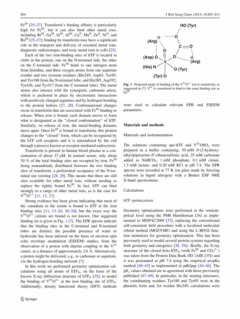
Strong evidence has been given indicating that most of
the vanadium in the serum is bound to hTF at the iron
binding sites [11, 13–24, 30–34], but the exact way the
VIVO2? cations are bound is not known. One suggested
binding set is given in Fig. 1 [7]. The EPR spectra indicate
that the binding sites in the C-terminal and N-terminal
lobes are distinct; the possible presence of water or
hydroxide has been inferred on the basis of electron spin
echo envelope modulation (ESEEM) studies, from the
observation of a proton with dipolar coupling to the VIV
center, at a distance of approximately 2.6 A. Alternatively,
a proton might be delivered, e.g., to carbonate or aspartate,
via the hydrogen-bonding network [7].
In this work we performed geometry optimization cal-
culations using all atoms of hTFN, on the basis of the
known X-ray diffraction structure of hTFN [35], to model
the binding of VIVO2? at the iron binding site of hTFN.
Additionally, density functional theory (DFT) methods
were used to calculate relevant EPR and ESEEM
parameters.
Materials and methods
Materials and instrumentation
The solutions containing apo-hTF and VIVOSO4 were
prepared in a buffer containing: 50 mM N-(2-hydroxy-
ethyl)piperazine-N0-ethanesulfonic acid, 25 mM carbonate
added as NaHCO3, 1 mM phosphate, 0.1 mM citrate,
1.5 mM lactate, and 0.20 mM KCl at pH 7.4. The EPR
spectra were recorded at 77 K (on glass made by freezing
solutions in liquid nitrogen) with a Bruker ESP 300E
X-band spectrometer.
Calculations
hTF optimizations
Geometry optimizations were performed at the semiem-
pirical level using the PM6 Hamiltonian [36] as imple-
mented in MOPAC2009 [37], replacing the conventional
self-consistent field procedure with a localized molecular
orbital method (MOZYME) and using the L-BFGS func-
tion minimizer for geometry optimization. This has been
previously used to model several protein systems regarding
both geometry and energetics [38, 39]). Briefly, the X-ray
structure of the closed holo-hTFN (with FeIII and CO32-)
was taken from the Protein Data Bank (ID 1A8E [35]) and
it was protonated at pH 7.4 using the empirical propKa
method [40–43] as implemented in pdb2pqr [44–46]. The
pKa values obtained are in agreement with those previously
published [47–49]. In particular, in the starting structures,
the coordinating residues Tyr188 and Tyr95 were in the
phenolic form and, for residue His249, calculations were
V
O (Tyr)
(Tyr) O
O
(Asp) O
(Arg)
(His) N
C
O
O
HO (Tyr)
(Arg)
O
Fig. 1 Proposed mode of binding of the VIVO2? ion to transferrin, as
suggested in [7]. VIV is considered to bind to the same binding site as
FeIII
804 J Biol Inorg Chem (2013) 18:803–813
123
Page 3
done with both tautomeric forms of the aromatic ring in
order to ascertain the most relevant one, which is the one
with the imidazole nitrogen-bound hydrogen atom further
away from the vanadium atom. Calculations were per-
formed both in the gas phase and with a polarizable con-
tinuum model approach, the conductor-like screening
model, to mimic the effects of water as an implicit solvent.
The VIVO2? ion as well as carbonate anions were placed
in the FeIII binding site. Briefly, the VIVO2?–CO32- hTF
model was built by replacing the iron atom with a vana-
dium atom and attaching to it the oxido oxygen atom; from
this model, the carbonate ion was removed to obtain a
starting VIVO2?–hTF model, and the VIVO2?–HCO3-–
hTF model was derived from the VIVO2?–CO32-–hTF
model by including a hydrogen atom on each of the two
oxygen atoms pointing away from the metal; a hydrogen
atom was also initially placed on the coordinating oxygen
atom but this led to a fast and marked increased in the total
energy of the system. For geometry comparison, an octa-
hedral [VO(H2O)5]2? complex was also studied in the
same conditions as the protein systems. An initial optimi-
zation was performed only on the hydrogen atoms, fol-
lowed by an unrestricted optimization of the whole protein.
A cutoff value (interatomic distance where the neglect of
diatomic differential overlap approximation stops) of 9 A
was used for all optimizations; larger values (12, 18, and
24 A) were also used, but no geometry or energy
improvements were obtained. Geometries were considered
optimized when the heat of formation (as calculated by
MOPAC) changed by less than 0.1 kcal mol-1 for 20
consecutive optimization cycles. Heats of formation, bond
parameters, and molecular orbitals were obtained by a
single-point calculation on the optimized geometry in order
to reorthogonalize the localized molecular orbitals.
Heats of formation of the species involved in the for-
mation of complexes of maltolate (mal-) and picolinate
(pic-) with VIVO2?, VIVO(mal)2(H2O), and VIVO(-
pic)2(H2O), respectively [1, 2, 11, 13, 14, 16–18, 20, 21,
24, 33, 34], were also calculated in order to compare their
corresponding association energetics with those of the
VIVO2?–HCO3-–hTF and VIVO2?–CO3
2-–hTF systems.
DFT calculations
The calculations for 51V and 14N tensors (AV and AN) were
performed with the program Gaussian 09 (revision C.01) [50].
Of the structures optimized at the semiempirical level,
only the residues less than 10 A from vanadium were
considered. To the CO–NH bonds that were fragmented in
the process a hydrogen atom was added: thus, these resi-
dues start with NH2– and end with a –CONH2 group.
Subsequently, the position of the hydrogen added was
optimized at B3P86/6-311g level of theory [51], freezing
all the other atoms. For the resulting structures, 51V (ANiso,
AVx , AV
y , and AVz ) and 14N (AN
iso, ANx , AN
y , and ANz ) hyperfine
coupling constants were calculated in the gas-phase using
the spin-unrestricted formalism.
The method used was tested on the model where CO32-
is introduced in the VIVO2?–hTF system (model 2, see
later), considering in the gas phase (1) only residues less
than 12 A from vanadium and (2) only residues interacting
with vanadium (Asp63, Tyr95, Tyr188, His249, and
CO32-). The results were comparable, confirming that the
values of the 51V and 14N hyperfine coupling constants
depend mainly on the first-sphere coordination environ-
ment of vanadium [52]. This is in agreement with the
additivity relationship, largely used over the last 30 years
for the characterization of VIVO2? species, which allows
one to predict the value of Az from the contribution of the
four donors bound to vanadium in the equatorial plane,
independently of the ligand complexity [53, 54]. Thus, to
predict correctly the spectroscopic parameters, it is
important that the semiempirical simulation provide a
reasonable structure; in particular, the coordination envi-
ronment of vanadium must be adequately described.
The 51V hyperfine coupling constants were calculated
with BHandHLYP functional and the 6-311g(d,p) basis set,
according to the procedure established in the literature [55–
60], the half-and-half functional BHandHLYP being
incorporated in Gaussian 09. In the first-order approxima-
tion, the hyperfine coupling tensors Ai, where i is V or N,
have one isotropic contribution deriving from the Fermi
contact (Aiiso) and another from the dipolar hyperfine
interaction (tensor Ti): Ai ¼ Aiiso1þ Ti. As demonstrated in
the literature, DFT simulations are a valid tool to predict
EPR parameters of VIVO2? complexes [55–67]. It must
also be remembered that for a VIVO2? species the AVz value
is usually negative, but in the literature its absolute value is
usually reported. The 14N hyperfine coupling constants
were calculated at the BHandH/6-311g(d,p) level of theory
[52], with the BHandH functional incorporated in the
Gaussian 09 package.
BHandHLYP and BHandH include a mixture of exact
Hartree–Fock (HF) and DFT methods to calculate the
exchange–correlation energy (EXC), and this seems to be
necessary to simulate correctly AViso and AN
iso, which depend
on the indirect core level spin polarization arising from the
unpaired spin density in the metal d orbitals. In particular,
BHandHLYP is defined as 0:5� EHFX þ 0:5� ELSDA
X þ0:5� EB
X þ ELYPC and BHandH is defined as 0:5� EHF
X þ0:5ELSDA
X þ ELYPC , where EHF
X ; ELSDAX ; EB
X , and ELYPC are
the energies due to the HF exchange, the local spin density
approximation exchange functional, the gradient-corrected
J Biol Inorg Chem (2013) 18:803–813 805
123
Page 4
Becke 88 exchange functional, and the gradient-corrected
Lee–Yang–Parr correlation functional, respectively.
Results and discussion
Computational modeling of hTF–VIVO binding
The binding site responsible for accommodating the FeIII
and carbonate species is well described in the literature
[26–29]. In the X-ray structure used, iron is coordinated to
two oxygen atoms from the carbonate anion (at 2.1 and 2.2
A), by the phenolate oxygen atoms of Tyr95 and Tyr188 (at
2.0 and 1.8 A, respectively), by one carboxylate oxygen
from Asp63 (2.0 A) and by the Ns atom of His249 (2.0 A).
For the VIVO2? ion (no carbonate present, model 1), the
modeling results indicate that the vanadium atom is coor-
dinated to the same aspartate, tyrosine, and histidine resi-
dues, at distances of 2.0 A (Asp63 O atom), 2.2 A (Tyr95
O atom), 1.8 A (Tyr188 O atom), and 2.0 A (His249 Nsatom), and that the V=O bond has a length of 1.6 A (see
Table 1). Noticeably, Tyr188 is present in the phenolate
form, whereas Tyr95 remains in the phenolic form, and the
phenolic hydrogen is bonded to the terminal nitrogen atom
of Lys296, which is thus present in the protonated Lys–
NH3? form. The modeled structure shows C–O–V angles
of 96.9� for Tyr95, 126.9� for Tyr188, and 131.6� for
Asp63, whereas with FeIII the three C–O–Fe angles are in
the 132–146� range; the major difference for Tyr95 may be
due to the presence of the hydroxylic hydrogen atom.
The oxido oxygen atom is able to participate in a salt
bridge to the guanidinium Nd atom of Arg124
(rON = 3.1 A), and in a hydrogen bond with the Arg124
terminal NH2 group (rOH = 2.0 A, rON = 2.8 A,
hNHO = 133.9�). In ferric transferrin, Arg124 is bound to
the synergistic carbonate anion [68]. Oxido oxygen inter-
actions with the atoms coordinated to vanadium are also
possible, although at longer distances: 2.8 A to the car-
boxylate oxygen atom of Asp63, 3.0 and 2.7 A to the
phenolate oxygen atoms of Tyr95 and Tyr188, respectively,
and 2.8 A to the Ns atom of His249. The OAsp63OTyr95-
OTyr188V dihedral is 20.8� and the NsHis249OTyr95OoxidoV
dihedral is -10.2�. The VIVO2? interactions with the pro-
tein are summarized in Fig. 2a, where the relevant residues
and major interactions are depicted.
The potential role of CO32- and HCO3
- was also taken
into account by introducing each of these anions in the
system. Carbonate was modeled from an initial orientation
similar to that in the known X-ray structure of
hTF ? FeIII ? CO32-, where it coordinates FeIII in a
bidentate mode [69].
When CO32- is introduced in the VIVO–hTFN system, in
the modeling calculation (model 2) it assumes a position
where all oxygen atoms are able to establish dipolar
Table 1 Distances between atoms of VIVO2? and the most relevant atoms involved in its binding to human serum transferrin (hTF)
Model 1 (without carbonate) Model 2 (with CO32-) Model 3b (with HCO3
-)
V=O 1.55 (2.49) 1.58 (2.31) 1.56 (2.34)
V–N (His249) 2.00 (0.75) 2.13 (0.55) 2.11 (0.58)
V–O (Tyr95) 2.25 (OH) (0.43) 3.28 (OH)a (0.05) 2.69 (OH)a (0.17)
V–O (Tyr188) 1.80 (1.22) 1.92 (0.91) 1.86 (1.04)
V–O (Asp63) 1.98 (0.72) 1.92 (0.78) 2.00 (0.69)
V–O1 (carbonate) – 3.60 (0.03) 3.80 (0.02)
V–O2 (carbonate) – 1.79 (1.09) 3.90 (0.02)
V–O3 (carbonate) – 4.00 (0.02) 1.88 (0.88)
Ooxido–N (His249) 2.78 (0.02) 2.87 (0.01) 2.76 (0.01)
Ooxido–O (Tyr95) 2.97 (0.01) 4.86 (0.00) 4.23 (0.01)
Ooxido–O (Tyr188) 2.67 (0.02) 2.82 (0.03) 2.77 (0.03)
Ooxido–O (Asp63) 2.79 (0.01) 2.89 (0.02) 2.93 (0.01)
Ooxido–NH2 (Arg124) (H bond) rON = 2.85 rON = 2.63 rON = 2.66
rOH = 2.05 rOH = 2.17 rOH = 2.28
h = 133.9 h = 104.9 h = 100.0
All bond lengths are given in angstroms; in the case of the hydrogen bond between the oxido oxygen atom and the NH2 group of Arg124, the
angle is given in degrees. The bond orders as computed with MOPAC [37] for the various atom pairs are given in parenthesesa The V–L(axial) internuclear distances, L(axial) being an atom coordinated trans to the oxido oxygen donor, are normally significantly longer
than the V–L(equatorial) distances, being up to approximately 2.4 A. In, e.g., model 2, the O=V–V–OH(Tyr95) angle is 178�, but the V–
O(Tyr95) distance is 3.28 A, which corresponds to a bond order of 0.05 (thus strictly it should not be considered a bond). For V–OH(Tyr95) the
internuclear distances are 3.28 and 2.69 A (bond orders of 0.05 and 0.17, respectively); whereas the latter may be considered a very weak bond,
the internuclear distance of 3.28 A corresponds to a very weak interaction
806 J Biol Inorg Chem (2013) 18:803–813
123
Page 5
interactions with groups of the protein (see Fig. 2b). The
V=O bond length remains at 1.6 A, the vanadium atom is
coordinated to an Asp63 carboxylate oxygen (1.9 A), to the
phenolate oxygen of Tyr188 (1.9 A), to Ns of His249
(2.1 A) and to the O3 atom of carbonate (1.8 A). The OH
group of Tyr95 is located at a distance of 3.3 A and it does
not participate in the coordination. CO32- is parallel to the
V=O bond, the dihedral angle between the V=O and C=O
directions being -5.6�, and the Ooxido–V–OH(Tyr95) angle
is 178�.
The oxido oxygen atom also interacts with Arg124
through both a dipolar interaction with the guanidinium Ndatom (3.2 A) and a hydrogen bond with a terminal NH
group (rOH = 2.1 A, rON = 2.6 A, hNHO = 104.9�); the
oxido oxygen atom also has rather long range interactions
with the vanadium-coordinating atoms from Asp63
(2.9 A), Tyr188 (2.8 A), and His249 (2.9 A); the phenolic
oxygen atom of Tyr95 is too distant (4.9 A) to allow
interactions to occur.
Although each FeIII in (Fe)2hTF is coordinated to two of
the carbonate oxygen atoms, the carbonate anion in VIVO-
bound transferrin in model 2 only coordinates to VIV
through one carbonate oxygen atom, and assumes a
geometry that is roughly 45� to that of the FeIII case; this
monodentate coordination of carbonate to VIV is in
agreement with previous experimental results for both
transferrin and lactoferrin and is also suggested in Fig. 1.
The angle between the aromatic ring of His249 and the
V=O bond is approximately 35�.
An HCO3- anion was also considered in the VIVO–hTF
system (model 3), and two possible locations for the
hydrogen atom were obtained. In one case, the hydrogen
atom is located on an oxygen atom close to VIVO (model
3a), and in the other case, it is located on the most distant
oxygen atom, pointing away from the VIVO2? ion (model
3b). In model 3a HCO3- is parallel to the V=O bond as in
model 2 (dihedral angle between the V=O and C=O
directions of -1.5�); in model 3b it is perpendicular
Fig. 2 Calculated structures of the human serum transferrin complex
with VIVO2?. The three different possibilities obtained are shown:
a in the absence of carbonate (model 1), b with CO32- (model 2), and
c with HCO3- (model 3b). A superposition of the three modeled
structures is shown in d, where the most relevant residues of model 1
are shown with a cyan carbon skeleton, the most relevant residues of
the model 2 are shown with a magenta carbon skeleton, and the most
relevant residues of the model 3b are shown with a yellow carbon
skeleton. The atoms of VIVO are shown as spheres at the center of a–
d (gray for vanadium and red for oxygen), and relevant residues are
depicted as sticks (Asp63, Tyr95, Tyr188, and His249) and as lines. In
a–c, the residues labeled in blue are those involved in iron
coordination in ferric transferrin, and residues labeled in orange
were also found to be relevant in VIV binding in this study. Vanadium
coordinating bonds are shown in orange, bonds to oxido oxygen are
shown in yellow, and bonds from CO32- or HCO3
- to the protein are
shown in red. Further possible bonds to vanadium in the absence of
carbonate are shown in magenta. The protein backbone is depicted in
a transparent cartoon mode. Bonding details are given in the text
J Biol Inorg Chem (2013) 18:803–813 807
123
Page 6
(dihedral angle of 86.7�). Energetically, there is a
1.3 MJ mol-1 difference in favor of the latter owing to the
local rearrangement of atoms and to a higher number of
hydrogen bonds, three in model 3b and just one in
model 3a.
In the energetically favored case (model 3b; Fig. 2c), the
V=O bond length is 1.6 A and the vanadium atom is
coordinated to Asp63 (carboxylate O, at 2.0 A), Tyr188
(phenolate O, at 1.9 A), His249 (Ns atom, at 2.1 A), and an
HCO3- oxygen atom at 1.9 A (see Table 1). The phenolic
oxygen atom of Tyr95 is located at 2.7 A, thus corre-
sponding to a weak interaction.
The oxido oxygen atom may also interact with Arg124
through either a hydrogen bond with a terminal NH2 group
(rOH = 2.3 A, rNO = 2.7 A, hNHO = 100.0�) or a salt
bridge to the same nitrogen atom; a salt bridge to the Nd atom
of Arg124 (3.2 A) is also possible. There are also long-range
interactions with the vanadium-coordinating atoms: with
Asp63 (2.9 A), Tyr188 (2.8 A), His249 (2.8 A), and HCO3-
(2.7 A). In model 3b the OAsp63OTyr95OTyr188V dihedral is
1.9� and the NsHis249OTyr95OVOV dihedral is -55.2�. The
HCO3- ion interacts with the protein by various hydrogen
bonds and salt bridges (see Fig. 2c), in particular from
hydrogen of the HCO3- ion to the OH group of Thr120
(rOH = 1.6 A, rOO = 2.6 A, hOHO = 158.7�) and from the
noncoordinating oxygen of HCO3- to the amide groups of
Ala126 (rOH = 1.9 A, rNO = 2.8 A, hNHO = 147.2�) and
Gly127 (rOH = 2.1 A, rNO = 3.1 A, hNHO = 154.2�).
A summary of the most important interactions involving
VIVO2? are presented in Table 1, and Fig. 1 depicts some
of the modeled VIVO–hTFN binding sites.
The bond lengths and the distances between vanadium
and the coordinating atoms obtained in our study of the
VIVO–hTFN system, performed at the semiempirical level
with the PM6 Hamiltonian, are in agreement with the dis-
tances computed for analogous systems at the DFT level. In
VIVO2?, the V=O bond length is approximately 1.57 A at
the B3LYP/6-311g(d) level for VIVO complexes with
nitrogen and oxygen coordinating ligands [56] and ranges
from 1.59 to 1.62 A at the same level of theory for a set of
peptidic ligands for which the experimentally determined
V=O bond lengths range from 1.58 to 1.62 A [55]; in our
studies, we obteined a V=O bond length of 1.6 A. Micera
and Garribba determined V–O coordinating distances from
1.8 to 2.1 A [56, 58–60], and in the present cases they range
from 1.8 to 2.2 A. For the V–N coordinating distances, the
DFT values are in the ranges 1.97–2.08 A for an amide N-,
2.15–2.19 A for an amine NH2, and 2.05–2.14 A for an
imidazole nitrogen atom [55]; our values for the imidazole
nitrogen atom are between 2.0 and 2.1 A.
A study of the octahedral water–VIVO complex was
performed in the same conditions as for the protein studies;
the geometry obtained [V–Ooxido = 1.56 A, V–
O(H2O)equatorial = 2.00 (±0.05) A, V–O(H2O)axial =
2.21 A, average Ooxido–V–O(H2O)equatorial angle =
(103.22 ± 3.2)�] is similar to the DFT geometry at the
B3LYP/3-21G (1.57 A, 2.03 A, 2.17 A, 97.8�, respec-
tively) that accurately describes the electronic structure of
this species [67].
Smith et al. [70] proposed a model for VIVO2? binding to
the C-terminal lobe of human lactoferrin in the presence of
carbonate, where vanadium is coordinated to Asp60, Tyr92,
Tyr192, and His253 (which are the residues equivalent to
Asp63, Tyr95, Tyr188, and His249 from the N-terminal
lobe), and the oxido oxygen atom is considered to establish a
hydrogen bond with a side-chain NH2 of Arg121 (Arg124 in
hTFN), carbonate being bound to both the NH2 and the
guanidinium Ns hydrogen atom of the side chain of Arg121,
as well as to the hydroxylic hydrogen atom of Thr117
(Thr120 in hTFN) [70]. In our calculated model, the carbonate
anion (protonated and deprotonated) is closer to Ser125 than
to Arg124, and VIVO2? is located between the side-chain
nitrogen atoms of Arg124 and the synergistic ions.
Our models, as described above, are capable of
accommodating the VIVO2? ion with a synergistic CO32-
or HCO3- ion without inducing major structural changes in
the protein, with the exception of the side chain of the
Tyr188 residue, which moves about 1.7 A,, and the Asp63
residue, which moves around 1.4 A, both due to the steric
effect of the presence of the carbonate ion.
As determined from the X-ray structure, in (Fe)2hTF the
FeIII is coordinated to six atoms that form an octahedral
environment with low distortion. Overall, in the case of
transferrin-bound VIVO2? without HCO3- or CO3
2- added
(Fig. 2a), vanadium is bound to five atoms in a distorted
arrangement (model 1). We analyzed this coordination
geometry using the s trigonality parameter of Addison
et al. [71]; this parameter is 0 for a perfect square pyramid
and 1 for a perfect trigonal bipyramid; intermediate values
reflect the structural continuum between the two structures.
In the case of model 1, s = 0.06, indicating that the
coordination geometry is essentially a square pyramid.
In the presence of either CO32- or HCO3
-, vanadium is
also bound to five atoms, in a distorted octahedral geom-
etry; in both cases the amine groups of Arg124 are not
spatially oriented toward the vanadium atom. Taking the
oxido oxygen atom as forming the main bond, we find the
coordination geometry is again essentially square pyrami-
dal, although with a higher trigonal bipyramid distortion.
The calculated trigonality indexes are s = 0.23 for hTF/
VIVO/CO32- (model 2) and s = 0.32 for hTF/VIVO/
HCO3- (model 3b), indicating that these two structures
also have a predominantly square pyramidal character,
which is more evident in the CO32- case. Figure 3 depicts
a schematic of the binding of VIV to the residues of the iron
binding site of hTFN for this particular model.
808 J Biol Inorg Chem (2013) 18:803–813
123
Page 7
Energetics
After geometry optimization, a single-point calculation was
performed to reorthogonalize the localized molecular
orbitals and to obtain the reaction energies of the various
systems, which are presented in Table 2. For comparison,
the same approach was applied to the formation of
VIVO2?–mal- [72] and VIVO2?–pic- [73] complexes,
which are systems previously characterized by potentio-
metric and spectroscopic studies [72, 73] and DFT calcu-
lations [18, 20]. The values obtained indicate that the
presence of the CO32- ion or HCO3
- ions favors VIVO2?
binding to transferrin, clearly indicating that the hTF–
VIVO2? system is stabilized by the presence of carbonate
anions. This confirms that carbonate also plays a syner-
gistic role in VIVO2? binding to hTF, similar to that
observed for FeIII binding. The protonation state of the
carbonate species is also important, as this stabilizing
effect is larger in the system with a bianionic CO32- than
with a monoanionic HCO3- (although this depends on the
hydrogen atom orientation). We emphasize that we based
our choices on the calculated heats of formation and not on
the free energy of binding. We also did not take into
account that in blood HCO3- is present in a much higher
concentration than CO32-. However, the values obtained
suggest the tendency for the preference of CO32-.
The use of heats of formation to establish favorable
forms in metal–protein complexes may be questionable,
and free energies of binding and proper consideration of
the relative concentrations of CO32- and HCO3
- in solu-
tion should have been taken into account to fully support
the conclusions. However, the heats of formation (and
DrE values) do reveal a tendency for the binding.
In fact, the DrE values obtained for VIVO(pic)2(H2O),
-0.1154 MJ mol-1, which corresponds to a formation
constant b2 = 1012.1 [73], and for VIVO(mal)2(H2O),
-1.3695 MJ mol-1, which corresponds to b2 = 1016.3
[72], do conform with this tendency.
The open and closed forms of hTFN are distinguishable
by specific bonds that are present in each case. It is
important to emphasize that since our modeling calculation
started from the closed conformation of ferric transferrin, it
is unlikely to obtain hTF in the open conformation. In fact,
we used energy minimization approaches that, unlike the
molecular dynamics methods, are not adequate to study
events such as protein hinge rotation that may open the
transferrin cleft. In our modeling calculations the modeled
system remains in the closed form on carbonate and VIVO
binding. Recent results obtained by some of us indicate that
hTF in hTF(VIVO)2 assumes a closed conformation [34].
Prediction of EPR and ESEEM spectra
The structures optimized at the semiempirical level were
used to calculate 51V and 14N tensors, AV and AN,
respectively (see ‘‘DFT calculations’’).
Figure 4 depicts the EPR spectra recorded for the
VIVO2?-hTF system, and the gz and AVz values simulated
using the computer program ROKI [74] are reported in
Table 3. The EPR spectrum of frozen aqueous solution
containing VIVO2? and hTF at physiological pH is composed
of two sets of resonances (A and B), corresponding to at least
two slightly different VIV environments whose relative
intensity is pH-dependent. The A and B resonances were
attributed to VIVO2? bound in the N-terminal (our simula-
tions) and C-terminal [75] sites, respectively. Various values
for AVz of A resonances have been reported in the literature,
ranging from 166 9 10-4 to 168.5 9 10-4 cm-1 [32, 53,
76–78], so a mean value around 168 9 10-4 cm-1 can be
used as a reference. No reliable accurate measure of AVx and
AVy exists, owing to the contemporaneous presence of EPR
resonances belonging to the VIV–hTFN and VIV–hTFC sites.
The DFT methods used for such calculations were val-
idated recently [52, 55]. Among several functionals tested,
the order of accuracy in the prediction of 51V AVz is
BHandHLYP [ PBE0 � B3PW [ TPSSh [ B3LYP �BP86 [ VWN5 [55].1 The better performance of half-and-
half functionals such as BHandHLYP than the hybrid
functionals is related to the prediction of the Fermi contact
term, which depends on the indirect core level spin
polarization arising from the unpaired spin density in the
metal d orbitals [55, 56]. The spin polarization is difficult
to simulate with high accuracy, and is significantly
underestimated by most of the functionals [61, 63]. The
higher fraction of HF exchange in half-and-half functionals
V
O
(Tyr95) HO
(Tyr188) O
(His249) N
O
COO
Ser125Ala126
HO (Thr120)
O (Asp63)
Arg 124
Fig. 3 Binding of VIVO2? ion to the residues of the iron binding site
of the N-terminal lobe of human serum transferrin (hTF) as obtained
in computed model 2 (CO32- as a synergistic anion)
1 For the acronyms of the functionals and their meaning, the reader is
referred to [55].
J Biol Inorg Chem (2013) 18:803–813 809
123
Page 8
such as BHandHLYP, which is available in Gaussian 09,
improves considerably the prediction of the Fermi contact
and, hence, that of AVz .
Our results for the calculation of the 51V AV tensor
components of the modeled structures of VIVO–hTFN are
presented in Table 4. They can be summarized as follows:
(1) the maximum value of the percent deviation is observed
when carbonate or hydrogen carbonate is not included in
the structure; (2) when carbonate or hydrogen carbonate is
taken into account in the calculations AVz
����calcd
increases
significantly and approaches AVz
����exptl
; (3) the structure that
yields the minimum deviation is model 2, with carbonate
coordinated to vanadium; (4) the deviation of 2.6 % for
model 2 is of the same order as the deviations obtained for
simple VIVO2? complexes and is in agreement with pre-
vious publications [52, 55, 57]; (5) models 3a and 3b, with
HCO3- included in the simulations, cannot be distin-
guished from these data.
The short relaxation times in EPR spectroscopy produce
lines that are considerably broad and in most of cases this
precludes the resolution of the coupling of the unpaired
electron on vanadium with the nuclei in the ligand sphere,
quantified by the superhyperfine coupling constant AL
(where L indicates a ligand). Information on this super-
hyperfine coupling can be provided by ESEEM spectros-
copy, a variant of EPR spectroscopy [79–81]. The
superhyperfine coupling constant reported for 14N are in
the range 1–8 MHz and vary with the nature, position, and
orientation of the ligands [82–84]. In the studies on the
identification of nitrogen donors bound to vanadium in
VIVO species, both ANiso and AN
z are used. Very few com-
putational studies have been published in which the ligand
superhyperfine coupling constants for VIVO2? complexes
were simulated [52, 85]. A recent study showed that the
half-and-half hybrid BHandH, which is available in the
Gaussian package, performed better than the other func-
tionals tested. The order of accuracy is
BHandH & B3PW91 � B3P86 [ B3LYP [48]. It was
also demonstrated that the 14N AN tensor shows a func-
tional dependence on the dihedral angle h between the
Table 2 Reaction energies of the various systems studied, computed with the semiempirical PM6 Hamiltonian
System DrE (MJ mol-1)
VIVO H2Oð Þ2þ5 þ2pic��VIVO picð Þ2 H2Oð Þ þ 4H2O -0.1154
VIVO H2Oð Þ2þ5 þ2mal��VIVO malð Þ2 H2Oð Þ þ 4H2O -1.36951
VIVO H2Oð Þ2þ5 þhTFN�VIVO�hTFN þ 5H2O (model 1) -9.17391
VIVO H2Oð Þ2þ5 þhTFN þ CO2�3 �VIVO�hTFN�CO2�
3 þ 5H2O (model 2) -9.3776
VIVO H2Oð Þ2þ5 þhTFN þ HCO�3 �VIVO�hTFN�HCO�3 þ 5H2O (model 3a) -8.00596
VIVO H2Oð Þ2þ5 þhTFN þ HCO�3 �VIVO�hTFN�HCO�3 þ 5H2O (model 3b) -9.32996
DrE values were computed as the difference between the sum of the heats of formation of the products and those of the reactants. The
VIVO H2Oð Þ2þ5 reactant is considered to lose four water molecules to form the VIVO(pic)2(H2O) and VIVO(mal)2(H2O) complexes (the species
stable in aqueous solution; mal- is maltolate and pic- is picolinate) and five water molecules to form the N-terminal lobe of hTF (hTFN)
complexes
1.551.751.952.152.352.55
g-value
Fig. 4 First-derivative X-band frozen solution EPR spectra of
solutions containing apo-hTF (ChTF = 750 lM) and VIVO2? with
hTF to VIVO molar ratios of 1:1 (black line) and 1:2 (gray line)
Table 3 Spin Hamiltonian parameters obtained from the simulation
of the recorded EPR spectra
gz AVz � 104cm�1 Reference
VO–hTF species Aa 1.939 168.5 This work
1.937 168.3 [32]
1.938 168.0 [70]
1.938 168.0 [72]
1.940 166.8 [71]
VO–hTF species Ba 1.940 171.1 This work
1.941 170.3 [70]
1.937 172.4
1.934 170 [71]
1.941 170.5 [32]
1.935 171.8
a See the text for the assignment of species A and B
810 J Biol Inorg Chem (2013) 18:803–813
123
Page 9
V=O and N–C bonds (where C is the carbon that bridges
the two nitrogen atoms in the imidazole ring) and on the
angle u between the O=V and V–N bonds (where N is the
coordinated aromatic nitrogen atom) [52]. The data
obtained from the simulations are listed in Table 5.
From an examination of Table 5, it can be observed that
h and u for the four models studied are comparable. Thus,
the values of ANcalcdiso are very similar. Overall, the model that
gives the best results is the one with CO32- bound to
vanadium (model 2), followed by the models with HCO3-
(models 3a and 3b). This conclusion is again in line with the
results of EPR simulations, but the differences are so small
that they cannot be used to evaluate the best model. Only
the combined analysis of the energy and EPR data allows us
to suggest which of the four structures is the stablest.
Conclusions
The geometry optimization calculations performed indicate
that in the binding of VIVO2? to hTFN, in the presence of
CO32- or HCO3
-, vanadium is coordinated to five atoms,
in a distorted geometry; moreover, between the CO32- and
HCO3-, the DrE values (computed as the difference
between the sum of the heats of formation of the products
and those of the reactants; Table 2), obtained in the
modeling calculations suggest that the preferred synergistic
anion is CO32-. Free energies of binding and correct
consideration of the relative concentrations of CO32- and
HCO3- in solution would be required to fully support this
conclusion, but the DrE values do reveal a tendency for the
preference of CO32- as a synergistic anion.
Both VIVO2?–hTF–HCO3- and VIVO2?–hTF–CO3
2-
structures were modeled. For the latter, the V=O bond
length is approximately 1.6 A, and the vanadium atom is
also coordinated to the Tyr188 phenolate oxygen atom (at
approximately 1.9 A), the His249 Ns atom (at approxi-
mately 2.1 A), an Asp63 carboxylate oxygen atom (at
approximately 1.9 A), and a carbonate oxygen atom at
approximately 1.8 A. The Tyr95 phenolic oxygen atom is
approximately 3.3 A from the metal center, and thus at best
very weakly interacts with VIV. All oxygen atoms are able
to establish dipolar interactions with protein groups.
The structures optimized at the semiempirical level were
used to calculate 51V AV and 14N AN tensors by DFT
methods, and 51V AVz , 14N AN
iso, and 14N ANz were compared
with the reported experimental values. Of the calculated
VIVO–hTFN structures, the one that yields both the lowest
calculated heats of formation and the minimum deviations
from the experimental values of 51V AVz , 14N A AN
iso, and14N AN
z is the structure that includes CO32- as a synergistic
anion.
Table 4 Calculated 51V AV tensor components for the calculated structures of VIVO2?–hTF
Simulation AViso TV
x TVy TV
z AVcalcdx AVcalcd
y AVcalcdz AVexptl
zDeviation (%)a
Model 1 -88.0 33.7 36.1 -69.8 -54.3 -51.8 -157.8 -168.0 -6.1
Model 2 -92.8 32.2 38.6 -70.9 -60.6 -54.1 -163.6 -168.0 -2.6
Model 3a -90.1 34.4 36.5 -71.0 -55.7 -53.6 -161.0 -168.0 -4.1
Model 3b -90.1 33.3 37.9 -71.2 -56.9 -52.2 -161.3 -168.0 -4.0
All values are given in 10-4 cm-1
a Percent deviation of AVx
����calcd
from AVz
����exptl
, expressed as 100� AVz
����calcd� AV
z
����exptl
� �
= AVz
����exptl
Table 5 Calculated 14N AN tensor components for the calculated structures of VIVO2?–hTF
Simulation h u ANcalcdiso A
Nexptliso
a Deviation (%)bANexptl
zc ANexptl
zDeviation (%)d
Model 1 6.8 102.4 -6.2 -6.6 -6.1 -7.8 -7.1 9.9
Model 2 8.4 100.2 -6.3 -6.6 -4.5 -7.1 -7.1 0.0
Model 3a -4.7 100.7 -6.3 -6.6 -4.5 -7.6 -7.1 7.0
Model 3b 10.8 95.8 -6.1 -6.6 -7.6 -7.1 -7.1 -0.0
All values are given in megahertza Values taken from [86]
b Percent deviation of ANiso
��
��calcd
from ANiso
��
��exptl
, expressed as 100� ANiso
��
��calcd� AN
iso
��
��exptl
� �
= ANiso
��
��exptl
c Values taken from [87]
d Percent deviation of ANz
����calcd
from ANz
����exptl
, expressed as 100� ANz
����calcd� AN
z
����exptl
� �
= ANz
����exptl
J Biol Inorg Chem (2013) 18:803–813 811
123
Page 10
The present modeling calculations taken together with the
results obtained recently by some of us [34], indicating that
on binding of VIVO2? to apo-hTF the protein closes its
conformation similarly to (FeIII)2hTF, suggest that
(VIVO)2hTF may be recognized by hTF cell receptors and
may thus be taken up by endocytosis (also similarly to FeIII).
Acknowledgments The authors thank the Portuguese Foundation
for Science and Technology and the FEDER and POCI programs,
namely, PEst-OE/QUI/UI0100/2013 and SFRH/BPD/68789/2010, for
financial support. G.C.J. acknowledges research grant SFRH/BPD/
27536/2006 from the Portuguese Foundation for Science and Tech-
nology. We also thank G. Goncalves for the measurement of the EPR
spectra in Fig. 4.
References
1. Thompson KH, Orvig C (2000) J Chem Soc Dalton Trans
2885–2892
2. Thompson KH, Orvig C (2001) Coord Chem Rev
219–221:1033–1053
3. Sakurai H, Kojima Y, Yoshikawa Y, Kawabe K, Yasui H (2002)
Coord Chem Rev 226:187–198
4. Evangelou AM (2002) Crit Rev Oncol Hematol 42:249–265
5. Costa Pessoa J, Papaioannou A, Manos M, Karkabounas S,
Liasko R, Evangelou AM, Correia I, Kalfakakou V, Kabanos T
(2004) J Inorg Biochem 98:959–968
6. Benitez J, Guggeri L, Tomaz I, Costa Pessoa J, Moreno V,
Lorenzo J, Aviles FX, Garat B, Gambino D (2009) J Inorg Bio-
chem 103:1386–1394
7. Rehder D (2008) Bioinorganic vanadium chemistry. Wiley, New
York
8. Gambino D (2011) Coord Chem Rev 255:2193–2203
9. Gambino D, Noblia P, Vieites M, Parajon-Costa BS, Baran EJ,
Cerecetto H, Draper P, Gonzalez M, Piro OE, Castellano EE,
Azqueta A, Cerain AL, Monge-Veja A (2005) J Inorg Biochem
99:443–451
10. Maurya MR, Khan AA, Azam A, Ranjan S, Mondal N, Kumar A,
Avecilla F, Costa Pessoa J (2010) Dalton Trans 39:1345–1360
11. Kiss T, Jakusch T, Hollender D, Dornyei A, Enyedy EA, Costa
Pessoa J, Sakurai H, Sanz-Medel A (2008) Coord Chem Rev
252:1153–1162
12. Willsky GR, Chi LH, Godzala M, Kostyniak PJ, Smee JJ, Trujillo
AM, Alfano JA, Ding WJ, Hu ZH, Crans DC (2011) Coord Chem
Rev 255:2258–2269
13. Jakusch T, Costa Pessoa J, Kiss T (2011) Coord Chem Rev
255:2218–2226
14. Liboiron BD, Thompson KH, Hanson GR, Lam E, Aebischer N,
Orvig C (2005) J Am Chem Soc 127:5104–5115
15. Sanna D, Micera G, Garribba E (2009) Inorg Chem
48:5747–5757
16. Costa Pessoa J, Tomaz I (2010) Curr Med Chem 17:3701–3738
17. Jakusch T, Hollender D, Enyedy EA, Gonzalez CS, Montes-
Bayon M, Sanz-Medel A, Costa Pessoa J, Tomaz I, Kiss T (2009)
Dalton Trans 2428–2437
18. Sanna D, Micera G, Garribba E (2010) Inorg Chem 49:174–187
19. Jakusch T, Dean T, Oncsik T, Benyei AC, Di Marco V, Kiss T
(2010) Dalton Trans 39:212–220
20. Sanna D, Biro L, Buglyo P, Micera G, Garribba E (2012) Me-
tallomics 4:33–36
21. Sanna D, Buglyo P, Micera G, Garribba E (2010) J Biol Inorg
Chem 15:825–839
22. Sanna D, Bıro L, Buglyo P, Micera G, Garribba E (2012) J Inorg
Biochem 115:87–99
23. Sanna D, Micera G, Garribba E (2011) Inorg Chem
50:3717–3728
24. Bordbar AK, Creagh AL, Mohammadi F, Haynes CA, Orvig C
(2009) J Inorg Biochem 103:643–647
25. Battin EE, Lawhon A, Brumaghim JL, Hamilton DH (2009) J
Chem Educ 86:969–972
26. Quarles CD Jr, Brumaghim JL, Marcus RK (2010) Metallomics
2:154–161
27. Sun H, Li H, Sadler PJ (1999) Chem Rev 99:2817–2842
28. Evans RW, Kong XL, Hider RC (2012) Biochim Biophys Acta
Gen Subj 1820:282–290
29. Williams K, Moreton K (1980) Biochem J 185:483–485
30. Nagaoka MH, Akiyama H, Maitani T (2004) Analyst 129:51–54
31. De Cremer K, Van Hulle M, Chery C, Cornelis R, Strijckmans K,
Dams R, Lameire N, Vanholder R (2002) J Biol Inorg Chem
7:884–890
32. Sanna D, Garribba E, Micera G (2009) J Inorg Biochem
103:648–655
33. Kiss T, Kiss E, Garribba E, Sakurai H (2000) J Inorg Biochem
80:65–73
34. Mehtab S, Goncalves G, Roy S, Tomaz AI, Santos-Silva T,
Santos MFA, Romao MJ, Jakusch T, Kiss T, Costa Pessoa J
(2013) J Inorg Biochem 121:187–195
35. MacGillivray RT, Moore SA, Chen J, Anderson BF, Baker H,
Luo Y, Bewley M, Smith CA, Murphy ME, Wang Y, Mason AB,
Woodworth RC, Brayer GD, Baker EN (1998) Biochemistry
37:7919–7928
36. Stewart JJP (2007) J Mol Model 13:1173–1213
37. Stewart JJP (2008) MOPAC2009. Stewart Computational
Chemistry, Colorado Springs. http://openmopac.net)
38. Stewart JJP (2009) J Mol Model 15:765–805
39. Stigliani JL, Bernardes-Genisson V, Bernadou J, Pratviel G
(2012) Org Biomol Chem 10:6341–6349
40. Li H, Robertson AD, Jensen JH (2005) Proteins 61:704–721
41. Bas DC, Rogers DM, Jensen JH (2008) Proteins 73:765–783
42. Olsson MHM, Søndergard CR, Rostkowski M, Jensen JH (2011)
J Chem Theory Comput 7:525–537
43. Søndergaard CR, Olsson MHM, Rostkowski M, Jensen JH (2011)
J Chem Theory Comput 7:2284–2295
44. Dolinsky TJ, Czodrowski P, Li H, Nielsen JE, Jensen JH, Klebe
G, Baker NA (2007) Nucleic Acids Res 35:W522–W525
45. Dolinsky TJ, Nielsen JE, McCammon JA, Baker NA (2004)
Nucleic Acids Res 32:W665–W667
46. Li H, Robertson AD, Jensen JH (2005) Proteins 61:704–721
47. Bas DC, Rogers DM, Jensen JH (2008) Proteins 73:765–783
48. Olsson MHM, Sondergard CR, Rostkowski M, Jensen JH (2011)
J Chem Theory Comput 7:525–537
49. Sondergaard CR, Olsson MHM, Rostkowski M, Jense JH (2011)
J. Chem Theory Comput 7:2284–2295
50. Frisch MJ, Trucks GW, Schlegel HB, Scuseria GE, Robb MA,
Cheeseman JR, Scalmani G, Barone V, Mennucci B, Petersson
GA, Nakatsuji H, Caricato M, Li X, Hratchian HP, F. Izmaylov
A, Bloino J, Zheng G, Sonnenberg JL, Hada M, Ehara M, Toyota
K, Fukuda R, Hasegawa J, Ishida M, Nakajima T, Honda Y,
Kitao O, Nakai H, Vreven T, Montgomery Jr JA, Peralta JE,
Ogliaro F, Bearpark M, Heyd JJ, Brothers E, Kudin KN, Staro-
verov VN, Keith T, Kobayashi R, Normand J, Raghavachari K,
Rendell A, Burant JC, Iyengar SS, Tomasi J, Cossi M, Rega N,
Millam JM, Klene M, Knox JE, Cross JB, Bakken V, Adamo C,
Jaramillo J, Gomperts R, Stratmann RE, Yazyev O, Austin AJ,
Cammi R, Pomelli C, Ochterski JW, Martin RL, Morokuma K,
Zakrzewski VG, Voth GA, Salvador P, Dannenberg JJ, Dapprich
S, Daniels AD, Farkas O, Foresman JB, Ortiz JV, Cioslowski J,
Fox DJ (2010) Gaussian 09, revision C.01. Gaussian, Wallingford
812 J Biol Inorg Chem (2013) 18:803–813
123
Page 11
51. Micera G, Garribba E (2012) Int J Quantum Chem 112:
2486–2498
52. Sanna D, Pecoraro VL, Micera G, Garribba E (2012) J Biol Inorg
Chem 17:773–790
53. Chasteen ND (1981) In: Berliner LJ, Reuben J (eds) Biological
magnetic resonance, vol 3. Plenum, New York, pp 53–119
54. Smith TS II, LoBrutto R, Pecoraro VLM (2002) Coord Chem Rev
228:1–18
55. Micera G, Garribba E (2011) J Comput Chem 32:2822–2835
56. Gorelsky S, Micera G, Garribba E (2010) Chem Eur J 16:
8167–8180
57. Micera G, Garribba E (2009) Dalton Trans 1914–1918
58. Micera G, Garribba E (2010) Eur J Inorg Chem 2010:4697–4710
59. Lodyga-Chruscinska E, Micera G, Garribba E (2011) Inorg Chem
50:883–899
60. Micera G, Garribba E (2011) Eur J Inorg Chem 2011:3768–3780
61. Munzarova ML, Kubacek P, Kaupp M (2000) J Am Chem Soc
122:11900–11913
62. Costa Pessoa J, Calhorda MJ, Cavaco I, Correia I, Duarte MTL,
Felix V, Henriques RT, Piedade MFM, Tomaz I (2002) J Chem
Soc Dalton Trans 4407–4415
63. Saladino AC, Larsen SC (2003) J Phys Chem A 107:1872–1878
64. Aznar CP, Deligiannakis Y, Tolis EJ, Kabanos TA, Brynda M,
Britt RD (2004) J Phys Chem A 108:4310–4321
65. Neese F (2003) J Chem Phys 118:3939–3948
66. Adao P, Costa Pessoa J, Henriques RT, Kuznetsov ML, Avecilla
F, Maurya MR, Kumar U, Correia I (2009) Inorg Chem 48:
3542–3561
67. Grant CV, Cope W, Ball JA, Maresch GG, Gaffney BJ, Fink W,
Britt RD (1999) J Phys Chem B 103:10627–10631
68. Wally J, Halbrooks PJ, Vonrhein C, Rould MA, Everse SJ,
Mason AB, Buchanan SK (2006) J Biol Chem 281:24934–24944
69. MacGillivray RT, Moore SA, Chen J, Anderson BF, Baker H,
Luo Y, Bewley M, Smith CA, Murphy ME, Wang Y, Mason AB,
Woodworth RC, Brayer GD, Baker EN (1998) Biochemistry
37:7919–7928
70. Smith CA, Ainscough EW, Brodie AM (1995) J Chem Soc
Dalton Trans 1121–1126
71. Addison AW, Rao TN, Reedijk J, van Rijn J, Verschoor GC
(1984) J Chem Soc Dalton Trans 1349–1356
72. Kiss T, Kiss E, Micera G, Sanna D (1998) Inorg Chim Acta
283:202–210
73. Kiss E, Garribba E, Micera G, Kiss T, Sakurai H (2000) J Inorg
Biochem 78:97–108
74. Rockenbauer A, Korecz L (1996) Appl Magn Reson 10:29–43
75. Chasteen ND, Grady JK, Holloway CE (1986) Inorg Chem
25:2754–2760
76. White LK, Chasteen ND (1979) J Phys Chem 83:279–284
77. Kiss T, Jakusch T, Bouhsina S, Sakurai H, Enyedy EA (2006) Eur
J Inorg Chem 2006:3607–3613
78. Mustafi D, Galtseva EV, Krzystek J, Brunuel LC, Makinen MW
(1999) J Phys Chem A 103:11279–11286
79. Schweiger A (1991) Angew Chem Int Ed Engl 30:265–292
80. Smith TS, LoBrutto R, Pecoraro VL (2002) Coord Chem Rev
228:1–18
81. Deligiannakis Y, Louloudi M, Hadjiliadis N (2000) Coord Chem
Rev 204:1–112
82. Fukui K, Ohya-Nishiguchi H, Kamada H (1997) Inorg Chem
36:5518–5529
83. Fukui K, Ohya-Nishiguchi H, Kamada H, Iwaizumi M, Xu Y
(1998) Bull Chem Soc Jpn 71:2787–2796
84. LoBrutto R, Hamstra BJ, Colpas GJ, Pecoraro VL, Frasch WD
(1998) J Am Chem Soc 120:4410–4416
85. Saladino AC, Larsen SC (2003) J Phys Chem A 107:4735–4740
86. Eaton SS, Dubachs J, More KM, Eaton G, Thurmanv G, Ambruso
DR (1989) J Biol Chem 264:4776–4781
87. Hanna M, Chasteen ND, Rottman GA, Aisen P (1991) Bio-
chemistry 30:9210–9216
J Biol Inorg Chem (2013) 18:803–813 813
123