Bioavailability and Recent Advances in the Bioactivity of Flavonoid and Stilbene Compounds
*Chin Giaw Lim and Mattheos A. G. Koffas
Department of Chemical and Biological Engineering, University at Buffalo, Buffalo, New York 14260.
ABSTRACT:
Polyphenols such as flavonoids and stilbenes are abundant in our daily diet, and their roles in the protection and prevention of
various diseases are substantial. However, the bioavailability varies among polyphenols and the actual compounds acting on the
designated tissues are often not the native molecules consumed in the diet. Hereby, we review the bioavailabilty of the main
classes of stilbenes and flavonoids, namely flavanones, flavones, isoflavones, flavanols, flavonols and anthocyanins, emphasizing
on their absorption, distribution, metabolism and excretion (ADME). The bioavailability summary can be useful for future
experimental design, especially the emphasis on the bioactivity on targeted tissues and organs. In addition, we review the
bioefficacy of the polyphenols, emphasizing recent advances on health benefits both in vivo and in vitro. Other issues of importance, such as structure, food source and synthesis methods, are also considered.
KEYWORDS: Polyphenols, flavonoids, resveratrol, bioavailability, absorption, distribution, metabolism, excretion, bioactivity
INTRODUCTION
Flavonoids are plant secondary metabolites found in the plant
kingdom and the largest phenolic group in nature. They are common
in our daily diet such as fruits, vegetables, herbs, red wine and tea. A
major part of their function in plant is related to their interactions
with extreme environments. For example, flavonoids accumulated in
epidermal cells by UV induction can act as a UV protectant by
absorbing the radiation. Upon wounding or infection by pathogens,
flavonoids also act as toxin and fungal pathogens as well as
antimicrobial compounds. The high concentration and wide variety of
flavonoids found in seed coats is attributed to the antifungal and
antimicrobial properties of flavonoids to protect the seeds and
indirectly facilitate plant reproduction[1]. Microbial is not necessary
harmful to plants, especially in the case with leguminous plant, when
they are in their nutrient-limited condition or more specific nitrate-
deficient state. The plants induce flavonoids accumulation to attract
nitrogen-fixing bacteria, Rhizobium[2], which convert nitrogen from
the atmosphere into plant usable ammonium in the nodule. In return,
the plant provides microaerobic environment and offers organic acids
necessary for the bacteria as carbon sources[3]. This form of
relationship between the plant and the bacteria is known as legume-
Rhizobium symbiosis. Other proposed functions of flavonoids in
plants include reproductive tissues protection, pollinator attraction,
seed dispersal, coloration, feed deterrent and enzyme inhibition.
Flavonoids are also involved in photosynthesis, morphogenesis and
sex determination[1, 2, 4]. From all the benefits flavonoids possess,
even though these compounds are not essential for cell survival, they
serve a key role in giving the organism an evolutionary advantage to
survive and reproduce[5], both in terms of physiological function and
biochemical properties.
Stilbenes do not belong to the flavonoids but they share high
resemblance to flavonoids in both functions in plant and chemical
structure. Stilbenes are synthesized naturally by distinct plants and
they are synthesized in response to infection by pathogens
(phytoalexins) and ultraviolet light exposure, and are also involved in
bacterial root nodulation and coloration[6, 7].
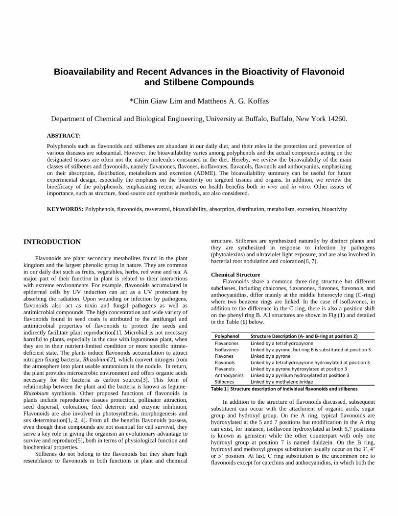
Chemical Structure
Flavonoids share a common three-ring structure but different
subclasses, including chalcones, flavanones, flavones, flavonols, and
anthocyanidins, differ mainly at the middle heterocyle ring (C-ring)
where two benzene rings are linked. In the case of isoflavones, in
addition to the difference in the C ring, there is also a position shift
on the phenyl ring B. All structures are shown in Fig.(1) and detailed
in the Table (1) below.
Polyphenol Structure Description (A- and B-ring at position 2)
Flavanones Linked by a tetrahydropyrone Isoflavones Linked by a pyrone, but ring B is substituted at position 3 Flavones Linked by a pyrone Flavonols Linked by a tetrahydropyrone hydroxylated at position 3 Flavanols Linked by a pyrone hydroxylated at position 3 Anthocyanins Linked by a pyrilium hydroxylated at position 3 Stilbenes Linked by a methylene bridge
Table 1| Structure description of individual flavonoids and stilbenes
In addition to the structure of flavonoids discussed, subsequent
substituent can occur with the attachment of organic acids, sugar
group and hydroxyl group. On the A ring, typical flavonoids are
hydroxylated at the 5 and 7 positions but modification in the A ring
can exist, for instance, isoflavone hydroxylated at both 5,7 positions
is known as genistein while the other counterpart with only one
hydroxyl group at position 7 is named daidzein. On the B ring,
hydroxyl and methoxyl groups substitution usually occur on the 3‟, 4‟
or 5‟ position. At last, C ring substitution is the uncommon one to
flavonoids except for catechins and anthocyanidins, in which both the
Category Source Polyphenol (content in mg/kg or mg/L)
Fruits Orange Flavanones (400-600) Flavonols (0-50) Lemon Flavanones (150-250) Flavonols (0-28) Flavones (0-15)
Lime Flavanones (450) Flavonols (5)
Blackcurrant Anthocyanins (1300-4000) Flavonols (30-130) Flavanols (10)
Strawberry Anthocyanins (150-750) Flavanols (40) Flavonols (10-20)
Olives Flavones (80-200) Flavonols (60-120)
Avocado Flavanols (0.2-5)
Plum Anthocyanins (20-250) Flavanols (50-100) Flavonols (10-15)
Black Grape Anthocyanins (300-7500) Stilbenes (1.5-10)
Flavanols (30-175) Flavonols (5-40)
Vegetables Soybean Isoflavones (200-900) Flavonols (5-35)
Celery Flavones (20-140)
Broccoli Flavonols (50-100)
Peanut Stilbenes (0.4-5.1)
Onions Flavonols (300-1500)
Kale Flavonols (300-600)
Tomato Flavonols (3-20) Flavanones (6-15)
Cabbage Anthocyanins (250) Flavonols (5)
Beverages Green Tea Flavanols (100-800) Flavonols (40)
Black Tea Flavanols (60-500) Flavonols (10-60)
Beer Flavonols (1-10) Flavanols (0.5-10)
Cider Flavanols (40)
Soy milk Flavanols (30-175)
Red Wine Anthocyanins (200-350) Stilbenes (5-18)
Flavanols (80-300) Flavonols (2-30)
Miscellaneous Foods
Chocolate Flavanols (35-600)
Honey Flavonols (1-60) Flavones (3-50)
Fruit Jams Flavonols (1-380) Flavanols (10-100)
Rosemary Flavones (10)
Parsley Flavones (200-2400) Flavonols (5-10)
structure lack oxygen grouyp at 4-position and the presence of
hydroxyl group at 3-position on the C ring. For anthocyanidin,
different sugar like glucose, arabinose and galactose can be attached
to this 3-position while for flavanols, gallic acid can attach to the
same position forming gallic acid esters.
Stilbene chemical structure consists of 2 aromatic rings (ring A
and B) joined by a methylene bridge[7]. One of the most well known
compounds in the stilbene family is trans-resveratrol, which is
hydroxylated at 3, 5and 4‟ position. Substituent can occur both on the
A and B ring. For substituent on the A ring, piceid is one typical
example. It is the glycosidic form of resveratrol with substituent on
the 5 position on the A ring. On the other hand, compounds with
varied substituent on the B ring are pinosylvin with no substituent
and piceatannol with hydroxyl groups at position 3‟ and 4‟.
Presence in food
Flavonoids and stilbenes are presented in an abundant amount in our
daily diet. Table (2) presents the polyphenols distributions in a few
common foods.
Flavanones
Flavanones main dietary sources are restricted to citrus products such
as orange (50-60mg/100g), grapefruit (30-55mg/100g), lemon (17-
26mg /100g) and lime (17-47mg/100g)[8-10]. The main aglycones of
flavanones are naringenin and hesperetin, but they are more
commonly found in glycosides[11].
Isoflavones
In human diet, isoflavones are primarily found in leguminous plant.
Soy bean is a major source of isoflavones, comprise of genistein,
daidzein and their glucosides, genistin, daidzin. In both soybean and
soybean related products like miso, soymilk and soy flour,
isoflavones exist mostly in glucosides rather than the aglycone[12].
In miso, the concentration of genistein is higher than other soy
product due to the microbes in the fermentation process which help
convert glucosides to the unconjugated form.
Flavones
Flavones are present in human diet as glycosides[13]. Major flavones
in plant are luteolin and apigenin. In plant, parsley and celery are two
major sources of flavones (240-1850mg/kg and 20-950mg/kg
respectively)[10, 11].
Flavonols
Flavonols are widely distributed in human diet and similar to
flavones, flavonols usually occur in diet as glycosides[13]. The major
aglycones of flavonols are kaempferol and quercetin. Flavonols
found in vegetables, fruits and beverages are generally low except for
kale (300-450mg/kg), onion (300-1300mg/kg), broccoli, leeks, beans,
apple, red wine and tea[11, 13].
Flavanols
Unlike most of flavonoids, the presence of flavanol glucosides are
rare[13]. The most abundant flavanols in plant are catechins,
gallocatechins and catechin gallates. Catechins can be found in a
variety of fruits, red wine, tea and chocolate. Apricot is reported to
have the highest amount of flavanols among fruits (67- 250 mg/kg),
while in beverages, flavonols content can reach up to 800 mg/L in
green tea and 300mg/L in red wine[11].
Anthocyanins
Due to the fact that anthocyanins are responsible for most of the red,
blue and purple colors of different parts of plant, there is no surprise
that anthocyanin are abundant in berries, grapes, grains and red wine.
The richest source of anthocyanins is in berries, in which their
content can top 10mg per 100g of edible portion of fruits[10]. Red
wine also contain anthocyanin but in a minute amount. The reason
the glycoside forms, anthocyanins are presented in this review instead
of the aglycones, anthocyanidin is because the aglycones are highly
unstable and rarely found in the plant. In recent years, anthocyanins
have drawn huge interest in their function as natural food colorants,
much preferred than synthetic dyes. As a result, small amount of
anthocyanins could also be consumed through healthy food colorant.
Table 2| Flavonoids and stilbene distribution in common foods[8-11].
Stilbenes
One of the most recognized stilbene, resveratrol is found in tiny
quantities in human diet and its primary dietary sources are peanuts
(0.02-1.92mg/kg), cranberries and grapes(0.16-3.54 mg/kg)[14].
Therefore, there is no surprise that resveratrol is also found in wine
(0.1-14.3mg/L)[14]. Red wine possesses a higher concentration of
resveratrol compared to white because red wine is fermented together
with the grape skin. This revealed that resveratrol is accumulated
mostly in the skin and is consistent with the „red-better-than white‟
hypothesis[15]. Generally, stilbene does not exist as single compound
but as a mixture of multiple isomers or metabolites[7]. Besides trans-
resveratrol, its cis isomer, resveratrol glycosides: trans- and cis-piceid
also exist.
Biosynthesis in Nature
Individual pathways for the biosynthesis of flavonoids and stilbene
are shown in Fig. (1). In plant, hydroxycinnamates-CoA ester,
particularly p-coumaroyl-CoA serves as precursor for the
biosynthesis of various plant secondary products, such as stilbenes
and flavonoids. The ester is synthesized from amino acids building
blocks, phenylalanine or tyrosine. First, the enzyme phenylalanine
ammonia lyase (PAL) or tyrosine ammonia lyase (TAL) converts the
amino acids into phenylpropanoic acids followed by another pivotal
enzyme 4-coumaroyl-CoA ligase (4CL) that converts the
phenylpropanoic acids to their CoA-esters.
Stilbenes and Chalcones
To synthesize both stilbenes and chalcones, the CoA-esters are
extended by three rounds of stepwise decarboxylative condensation
with malonyl-CoA to form a tetraketide intermediate. This is where
two closely related enzymes, stilbene synthase (STS) and chalcone
synthase (CHS) take place. Austin et al. published an excellent article
that clarifies the intramolecular condensation mechanisms of the two
highly similar sequence enzymes, CHS and STS which lead to the
fabrication of two different products[16]. CHS mediates the claisen
cyclization of the tetraketide intermediate (carbon 6 to carbon 1) and
offloading naringenin chalcone, which is the first flavonoid
compound formed. On the other hand, stilbene synthase (STS) is
responsible for the aldol-type cyclization (carbon 2 to carbon 7) and
decarboxylation to form trans-3, 5, 4‟-trihydroxystilbene or
resveratrol[17].
Flavanones
Flavanones are the direct precursor of different subgroups in the
flavonoids pathway. After the naringenin chalcones are formed by
4CL and CHS enzymes, the chalcones are stereospecifically
isomerized into (2S)-flavanones by chalcone isomerase (CHI).
Combining 4CL, CHS and CHI, they are known as the “upper
pathway”.
Isoflavones
One main feature that differentiates isoflavone from other flavonoids
is the aryl group location on the C ring (position 3 instead of position
2). Isoflavone synthase (IFS) is responsible for the oxidative attack
and the ring migration of the flavanone precursor[18]. IFS, a type II
P450 enzyme requires bounding to the intracellular membrane and
association with another membrane bound protein CPR that provides
an electron to IFS via flavin mononucletide (FMN) and flavin
adenine dinucleotide FAD. As a result, 2-hydroxyisoflavanones are
formed with 3-hydroxyflavanones as the byproduct. Subsequently,
dehydration occurs spontaneously to form the final product,
isoflavone. This 1,2-elimination of a water can be facilitated by a
third enzyme, namely2-hydroxyisoflavanone dehydratase (HID)[19].
Figure 1| Flavonoids and stilbene
biosynthetic pathway. Enzyme abbreviations
in text are as follows: PAL: phenylalanine ammonia-lyase; C4H: cinnamate 4-hydroxylase;
TAL: tyrosine ammonia-lyase; 4CL: 4-
coumaroyl:coenzyme A ligase; STS: stilbene synthase; CHS: chalcone synthase; CHI:
chalcone isomerase; IFS: isoflavones synthase;
FSI: flavone synthase; FHT: flavanone 3β-hydroxylase; FLS: flavonol synthase; DFR:
dihydroflavonol 4-reductase; LAR: leucoanthocyanidin reductase; ANS:
anthocyanidin synthase; 3GT: UDPG-flavonoid
3-O-glucosyl transferase.
Flavones
Flavones are another subgroup of flavonoids that utilize flavanones
as the precursors. In plant, the flavones biosynthesis is governed by
two distinct enzymes: flavone synthase I (FSI), which is found only
in plants belong to the Apiceae family, and flavone synthase II
(FSII), a membrane-bound protein ubiquitous in plants[18].
Flavonols
Two vital enzymes are required to produce flavonols. First,
flavanones are converted to dihydroflavonols (DHF) by flavanone
3-hydroxylase (F3H or FHT). DHFs are then used as the substrate
for the second enzyme, flavonol synthase (FLS) to synthesize
flavonols. Both the enzymes are 2-oxoglutarate-dependent enzymes.
Flavanols and Anthocyanins
The further down the molecules in the flavonoid biosynthesis
pathway, the more tedious it is to synthesize. This is the case for
anthocyanins in which five enzymes are required prior to the
formation of flavanone (FHT, DFR, LAR, ANS and 3GT). Both
anthocyanin and flavanol are grouped into the same section because
flavanol can be one of the intermediates for anthocyanin biosynthesis.
Similar to the first step towards producing flavonols, the biosynthesis
of anthocyanidins utilize FHT to form DHKs. DHKs are used as
substrates for dihydroflavonol 4-reductase (DFR) to produce
leucoanthocyanidins. Subsequently, catechins are formed using
leucoanthocyanidin reductase (LAR), in which it removes the
hydroxyl group at position 4 from leucoanthocyanidins. Next,
anthocyanidin synthase (ANS) takes place and this enzyme catalyzes
the reduction of either catechins or leucoanthocyanidins to form
anthocyanidins. At last, anthocyanidins are converted to anthocyanins
with UDPG-flavonoid 3-O-glucosyl transferase (3GT).
Biosynthesis Methods
In the past few decades, different attempts have been made to
synthesize flavonoids and stilbenes with the purposes of utilizing
them as valuable nutraceuticals and colorants. Chemical synthesis or
to be more precise organic synthesis is one of the more common
approaches for polyphenols biosynthesis and often, it takes multiple
steps to synthesize the product of interest. However, this approach
has a few limitations: first, being the starting materials for some of
the reactions are cost prohibitive for large scale synthesis and second,
in some instances, products are formed as racemic mixtures, which
are highly unfavorable for purification. By-products of each synthesis
steps can also be problematic and they have to be removed to prevent
contamination of the final products. On the other hand, the main
benefit of this method is it offers diverse structure modification to
bioactive polyphenols. By using the polyphenols as a lead, this
approach offers huge potentials in fabricating more desirable
molecules, potentially with a better potency and more desirable traits,
for instance higher binding affinity, less toxicity, altered lipophilicity
and higher oral bioavailability. Biddle et al. recently reported the
discovery of a bifunctional quinine-derived thiourea catalyst that
effectively regulates the chemical outcome and produces either a left-
handed molecule or a right-handed molecule, not a one-to-one
mixture of both. This catalyst was used in flavanones and
chromanones synthesis and high yields were reported with excellent
enantioselectivity, in addition to the advantages of further
modification capability[20]. Apart from chemical synthesis, different
polyphenols biosynthesis approaches with each offers advantages and
disadvantages are available, for example plant extract, plant cell
cultures, and recombinant microororganisms.
Plant extracts
Figure 2| Recombinant bacteria and
anthocyanidins colored compounds. (A) Schematic representation of gene of interest
isolated from the origin, expressing it in a plasmid and transforming into bacteria host cell. (B)
Biosynthesis of diverse chemistry of natural and
unnatural anthocyanidins utilizing recombinant microorganism.
Plant kingdom is the origin of many potent drugs, and plant extract
remains one of the most abundant and classic way to produce natural
compounds. The disadvantage of using plant extract is the hefty
amount of plant tissues required to produce a significant amount of
individual flavonoid. This method can be very costly and time
consuming. In addition, the “browning effect” of flavonoids extract
remains one of the biggest obstacles in the field[18].
Plant cell cultures
Plant cell cultures can be one good alternative to plant extracts as
there is no heavy depletion of environmental components; instead,
plant cells are mass produced in the bioreactor. The major challenge
of this production method is with the plant cells. They tend to form
cells aggregates mainly due to the affinity of the cells not to separate
after cell division[21]. This issue leads to a fraction of the cells
within the aggregates not uniformly exposed to lightning. In the
nature of plant cells, a few enzymes along the flavonoid and stilbene
biosynthetic pathway are induced primarily by UV[22, 23], and it had
been shown that the intensity of lightning improved the overall
flavonoids productivity[24]. Without uniform lightning, there are
limited induction and subsequently low protein expression within the
cells, which will directly affect the output of the bioactive
compounds. Until this issue is solved, this method remains
economically non-feasible.
Recombinant microorganisms
Microbial production of polyphenols is one of the more popular
methods which can involve both bacteria and yeast. Fig. (2A)
demonstrates the essential steps of embedding a gene of interest
isolated from plant or animal cells, cloning the gene into a functional
plasmid, transforming the whole plasmid into the microorganisms
and subsequently subjecting the microorganisms for fermentation.
Microbial production possesses a few distinctive advantages. For one,
microbial does not form aggregates and has a significant shorter
doubling time compared to the plant counterpart. Secondly, plant
biosynthetic pathways cloned have to be under the respective
microbial promoters, which indeed exclude the dependent of UV
induction exist in plants. By using this method the end products
generated are not only limited to natural polyphenols, but also a
diverse library of high-valued unnatural compounds. Fig. (2B) shows
the potential of microbial to create diverse natural and unnatural
dihydroflavonol through combinatorial mutasynthesis and
subsequently generate a plethora of different colored anthocyanidin
[Chemler et al. unpublished].
The only drawback using microbial for natural compound
productions is the complication involved in embedding the
biosynthetic pathways and to functionally express individual pathway
in the microorganism. Escherichia coli strains have been engineered
to produce natural compounds primarily due to the ease of gene
manipulation and their rapid cell division. One major disadvantage of
using the bacteria platform is the absence of intracellular membrane
or endoplasmic reticulum that lead to the non-fuctionality of many
key enzymes, for example the membrane bound cytochrome P450
family, which in some cases requires the association of another
membrane bound protein or redox partner, namely cytochrome P450
reductases (CPRs). Effendi et al. successfully designed an artificial
P450 system that is functionally expressed in E. coli by mimicking
the architecture of the bacterial P450BM-3[25, 26]. Other than the
slight lengthy doubling time, yeast or Saccharomyces cerevasiae can
be advantageous over bacteria especially of its capability to support
the challenge of P450 enzymes with the existence of its endoplasmic
reticulum. It also contains endogenous CPR that can facilitate
electron transfer to the cytochrome P450 enzymes.
Both flavonoids and stilbenes have been shown to possess a
range of beneficial effects across distant species and disease models.
Oxidation in the body is detrimental to the health and it has been
proved to be responsible for a few major degenerative diseases,
including arteriosclerosis, cancer and heart disease. Polyphenols are
known to prevent many of the oxidative processes by acting as potent
metal chelators, free radical scavengers and chain-breaking
antioxidant[4]. The number of flavonoids antioxidant activities
related publications have increased exponentially in these past two
decades covering activities in vitro, ex vivo and also in vivo[27].
Since the polyphenols‟ antioxidant properties have been extensively
studied, only the ones with higher antioxidant activity will be
discussed in this review.
Flavonoids and stilbenes associated health benefits are apparent
but there are still a lot of inquiries on how these small molecules
proceed in the body once ingested, followed by promoting their vital
bioactivities. This can be addressed by the study of absorption,
distribution, metabolism and excretion (ADME), which is crucial in
defining the pharmacokinetics and pharmacodynamics of
polyphenols, most importantly addressing the fate of the glycosides
and aglycones. Reviews by Walle et al., Zhang et al. and Prasain et
al. provide detailed explanations on the possible transport
mechanisms and metabolisms of polyphenols in the body. The
absorption patents between glycosides and the aglycones are
distinctively different mainly due to the variation in size and polarity.
Polyphenol aglycones are more permeable across human intestinal,
therefore more readily absorbed while the glycosides are hydrolyzed
into their aglycones prior to absorption. Hydrolysis of these
glycosides could be accomplished by β-glucosidase in the small
intestinal epithelium of human or lactase phlorizin hydrolase (LPH)
bound to the intestinal lumen[28]. LPH was initially known only to
hydrolyze lactose. Besides being metabolized, the glycosides can
also remain intact and directly be absorbed using the sodium-
dependent glucose transporter 1 (SGLT-1)[28, 29], with absorption
efficiency greatly determined by the sugar moiety of individual
molecules[28]. These absorbed glycosides could possibly be
transported back into lumen by the multidrug resistance protein 2
(MRP2). Although the majority of the flavonoids absorbed are
aglycones followed by a minute amount of glycosides, both groups
are found in very low concentrations in the plasma. Compounds that
predominate the blood circulation are primarily phase II metabolites
such as glucuronides, sulfo and methyl conjugates as a result of
glucuronidation, sulfonation and methylation[30]. Glucuronidation
reaction is mediated by UDP-glucuronosyltransferases (UGTs),
sulfonation is mediated by sulfotransferases (SULTs) while
methylation is regulated by catechol-O-methyl transferases
(COMTs)[29, 30]. Walle et al. also suggested a few additional
metabolic pathways including bacteria metabolism that happened
mostly in the colon and metabolites oxidation by reactive oxygen
species (ROS)[29]. Flavonoids and their metabolites that reach the
colon may be further metabolized and degraded by bacterial enzymes
into phenolics, carboxylic acids and carbon dioxide. On the other
hand, oxidized flavonoids were reported to be able to covalently bind
to DNA and protein in human intestinal and hepatic cells[31].
This review focuses on investigating the bioavailability of
individual flavonoids/stilbene, the different approaches attempted to
improve polyphenols‟ bioavailability as well as their current
advances in biological activities. This review includes in vitro
studies, in vivo studies in both animals and humans as well as a few
clinical trials. This study seeks to offer a greater understanding of the
bioavailability of different polyphenols and their metabolites, their
role in preventing and curing diseases, and ultimately help improve
human well-being.
BIOAVAILABILITY AND BIOEFFICACY
FLAVANONES
Flavanones are among the most prominent phenolics due to their
role as the direct precursors of all other flavonoids. The most
representative flavanones are naringenin, hesperetin as well as their
glycosides. Flavanones are most abundant in citrus fruit and juice
compared to other food sources[32], with hesperidin and narirutin,
the 7-O-rutinoside version of the aglycone hesperetin and naringenin
respectively, as the major flavanone glucosides in oranges. Several
studies have looked into the in vivo bioavailibililty of flavanone after
consumption of citrus fruit or juice and cooked tomato paste[33-35].
In the plasma and urine, flavanones and their metabolites were
determined by measuring their concentration throughout the course of
the studies.
Overall, flavanones are poorly bioavailable and their reported
peak plasma concentrations are in the low µM range[32, 33, 36].
Manach et al. conducted a human study in which 0.5 or 1 L of
commercial orange juice (444mg/L of hesperidin and 96.4 mg/L
narirutin) was ingested. The peak plasma concentration of aglycones
hesperetin and naringenin were 0.46µM and 1.28µM,
respectively[33]. In another human study, 150 subjects whose age
ranged from 18 to 80-year-old were given either orange fruit (150g)
or juice (300g). The reported mean peak plasma concentration of
flavanone was in the range of 0.05-0.1µM. This particular study
reported a few important observations: first, there were no significant
bioavailability variation in flavanones ingested from fruit or juice and
second, there was a huge inter-individual flavanone excretion
variation but it was independent of age, sex, body mass index, or use
of contraceptive pill[32]. The major reason behind the low
bioavailability and absorption of flavanone glycosides in human is
the individual sugar moieties[37]. Hesperidin with a rutinoside is one
of the glycosides with low bioavailability. By treating the consumed
orange juice with hesperidinase enzyme to remove the rhamnose
group yielding hesperetin-7-glucoside, Nielsen et al. were able to
improve the bioavailability of hesperetin by 4-fold compared to
volunteers who consumed orange juice with natural hesperidin alone,
and by 1.5-fold compared to those who consumed modified orange
juice with 3 times more hesperidin. The researcher proposed that the
improved bioavailability was caused by the change in the absorption
site from the colon to the small intestine[38].
In order to eliminate doubts that other active compounds present
in orange that decrease the overall flavanone bioavailability, some
studies used pure flavanones to scrutinize flavanone bioavailability.
Six healthy volunteers were orally administered with 135 mg of
hesperetin or naringenin under fasting conditions. Both the flavanone
aglycones were rapidly absorbed and detected in the blood samples
20 minutes after dosing. Their mean peak plasma concentrations were
about 2.3µM and 7.3µM respectively; however, their urinary
excretions were merely 3.26 percent and 5.81 percent the
administered dose, respectively[39]. These results confirmed the low
bioavailability of flavanone aglycones.
The flavanone metabolites found in the circulating plasma,
urinary excretion were mainly glucuronides and
sulfoglucuronides[40]. Manach et al. reported four major metabolites:
4‟- and 7-O-monoglucuronides of naringenin and the 3‟- and 7-O-
monoglucuronides of hesperetin in addition to two hesperetin
diglucuronides and a hesperetin sulfo-glucuronide in plasma and
urine[33]. Phenolic acids were also reported as flavanone metabolites
resulted from intestinal microflora degradation[33, 41]. Kanaze et al.
attributed the low cumulative flavanone urinary recovery to possible
cleavage of flavanone aglycones on the C ring[39].
Even though flavanone aglycone and glycosides possess very
low bioavailability, numerous in vivo animal and human as well as in
vitro studies have shown that flavanones exhibit an array of health
benefits including antioxidant, anti-inflammatory, anti-tumor and
anti-carcinogenic activities. There have been reports in the past two
decades suggesting the antioxidant activities of flavanone in vitro[42,
43]. On the contrary, Andrade et al. reported no antioxidant or pro-
oxidant effects in the rat model when the rats were fed with 3
increasing concentrations of naringenin (30, 60 and 120mg/kg or
diet)[44].
Several in vivo animal studies looked into the function of
flavanones as anti-carcinogenic and anti-cancer agents. Oral
administration of hesperidin on azoxymethane (AOM)-induced colon
carcinogenesis in rats, significantly decreased the incidence and
multiplicity of neoplasms in the rats‟ large intestines, reduced the 5'-
bromodeoxyuridine-labeling index and argyrophilic nuclear organizer
region's number in crypt cells, colonic mucosal ornithine
decarboxylase activity, and polyamine levels in the blood, suggesting
hesperidin possible chemoprevention effect against colon
carcinogenesis[45]. They later reported a similar chemopreventive
effect of a commercial Satsuma mandarin (Citrus unshiu Marc.)
juice, which is rich in hesperidin (3.58 percent) and β-cryptoxanthin
(0.67 percent)[46]. Later studies were conducted to investigate
whether citrus juices could modify carcinogenesis in other organs. A
modified Satsuma mandarin juice with higher hesperidin (100mg)
and β-cryptoxanthin (3.9mg) was reported to possess a
chemopreventive ability against 4-(methylnitrosamino)-1-(3-pyridyl)-
1-butanone-induced mouse lung tumorigenesis[47]. Apart from the
glycosides, flavanone aglycones themselves were also reported
similar anti-carcinogenic effect. In 1,2-dimethylhydrazine-induced
colon carcinogenesis rats, hesperetin reduced the formation of
preneoplastic lesions and modulated the xenobiotic-metabolizing
enzymes in rats[48], whereas in N-methyl-N‟-nitro-N-
nitrosoguanidine-induced gastric carcinogenesis rats, naringenin
significantly up-regulated the redox state in rats to decrease the risk
of cancer[49]. Liquiritigenin, a naringenin analog lacking the 5-
hydroxyl group also showed potent cytotoxic effect against human
cancer cells in vitro via reactive oxygen species production and
subsequently lead to cell apoptosis[50, 51].
Besides the evidence on the anticarcinogenic and anti-tumor
activities in animals, flavanones have been shown to protect against
various inflammatory injuries. Vafeiadou et al. studied the role of
flavanones as neuroprotective agents. In vitro assays performed to
assess flavonoids‟ role in glial cells suggested that naringenin had
one of the strongest activity among flavonoids tested against
attenuating LPS/IFN-γ-induced TNF-α production and inhibiting
LPS/IFN-γ-induced iNOS expression, p38 mitogen-activated protein
kinase phosphorylation and nitric oxide production. Moreover, it also
protected inflammatory-induced neuronal death in a primary
neuronal-glial co-culture system[52]. In periodontitis, an
inflammatory disease of tooth supporting tissues, naringenin could
act as a potent inhibitor of the pro-inflammatory cytokine response
induced by lipopolysaccharide in both macrophages and in human
whole blood model[53]. Both naringin and the aglycones were
evaluated for their anti-inflammatory effect on acute model of
induced colitis in mice, a disease affecting the tissue that lines the
gastrointestinal system. Results revealed both the flavanones were
capable of reducing edema of the gut and the levels of nitrate/nitrites
through inhibition of inducible nitric oxide synthetase, and
minimized lipid peroxidation by oxygen radicals scavenging; hence
suggesting anti-inflammatory potentials[54]. Another flavanone
derivative, isopedicin also showed potent anti-inflammatory function
in inhibiting superoxide anion production in formyl-l-methionyl-l-
leucyl-l-phenylalanine (FMLP)-activated human neutrophils[55].
Hypocholesterolemic and cardioprotective activities of
flavanones also attracted a few animal studies and human clinical
trials. Supplementation of naringenin in 1 percent high-cholesterol
diet-fed rats effectively improved the plasma or hepatic lipid profiles
and antioxidant capacities compared to the control group[56].
Naringin also showed cardioprotective role in ISO-induced
myocardial infarction in rats that were pretreated with 10 to 40mg/kg
for a period of 56 days[57]. In contrast, one study contradicted the
benefits declared. 50mg/kg of naringenin ingested in both control and
ethanol-drinking rats for 2 weeks only exerted minor influence on
lipid metabolism. Likewise, isolated adipocytes incubated with
naringenin revealed similar lack of effect[58]. In a human clinical
trial, orange juice fed three times daily to thirteen healthy subjects for
three weeks successfully decreased the low density lipoprotein to
high density lipoprotein cholesterol (LDL/HDL) ratios but
cholesterol concentrations in the plasma did not vary
significantly[59].
In anti-diabetic related area, Ghanim et al. employed a different
approach using orange juice as possible glucose substitution. Glucose
intake could induce oxidative stress and inflammation but these
effects were absent following fructose, orange juice and water
ingestion. In vitro proved that reactive oxygen species in
mononuclear cells was suppressed by naringenin and hesperetin in
orange juice but not by fructose or ascorbic acid[60]. Naringenin was
also shown to facilitate extra-pancreatic action and to inhibit
carbohydrate absorption from intestine, thereby promoting anti-
diabetic effect on diabetic rat models[61].
ISOFLAVONES
Leguminous plants such as soya are major source of isoflavone.
Isoflavones glycosides predominate in soy compared to the
aglycones, but once it is fermented, the soya product primarily
contains the aglycones.
When it comes to the bioavailability of isoflavones (aglycones
and glycosides), the results are highly contradictory. There are
reports suggested that isoflavone aglycones are more rapidly
absorbed than their glucosides[62-64]; whereas Setchell et al.
reported higher bioavailability of glucosides form of isoflavones[65].
More interestingly, Richelle et al. and Zubik et al. did not observe any
absorption difference when either aglycones or glucosides were
administered[66, 67]. Discrepancy among the reports could be
attributed to the used of soybean extract (mixture of isoflavones)
compared to pure compounds in some other studies. Setchell credited
the inconsistent results to the importance of multiple time points
sampling during the elimination phase and after attaining steady-state
rather than sampling merely 2 time points in the elimination
phase[65].
In the plasma, concentration of genistein is consistently higher
than daidzein[64, 65]. Similar to most other flavonoids, isoflavones
glycosides were not found in human plasma after ingesting either 50
mg of isoflavone glycosides (daidzin or genistin) or 250mL of
soymilk[68]. Isoflavone aglycones could be detected when pure
daidzein and genistein were administered[65]. In addition, data of
individual conjugates of daidzein and genistein have also been
reported. Glucuronides predominates in human plasma after
metabolism. Clarke et al. determined the intact isoflavone conjugates
in human urine and the average pattern was found to be 54 percent 7-
glucuronide, 25 percent 4′-glucuronide, 13 percent monosulfates, 7
percent free daidzein, 0.9 percent sulfoglucuronides, 0.4 percent
diglucuronide, and less than 0.1 percent of disulfate[69].Cassidy et al.
investigated the effect of age, gender, and the food matrix on the
bioavailability of isoflavones. Firstly, they showed gender differences
in peak concentrations, in which women attained higher levels of
daidzein in the plasma. Consumption of predominantly isoflavone
aglycones (tempeh) attained a higher serum peak and area than the
administration of corresponding isoflavones glucosides (textured
vegetable protein). At last, consuming isoflavone through liquid
matrix reached a higher absorption rate and earlier peak than solid
soy food[64]. Another study looked into the role of ethnical
difference in isoflavones pharmacokinetic and bioavailability. On
acute intake of soya cheese, Asians absorbed daidzein and genistein
better than Caucasians, while on chronic ingestions, the concentration
of isoflavones in the plasma increased in Caucasians but not in
Asians[70].
Isoflavone family attracted a lot of in vivo studies and they
remain one of the most investigated polyphenols mainly due to their
estrogen-like structures[71]. They belong to the phytoestrogen family
and have been demonstrated to have a plethora of pharmacological
benefits in illnesses such as cardiovascular disease, breast and
prostate cancer, post-menopausal syndrome and osteoporosis. In
cardiovascular-related area, genistein was shown to improve
endothelium function in postmenopausal women after one year of
therapy with dose of 54 mg/day, and the result was comparable to a
similar extent as the estrogen/progestin treatment[72]. A review by
Siow et al. concluded that isoflavones are able to activate
intracellular signaling pathways, leading to increased NO
bioavailability and improving antioxidant gene expression via key
transcription factors NFκB and Nrf2, and hence protecting ones from
cardiovascular-related diseases[73].
Breast cancer is the most common and second leading cause of
cancer deaths among women in the United States. One possible
reason Asian women are less prone to breast cancer compared to
American women could be that traditional Asian diet is rich in soy
products such as miso, tofu and soymilk. This is supported by the fact
that Asian population has a higher mean daily soy intake of 10 to 50g
compared to the intake of 1 to 3g in the USA[74]. In a rodent study,
Lamartiniere et al. looked into the mammary glands of immature rats
after genistein treatment. The expression of epidermal growth factor
was up-regulated shortly after treatment, suggesting that early
genistein action promotes cell differentiation that might result in a
less active epidermal growth factor signaling pathway in adulthood,
and eventually lead to the suppression of the development of
mammary or breast cancer[75]. In a clinically-relevant breast tumor
animal model, bioactive genistein was shown to enhance Tamoxifen
(an estrogen antagonist that interferes with the activity of estrogens in
most estrogen sensitive tissues) activity in a synergistic manner via
induction of tumor cell apoptosis and inhibition of tumor cell
proliferation[76]. In addition, isoflavones have also shown positive
effects in decreasing the likelihood of prostate cancer. Dietary
genistein dose of up to 250mg/kg was shown to decrease human
prostate cancer metastasis in a dose-dependent manner through
mediated inhibition of prostate cancer cell detachment[77]. Another
cancer related case-control study was conducted in Japan on
colorectal adenoma or colon cancer. The researchers observed a
ceiling effect associated with higher isoflavone intake on colon
cancer and showed an inverse association between isoflavone intake
and the risk of colorectal adenoma[78].
In obese Zucker rat (OZR), high isoflavones soy protein
consumption resulted in an improved lipid metabolism and activated
peroxisome-proliferator activated receptors (PPARs), which
subsequently led to an anti-diabetic effect[79]. Besides the potent
effects of various illnesses, isoflavones could also decrease the risk of
osteoporosis[80, 81], improve cognitive performance[82, 83] and
reduce LDL cholesterol and LDL/HDL ratio[84, 85].
Despite all the aforementioned positive reports, there are also
studies that presented contradictory results. Taku et al. reported no
improvement of LDL cholesterol levels in normocholesterolemic
menopausal women ingested with 70 mg of extracted soy
isoflavones/day for 1–3 months[86]. Kreijkamp-Kaspers et al. found
no apparent correlation between dietary isoflavone intake and
cognition functions such as memory, processing capacity and speed,
and executive function[87]. Furthermore, no beneficial effects of
isoflavones were detected on vascular function in older
postmenopausal women[88]. In a review, Cassidy et al. concluded
that the evidence of isoflavone potentials on cancer, diabetes and
cognitive function are inconclusive due to the lack of appropriately
designed studies and inconsistent methodological approaches[89].
FLAVONES
Flavones present in diet primarily as 7-O-glycosides and C-
glycosides[13]. Chrysin, apigenin and luteolin are the most popular
flavones with 0, 1 and 2 hydroxyl substitutions on the B ring
respectively. To date, only a handful of studies have reported the
absorption and bioavailability of flavone family. Shimoi et al. looked
into the bioavailability of luteolin in both rats and humans[90].
Plasma of rats orally administered with luteolin contained free
luteolin, glucuronide, sulfate and methyl conjugates. For the rats
administered with luteolin 7-O-β-glucoside, the glucoside was not
detected in the plasma but luteolin glucuronides were identified. The
author suggested that flavone glucoside was hardly absorbed by
itself; it was hydrolyzed to luteolin by the microbacteria presented on
the surface of the intestinal mucosa and later glucuronidated.
Pharmacokinetic studies revealed the rats‟ luteolin plasma
concentration was about 15µM, 30 minutes after 50µmol/kg was
administered. In the same study, two volunteers were involved in
which they ingested 50mg of luteolin. In the serum of both
volunteers, free luteolin and its monoglucuronide were detected[90].
Similar study with human subjects was conducted, in which two
different artichoke leaf extracts with varied luteolin glucoside
concentrations (equivalent to 14.4mg and 35.2mg of luteolin
respectively) were administered to 14 healthy volunteers. The
reported peak luteolin plasma concentration was 206nM (extract 1)
and 546nM (extract 2) after 0.36 and 0.46 hours respectively[91]. In
another human study, 11 healthy subjects were administered a single
oral bolus of 2g/kg blanched parsley (65.8µmol equivalent of
apigenin) to scrutinize the bioavailability of apigenin. The peak
apigenin plasma concentration in urine was reported to be 127 nM
after about 7.2 hours, and the average apigenin content for the first
24hour was 144nmol or 0.22 percent of the ingested dose[92]. Walle
et al. looked into the bioavailability of chrysin, after 7 subjects
ingested two 200mg capsules of chrysin. The highest chrysin peak
plasma concentration among the volunteers was 16mg/mL or 64nM
and surprisingly, the chrysin sulphate concentratrions were found to
be 30-fold higher than the aglycone concentration; whereas in urine,
free chrysin and its glucuronide accounted for 0.2–3.1mg and 2–
26mg, respectively and only trace amounts of chrysin sulphate were
found[93]. In their earlier in vitro study in human intestinal Caco-2
and hepatic Hep G2 cells, the only detected metabolites were flavone
conjugates[94, 95].
In order to overcome the low bioavailability bottleneck that
flavones possess, Walle et al. proposed methyl-capping all free
hydroxyl groups of flavones. In rats, the oral bioavailability of
methylated flavone was tested by comparing chrysin and its
methylated version 5,7-dimethoxyflavones. When both compound
were co-administered, only the methylated flavone was detected in
the plasma with a peak concentration of 2.3 µM at 1 hour, and it was
found to be accumulated highly in tissue, especially in the liver[96].
In conclusion, methylated flavone could resulted in higher
bioavailability and improved metabolic stability compared to the
aglycones[97-99]. This approach might be a way to overcome the
bottleneck of flavonoids being such low bioavailable polyphenols.
Similar to the bioavailability studies, the most investigated
flavones are the aglycones luteolin, chrysin, apigenin, and their
derivatives. Based on numerous in vitro studies and animal
experiments, flavones exhibit a wide range of biological and
pharmacological benefits. Michels et al. used H4IIE rat hepatoma
cells as a model system for studying luteolin capability on inducing
oxidative stress and apoptosis. It was reported that luteolin is
relatively toxic and it triggered cell apoptosis through the
mitochondria pathway. At a concentration of 250µM, luteolin was
capable of activating and at the same time increasing the activity of
different caspases(used in cell apoptosis)[100]. Besides the capability
of inducing oxidative stress, surprisingly flavone apigenin was found
to have a protective role against oxidative stress and DNA damage in
N-nitrosodiethylamine (NDEA)-induced hepatocellular
carcinogenesis in Wistar albino rats[101]. Apigenin was also
demonstrated to successfully manage the evasiveness of tumor cells
by preventing them from penetrating healthy tissues[102]. In human
pancreatic cancer cells MiaPaCa2, 20µM of luteolin successfully
blocked the epidermal growth factor receptor (EGFR) tyrosine kinase
activity, inhibited the growth of tumor cells and induced
apoptosis[103]. In human prostate cancer DU145 and PC-3 cells,
apigenin was reported to significantly inhibit insulin like growth
factor (IGF)-I-stimulated cell proliferation, dephophorylate Akt and
induced cell death, both in vitro and in vivo[104, 105]. Flavones also
showed potent effects on many other cancer diseases, for example
leukemia[106], breast cancer[107] and etc.
In cardiovascular-related diseases, Jin et al. investigated
apigenin potential on endothelium-dependent vasorelaxation in
isolated rat aortic rings exposed to superoxide anion. 0.5 to 72.0 μM
of apigenin were used and resulted in a concentration-dependent
relaxation in aortas and also provided protection against the
dysfunction caused by the superoxide anion[108]. Similarly, Ma et al.
verified superoxide anion capability to impair acetylcholine-induced
relaxation and hyperpolarization of smooth muscle cells in resistance
arteries, and both luteolin and apigenin protected resistance arteries
from injury, which further verified flavonols‟ vasoprotective
activity[109]. Another article looked into the effect of flavones as
angiotensin converting enzyme (ACE) inhibitors, which are
commonly used to treat cardiovascular diseases. Among the flavones
tested, apigenin, luteolin and luteolin-7-O-β-glucopyranoside
appeared to be potential inhibitor candidates with IC50 values of 280,
290 and 280µM respectively, as compared to the therapeutic drug
captopril (IC50 0.02 µM). Yamagata revealed apigenin function as a
arteriosclerotic vascular disorder protectant by regulating the
activation of NF-κB[110]. Some studies also underlined the anti-
inflammatory effect of flavones. Chrysin concentrations of 1, 5 and
10mg/kg were administered orally to dextran sodium sulfate (DSS)-
induced colitis in mice and flavone was able to attenuate symptoms
and colitis DAI scores of DSS-induced colitis, and inhibited NF-κB
activation in TNF-α-stimulated intestinal epithelial cells[111]. These
results suggested chrysin as a potential therapeutic agent for intestinal
inflammation. Also in mouse model of LPS-induced acute pulmonary
inflammation, two less common flavones, fisetin (3,3',4',7-
tetrahydroxyflavone) and tricetin (3',4',5,5',7-pentahydroxyflavone)
were compared to an anti-inflammatory glucocorticoid
dexamethasone as potential therapeutic agents against pulmonary
inflammation. Fisetin was shown to have the highest potential and it
significantly reduced lung myeloperoxidase-levels and gene-
expression of different inflammatory mediators[112]. Other studies
also examined flavone therapeutic effects on anti-aging, antibacterial,
neuprotective, etc.
Lack of medicinal effects of flavones in some studies was also
reported. Human leukemia cells HL60 and their MDR-1 resistant
subline HL60/VCR were treated with both luteolin and apigenin, but
neither of them regulated the cell apoptotic program[113]. Janssen et
al. reported that at a relatively high concentration of 2500 µmol/L in
vitro, apigenin inhibited collagen- and ADP-induced platelet
aggregation; whereas at lower concentration, it showed no effect. The
hypothesis was again tested in vivo and no significant effects were
found on platelet aggregation, thromboxane B2 production, factor
VII, or other hemostatic variables[114].
In more recent years, methylated flavones have attracted
voluminous studies mainly due to their improved bioavailability
compared to the flavone aglycones. The downside of using the
methylated compounds is associated with their antioxidant potentials.
The major determinants for radical-scavenging ability are based on
the presence of catechol group in the B ring, 2,3-double bond
conjugated with the 4-oxo group in the C ring[115]. O-methylation of
the hydroxyl groups particularly on the B-ring greatly reduces the
overall antioxidant capability of the molecules. Michels et al.
demonstrated that the anti-oxidative activity of luteolin was lowered
by methylation (5,3‟-dimethylflavone and 5,7,3‟,4‟-
tetramethylflavone) in H4IIE rat hepatoma cells, and hence led to the
decline of radical-scavenging activity and to the reduction of luteolin
aglycone‟s inducing-apoptotic potential[116]. Walle et al. who
suggested the methyl-capping approach confuted the fact that even
though the methylated molecules lack antioxidant properties, their
effects as modulators of protein and lipid kinase signaling remain and
in some cases even more potent[97, 117]. For instance, 30µM of
nobiletin (3',4',5,6,7,8-hexamethoxyflavone) was capable of
attenuating the apoptosis induced by H2O2 exposure in human
neuroblastoma SH-SY5Y cells[118]. Methylated flavone also showed
potent inhibitory activity on carcinogen bioactivating enzymes
CYP1A1 and CYP1B1 at low concentration[119, 120]. On cancer
chemoprevention in vitro, methylated flavones were ten times more
potent inhibitors of cell proliferation than the unmethylated flavones
on humanoral SCC-9 cancer cells[96]. Likewise, Cai et al. revealed
the effectiveness of the methoxy group in promoting gastrointestinal
cancer chemopreventive efficacy both in vitro and in vivo[121]. The
issue of using methyl-capping to significantly improve the
bioavailability of not just flavones but also flavonoids and stilbene
requires further investigation as its possible consequences: lower
antioxidant activity might jeopardize a handful of polyphenols vital
bioactivities.
FLAVONOLS
Flavonols occur as glycosides in diet. Onion, kale and apple are
a few major sources of flavonols and they are best represented by
quercetin, kaempferol and myricetin. It has long been known that
flavonol aglycones‟ bioavailability is low. A number of studies have
looked into the bioavailability of quercetin, mostly by using onion as
the source. Quercetin glycosides were generally more efficiently
absorbed than quercetin aglycones, except rutin (quercetin 3-O-
rutinoside), which was reported to be less rapidly absorbed mainly
due to its sugar moeity[122], and these results were confirmed by
Graefe et al.‟s study. A four way crossover study was conducted, in
which 12 subjects were administered with two isolated quercetin
glucosides (quercetin-4‟-O-glucoside and quercetin-3-O-rutinoside)
and two plant extracts (onion and buckwheat tea). Quercetin
glucuronides were found in human plasma but not the aglycones and
the peak plasma concentrations were 7.0, 1.1, 7.6 and 2.1µM
respectively. It was concluded that sugar moiety, glycosylation
position and the dietary sources contribute to the overall flavonols‟
bioavailability[123]. Flavonol bioavailability studies up to date
concluded that flavonols are less bioavailable than their glucosides;
however, this conclusion have defied the typical assumption of
aglycone should be more bioavailable than its glucosides due to the
requirement of either preliminary hydrolysis to aglycones or active
transport for absorption. Wiczkowski et al. attributed the low
bioavailability of the aglycones to their low solubility in the digestive
tract, and they suggested dispersing flavonol quercetin from shallot
into the food matrix. As a result, a higher quercetin bioavailability
than its glucosides was reported[124].
Similar to most flavonoids, flavonol glucuronides and sulfates
occurred predominantly in the circulating plasma. Major metabolites
of quercetin detected in human plasma 1.5 hours after consumption
of onions were quercetin-3-glucuronide, 3'-methylquercetin-3-
glucuronide and quercetin-3'-sulfate[125]. Isorhamnetin (3‟-
methylquercetin) contributed to 20 percent of the absorbed quercetin,
one third was sulfated and the remaining were quercetin glucuronides
or mixed conjugates[40]. Walle et al. also found oxidized quercetin
as a result of ROS oxidation and these quercetin were reported to
covalent bound to DNA and cellular protein in hepatic cells and
human intestinal[31].
There are also some studies that looked into amending the
bioavailability of flavonols, intending to maintain a higher flavonol
plasma concentration for greater distribution and subsequently
greater bioactivity. In a review, Manach et al. reported the baseline
quercetin plasma concentration after overnight fasting and promising
improvement on the baseline after prolonged supplement of flavonols
was reported[126]. A rat study was conducted to further elucidate the
phenomenon. Male CD rats were fed with two distinct administration
procedures: intragastric administration (single 50mg/kg dose) and
free access administration (diet with 1 percent quercetin for 4 weeks).
The total quercetin concentration was higher in the free access group
(7µM) compared to intragastric group (2.5µM), which supported the
improved flavonol baseline hypothesis. The methylated to non-
methylated metabolites ratio was also accounted in which the free
access group had a much higher ratio of 3.79 compared to 0.39 for
the intragastric group[127]. On the other hand, experiments by
Bieger et al. in pig reported no greater flavonol accumulation in any
tissue or plasma between orally administered a single dose quercetin
aglycone (25mg/kg) and twice a day orally administered at the same
dosage for 4 weeks[128]. These results indicated possible
bioavailability variation among organisms of interest and this issue
requires further elucidation. Dietary quercetin combined with pectin
(a heteropolysaccharide used as a thickening agent) could
significantly enhance the intestinal absorption of quercetin. Male
mice fed with pectin-rutin(PR) diet or cellulose-rutin(CR) diet for 14
days resulted in varied plasma concentration of quercetin (PR 2.5µM
versus CR 0.89 µM) and of isorhamnetin (PR 6.66 µM versus CR
0.64 µM)[129]. Similarly, the effect of apple pectin (AP) on
quercetin and rutin bioavailability was investigated. Suprisingly, for
rats with quercetin administration, the maximum plasma
concentration of quercetin metabolites was significantly higher in
AP-fed rats. In contrast, this phenomena was not seen in AP-fed rats
administered with rutin. In short, the apple pectin did not affect rutin
bioavailability[130]. The researchers attributed the disagreement
between the 2 studies to the manner in which rutin was fed.
Flavonols have prominent antioxidant properties which are
among the best among flavonoids, particularly with quercetin. This
compound fulfilled all requirements as a potent free radical
scavenger, with 3‟ and 4‟ hydroxyls on the B ring (catechol group),
2,3-double bond conjugated with the 4-oxo group and 3-hydroxyl on
the heterocyclic C ring[115]. In a recent study, rats with nicotine-
induced prooxidant and antioxidant imbalance in circulation were
given quercetin by intragastric intubations for 22 weeks. Quercetin
modulated the lipid peroxidation and antioxidant status of nicotine-
induced rat, decreased DNA damage and showed protective effect
against nicotine toxicity to the extent of N-acetylcysteine (NAC), a
popular antioxidant[131]. In plant, it has long been demonstrated that
adaptation of plants to high light stress involves accumulation of
protective pigments namely, flavonols which absorb solar radiation in
broad spectral ranges extending from UV to the green and, in some
cases, to the red regions of the spectrum. The build-up of such
substances in specific cell and tissue structures reduces the fraction of
radiation absorbed by potent photosensitizers, and thereby diminishes
light-induced damage. As an important defence mechanism against
the deleterious effects of solar radiation, long-term adaptation of
higher plants involves synthesis of relatively stable flavonols capable
of serving as light screens and/or internal traps. Similar
photoprotection effect was also demonstrated on mice skin by
Campos et al., in which flavonols was applied on dorsal skin of
hairless mice irradiated by UVA/B for 15 minutes[132]. The
association of flavonols to the vitamin was shown to further improve
the antioxidant activity in vitro and protect the mice skin from UV
damage by reducing the number of sunburn cells. Similar outcome
was seen when topical application of 0.2mg/cm2 standardized black
tea extract in SKH-1 hairless mice prior to UVB exposure resulted in
40 percent reduced incidence, 64 percent reduced severity of
erythema and 50 percent reduction in skinfold thickness by day 6
when compared to nontreated UVB-exposed mice[133]. In addition to antioxidant and photoprotective potentials,
flavonols also possess anti-inflammatory effect. In a very recent
study, kaempferol, quercetin and other flavonoids were found to
block tumor necrosis factor (TNF)-α induced interleukin-8 promoter
activation and gene expression in HEK 293 cells. Interestingly,
kaempferol was the only flavonoid compound that did not affect the
cell viability during pretreatment[134]. Similarly, quercetin effect as
an anti-inflammatory compound on neuroinflammation was
investigated and it showed potent effect on lipopolysaccharide (LPS)-
induced mRNA levels of two proinflammatory genes, interleukin 1-α
and tumor necrosis factor-α, in which both were greatly reduced.
Moreover, the inflammation-mediated apoptotic death of neuronal
cells was also inhibited[135]. During inflammation, nitric oxide (NO)
formation is enhanced and Hamalainen et al. revealed that both the
flavonols: kaempferol and quercetin repressed inducible nitric oxide
synthase (iNOS) protein and mRNA level as well as NO production
by first inhibiting the activation of nuclear factor-kappaB (NF-κB) as
well as signal transducer and activator of transcription 1 (STAT-1),
both transcription factor of iNOS[136].
Quercetin is a potent molecule for treating or preventing cancer
due to its anti-mutagenic, anti-proliferative, antioxidative activities
and its role in cellular receptor interactions and modification of signal
transduction[137]. Ginkgo biloba extract was proven to induce
apoptosis by activation of caspase-3 in oral cavity cancer cells[138]
and the individual molecules responsible for the anti cancer potential
were the flavonols, kaempferol and quercetin. Both at concentration
of 40µM were able to induce apoptosis on oral cancer cell lines SCC-
1483, SCC-25 and SCC-QLL1, and showed cleavage of poly (ADP-
ribose) polymerase[139]. Quercetin also inhibited cell proliferation
and induced apoptosis in HepG2 cells in dose- and time-dependent
manner through activation of caspases and down-regulation of two
anti-apoptosis gene, survivin and Bcl-2[140]. Shan et al.
demonstrated quercetin‟s antitumor potential in SW480 colon cancer
cells by inhibiting expression of cycline D1 and survivin through
Wnt/beta-catenin signaling pathway[141]. Reports on quercetin anti-
cancer potentials in vivo have been published. Administration of a
diet containing 4.5g/kg of quercetin was capable of suppressing the
formation of early preneoplastic lesions in azoxymethane (AOM)-
induced rat colon carcinogenesis, together with inhibition of cell
proliferation and induction of apoptosis[142]. Similar studies were
done by comparing the anti-cancer properties of rutin to quercetin but
results are strongly contradictory. Compared to the control (no test
reagent) in AOM-induced rat colon cancer model, quercetin and rutin
decreased the number of aberrant crypt foci, a histological tumor
marker by 4 and 1.2 fold respectively[143]. In contrast, with dextran
sulfate sodium (DSS)-induced colitis in mice, diet containing 0.1
percent of rutin but not quercetin attenuated DSS-induced body
weight loss and shortening of the colorectum, improved colitis
histological scores and significantly blunted colonic mucosal IL-1β
levels[144]. The author attributed the discrepancy again to the model
of study and pointed out that there are differences among mouse, rat
and human gut with respect to the mechanisms each organism utilize
or exclude luminal flavonoids. Quercetin also showed promising
effect on breast cancer[107, 145] and prostate cancer[146, 147].
Apart from inducing apoptosis in flavonols‟ anticancer activity,
they can also reverse apoptosis when cells are under oxidative
stress[137]. Chow et al. showed that quercetin prevented H2O2-
induced apoptosis in macrophages through its antioxidant potential
and heme oxygenase 1 expression; but similar effects were not seen
in both rutin and quercitrin (quercetin 3-O-rhamose)[148]. Quercetin
also facilitated anti-apoptosis in its anti-neurodegenerative potential,
in which it regulated the mRNA levels and protein expression of pro-
apoptotic (Bax) and anti-apoptotic Bcl-2 genes and as a result, the
apoptotic neuronal cell death was diminished[149]. Similar effect of
quercetin was observed in neuronal cultures treated with amyloid
beta (Aβ), an Alzheimer disease protein[150, 151]. Apart from
inhibiting apoptotic cell death, quercetin also attenuated the peptide-
induced cytotoxicity, protein oxidation and lipid peroxidation.
Interestingly, this promising Alzheimer disease treating approach was
only attained at lower quercetin doses (5µM); higher doses (20 µM or
40 µM) were not only non-neuroprotective but toxic effects are
observed[152].
In cardiovascular-related diseases, flavonol has also been shown
to be a powerful cardioprotective agent. Daunorubicin (DNR) was
commonly used for cancer treatment, but its use was limited because
of its association with the development of cardiac toxicity. Mojzisova
et al. revealed quercetin at concentrations between 10µM and 100
µM were able to protect H9c2 cardiomyocyte cells against DNR-
induced damage[153]. As mentioned earlier, flavonol metabolites
predominate in plasma while the aglycone exists in a tiny amount.
Lodi et al. assessed quercetin and its glucuronidated, methylated and
sulfated metabolites vasorelaxant effects, and their roles on NO
bioavailability and endothelial function in rat aorta. It was found that
the free quercetin and its metabolites offered protection against
endothelial dysfunction. The only downside was these conjugates
lack direct vasorelaxant effects, unlike the quercetin aglycone[154].
Flavonols‟ other reported health benefits involved life-
prolonging property in Caenorhabditis elegans[155], anti-
leishmanicidal activity[156], anti-viral activity[157] etc. A recent
review by Bischoff et al. provides a comprehensive assessment of the
biological effects of quercetin[158].
Despite promising results related to the effects of flavonol on
human health, possible adverse outcomes or lack of effects are also
matters of debate. Back in 1997, Hertog et al. investigated the
hypothesis that high flavonols intake resulted in low rate of ischemic
heart disease (IHD). This was done in 1900 Welsh man aged range
from 45 to 59 years old but surprisingly, no correlation was
found[159]. Similarly, effects of the dietary antioxidant
supplementation (quercetin and vitamin C) at 900mg/day on the
blood inflammatory biomarkers and on the severity of rheumatoid
arthritis in patients were assessed after treatment. Again, no
significant differences of pro-inflammatory cytokines such as C-
reactive protein (CRP) in the plasma after 4 weeks of
supplementation were observed. Moreover, the scores of disease
severity measurement were not significantly decreased[160].
Kääriäinen et al. investigated quercetin antioxidant and cytotoxicity
in vitro and neurotoxicity in vivo. Quercetin showed significant
antioxidant effect against 6-hydroxydopamine(6-OHDA)-induced
oxygen radical formation in catecholaminergic SH-SY5Y
neuroblastoma cells. At low concentration in the range of 10 to
100μM, quercetin protective effect was seen by a reduction of
caspase-3 like activity, but at the highest concentration tested (100
μM), enhanced toxicity occurred reversing the protective effect. In
vivo results showed no consistent neuroprotective effect of quercetin
concentration of up to 200mg/kg in 6-OHDA rat lesion models of
Parkinson's disease[161]. In a later study, it was revealed that
quercetin‟s protective in vitro was time-dependent, in which initially,
quercetin protected against 6-OHDA-induced cell death, but after
prolonged treatment, the protective effect began to diminish[162].
The author attributed the discrepancy to the fact that articles reporting
quercetin anti-neuroprotective property only analyzed results in the
early protective stage, missing the toxicity effect that might have
followed. In a phase I clinical trial, cancer patients who were given
intravenous quercetin at escalating doses reported renal toxicity,
chest pain, nephrotoxicity, nausea[163].
FLAVANOLS
One unique characteristic of flavanols that distinguish them
among other flavonoids is instead of being present in glycosides, they
can occur in diet as aglycones. Flavanols are abundant in tea,
chocolate, fruits and red wine[13, 126], and the most representative
molecules of flavanols are (-)-epicatechin and (+)-catechin epimers,
gallocatechins, and catechin gallates, the gallic acid esters of
catechins. Bioavailability and pharmacokinetic studies of flavanols
have been investigated mainly after the administration of tea, cocoa
or red wine[126]. In some studies, pure flavanol compounds were
used. In tea, the most abundant flavanols are (-)-epicatechin gallate
(ECg), (-)-epigallocatechin (EGC) and (-)-epigallocatechin gallate
(EGCg)[13, 164, 165]. In red wine, relatively high levels of (-)-
epicatechin and (+)-catechin epimers were found[13], while in cocoa
(-)-epicatechin and (-)-catechin, instead of the more common (+)-
enantiomer, were reported[166].
In a human pharmacokinetic study, the peak plasma
concentrations (Cmax) of EGCg, EGC, and EC were found to be 0.17,
0.73 and 0.43µM, respectively at Tmax 1.3 to 1.6 hours after a single
dose of 20mg green tea solids/kg was administered[165]. The results
elucidated the relatively low bioavailability of all three catechin
found in green tea. However, Nakagawa et al. report a much higher
EGCg Cmax of 4.3 and 4.4µM, 1.5 hours after ingesting 375mg and
525mg EGCg equivalent of green tea capsules respectively[167].
Flavanol bioavailability was also tested in cocoa beverage and red
wine. After 5 healthy adults were given 0.375 g cocoa/kg as a
beverage, the Cmax for (-)-epicatechin, and (+)-catechin were 5.92 µM
and 0.16 µM, respectively after 2 hours. Interestingly, epicatechin
was the predominant plasma flavanol even though the measured ratio
during cocoa preparation of epicatechin to catechin was 1:1[168].
Possible explanation for this observation can be that the cleavage of
dimers or other procyanidin oligomers that form epicatechin
monomers were then absorbed[168, 169]. In another study that
involved the consumption of black chocolate, much lower Cmax of
0.383 and 0.7µM were reported after the administration on
epicatechin equivalent dose to 82 and 164mg in chocolate. In wine,
the bioavailability of catechin is extremely low compared to other
flavanol food sources. An intake of 120mL of red wine (35mg
catechin) resulted in a catechin Cmax of 91nM[170].
Investigations on flavanols using pure individual tea catechins
also revealed low bioavalability of each flavanol. It was reported that
no EGC or EC in the plasma after administration of pure EGCg and
the reported EGCg Cmax was 0.96µM[164]. Van Amelsvoort et al.
reported similar Cmax of 1.3µM, but traces of EGC (5.1 percent of
AUC) was detected in the plasma, while after ingesting 663mg of
ECg, its Cmax was found to be 3.1µM with traces of the
epicatechin(3.3 percent of AUC). Interestingly, after the consumption
of 459mg of EGC (the only tea polyphenols without gallic acid
substituent), a separate peak was found in addition to EGC, which
was confirmed as the O-methylated EGC. EGC Cmax was 5.0 µM
whereas the Cmax for the methylated EGC was reported to be 1.9 µM.
In urine, no traces of EGCg or ECg was found but 13.6 percent of
EGC was excreted (9.8 percent aglycone and 3.8 percent
methylated)[171].
In plasma, flavanols can appear as aglycones or as glucuronited,
sulfated or methylated conjugates[13]. For (+)-catechin, Donovan et
al. reported the formation of 3'-O-methylcatechin and their sulfate,
glucuronide, sulfo-glucuronide conjugates after consuming 35mg of
catechin in 120mL of red wine[172]. On the other hand, for (-)-
epicatechin, three metabolites namely (-)-epicatechin-3′-O-
glucuronide, 4′-O-methyl-(-)-epicatechin-3′-O-glucuronide, and 4′-O-
methyl-(-)-epicatechin-5 or 7-O-glucuronid were purified from
human urine[173]. Lu et al. investigated extensively the enzymology
of the human cytosol catechol-O-methyltransferase (COMT)-
catalyzed methylation of EGCg and EGC. This enzyme catalyzed the
methylation of EGC to form 4‟-O-methylEGC and at a lower
concentration, 3‟-O-methyl-EGC. For EGCG, it was first metabolized
to 4”-O-methyl-EGCG, then to 4‟-4”-di-O-methyl-EGCG. The
possible glucuronidation sites of EGCG was reported at 7 position on
the A ring, 3‟ position on the B ring and 3”,4” position on the gallate
ester D ring. Besides, the structure-inhibition activity was also
proposed. It was found that glucuronidated EGCG on the B- or the D-
ring greatly inhibited methylation on the same ring but
glucuronidation on the A ring of both EGC and EGCG did not affect
methylation[174]. Apart from the flavanol conjugates, phenolic acids
were also reported after the consumption of flavanol-rich food.
Colonic microflora has been reported to degrade flavanols to simple
phenolic acids or ring-fission metabolites, valerolactones. 6 phenolic
acids were found in the urine after chocolate intake: m-
hydroxyphenylpropionic acid, ferulic acid, 3,4-
dihydroxyphenylacetic acid, m-hydroxyphenylacetic acid, vanillic
acid, and m-hydroxybenzoic acid[175] while 2 ring-fission
metabolites (-)-5-(3',4',5'-trihydroxyphenyl)-γ-valerolactone and (-)-
5-(3',4'-dihydroxyphenyl)-valerolactone were also identified[165].
Higher and repeated dosing were shown to improve flavanol‟s
bioavailability[164, 176]. Compared to a single grape seed
polyphenolic extract (GSPE) dose, repeated daily exposure to GSPE
in mouse models was found to significantly improve the
bioavailability of catechin and epicatechin by 253 and 282 percent
respectively[176]. The effects of alcohol and milk on the
bioavailability of flavanols were also scrutinized. No difference was
seen in the proportion of individual flavanol metabolites after the
consumption of red wine and de-alcoholized red wine, but ethanol
was able to reduce the elimination half-life of catechin in plasma by
facilitating elimination in urine[172]. Results on the effect of milk on
plasma flavanol are mixed. Serafini et al. reported the absorption of (-
)epicatechin into the bloodstream after ingestion of chocolate was
significantly less when chocolate was accompanied by milk (-
46.4percent) or chocolate itself containing milk(-69.1percent). The
author hypothesized a formation of secondary bonds between the
flavanols and milk proteins that reduced the absorption of
flavanols[177]. Other studies clearly oppose the reported effects.
Roura et al. demonstrated that milk did not affect the bioavailability
of flavanols[178]. Likewise, Mullen et al. revealed negligible effects
on the plasma pharmacokinetics of catechin metabolites when cocoa
beverage was made with water or milk. However, milk had a major
effect on the flavanol metabolite concentrations in urine, with
significant reductions in the amounts excreted of the 4 flavanol
metabolites[179]. The author credited such discrepancies to the
amount of flavanols available in cocoas. With high flavanol
concentrations, milk that reduces absorption have minimal effect
compared to typical commercial cocoas with lower flavanol content
in which milk is capable to affect the absorption.
Besides flavonol, flavanols also possess prominent antioxidant
properties, with both ECg and EGCg being the best of breed,
comparable to or even greater than that of quercetin. The presence of
pyrogallol group in the B ring greatly increases the radical
scavenging activity and this pyrogallol ring is known to outperform
the typical catechol ring. In addition, galloylation of the 3 position
further enhances the radical scavenging capacity. Catechin, which
lacks all these features, had a 50 percent lower Trolox Equivalent
Antioxidant Capacity (TEAC) than both EGCg and ECg[115]. Tea
flourishing with flavonoids, especially flavanols, is not surprising to
possess potent beneficial antioxidant effects. In a recent study on
experimentally induced cerebral hypoperfusion rat models, 400mg/kg
of green tea polyphenols were found to scavenge oxygen free
radicals, enhance antioxidant potential, decrease lipid peroxide
production and reduce oxidative DNA damage in rats. In addition,
the green tea treated rats had better spatial learning and memory
compared to the control rats[180]. Similarly, on cholesterol-fed rats,
green tea polyphenol reduced the susceptibility of LDL to oxidation,
decreased malondialdehyde (oxidative stress marker), improved
antioxidative activity of serum and increased HDL cholesterol
level[181]. However, an in vivo human study showed that single or
double dosage of tea polyphenol extract did not improve the
antioxidant activity measured by ferric reducing antioxidant power
(FRAP) assay. No antioxidant activity increment was detected even
after 7 days of continuous consumption of tea catechins but
significant decrease in the activity was observed 7 days after the
withdrawal from the intake[182]. Cocoa, another food source that
contains abundant flavanols was also shown to exert strong
antioxidant effects. After heat exposure, flavanols in rats fed with
cocoa polyphenolic extract inhibited the generation of free radicals
by activated leukocytes and protected the rat from subsequent
cognitive impairment[183]. Consumption of cocoa also contributed to
the inhibition of lipid peroxidation in human[184] and attenuated in
vitro LDL oxidation[185].
Besides boosting individuals‟ antioxidant defense system,
flavanol-rich green tea and cocoa can affect cardiovascular health and
function, by modulating inflammation, platelet aggregation, nitric
oxide availability and lipid profile that directly affects blood
pressure[186, 187]. In a human study, one week of dark chocolate
consumption elucidated flavanols‟ cardioprotective properties, in
which they improved lipid profile (lowered LDL by 6 percent,
increased HDL by 9 percent) and decreased platelet reactivity[188].
Similarly, a 5-week treatment with green tea extract, or equivalent of
270 mg of EGCg on 14 healthy women resulted in 37.4 percent
reduction in the oxidized LDL concentration[189]. The role of platlet
in the development and manifestation of atherosclerosis, myocardial
infarction and stroke is well documented[186]. Compared to the
placebo group, Murphy et al. reported significantly decreased platelet
function, P-selectin expression, and ADP- as well as collagen-
induced aggregation after healthy volunteers were administered with
234 mg cocoa flavanols and procyanidins for 28 days[190]. When
human umbilical-vein endothelial cells were treated with pure EC,
ECg, EGC and EGCg, only EGCg showed inhibition properties on
endothelial exocytosis in a dose-dependent manner. Besides, EGCg
increased Akt phosphorylation, eNOS phosphorylation, and nitric
oxide (NO) production, which help attenuated vascular
inflammation[191].
Many experimental animal studies and in vitro cell culture
studies have also tested the antimutagenic, ant-carcinogenic and
anticancer potential of flavanols. Kavanagh et al. revealed the effect
of green tea on breast cancer both in vivo and in vitro. Green tea
ingested by 7,12-dimethylbenz(a)anthracene (DMBA)-induced
mammary cancer in rats significantly improved the mean latency of
first tumor, reduced the tumor burden and decreased the number of
invasive tumors. In vitro experiment with the treatment on EGCg on
breast cancer cell lines further confirmed that it inhibited cell
proliferation[192]. Similar in vitro studies on the effect of flavanols
on mammary cancer elucidated two diverse mechanisms of EGCg
and EGC independently inhibited heregulin-beta1 (HRG)-induced
migration/invasion of MCF-7 human breast carcinoma cells and
prevented the metastasis of the cancer cells. EGCg inhibited the
migration/invasion through downregulation of
ErbB2/ErbB3/PI3K/Akt signaling, while EGC functioned through the
disruption of the HRG-stimulated activation of ErbB2/ErbB3 but not
Akt[193]. Aqueos black tea extract reversed testosterone-induced
oxidative stress in Wistar rats that if untreated might result in
development of prostate cancer[194]. In vitro studies also showed
that at 0.2 percent, cocoa polyphenols extracts had a growth
inhibition effect on two prostate cancer cell lines (nonmetastatic
22Rv1 cells and metastatic DU145 cells) but not on normal prostate
cells. The anti-proliferative activity was significantly greater than β-
sitosterol, the most common phytosterol used which only showed
minor growth inhibition. Interestingly, there was no synergistic effect
when both cocoa extract and β-sitosterol were combined for
treatment[195]. Lee et al. presented a detailed review article on the
possible effects of tea and coffee on cancer, particularly prostate
cancer[196]. Green tea, cocoa and pure flavanols have also been
reported to treat other cancer diseases, for instance oral and
gastrointestinal cancer[197-199], lung cancer[200], pancreatic
cancer[201] and liver cancer[202, 203].
Flavanol consumption has also been associated with life
prolonging benefits. Saul et al. found that catechin treatment caused a
marked reduction in Caenorhabditis elegans‟ body length, but an
increase in lifespan, which supported the “Disposable Soma” theory.
Further investigation revealed the importance of catechin in assisting
modulation of stress response and repair system that resulted in a
prolonged lifespan[204]. This result was replicated by Abbas et al.
with the use of main tea polyphenols, EGCg[205]. Flavanols are
also considered to be beneficial for providing ultraviolet protection,
especially with EGCG[206]. Some studies also suggest that
flavanols‟ neuroprotective and anti-neurodegenerative activities[207,
208]. In addition, flavanols administration led to improved body
composition and possible anti-obesity potential[209, 210]. Finally,
flavanols are also associated with anti-diabetic effect, such as
improved glucose tolerance and insulin sensitivity[211, 212].
There are also studies that reported unfavorable or lack of
beneficial effects associated with flavanols. Ried et al. reported no
blood pressure lowering effect of flavonol-rich dark chocolate, after
50g or 750 mg polyphenols equivalent of consumption for 8
weeks[213]. Moreoever, neuroprotective effect of catechin was not
observed in nigrostriatal dopaminergic neurons in Parkinson‟s
disease rat model, unlike other flavonoids, such as flavones,
procyanidins and isoflavones that could attenuate the 6-
hydroxydopamine-induced dopaminergic loss[214]. A cohort study
conducted by Kikuchi et al. revealed no association of green tea
consumption and prostate cancer incidences among 19561 Japanese
men, who on average consumed more tea than the Westerners[215].
Some adverse effects after ingestion of flavanols were reported.
Bonkovsly et al. and Stevens et al. both demonstrated hepatotoxicity
following the consumption of supplements containing tea extracts:
green tea extract from Camellia sinensis and Hydroxycut, a
concoction of plant extracts respectively[216, 217].
ANTHOCYANINS
Studies investigating the bioavailability of anthocyanins that
looked into the pharmacokinetics, absorption, metabolic fate and
excretion have increased exponentially over the past decades in both
human and animal models. In plants, both cyanidin aglycone and its
glycosides, with hydroxyl on the 3-position on the C ring of the
aglycone being occupied by different sugar groups, exist in leaves,
fruits, vegetables and flowers. Anthocyanins pH dependent
colorations from red, purple to blue have attracted interest from food
industries due to their promising properties as natural food colorants.
They can be used to replaced artificial food color that was reported to
increase hyperactivity in children[218]. In order to study the
bioavailability of anthocyanins, animal or volunteers are generally
administered with anthocyanin-abundant berries (elderberry,
blackcurrant, blackberries, chokeberry, etc), juices, wines, extracts or
pulps. After a certain period, plasma and urinary excretion are
collected for analysis. In general, the peak plasma concentrations
(Cmax) of anthocyanins were extremely low, varying from the mid to
low nM range[219]. The highest reported anthocyanin Cmax at 96nM
was attained 2.8 hours after volunteers were administered with 7.1g
of encapsulated chokeberry extract containing 721.4mg of cyanidin 3-
O-glycosides[220]. A merely 5.17nM and 2.53nM Cmax were
reported after consumption of acai pulp and clarified acai juice,
respectively[221]. Similarly, the anthocyanin urinary excretions were
relatively low. Elimination of plasma total anthocyanins followed
first-order kinetics, with rate of urinary excretion reaching maximum
3-4 hours after post-administration and then decreased
exponentially[222, 223]. Kay et al. reported the total urinary
excretion of metabolites and parent compounds of merely 0.15
percent of the initial dose over the course of 24 hours[220]. An even
lower total anthocyanin urinary excretion of 0.05 percent of ingested
dose was reported after the consumption of 12g of vitis vinifera grape
peel extract (equivalent anthocyanin dose of 183.9mg)[224].
Unlike flavonoids, in which glucuronidated, sulfated or
methylated conjugates are generally uncovered in plasma and urine
with little or no native forms found, both intact anthocyanin
glycosides and the metabolized derivatives have been identified in
both plasma and urine[126, 219]. Various glycosides (glucoside,
galactoside, rutinoside, sambubioside) glycosylated at the 3-position
and anthocyanin derivatives on the B ring (delphinidin, malvidin, etc)
could be absorbed directly into the plasma. Miyazawa et al. showed
that in both humans and rats, cyanidin-3-O-glycoside and cyanidin-
3,5-diglycoside were absorbed from the digestive tract into the blood
circulation system in structurally intact forms[225]. In addition,
cyanidin 3-galactoside, cyanidin 3-rutinoside, cyanidin 3-
sambubioside, cyanidin-3-sambubioside-5-glucoside have also been
detected unaltered in plasma, urine or both[226-228]. In a recent
human study, following 12g of grape extract oral ingestion, 3-
monoglucosides of delphinidin, cyanidin, petunidin, peonidin and
malvidin, were detected in their intact forms in both plasma and
urine, except for cyanidin 3-glucosides, which could not be identified
in the plasma alone[224]. No further explanation was given on the
undetected glucosides.
In addition to the intact anthocyanin glycosides, their
metabolites have also been profoundly investigated. Tian et al.
showed that the methylation patterns of anthocyanin glycosides were
primarily affected by the structures of the glycoside moieties
(monoglycosides, diglycosides or triglycosides). Methylation
occurred on the 3‟-hydroxy on the B-ring of anthocyanin
monoglycoside and diglycoside, whereas for anthocyanin
triglycosides like cyanidin 3-xylosylrutinoside, methylation could
occur at either 3‟- or 4‟-hydroxyl[219, 229]. Both glucuronide and
sulfate conjugates of cyanidin have been studied. In most of the
studies, mono-glucoronidated anthocyanin like peonidin, isopeonidin,
cyanidin and cyanidin-3-glucoside monoglucuronides were
reported[220, 224, 230-233]. The urinary excretion of cyanidin 3-
glucoside diglucuronide was also detected after consumption of
steamed red cabbage[234]. In the case of sulfoconjugates,
pelargonidin- and cyanidin-sulfates have been identified in the
urine[235, 236]. Anthocyanin aglycones or anthocyanidins have been
identified only in a appreciable amount in a few studies due to their
relative instability. Hassimotto et al. attributed the trace amounts of
the aglycones in small and large intestines to the action of
endogenous and microbial β-glycosidases, which hydrolyze or
deglycosylate flavonoid glycosides for subsequent glucuronidation
and sulfoconjugation[230].
The main metabolites resulted from the degradation by the
intestinal microflora are phenolic acids and aldehydes. Woodward et
al. elucidated rapid degradation of anthocyanins to phenolic acids (4-
hydroxybenzoic acid, protocatechuic acid, gallic acid) and aldehydes
(phloroglucinol aldehyde) under simulated (in vitro) physiological
conditions. In addition, the author also revealed the inversed
correlation between the number of hydroxyl groups on the B-ring and
the stability of anthocyanin molecules[237]. In an earlier study
conducted by Vitaglione and colleagues, protocatechuic acid was
found to be the major metabolite of cyanidin-glucosides in humans,
accounting for 44 percent of the ingested cyanidin-3-glucosides in the
plasma (6 hours) and 28.1 percent in the feces (24 hours) after
administration of blood orange juice[238]. However, no
protocatechuic acid was detected by Hassimotto et al. after Wistar
rats were orally administered with anthocyanin-rich extract from wild
mulberry[230].
The diverse result among studies in the area of bioavailability,
including plasma and urine concentrations as well as the availability
of certain compound can be attributed to a few factors including
source of anthocyanins, food matrix, acylation, dosage, individual
variation, analytical methodology and the sensitivity of detection
method[219, 234]. A very recent study revealed the effect of dosage,
acylation and plant matrix on the bioavailability of anthocyanin in
purple carrot juice. Highest peak anthocyanin plasma concentration
was obtained with 250mL of carrot juice containing 380.2µM of
anthocyanin compared to 2 other lower doses of 50mL (76.1µM) and
150mL (228.1µM). Two major findings in this report were
nonacylated anthocyanins were more bioavailable than acylated
anthocyanins and secondly, increased administration dose resulted in
a increased absorption but decreased absorption efficiency[234]. On
an even greater administration dosage of 714µM equivalent
anthocyanin published in later publications, no peak plasma
concentration was observed suggesting saturation of anthocyanin
occurred at dosage range from 250 to 350µM[234, 239]. These
results highlighted the fact that increased dosage resulted in improved
peak plasma concentration up until a saturation point. In addition,
sample preparation techniques and the molecule structure can also
contribute to the overall recovery of anthocyanins. Woodward et al.
demonstrated that solid-phase extraction (SPE) adopted by different
studies was seriously affected by hydroxylation on the B-ring and
resulted in compound degradation and lost of anthocyanins[237].
Similar to other polyphenols, anthocyanins affect a huge array of
biological activities and many of the potentials are related to their
inherent antioxidant capacities[71]. Anthocyanins and the aglycones
antioxidant activities are equipotent to quercetin and catechin gallates
(EGCg), especially with the aglycone cyanidin. Similar to other
antioxidants, catechol structure of the B-ring like cyanidin drastically
affects the overall radical-scavenging capacity of the molecules; for
example pelargonidin with the missing 3‟-hydroxyl group had a
lower overall antioxidant capacity by more than 3-fold compared to
cyanidin[115]. These results were further confirmed by Fukumoto et
al. who also reported improved antioxidant activity with increased
number of hydroxyl groups on the same ring whereas decreased
activity with the glycosylation of anthocyanidins[240]. However, in
vitro and in vivo studies have presented anthocyanins compelling
potentials on modulating nitric oxide(NO) production[241, 242],
protecting cells against oxidative stress[243] and oxidative
damage[244], inhibiting lipid peroxidation[245], reducing reactive
oxygen species (ROS)[246].
Anthocyanins‟ anti-carcinogenic, anti cancer and
chemopreventive activities were extensively assessed in in vitro,
animal cancer models and also controlled human dietary intervention
studies. In vitro tests have been performed on different human cancer
cell lines under different experimental conditions to show
anthocyanins‟ anti-proliferative, pro-apoptotic and pro-oxidant
potentials by regulating gene and protein expression[71, 219]. Hafeez
et al. showed that delphinidin was able to induce apoptosis on human
prostate cancer cells both in vitro and in vivo. Delphinidin treatment
of human prostate cancer LNCaP, C4-2, 22Rnu1, and PC3 cells gave
rise to cell growth inhibition and induced apoptosis in a dose-
dependent manner by activation of caspases. Similar results were also
presented in vivo in athymic nude mice implanted with PC3 cells, in
which delphinidin significantly inhibited tumor growth[247]. In rats,
black raspberries and anthocyanin-rich fractions were shown to
prevent esophageal tumorigenesis by inhibiting cell proliferation,
inflammation, and angiogenesis as well as inducing cell death in both
preneoplastic and papillomatous esophageal tissues[248].
Mirtoselect, an anthocyanin-rich standardized bilberry extract as well
as isolated cyanidin-3-glucoside were tested on ApcMin mouse models
of intestinal carcinogenesis for 12 weeks at a concentration of up to
0.3 percent in the diet. As a result, the number of intestinal adenomas
was decreased by 45 and 30 percent, respectively compared to the
untreated mouse[249]. Similarly, the chemopreventive role of
Mirtocyan (previously Mirtoselect) was later elucidated on 25
colorectal cancer patients. The consumption of up to 2g of
anthocyanins daily for 7 days before surgery inhibited the tumor
tissue proliferation by 7 percent compared to pre-intervention
values[250].
Several studies looking into anthocyanins‟ cardioprotective
effects were intended to investigate the underlying mechanisms in the
vascular system. Human umbilical vein endothelial cells (HUVECs)
were pretreated with Aronox, an anthocyanin-rich extract from
Aronia melanocarpa E before 7β-hydroxycholesterol-induced
apoptosis treatment. Aronox significantly decreased apoptosis by
inhibiting cytochrome c release, reversing both the down-regulation
of Bcl-2 and up-regulation of caspase-3[251]. In another study, pure
cyanidin- and peonidin-3-glucosides were treated on CD40-mediated
endothelial activation and apoptosis in HUVECs. Similarly, both
anthocyanins inhibited CD40-induced apoptosis, JNK and p38
activation whereas endothelial activation was also prevented by
limiting the production of pro-inflammatory cytokines and matrix
metalloproteinases[252]. Anthocyanins‟ cardioprotective effects were
also confirmed in vivo. Wistar rats fed with anthocyanin-rich diet for
8 weeks successfully protected the myocardium from ischemia-
reperfusion injury ex vivo as well as in vivo by possibly improving the
heart endogenous antioxidant defenses[253]. Atherosclerosis-induced
mice fed with anthocyanin-rich extracts from black rice for 20 weeks
had significantly improved lipid profile by decreasing serum
triglyceride, total cholesterol and non-HDL cholesterol, and hence,
resulted in a smaller atherosclerotic plaque area[254]. In dyslipidemic
patients, the intake of berry-derived anthocyanin supplement was
capable of increasing HDL-cholesterol concentrations, decreasing
LDL-cholesterol concentrations and improved the cellular cholesterol
efflux to serum[255].
Significant life-prolonging effect of anthocyanin was also
demonstrated by Butelli et al.. The group engineered tomato intended
to boost the suboptimal anthocyanin level. By expressing two
transcription factors of snapdragon in tomato, they successfully
induced the high accumulation of anthocyanin in tomato to the level
found in berries, which enhanced the overall antioxidant capacity of
tomato fruit by 3-fold and resulted in fruit with intense purple
coloration in both peel and flesh. The life span of cancer-susceptible
Trp53(-/-) mice fed with 10 percent of the engineered tomato powder
was significantly enhanced with average lifespan of 182.2 days
compared to the control diet (142 days) and red tomato diet
(145.9days)[256]. Anti-inflammatory effects exhibited by dietary
anthocyanins have also been evaluated. Recently, a study by DeFuria
et al. utilizing mice on high-fat diet showed that blueberry powder
anthocyanin displayed protective potential against adipose tissue
inflammation by attenuated the up-regulation of inflammatory genes
including tumor necrosis factor-α, interleukin-6, monocyte
chemoattractant protein 1 and inducible nitric oxide synthase[257].
Evidences suggesting anthocyanin vision improvement potential have
also been demonstrated. Matsumoto et al. verified the effects of
purified cyanidin 3-glucoside and cyanidin 3-rutinoside on
accelerating the regeneration of rhodopsin, most probably through
improved formation of a regeneration intermediate[258]. In a human
study, administration of high doses of anthocyanoside oligomer daily
for 4 weeks, improved morphoscopic objective contrast sensitivity in
myopia subjects[259]. In addition to the plethora of bioactivities
discussed, anthocyanins also possess anti-obesity and anti-
diabetic[257, 260], neuroprotective[246, 261], and gastroprotective
properties[262].
Despite all the health benefits, Galvano et al. doubted the
relevance of in vitro culture experiments performed exclusively with
anthocyanin aglycones and the significance of in vivo studies
evaluating the bioactivity anthocyanins possess. This is mainly due to
the nature of anthocyanins at physiological pH that can be easily
degraded to protocatechuic acid and other benzoic acids, resulting in
the low amount of actual anthocyanins to act on cells as well as
tissues, and promote their health benefits[263]. Similarly, there are
also reports declaring absolutely no effect on certain reported
bioactivities. Moller et al. showed no effect of blackcurrant juice or
an anthocyanin drink prepared from blackcurrant anthocyanin
concentrate on DNA damage markers in 57 healthy volunteers[264].
Possible explanation to this observation was that the antioxidant
activity might only show protective effect on oxidatively stressed
subjects.
STILBENES
Trans-resveratrol is by far the most extensively investigated and
reported stilbene, as compared to other analogs, like its cis
counterpart, pinosylvin (trans-3,5-dihydroxystilbene) and piceatannol
(trans-3,5,3',4'-tetrahydroxystilbene). Pharmacokinetic studies
indicate that circulating resveratrol in the plasma is extensively
metabolized in human body and the oral bioavaibility of resveratrol is
close to zero[265], being restricted by limited absorption, limited
chemical stability, and degradation by intestinal microflora and
intestinal enzymes[94, 95, 266]. In Walle et al.‟s bioavailability
study, when 14C-resveratrol doses of 25 mg were orally administered
to six healthy volunteers, the peak resveratrol and metabolite plasma
concentration was 491 ng/mL or equivalent to 2µM after an hour,
followed by a second peak of 1.3 µM and plasma concentrations
declined exponentially thereafter[267]. Similarly, Yu et al. reported
that virtually no unconjugated form of resveratrol was found in the
plasma or urine samples[268]. Resveratol is mainly distributed to
various tissues in its conjugated forms in humans, and resveratrol
glucuronides as well as sulfates predominate the plasma[267, 269].
The fact that resveratrol remains intact after incubation with human
liver microsomes shows that phase I enzymes are inactive in
resveratrol metabolism[268, 270]. Phase II enzymes, which are active
in conjugation reaction, are the ones that actively metabolizing the
intact resveratrol and resulting in the formation of the conjugates.
Resveratrol received little interest until 1992, when it was
postulated to explain the “French paradox”, the phenomenon that
describes the inverse correlation between the highly saturated fat
consumption and the number of cardiovascular diseases in France
compared to the USA. This led to the hypothesis that consumption of
relatively high amount of red wine might lead to cardioprotective
effects. Since then, dozens of publications have revealed different
human related health benefits associated with resveratrol, including
antioxidant, anticancer, anti-inflammatory, antiviral, anti-aging and
life-prolonging activities[14, 271-273].
Out of all the health associated benefits resveratrol possesses,
the most intriguing properties are its anti-aging and life-prolonging
potentials. A discovery in the 1930s revealed the inverse relationship
between calorie intake and lifespan, and recently, sirtuin, a highly
conserved class of NAD+-dependent deacetylase, has been shown to
play a significant role in calorie restriction[274, 275]. Up to 20000
compounds were screened for sirtuin activator compounds (STAC)
using fluorescent deacetylation assay and resveratrol was found to be
the most potent sirtuin activator, up to 14-fold improvement[271].
Howitz et al. also demonstrated resveratrol potential in yeast on
mimicking calorie restriction, increasing DNA stability and
eventually extending their lifespan by up to 70 percent[271]. Besides,
resveratrol has been demonstrated to extend the lifespan of
evolutionary distant species such as C. elegans, D. melanogaster and
vertebrate fish[276-278]. In a more recent publication by Baur et al.,
mice models were used to study the life prolonging properties of
resveratrol. They were divided into 3 main groups: standard diet mice
(SD), high calorie diet mice (HC) and high calorie diet mice with
resveratrol (HCR). After 114 weeks, resveratrol successfully reduced
the risk of death from HC by 31 percent to a point similar to the SD
or in other words, resveratrol successfully shifted the physiology of
HC towards that of mice on a SD without the need of calorie
restriction. Furthermore, resveratrol opposed the effects of high
calorie diet in 144 out of 153 significantly altered pathways and was
found to improve insulin sensitivity, improve liver histology, increase
mitochondrial number, etc[14]. It was later found that resveratrol did
not actually extending the lifespan of mice, but it delayed age-related
deterioration, such as reduction of osteoporosis, cataracts, vascular
dysfunction and declined in motor coordination[279].
There are a few limitations involved in the anti-aging effect of
resveratrol. First, these results are yet to be replicated in human and a
human clinical trial that is specifically for anti-aging study would
take a considerable long period of time. An alternative to the
difficulties is instead of anti-aging, focuses could be directed to the
potentials on age-related diseases, such as diabetes, heart attack and
cancer. Second, to achieve the equivalent dose of resveratrol fed to
the high calorie diet mice that resulted in a longer lifespan[271, 276-
278], a equivalent human dose of about 1000 bottles/day of red wine
or 60L/day of red wine with high resveratrol concentration is
required, which is unfeasible. Therefore, more efforts are needed to
be laid on elucidating the “French Paradox” because the apparent
cardioprotective potentials are probably unachievable through daily
consumption of wine. The only way to achieve the anti-aging and
cardioprotective dose has to be done through pharmacology
approach, for instance with resveratrol concentrated capsules or
supplements. However, clinical studies using high concentrated
polyphenols have showed potential toxicity effects in treated patients.
Williams showed that up to 700mg/kg of Resvida (high purity trans-
resveratrol) fed in rats daily for 90 days caused no adverse effects,
such as toxicity and it was also found that high concentration of
resveratrol was well-tolerated both in vivo and in vitro (rat
hepatocytes and Caco-2 cells)[280]. Human clinical studies also
showed no serious adverse effect after ingesting a single dose of up to
5g of resveratrol[281].
Besides strong evidence of its anti-aging and life-prolonging
properties, resveratrol also showed potential benefits in the treatment
of diabetes, neurodegenerative disorder and cancer. In an animal
study, streptozotocin-induced rats pretreated with resveratrol showed
an enhancement in catalase activities, nitric oxide and zinc levels, and
a decrease in lipid peroxidation product malondialdehyde (an
oxidative stress marker) and copper concentrations[282]. In addition,
resveratrol helped maintain insulin sensitivity in diet-induced obese
mice[283]. Regarding neurological disorders, Yousuf et al.
underlined the significance of resveratrol in the preservation of
ischemic neurovascular units and its ability in the treatment of
ischemic stroke in cerebral ischemia/reperfusion(I/R)-induced
mitochondrial dysfunctions in rats[284]. Also in Alzheimer‟s disease,
resveratrol reduced the plaque formation in a region specific manner,
including media cortex (-48 percent), striatum (-89 percent) and
hypothalamus (-90percent) after 45 days administration of resveratrol
in Alzheimer‟s disease transgenic mice[285].
Resveratrol also thrives in its anticancer activity at the initiation,
and progression stages[270]. This has been shown in both animal and
in vitro studies. Resveratrol suppressed prostate cancer in transgenic
adenocarcinoma mouse prostate when 625 mg resveratrol was
administered per kg diet for 28 weeks[286]. Similarly, resveratrol
reduced both the number and weight of the lung metastases in rat
models[287]. In human in vitro studies, a 48 hour treatment with
100µM resveratrol activated the “death pathway” in human colorectal
cancer DLD1 and HT29 cells[288]. Resveratrol also inhibited tumor
growth of in mouse xenograft models of human neuroblastoma. It
was reported that the loss of mitochondrial membrane potential was
an early response to resveratrol. When the isolated mitochondria were
treated with resveratrol, depolarization occurred, and mitochondria
released cytochrome c and Smac/Diablo and subsequently activated
the caspases[289]. Zhou et al. utilized human hepatocellular
carcinoma-derived HepG2 cells as a model to study resveratrol effect
on cell growth, cell cycle progression and apoptosis. At low
concentration (6.25 to 25µM), resveratrol did not cause cytotoxicity
or cell apoptosis but it slowed down cell cycle progression by
prolonging the synthesis phase (S phase). The slowing down of the
cell cycle at this stage allocated more time to repair damaged DNA,
hence reduced the chance of tumorigenesis and mutagenesis.
However, S-phase arrest was not observed at high concentration (50
to 100 µM); indeed resveratrol induced apoptosis and growth
inhibition mediated by mitochondria pathway[290].
Resveratrol has also been reported to show cardioprotective
effects. Recently, Yang et al. underlined the protective effect of
resveratrol in monocrotaline (MCT)-induced right ventricular
hypertrophy in rats by improving various detriment caused by MCT,
such as increased right ventricular wall thickness, systolic pressure
and hypertrophy, mitochondria swollen and cardiomyocyte
apoptosis[291]. In doxorubicin-induced cardiomyocyte death,
resveratrol also reversed the oxidative stress and cell death induced
by doxorubicin through manipulation of mitochondrial function[292].
Other possible therapeutic potentials of resveratrol are anti-
bacterial[293], anti-viral[294], anti-asthmatic[295],etc.
There were also articles reporting the lack of bioactivity on
several area of interest. 10ppm of resveratrol showed no activity on
pancreatic carcinogenesis in either the initiation or post-initiation
stages of pancreatic carcinogenesis models in hamsters, which
possibly due to the insufficient dosage used for pancreatic
protection[296]. In two eight-weeks long rat feeding experiments,
rats fed with either high resveratrol diet (300mg/kg body weight) or
low resveratrol diet (50mg/kg) revealed no effect on different
chemopreventive parameters, except for the total antioxidant
activity[297]. Researchers attributed the lack of effect to the
formation of resveratrol conjugates, which subsequently lowered the
resveratrol bioavailability.
CONCLUSION AND FUTURE PERSPECTIVES
Polyphenols are common in our daily diet and ample analytical
studies have provided good indications of individual flavonoids and
stilbene distribution in nature with flavanones found predominantly
in citrus fruits, flavones in parsley, isoflavones in leguminous plant,
flavonols in onion, flavanols in green tea and cocoa, anthocyanins in
berries, and stilbenes in red wine. Voluminous studies have identified
the tremendous health potentials of polyphenols varying from
cardioprotective, anti-carcinogenic, vasoprotective to lifespan
prolonging and anti-aging shown both in vivo and in vitro (Fig. [3]).
In order to benefit from these health potentials that polyphenols
offer, bioavailability studies are essential. Absorption, distribution,
metabolism and excretion (ADME) studies allow precise
investigation of the actual metabolites presented in the plasma and
urine, their peak plasma concentration (Cmax), the duration to reach
the respective peak (tmax), the duration the compound remains in the
body before excretion and the percentage of excretion compared to
the ingested amount. As presented in this review, most of the
flavonoids and stilbene showed low bioavailability mainly due to
efflux transporter and more importantly extensive metabolism
through glucuronidation, sulfation and methylation[98]. The free
hydroxyls exist in polyphenols are major targets for these UDP-
glucuronosyltransferases (UGTs), sulfotransferases (SULTs) and
catechol-O-methyl transferases (COMTs). In addition to flavonoids
metabolites, phenolics, carboxylic acids and carbon dioxide are
detected as a result human gut microbial degradation. Simons et al.
studied in vitro flavonoids degradation using cultivated gut
microflora[298]. The degradation rates for 5,7,4‟-trihydroxyl
flavonoids, such as naringenin, genistein, apigenin, and kaempferol
were significantly higher than other structural motifs. The structure-
activity relationship studies were generated to examine the effect of
A, B, C rings and substitution patterns on the degradation kinetics.
No rate differences were observed with A-ring variations; on the B-
ring, 4‟-hydroxyl was crucial for rapid degradation but only with the
present of 5- and 7-hydroxyls; on the C-ring, existence of the double
bond at the 2-3 position and 3-hydroxyl did not affect the rate of
microbial degradation while substitution of B-ring at position-3
(genistein) instead of position-2 (apigenin), did not affect the
degradation rate. Interestingly, daidzin (genistein glycosylated at
position-7) was rapidly hydrolyzed to daidzein while puerarin
(genistein glycosylated at position-8) was not. Puerarin was the only
tested compound that was resistant to degradation[298]. In
conclusion, polyphenol aglycones and glucosides that predominate in
food sources are not the primary compounds circulating the plasma
and subsequently acting on target tissues. Instead, the glucuronidated,
sulfoconjugated and methylated derivatives as well as the phenolic
acids, resulting from intestinal, hepatic conjugation and microflora
degradation, are the ones that possibly give rise to the reported health
potentials. The low bioavailability of flavonoids and stilbene can be
mainly attributed to the hydroxyl substitutions on the A and B rings.
Furthermore, sugar moieties, number of hydroxyls and position of
glycoside substitution can greatly affect the overall bioavailability of
polyphenols.
Figure 3. Summary of flavonoid and
stilbene bioefficacy and protective
potentials against various diseases in
humans.
In vitro studies looking into health benefits are useful in
identifying potential target molecules and understanding individual
mechanism of action. However, to identify therapeutic potential,
animal and human studies are more appropriate because most
concentrations used in vitro are significantly higher and unrealistic
compared to the actual concentration distributed to the target tissues
in vivo. This factor can explain the results of numerous human and
animal studies that cannot be extrapolated from in vitro observations.
This review provides a comprehensive discussion on the
bioavailability as well as the bioefficacy of stilbene and 6 subclasses
of flavonoids. In terms of bioavailability, they are generally low but
uncertainties still exist particularly in some contradictory results in
absorption, metabolism and tissue distribution that are yet to be
clarified. Also, given the contradictory in vivo results reported
previously, more efforts need to be laid on elucidating the issues
specifically on the bioactivity, toxicity and the adverse effects of
individual compounds.
Future in vitro studies exploring the benefits of polyphenols
should include main metabolites, and the concentration treated has to
be close to the concentration detected in plasma or at least
physiological viable. In vivo experiments should investigate the use
of well-defined mixtures of pure compounds in addition to whole
food or food extracts. This could possibly assist in identifying not
only the true effects of individual compounds, but also the possible
synergistic effects of different analogs that might exist. In terms of
bioavailability studies, future experiments can focus on more
strategies to improve bioavailability of different compounds, in
addition to the ones mentioned in this review: dispersing in food
matrix, methyl-capping exposed hydroxyls, combining with
thickening agent (pectin), prolong supplement, etc. Methyl-capping is
one promising approach that involves modifying the tested
compound. It has been shown to improve bioavailability but at the
same time decrease antioxidant potential. Improved polyphenols‟
bioavailability could help maintain a higher plasma concentration for
distribution to the targets and possibly improved bioactivity. Finally,
future studies should relate both the bioavailability and bioefficacy,
mainly on in vivo studies instead of addressing each of them
independently. Pharmacokinetics-pharmacodynamics relationships
are crucial and have to be clarified to avoid question on whether the
compounds themselves or their derivatives that are offering the
plethora of pharmacological benefits.
ACKNOWLEDGEMENTS
Chin Giaw Lim acknowledges the help of master student Lynn Wong
with the figures.
REFERENCES
[1] Lepiniec, L.; Debeaujon, I.; Routaboul, J.-M.; Baudry, A.; Pourcel, L.; Nesi, N.; Caboche, M., Genetics and biochemistry of seed flavonoids. Annual review of plant biology 2006, 57, 405-30.
[2] Gould, K. S.; Lister, C., Flavonoid functions in plants. Flavonoids 2006, 397-441.
[3] Jones, K. M.; Kobayashi, H.; Davies, B. W.; Taga, M. E.; Walker, G. C., How rhizobial symbionts invade plants: the Sinorhizobium-Medicago model. Nat Rev Micro 2007, 5 (8), 619-633.
[4] Middleton, E., Jr.; Kandaswami, C.; Theoharides, T. C., The effects of plant flavonoids on mammalian cells: implications for inflammation, heart disease, and cancer. Pharmacol Rev 2000, 52 (4), 673-751.
[5] Chemler, J. A.; Koffas, M. A. G., Metabolic engineering for plant natural product biosynthesis in microbes. Current Opinion in Biotechnology 2008, 19 (6), 597-605.
[6] Watts, K.; Lee, P.; Schmidt-Dannert, C., Biosynthesis of plant-specific stilbene polyketides in metabolically engineered Escherichia coli. BMC Biotechnology 2006, 6 (1), 22.
[7] King, R. E.; Bomser, J. A.; Min, D. B., Bioactivity of Resveratrol. Comprehensive Reviews in Food Science and Food Safety 2006, 5 (3), 65-70.
[8] Justesen, U.; Knuthsen, P.; Leth, T., Quantitative analysis of flavonols, flavones, and flavanones in fruits, vegetables and beverages by high-performance liquid chromatography with photo-diode array and mass spectrometric detection. Journal of Chromatography A 1998, 799 (1-2), 101-110.
[9] Peterson, J. J.; Beecher, G. R.; Bhagwat, S. A.; Dwyer, J. T.; Gebhardt, S. E.; Haytowitz, D. B.; Holden, J. M., Flavanones in grapefruit, lemons, and limes: A compilation and review of the data from the analytical literature. Journal of Food Composition and Analysis 2006, 19 (Supplement 1), S74-S80.
[10] Kyle, J. A. M.; Duthie, G. G., Flavonoids in foods. Flavonoids 2006, 219-262.
[11] Manach, C.; Scalbert, A.; Morand, C.; Remesy, C.; Jimenez, L., Polyphenols: food sources and bioavailability. Am J Clin Nutr 2004, 79 (5), 727-747.
[12] Fukutake, M.; Takahashi, M.; Ishida, K.; Kawamura, H.; Sugimura, T.; Wakabayashi, K., Quantification of genistein and genistin in soybeans and soybean products. Food and Chemical Toxicology 1996, 34 (5), 457-461.
[13] Hollman, P. C. H.; Arts, I. C. W., Flavonols, flavones and flavanols - nature, occurrence and dietary burden. Journal of the Science of Food and Agriculture 2000, 80 (7), 1081-1093.
[14] Baur, J. A.; Pearson, K. J.; Price, N. L.; Jamieson, H. A.; Lerin, C.; Kalra, A.; Prabhu, V. V.; Allard, J. S.; Lopez-Lluch, G.; Lewis, K.; Pistell, P. J.; Poosala, S.; Becker, K. G.; Boss, O.; Gwinn, D.; Mingyi, W.; Ramaswamy, S.; Fishbein, K. W.; Spencer, R. G.; Lakatta, E. G., Resveratrol improves health and survival of mice on a high-calorie diet. Nature 2006, 444 (7117), 337-342.
[15] Opie, L. H.; Lecour, S., The red wine hypothesis: from concepts to protective signalling molecules. Eur Heart J 2007, 28 (14), 1683-1693.
[16] Austin, M. B.; Bowman, M. E.; Ferrer, J. L.; Schroder, J.; Noel, J. P., An aldol switch discovered in stilbene synthases mediates cyclization specificity of type III polyketide synthases. Chem Biol 2004, 11 (9), 1179-94.
[17] Morita, H.; Noguchi, H.; Schroder, J.; Abe, I., Novel polyketides synthesized with a higher plant stilbene synthase. European Journal of Biochemistry 2001, 268 (13), 3759-3766.
[18] Chemier, J. A.; Fowler, Z. L.; Koffas, M. A.; Leonard, E., Trends in microbial synthesis of natural products and biofuels. Adv Enzymol Relat Areas Mol Biol 2009, 76, 151-217.
[19] Akashi, T.; Aoki, T.; Ayabe, S., Molecular and biochemical characterization of 2-hydroxyisoflavanone dehydratase. Involvement of carboxylesterase-like proteins in leguminous isoflavone biosynthesis. Plant Physiol 2005, 137 (3), 882-91.
[20] Biddle, M. M.; Lin, M.; Scheidt, K. A., Catalytic Enantioselective Synthesis of Flavanones and Chromanones. Journal of the American Chemical Society 2007, 129 (13), 3830-3831.
[21] Su, W. W., Bioreactor engineering for recombinant protein production using plant cell suspension culture. Focus on Biotechnology 2008, 6 (Plant Tissue Culture Engineering), 135-159.
[22] Wellmann, E., Ultraviolet dose-dependent induction of enzymes related to flavonoid biosynthesis in cell suspension cultures of parsley. FEBS Letters 1975, 51 (1), 105-7.
[23] Meyer, J. E.; Pepin, M. F.; Smith, M. A. L., Anthocyanin production from Vaccinium pahalae: limitations of the physical microenvironment. Journal of Biotechnology 2002, 93 (1), 45-57.
[24] Zhong, J. J.; Seki, T.; Kinoshita, S.; Yoshida, T., Effect of light irradiation on anthocyanin production by suspended culture of Perilla frutescens. Biotechnology and Bioengineering 1991, 38 (6), 653-8.
[25] Leonard, E.; Koffas, M. A. G., Engineering of artificial plant cytochrome P450 enzymes for synthesis of isoflavones by Escherichia coli. Applied and Environmental Microbiology 2007, 73 (22), 7246-7251.
[26] Hannemann, F.; Bichet, A.; Ewen, K. M.; Bernhardt, R., Cytochrome P 450 systems - biological variations of electron transport chains. Biochimica et Biophysica Acta, General Subjects 2007, 1770 (3), 330-344.
[27] Tomas-Barberan, F. A.; Garcia-Conesca, M. T.; Larrosa, M.; Cerda, B.; Gonzalez-Barrio, R.; Bermudez-Soto, M. J.; Gonzalez-Sarrias, A.; Espin, J. C., Bioavailability, metabolism, and bioactivity of food ellagic acid and related polyphenols. Recent Advances in Polyphenol Research 2008, 1, 263-277.
[28] Zhang, L.; Zuo, Z.; Lin, G., Intestinal and Hepatic Glucuronidation of Flavonoids. Molecular Pharmaceutics 2007, 4 (6), 833-845.
[29] Walle, T., Absorption and metabolism of flavonoids. Free Radic Biol Med 2004, 36 (7), 829-37.
[30] Prasain, J. K.; Barnes, S., Metabolism and Bioavailability of Flavonoids in Chemoprevention: Current Analytical Strategies and Future Prospectus. Molecular Pharmaceutics 2007, 4 (6), 846-864.
[31] Walle, T.; Vincent, T. S.; Walle, U. K., Evidence of covalent binding of the dietary flavonoid quercetin to DNA and protein in human intestinal and hepatic cells. Biochem Pharmacol 2003, 65 (10), 1603-10.
[32] Brett, G. M.; Hollands, W.; Needs, P. W.; Teucher, B.; R. Dainty, J.; Davis, B. D.; Brodbelt, J. S.; Kroon, P. A., Absorption, metabolism and excretion of flavanones from single portions of orange fruit and juice and effects of anthropometric variables and contraceptive pill use on flavanone excretion. British Journal of Nutrition 2009, 101 (05), 664-675.
[33] Manach, C.; Morand, C.; Gil-Izquierdo, A.; Bouteloup-Demange, C.; Remesy, C., Bioavailability in humans of the flavanones hesperidin and narirutin after the ingestion of two doses of orange juice. Eur J Clin Nutr 2003, 57 (2), 235-242.
[34] Mata-Bilbao Mde, L.; Andres-Lacueva, C.; Roura, E.; Jauregui, O.; Escribano, E.; Torre, C.; Lamuela-Raventos, R. M., Absorption and pharmacokinetics of grapefruit flavanones in beagles. Br J Nutr 2007, 98 (1), 86-92.
[35] Bugianesi, R.; Catasta, G.; Spigno, P.; D'Uva, A.; Maiani, G., Naringenin from Cooked Tomato Paste Is Bioavailable in Men. J. Nutr. 2002, 132 (11), 3349-3352.
[36] Stalmach, A. l.; Mullen, W.; Pecorari, M.; Serafini, M.; Crozier, A., Bioavailability of C-Linked Dihydrochalcone and Flavanone Glucosides in Humans Following Ingestion of Unfermented and Fermented Rooibos Teas. Journal of Agricultural and Food Chemistry 2009.
[37] Hollman, P. C. H.; Bijsman, M. N. C. P.; van Gameren, Y.; Cnossen, E. P. J.; de Vries, J. H. M.; Katan, M. B., The sugar moiety is a major determinant of the absorption of dietary flavonoid glycosides in man. Free Radical Research 1999, 31 (6), 569 - 573.
[38] Nielsen, I. L. F.; Chee, W. S. S.; Poulsen, L.; Offord-Cavin, E.; Rasmussen, S. E.; Frederiksen, H.; Enslen, M.; Barron, D.;
Horcajada, M.-N.; Williamson, G., Bioavailability Is Improved by Enzymatic Modification of the Citrus Flavonoid Hesperidin in Humans: A Randomized, Double-Blind, Crossover Trial. J. Nutr. 2006, 136 (2), 404-408.
[39] Kanaze, F. I.; Bounartzi, M. I.; Georgarakis, M.; Niopas, I., Pharmacokinetics of the citrus flavanone aglycones hesperetin and naringenin after single oral administration in human subjects. Eur J Clin Nutr 2006, 61 (4), 472-477.
[40] Clifford, M.; Brown, J. E., Dietary flavonoids and health - broadening the perspective. Flavonoids 2006, 319-370.
[41] Felgines, C.; Texier, O.; Morand, C.; Manach, C.; Scalbert, A.; Regerat, F.; Remesy, C., Bioavailability of the flavanone naringenin and its glycosides in rats. Am J Physiol Gastrointest Liver Physiol 2000, 279 (6), G1148-1154.
[42] Heo, H. J.; Kim, D.-O.; Shin, S. C.; Kim, M. J.; Kim, B. G.; Shin, D.-H., Effect of Antioxidant Flavanone, Naringenin, from Citrus junos on Neuroprotection. Journal of Agricultural and Food Chemistry 2004, 52 (6), 1520-1525.
[43] Pari, L.; Gnanasoundari, M., Influence of Naringenin on Oxytetracycline Mediated Oxidative Damage in Rat Liver. Basic & Clinical Pharmacology & Toxicology 2006, 98 (5), 456-461.
[44] Andrade, J. E.; Burgess, J. R., Effect of the Citrus Flavanone Naringenin on Oxidative Stress in Rats. Journal of Agricultural and Food Chemistry 2007, 55 (6), 2142-2148.
[45] Tanaka, T.; Makita, H.; Kawabata, K.; Mori, H.; Kakumoto, M.; Satoh, K.; Hara, A.; Sumida, T.; Ogawa, H., Chemoprevention of azoxymethane-induced rat colon carcinogenesis by the naturally occurring flavonoids, diosmin and hesperidin. Carcinogenesis 1997, 18 (5), 957-965.
[46] Tanaka, T.; Kohno, H.; Murakami, M.; Shimada, R.; Kagami, S.; Sumida, T.; Azuma, Y.; Ogawa, H., Suppression of azoxymethane-induced colon carcinogenesis in male F344 rats by mandarin juices rich in beta-cryptoxanthin and hesperidin. International Journal of Cancer 2000, 88 (1), 146-150.
[47] Kohno, H.; Taima, M.; Sumida, T.; Azuma, Y.; Ogawa, H.; Tanaka, T., Inhibitory effect of mandarin juice rich in [beta]-cryptoxanthin and hesperidin on 4-(methylnitrosamino)-1-(3-pyridyl)-1-butanone-induced pulmonary tumorigenesis in mice. Cancer letters 2001, 174 (2), 141-150.
[48] Aranganathan, S.; Panneer Selvam, J.; Sangeetha, N.; Nalini, N., Modulatory efficacy of hesperetin (citrus flavanone) on xenobiotic-metabolizing enzymes during 1,2-dimethylhydrazine-induced colon carcinogenesis. Chemico-Biological Interactions 2009, 180 (2), 254-261.
[49] Ekambaram, G.; Rajendran, P.; Magesh, V.; Sakthisekaran, D., Naringenin reduces tumor size and weight lost in N-methyl-N'-nitro-N-nitrosoguanidine-induced gastric carcinogenesis in rats. Nutrition Research 2008, 28 (2), 106-112.
[50] Falcao, M. J. C.; Pouliquem, Y. B. M.; Lima, M. A. S.; Gramosa, N. V.; Costa-Lotufo, L. V.; Militao, G. C. G.; Pessoa, C.; Odorico de Moraes, M.; Silveira, E. R., Cytotoxic Flavonoids from Platymiscium floribundum. Journal of Natural Products 2005, 68 (3), 423-426.
[51] Zhang, S.-p.; Zhou, Y.-j.; Liu, Y.; Cai, Y.-q., Effect of liquiritigenin, a flavanone existed from Radix glycyrrhizae on pro-apoptotic in SMMC-7721 cells. Food and Chemical Toxicology 2009, 47 (4), 693-701.
[52] Vafeiadou, K.; Vauzour, D.; Lee, H. Y.; Rodriguez-Mateos, A.; Williams, R. J.; Spencer, J. P. E., The citrus flavanone naringenin inhibits inflammatory signalling in glial cells and protects against neuroinflammatory injury. Archives of Biochemistry and Biophysics 2009, 484 (1), 100-109.
[53] Bodet, C.; La, V. D.; Epifano, F.; Grenier, D., Naringenin has anti-inflammatory properties in macrophage and ex vivo human whole-blood models. J Periodontal Res 2008, 43 (4), 400-7.
[54] Inês Amaro, M.; Rocha, J.; Vila-Real, H.; Eduardo-Figueira, M.; Mota-Filipe, H.; Sepodes, B.; Ribeiro, M. H., Anti-inflammatory activity of naringin and the biosynthesised naringenin by naringinase immobilized in microstructured materials in a model of DSS-induced colitis in mice. Food Research International 2009, 42 (8), 1010-1017.
[55] Hwang, T.-L.; Li, G.-L.; Lan, Y.-H.; Chia, Y.-C.; Hsieh, P.-W.; Wu, Y.-H.; Wu, Y.-C., Potent inhibition of superoxide anion production in activated human neutrophils by isopedicin, a bioactive component of the Chinese medicinal herb Fissistigma oldhamii. Free Radical Biology and Medicine 2009, 46 (4), 520-528.
[56] Jeon, S.-M.; Kim, H. K.; Kim, H.-J.; Do, G.-M.; Jeong, T.-S.; Park, Y. B.; Choi, M.-S., Hypocholesterolemic and antioxidative effects of naringenin and its two metabolites in high-cholesterol fed rats. Translational Research 2007, 149 (1), 15-21.
[57] Rajadurai, M.; Stanely Mainzen Prince, P., Preventive effect of naringin on isoproterenol-induced cardiotoxicity in Wistar rats: an in vivo and in vitro study. Toxicology 2007, 232 (3), 216-225.
[58] Szkudelska, K.; Nogowski, L.; Nowicka, E.; Szkudelski, T., In vivo metabolic effects of naringenin in the ethanol consuming rat and the effect of naringenin on adipocytes in vitro. Journal Of Animal Physiology And Animal Nutrition 2007, 91 (3-4), 91-99.
[59] Franke, A. A.; Cooney, R. V.; Henning, S. M.; Custer, L. J., Bioavailability and Antioxidant Effects of Orange Juice Components in Humans. Journal of Agricultural and Food Chemistry 2005, 53 (13), 5170-5178.
[60] Ghanim, H.; Mohanty, P.; Pathak, R.; Chaudhuri, A.; Sia, C. L.; Dandona, P., Orange Juice or Fructose Intake Does Not Induce Oxidative and Inflammatory Response. Diabetes Care 2007, 30 (6), 1406-1411.
[61] Ortiz-Andrade, R. R.; Sanchez-Salgado, J. C.; Navarrete-Vazquez, G.; Webster, S. P.; Binnie, M.; Garcia-Jimenez, S.; Leon-Rivera, I.; Cigarroa-Vazquez, P.; Villalobos-Molina, R.; Estrada-Soto, S., Antidiabetic and toxicological evaluations of naringenin in normoglycaemic and NIDDM rat models and its implications on extra-pancreatic glucose regulation. Diabetes Obes Metab 2008, 10 (11), 1097-104.
[62] Izumi, T.; Piskula, M. K.; Osawa, S.; Obata, A.; Tobe, K.; Saito, M.; Kataoka, S.; Kubota, Y.; Kikuchi, M., Soy Isoflavone Aglycones Are Absorbed Faster and in Higher Amounts than Their Glucosides in Humans. J. Nutr. 2000, 130 (7), 1695-1699.
[63] Kano, M.; Takayanagi, T.; Harada, K.; Sawada, S.; Ishikawa, F., Bioavailability of Isoflavones after Ingestion of Soy Beverages in Healthy Adults. J. Nutr. 2006, 136 (9), 2291-2296.
[64] Cassidy, A.; Brown, J. E.; Hawdon, A.; Faughnan, M. S.; King, L. J.; Millward, J.; Zimmer-Nechemias, L.; Wolfe, B.; Setchell, K. D. R., Factors Affecting the Bioavailability of Soy Isoflavones in Humans after Ingestion of Physiologically Relevant Levels from Different Soy Foods. J. Nutr. 2006, 136 (1), 45-51.
[65] Setchell, K. D. R.; Brown, N. M.; Desai, P.; Zimmer-Nechemias, L.; Wolfe, B. E.; Brashear, W. T.; Kirschner, A. S.; Cassidy, A.; Heubi, J. E., Bioavailability of Pure Isoflavones in Healthy Humans and Analysis of Commercial Soy Isoflavone Supplements. J. Nutr. 2001, 131 (4), 1362S-1375.
[66] Richelle, M.; Pridmore-Merten, S.; Bodenstab, S.; Enslen, M.; Offord, E. A., Hydrolysis of Isoflavone Glycosides to Aglycones by {beta}-Glycosidase Does Not Alter Plasma and Urine Isoflavone Pharmacokinetics in Postmenopausal Women. J. Nutr. 2002, 132 (9), 2587-2592.
[67] Zubik, L.; Meydani, M., Bioavailability of soybean isoflavones from aglycone and glucoside forms in American women. Am J Clin Nutr 2003, 77 (6), 1459-1465.
[68] Setchell, K. D.; Brown, N. M.; Zimmer-Nechemias, L.; Brashear, W. T.; Wolfe, B. E.; Kirschner, A. S.; Heubi, J. E., Evidence for lack of absorption of soy isoflavone glycosides in humans, supporting the crucial role of intestinal metabolism for bioavailability. Am J Clin Nutr 2002, 76 (2), 447-453.
[69] Clarke, D. B.; Lloyd, A. S.; Botting, N. P.; Oldfield, M. F.; Needs, P. W.; Wiseman, H., Measurement of intact sulfate and glucuronide phytoestrogen conjugates in human urine using isotope dilution liquid chromatography-tandem mass spectrometry with [13C3]isoflavone internal standards. Analytical Biochemistry 2002, 309 (1), 158-172.
[70] Vergne, S. b.; Sauvant, P.; Lamothe, V. r.; Chantre, P.; Asselineau, J.; Perez, P.; Durand, M. n.; Moore, N.; Bennetau-Pelissero, C., Influence of ethnic origin (Asian v. Caucasian) and background diet on the bioavailability of dietary isoflavones. British Journal of Nutrition 2006, Forthcoming (-1), 1-12.
[71] Espín, J. C.; García-Conesa, M. T.; Tomás-Barberán, F. A., Nutraceuticals: Facts and fiction. Phytochemistry 2007, 68 (22-24), 2986-3008.
[72] Squadrito, F.; Altavilla, D.; Crisafulli, A.; Saitta, A.; Cucinotta, D.; Morabito, N.; D'Anna, R.; Corrado, F.; Ruggeri, P.; Frisina, N.; Squadrito, G., Effect of genistein on endothelial function in postmenopausal women: a randomized, double-blind, controlled study. The American Journal of Medicine 2003, 114 (6), 470-476.
[73] Siow, R. C. M.; Li, F. Y. L.; Rowlands, D. J.; de Winter, P.; Mann, G. E., Cardiovascular targets for estrogens and phytoestrogens: Transcriptional regulation of nitric oxide synthase and antioxidant defense genes. Free Radical Biology and Medicine 2007, 42 (7), 909-925.
[74] Limer, J. L.; Speirs, V., Phyto-oestrogens and breast cancer chemoprevention. Breast Cancer Res. 2004, 6 (3), 119-127.
[75] Lamartiniere, C. A., Protection against breast cancer with genistein: a component of soy. Am J Clin Nutr 2000, 71 (6), 1705S-1707.
[76] Mai, Z.; Blackburn, G. L.; Zhou, J.-R., Soy phytochemicals synergistically enhance the preventive effect of tamoxifen on the growth of estrogen-dependent human breast carcinoma in mice. Carcinogenesis 2007, 28 (6), 1217-1223.
[77] Lakshman, M.; Xu, L.; Ananthanarayanan, V.; Cooper, J.; Takimoto, C. H.; Helenowski, I.; Pelling, J. C.; Bergan, R. C., Dietary genistein inhibits metastasis of human prostate cancer in mice. Cancer Res. 2008, 68 (6), 2024-2032.
[78] Akhter, M.; Iwasaki, M.; Yamaji, T.; Sasazuki, S.; Tsugane, S., Dietary isoflavone and the risk of colorectal adenoma: a case-control study in Japan. Br J Cancer 2009, 100 (11), 1812-1816.
[79] Mezei, O.; Banz, W. J.; Steger, R. W.; Peluso, M. R.; Winters, T. A.; Shay, N., Soy Isoflavones Exert Antidiabetic and Hypolipidemic Effects through the PPAR Pathways in Obese Zucker Rats and Murine RAW 264.7 Cells. J. Nutr. 2003, 133 (5), 1238-1243.
[80] Kim, D. W.; Yoo, K.-Y.; Lee, Y.-B.; Lee, K.-H.; Sohn, H.-S.; Lee, S.-J.; Cho, K.-H.; Shin, Y. K.; Hwang, I. K.; Won, M.-H.; Kim, D.-W., Soy Isoflavones Mitigate Long-Term Femoral and Lumbar
Vertebral Bone Loss in Middle-Aged Ovariectomized Mice. Journal of Medicinal Food 2009, 12 (3), 536-541.
[81] Kawakita, S.; Marotta, F.; Naito, Y.; Gumaste, U.; Jain, S.; Tsuchiya, J.; Minelli, E., Effect of an isoflavones-containing red clover preparation and alkaline supplementation on bone metabolism in ovariectomized rats. Clin Interv Aging 2009, 4 (1), 91-100.
[82] Thorp, A. A.; Sinn, N.; Buckley, J. D.; Coates, A. M.; Howe, P. R. C., Soya isoflavone supplementation enhances spatial working memory in men. British Journal of Nutrition 2006, Forthcoming (-1), 1-7.
[83] Monteiro, S.; de Mattos, C.; Ben, J.; Netto, C.; Wyse, A., Ovariectomy impairs spatial memory: prevention and reversal by a soy isoflavone diet. Metabolic Brain Disease 2008, 23 (3), 243-253.
[84] Zhuo, X.-G.; Melby, M. K.; Watanabe, S., Soy Isoflavone Intake Lowers Serum LDL Cholesterol: A Meta-Analysis of 8 Randomized Controlled Trials in Humans. J. Nutr. 2004, 134 (9), 2395-2400.
[85] Taku, K.; Umegaki, K.; Sato, Y.; Taki, Y.; Endoh, K.; Watanabe, S., Soy isoflavones lower serum total and LDL cholesterol in humans: a meta-analysis of 11 randomized controlled trials. Am J Clin Nutr 2007, 85 (4), 1148-1156.
[86] Taku, K.; Umegaki, K.; Ishimi, Y.; Watanabe, S., Effects of extracted soy isoflavones alone on blood total and LDL cholesterol: Meta-analysis of randomized controlled trials. Ther Clin Risk Manag 2008, 4 (5), 1097-103.
[87] Kreijkamp-Kaspers, S.; Kok, L.; Grobbee, D. E.; de Haan, E. H. F.; Aleman, A.; van der Schouw, Y. T., Dietary Phytoestrogen Intake and Cognitive Function in Older Women. J Gerontol A Biol Sci Med Sci 2007, 62 (5), 556-562.
[88] Kreijkamp-Kaspers, S.; Kok, L.; Bots, M. L.; Grobbee, D. E.; Lampe, J. W.; van der Schouw, Y. T., Randomized controlled trial of the effects of soy protein containing isoflavones on vascular function in postmenopausal women. Am J Clin Nutr 2005, 81 (1), 189-195.
[89] Cassidy, A.; Albertazzi, P.; Nielsen, I. L.; Hall, W.; Williamson, G.; Tetens, I.; Atkins, S.; Cross, H.; Manios, Y.; Wolk, A.; Steiner, C.; Branca, F., Critical review of health effects of soyabean phyto-oestrogens in post-menopausal women. Proceedings of the Nutrition Society 2006, 65 (01), 76-92.
[90] Shimoi, K.; Okada, H.; Furugori, M.; Goda, T.; Takase, S.; Suzuki, M.; Hara, Y.; Yamamoto, H.; Kinae, N., Intestinal absorption of luteolin and luteolin 7-O-[beta]-glucoside in rats and humans. FEBS Letters 1998, 438 (3), 220-224.
[91] Wittemer, S. M.; Ploch, M.; Windeck, T.; Müller, S. C.; Drewelow, B.; Derendorf, H.; Veit, M., Bioavailability and pharmacokinetics of caffeoylquinic acids and flavonoids after oral administration of Artichoke leaf extracts in humans. Phytomedicine 2005, 12 (1-2), 28-38.
[92] Meyer, H.; Bolarinwa, A.; Wolfram, G.; Linseisen, J., Bioavailability of apigenin from apiin-rich parsley in humans. Annals Of Nutrition & Metabolism 2006, 50 (3), 167-172.
[93] Walle, T.; Otake, Y.; Brubaker, J. A.; Walle, U. K.; Halushka, P. V., Disposition and metabolism of the flavonoid chrysin in normal volunteers. British Journal of Clinical Pharmacology 2001, 51 (2), 143-146.
[94] Galijatovic, A.; Otake, Y.; Walle, U. K.; Walle, T., Extensive metabolism of the flavonoid chrysin by human Caco-2 and Hep G2 cells. Xenobiotica 1999, 29 (12), 1241 - 1256.
[95] Walle, U. K.; Galijatovic, A.; Walle, T., Transport of the flavonoid chrysin and its conjugated metabolites by the human intestinal cell line Caco-2. Biochemical Pharmacology 1999, 58 (3), 431-438.
[96] Walle, T.; Ta, N.; Kawamori, T.; Wen, X.; Tsuji, P. A.; Walle, U. K., Cancer chemopreventive properties of orally bioavailable flavonoids--methylated versus unmethylated flavones. Biochem Pharmacol 2007, 73 (9), 1288-96.
[97] Walle, T., Methylation of Dietary Flavones Greatly Improves Their Hepatic Metabolic Stability and Intestinal Absorption. Molecular Pharmaceutics 2007, 4 (6), 826-832.
[98] Wen, X.; Walle, T., Methylated Flavonoids Have Greatly Improved Intestinal Absorption and Metabolic Stability. Drug Metab Dispos 2006, 34 (10), 1786-1792.
[99] Tsuji, P. A.; Winn, R. N.; Walle, T., Accumulation and metabolism of the anticancer flavonoid 5,7-dimethoxyflavone compared to its unmethylated analog chrysin in the Atlantic killifish. Chemico-Biological Interactions 2006, 164 (1-2), 85-92.
[100] Michels, G.; Wätjen, W.; Niering, P.; Steffan, B.; Thi, Q. H. T.; Chovolou, Y.; Kampkötter, A.; Bast, A.; Proksch, P.; Kahl, R., Pro-apoptotic effects of the flavonoid luteolin in rat H4IIE cells. Toxicology 2005, 206 (3), 337-348.
[101] Jeyabal, P. V.; Syed, M. B.; Venkataraman, M.; Sambandham, J. K.; Sakthisekaran, D., Apigenin inhibits oxidative stress-induced macromolecular damage in N-nitrosodiethylamine (NDEA)-induced hepatocellular carcinogenesis in Wistar albino rats. Mol Carcinog 2005, 44 (1), 11-20.
[102] Czy, J.; zdot; Zbigniew Madeja; Uwe Irmer; Wlodzimierz Korohoda; Dieter F. Hülser, Flavonoid apigenin inhibits motility and invasiveness of carcinoma cells <I>in vitro</I>. International Journal of Cancer 2005, 114 (1), 12-18.
[103] Lee, L. T.; Huang, Y. T.; Hwang, J. J.; Lee, P. P.; Ke, F. C.; Nair, M. P.; Kanadaswam, C.; Lee, M. T., Blockade of the epidermal growth factor receptor tyrosine kinase activity by quercetin and luteolin leads to growth inhibition and apoptosis of pancreatic tumor cells. Anticancer Res 2002, 22 (3), 1615-27.
[104] Shukla, S.; Gupta, S., Apigenin suppresses insulin-like growth factor I receptor signaling in human prostate cancer: An in vitro and in vivo study. Molecular Carcinogenesis 2009, 48 (3), 243-252.
[105] Kaur, P.; Shukla, S.; Gupta, S., Plant flavonoid apigenin inactivates Akt to trigger apoptosis in human prostate cancer: an in vitro and in vivo study. Carcinogenesis 2008, 29 (11), 2210-2217.
[106] Vargo, M. A.; Voss, O. H.; Poustka, F.; Cardounel, A. J.; Grotewold, E.; Doseff, A. I., Apigenin-induced-apoptosis is mediated by the activation of PKCdelta and caspases in leukemia cells. Biochem Pharmacol 2006, 72 (6), 681-92.
[107] Choi, E. J.; Kim, G. H., Apigenin Induces Apoptosis through a Mitochondria/Caspase-Pathway in Human Breast Cancer MDA-MB-453 Cells. J Clin Biochem Nutr 2009, 44 (3), 260-5.
[108] Jin, B.-h.; Qian, L.-b.; Chen, S.; Li, J.; Wang, H.-p.; Bruce, I. C.; Lin, J.; Xia, Q., Apigenin protects endothelium-dependent relaxation of rat aorta against oxidative stress. European Journal of Pharmacology 2009, 616 (1-3), 200-205.
[109] Ma, X.; Li, Y. F.; Gao, Q.; Ye, Z. G.; Lu, X. J.; Wang, H. P.; Jiang, H. D.; Bruce, I. C.; Xia, Q., Inhibition of superoxide anion-mediated impairment of endothelium by treatment with luteolin and apigenin in rat mesenteric artery. Life Sci 2008, 83 (3-4), 110-7.
[110] Yamagata, K.; Miyashita, A.; Matsufuji, H.; Chino, M., Dietary flavonoid apigenin inhibits high glucose and tumor necrosis factor [alpha]-induced adhesion molecule expression in human endothelial cells. The Journal of Nutritional Biochemistry In Press, Corrected Proof.
[111] Shin, E. K.; Kwon, H.-S.; Kim, Y. H.; Shin, H.-K.; Kim, J.-K., Chrysin, a natural flavone, improves murine inflammatory
bowel diseases. Biochemical and Biophysical Research Communications 2009, 381 (4), 502-507.
[112] Geraets, L.; Haegens, A.; Brauers, K.; Haydock, J. A.; Vernooy, J. H. J.; Wouters, E. F. M.; Bast, A.; Hageman, G. J., Inhibition of LPS-induced pulmonary inflammation by specific flavonoids. Biochemical and Biophysical Research Communications 2009, 382 (3), 598-603.
[113] Duraj, J.; Zazrivcova, K.; Bodo, J.; Sulikova, M.; Sedlak, J., Flavonoid quercetin, but not apigenin or luteolin, induced apoptosis in human myeloid leukemia cells and their resistant variants. Neoplasma 2005, 52 (4), 273-9.
[114] Janssen, K.; Mensink, R.; Cox, F.; Harryvan, J.; Hovenier, R.; Hollman, P.; Katan, M., Effects of the flavonoids quercetin and apigenin on hemostasis in healthy volunteers: results from an in vitro and a dietary supplement study. Am J Clin Nutr 1998, 67 (2), 255-262.
[115] Pietta, P.-G., Flavonoids as Antioxidants. Journal of Natural Products 2000, 63 (7), 1035-1042.
[116] Michels, G.; Mohamed, G. A.; Weber, N.; Chovolou, Y.; Kampkötter, A.; Wätjen, W.; Proksch, P., Effects of methylated derivatives of Luteolin isolated from Cyperus alopecuroides in rat H4IIE hepatoma cells*. Basic & Clinical Pharmacology & Toxicology 2006, 98 (2), 168-172.
[117] Williams, R. J.; Spencer, J. P. E.; Rice-Evans, C., Flavonoids: antioxidants or signalling molecules? Free Radical Biology and Medicine 2004, 36 (7), 838-849.
[118] Akao, Y.; itoh, T.; Ohguchi, K.; Iinuma, M.; Nozawa, Y., Interactive effects of polymethoxy flavones from Citrus on cell growth inhibition in human neuroblastoma SH-SY5Y cells. Bioorganic & Medicinal Chemistry 2008, 16 (6), 2803-2810.
[119] Wen, X.; Walle, T., Preferential induction of CYP1B1 by benzo[a]pyrene in human oral epithelial cells: impact on DNA adduct formation and prevention by polyphenols. Carcinogenesis 2005, 26 (10), 1774-81.
[120] Wen, X.; Walle, U. K.; Walle, T., 5,7-Dimethoxyflavone downregulates CYP1A1 expression and benzo[a]pyrene-induced DNA binding in Hep G2 cells. Carcinogenesis 2005, 26 (4), 803-9.
[121] Cai, H.; Sale, S.; Schmid, R.; Britton, R. G.; Brown, K.; Steward, W. P.; Gescher, A. J., Flavones as Colorectal Cancer Chemopreventive Agents--Phenol-O-Methylation Enhances Efficacy. Cancer Prev Res 2009, 2 (8), 743-750.
[122] Hollman, P.; de Vries, J.; van Leeuwen, S.; Mengelers, M.; Katan, M., Absorption of dietary quercetin glycosides and quercetin in healthy ileostomy volunteers. Am J Clin Nutr 1995, 62 (6), 1276-1282.
[123] Graefe, E.; Wittig, J.; Mueller, S.; Riethling, A.; Uehleke, B.; Drewelow, B.; Pforte, H.; Jacobasch, G.; Derendorf, H.; Veit, M., Pharmacokinetics and bioavailability of quercetin glycosides in humans. J Clin Pharmacol 2001, 41 (5), 492-499.
[124] Wiczkowski, W.; Romaszko, J.; Bucinski, A.; Szawara-Nowak, D.; Honke, J.; Zielinski, H.; Piskula, M. K., Quercetin from Shallots (Allium cepa L. var. aggregatum) Is More Bioavailable Than Its Glucosides. J. Nutr. 2008, 138 (5), 885-888.
[125] Day, A. J.; Mellon, F.; Barron, D.; Sarrazin, G.; Morgan, M. R.; Williamson, G., Human metabolism of dietary flavonoids: identification of plasma metabolites of quercetin. Free Radic Res 2001, 35 (6), 941-52.
[126] Manach, C.; Williamson, G.; Morand, C.; Scalbert, A.; Remesy, C., Bioavailability and bioefficacy of polyphenols in humans. I. Review of 97 bioavailability studies. Am J Clin Nutr 2005, 81 (1), 230S-242.
[127] Kawai, Y.; Saito, S.; Nishikawa, T.; Ishisaka, A.; Murota, K.; Terao, J., Different Profiles of Quercetin Metabolites in Rat Plasma: Comparison of Two Administration Methods. Biosci. Biotechnol. Biochem. 2009, 73 (3), 517-523.
[128] Bieger, J.; Cermak, R.; Blank, R.; de Boer, V. C. J.; Hollman, P. C. H.; Kamphues, J.; Wolffram, S., Tissue distribution of quercetin in pigs after long-term dietary supplementation. Journal of Nutrition 2008, 138 (8), 1417-1420.
[129] M. Tamura; H. Nakagawa; T. Tsushida; K. Hirayama; K. Itoh, Effect of Pectin Enhancement on Plasma Quercetin and Fecal Flora in Rutin-Supplemented Mice. Journal of Food Science 2007, 72 (9), S648-S651.
[130] Nishijima, T.; Iwai, K.; Saito, Y.; Takida, Y.; Matsue, H., Chronic Ingestion of Apple Pectin Can Enhance the Absorption of Quercetin. Journal of Agricultural and Food Chemistry 2009, 57 (6), 2583-2587.
[131] Muthukumaran, S.; Sudheer, A. R.; Menon, V. P.; Nalini, N., Protective effect of quercetin on nicotine-induced prooxidant and antioxidant imbalance and DNA damage in Wistar rats. Toxicology 2008, 243 (1-2), 207-215.
[132] Maia Campos, P. M.; Gianeti, M. D.; Kanashiro, A.; Lucisano-Valim, Y. M.; Gaspar, L. R., In vitro antioxidant and in vivo photoprotective effects of an association of bioflavonoids with liposoluble vitamins. Photochem Photobiol 2006, 82 (3), 683-8.
[133] Zhao, J.; Jin, X.; Yaping, E.; Zheng, Z. S.; Zhang, Y. J.; Athar, M.; DeLeo, V. A.; Mukhtar, H.; Bickers, D. R.; Wang, Z. Y., Photoprotective effect of black tea extracts against UVB-induced phototoxicity in skin. Photochem Photobiol 1999, 70 (4), 637-44.
[134] Lee, S.; Kim, Y. J.; Kwon, S.; Lee, Y.; Choi, S. Y.; Park, J.; Kwon, H. J., Inhibitory effects of flavonoids on TNF-alpha-induced IL-8 gene expression in HEK 293 cells. BMB Rep 2009, 42 (5), 265-70.
[135] Bureau, G.; Longpre, F.; Martinoli, M. G., Resveratrol and quercetin, two natural polyphenols, reduce apoptotic neuronal cell death induced by neuroinflammation. J. Neurosci. Res. 2008, 86 (2), 403-410.
[136] Hamalainen, M.; Nieminen, R.; Vuorela, P.; Heinonen, M.; Moilanen, E., Anti-inflammatory effects of flavonoids: genistein, kaempferol, quercetin, and daidzein inhibit STAT-1 and NF-kappa B activations, whereas flavone, isorhamnetin, naringenin, and pelargonidin inhibit only NF-kappa B activation along with their inhibitory effect on iNOS expression and NO production in activated macrophages. Mediat. Inflamm. 2007, 10.
[137] Murakami, A.; Ashida, H.; Terao, J., Multitargeted cancer prevention by quercetin. Cancer Lett 2008, 269 (2), 315-25.
[138] Kim, K.-S.; Rhee, K.-H.; Yoon, J.-H.; Lee, J. G.; Lee, J.-H.; Yoo, J.-B., Ginkgo biloba extract (EGb 761) induces apoptosis by the activation of caspase-3 in oral cavity cancer cells. Oral Oncol. 2005, 41 (4), 383-389.
[139] Kang, J. W.; Kim, J. H.; Song, K.; Kim, S. H.; Yoon, J. H.; Kim, K. S., Kaempferol and quercetin, components of Ginkgo biloba extract (EGb 761), induce caspase-3-dependent apoptosis in oral cavity cancer cells. Phytother Res 2009.
[140] Tan, J.; Wang, B.; Zhu, L., Regulation of survivin and Bcl-2 in HepG2 cell apoptosis induced by quercetin. Chem Biodivers 2009, 6 (7), 1101-10.
[141] Shan, B. E.; Wang, M. X.; Li, R. Q., Quercetin inhibit human SW480 colon cancer growth in association with inhibition of cyclin D1 and survivin expression through Wnt/beta-catenin signaling pathway. Cancer Invest 2009, 27 (6), 604-12.
[142] Warren, C. A.; Paulhill, K. J.; Davidson, L. A.; Lupton, J. R.; Taddeo, S. S.; Hong, M. Y.; Carroll, R. J.; Chapkin, R. S.;
Turner, N. D., Quercetin May Suppress Rat Aberrant Crypt Foci Formation by Suppressing Inflammatory Mediators That Influence Proliferation and Apoptosis. Journal of Nutrition 2009, 139 (1), 101-105.
[143] Volate, S. R.; Davenport, D. M.; Muga, S. J.; Wargovich, M. J., Modulation of aberrant crypt foci and apoptosis by dietary herbal supplements (quercetin, curcumin, silymarin, ginseng and rutin). Carcinogenesis 2005, 26 (8), 1450-1456.
[144] Kwon, K. H.; Murakami, A.; Tanaka, T.; Ohigashi, H., Dietary rutin, but not its aglycone quercetin, ameliorates dextran sulfate sodium-induced experimental colitis in mice: attenuation of pro-inflammatory gene expression. Biochemical Pharmacology 2005, 69 (3), 395-406.
[145] Du, G.; Lin, H.; Wang, M.; Zhang, S.; Wu, X.; Lu, L.; Ji, L.; Yu, L., Quercetin greatly improved therapeutic index of doxorubicin against 4T1 breast cancer by its opposing effects on HIF-1alpha in tumor and normal cells. Cancer Chemother Pharmacol 2009.
[146] Morris, J. D. H.; Pramanik, R.; Zhang, X.; Carey, A.-M.; Ragavan, N.; Martin, F. L.; Muir, G. H., Selenium- or quercetin-induced retardation of DNA synthesis in primary prostate cells occurs in the presence of a concomitant reduction in androgen-receptor activity. Cancer Letters 2006, 239 (1), 111-122.
[147] Lee, D.-H.; Szczepanski, M.; Lee, Y. J., Role of Bax in quercetin-induced apoptosis in human prostate cancer cells. Biochemical Pharmacology 2008, 75 (12), 2345-2355.
[148] Chow, J. M.; Shen, S. C.; Huan, S. K.; Lin, H. Y.; Chen, Y. C., Quercetin, but not rutin and quercitrin, prevention of H2O2-induced apoptosis via anti-oxidant activity and heme oxygenase 1 gene expression in macrophages. Biochem Pharmacol 2005, 69 (12), 1839-51.
[149] Bournival, J.; Quessy, P.; Martinoli, M. G., Protective Effects of Resveratrol and Quercetin Against MPP(+) -Induced Oxidative Stress Act by Modulating Markers of Apoptotic Death in Dopaminergic Neurons. Cell Mol Neurobiol 2009.
[150] Hou, Y.; Aboukhatwa, M. A.; Lei, D. L.; Manaye, K.; Khan, I.; Luo, Y., Anti-depressant natural flavonols modulate BDNF and beta amyloid in neurons and hippocampus of double TgAD mice. Neuropharmacology 2009.
[151] Akaishi, T.; Morimoto, T.; Shibao, M.; Watanabe, S.; Sakai-Kato, K.; Utsunomiya-Tate, N.; Abe, K., Structural requirements for the flavonoid fisetin in inhibiting fibril formation of amyloid beta protein. Neurosci Lett 2008, 444 (3), 280-5.
[152] Ansari, M. A.; Abdul, H. M.; Joshi, G.; Opii, W. O.; Butterfield, D. A., Protective effect of quercetin in primary neurons against A[beta](1-42): relevance to Alzheimer's disease. The Journal of Nutritional Biochemistry 2009, 20 (4), 269-275.
[153] Mojzisova, G.; Sarissky, M.; Mirossay, L.; Martinka, P.; Mojzis, J., Effect of flavonoids on daunorubicin-induced toxicity in H9c2 Cardiomyoblasts. Phytother Res 2009, 23 (1), 136-9.
[154] Lodi, F.; Jimenez, R.; Moreno, L.; Kroon, P. A.; Needs, P. W.; Hughes, D. A.; Santos-Buelga, C.; Gonzalez-Paramas, A.; Cogolludo, A.; Lopez-Sepulveda, R.; Duarte, J.; Perez-Vizcaino, F., Glucuronidated and sulfated metabolites of the flavonoid quercetin prevent endothelial dysfunction but lack direct vasorelaxant effects in rat aorta. Atherosclerosis 2009, 204 (1), 34-39.
[155] Kampkotter, A.; Timpel, C.; Zurawski, R. F.; Ruhl, S.; Chovolou, Y.; Proksch, P.; Watjen, W., Increase of stress resistance and lifespan of Caenorhabditis elegans by quercetin. Comp Biochem Physiol B Biochem Mol Biol 2008, 149 (2), 314-23.
[156] Sen, G.; Mukhopadhyay, S.; Ray, M.; Biswas, T., Quercetin interferes with iron metabolism in Leishmania donovani and targets ribonucleotide reductase to exert leishmanicidal activity. J Antimicrob Chemother 2008, 61 (5), 1066-75.
[157] Davis, J. M.; Murphy, E. A.; McClellan, J. L.; Carmichael, M. D.; Gangemi, J. D., Quercetin reduces susceptibility to influenza infection following stressful exercise. Am J Physiol Regul Integr Comp Physiol 2008, 295 (2), R505-9.
[158] Bischoff, S. C., Quercetin: potentials in the prevention and therapy of disease. Curr Opin Clin Nutr Metab Care 2008, 11 (6), 733-40.
[159] Hertog, M. G.; Sweetnam, P. M.; Fehily, A. M.; Elwood, P. C.; Kromhout, D., Antioxidant flavonols and ischemic heart disease in a Welsh population of men: the Caerphilly Study. Am J Clin Nutr 1997, 65 (5), 1489-94.
[160] Bae, S.-C.; Jung, W.-J.; Lee, E.-J.; Yu, R.; Sung, M.-K., Effects of Antioxidant Supplements Intervention on the Level of Plasma Inflammatory Molecules and Disease Severity of Rheumatoid Arthritis Patients. J Am Coll Nutr 2009, 28 (1), 56-62.
[161] Kääriäinen, T. M.; Piltonen, M.; Ossola, B.; Kekki, H.; Lehtonen, S.; Nenonen, T.; Lecklin, A.; Raasmaja, A.; Männistö, P. T., Lack of robust protective effect of quercetin in two types of 6-hydroxydopamine-induced parkinsonian models in rats and dopaminergic cell cultures. Brain Research 2008, 1203, 149-159.
[162] Ossola, B.; Kääriäinen, T. M.; Raasmaja, A.; Männistö, P. T., Time-dependent protective and harmful effects of quercetin on 6-OHDA-induced toxicity in neuronal SH-SY5Y cells. Toxicology 2008, 250 (1), 1-8.
[163] Ferry, D. R.; Smith, A.; Malkhandi, J.; Fyfe, D. W.; deTakats, P. G.; Anderson, D.; Baker, J.; Kerr, D. J., Phase I clinical trial of the flavonoid quercetin: pharmacokinetics and evidence for in vivo tyrosine kinase inhibition. Clin. Cancer Res. 1996, 2 (4), 659-668.
[164] Chow, H. H.; Cai, Y.; Alberts, D. S.; Hakim, I.; Dorr, R.; Shahi, F.; Crowell, J. A.; Yang, C. S.; Hara, Y., Phase I pharmacokinetic study of tea polyphenols following single-dose administration of epigallocatechin gallate and polyphenon E. Cancer Epidemiol Biomarkers Prev 2001, 10 (1), 53-8.
[165] Lee, M. J.; Maliakal, P.; Chen, L.; Meng, X.; Bondoc, F. Y.; Prabhu, S.; Lambert, G.; Mohr, S.; Yang, C. S., Pharmacokinetics of tea catechins after ingestion of green tea and (-)-epigallocatechin-3-gallate by humans: formation of different metabolites and individual variability. Cancer Epidemiol Biomarkers Prev 2002, 11 (10 Pt 1), 1025-32.
[166] Donovan, J. L.; Crespy, V.; Oliveira, M.; Cooper, K. A.; Gibson, B. B.; Williamson, G., (+)-Catechin is more bioavailable than (-)-catechin: relevance to the bioavailability of catechin from cocoa. Free Radic Res 2006, 40 (10), 1029-34.
[167] Nakagawa, K.; Okuda, S.; Miyazawa, T., Dose-dependent incorporation of tea catechins, (-)-epigallocatechin-3-gallate and (-)-epigallocatechin, into human plasma. Biosci Biotechnol Biochem 1997, 61 (12), 1981-5.
[168] Holt, R. R.; Lazarus, S. A.; Sullards, M. C.; Zhu, Q. Y.; Schramm, D. D.; Hammerstone, J. F.; Fraga, C. G.; Schmitz, H. H.; Keen, C. L., Procyanidin dimer B2 [epicatechin-(4 beta-8)-epicatechin] in human plasma after the consumption of a flavanol-rich cocoa. Am. J. Clin. Nutr. 2002, 76 (4), 798-804.
[169] Spencer, J. P. E.; Schroeter, H.; Shenoy, B.; S. Srai, S. K.; Debnam, E. S.; Rice-Evans, C., Epicatechin Is the Primary Bioavailable Form of the Procyanidin Dimers B2 and B5 after Transfer across the Small Intestine. Biochemical and
Biophysical Research Communications 2001, 285 (3), 588-593.
[170] Donovan, J. L.; Bell, J. R.; Kasim-Karakas, S.; German, J. B.; Walzem, R. L.; Hansen, R. J.; Waterhouse, A. L. In Catechin is present as metabolites in human plasma after consumption of red wine, Amer Inst Nutrition: 1999; pp 1662-1668.
[171] Van Amelsvoort, J. M.; Van Hof, K. H.; Mathot, J. N.; Mulder, T. P.; Wiersma, A.; Tijburg, L. B., Plasma concentrations of individual tea catechins after a single oral dose in humans. Xenobiotica 2001, 31 (12), 891-901.
[172] Donovan, J. L.; Kasim-Karakas, S.; German, J. B.; Waterhouse, A. L., Urinary excretion of catechin metabolites by human subjects after red wine consumption. British Journal of Nutrition 2002, 87 (01), 31-37.
[173] Natsume, M.; Osakabe, N.; Oyama, M.; Sasaki, M.; Baba, S.; Nakamura, Y.; Osawa, T.; Terao, J., Structures of (-)-epicatechin glucuronide identified from plasma and urine after oral ingestion of (-)-epicatechin: differences between human and rat. Free Radical Biology and Medicine 2003, 34 (7), 840-849.
[174] Lu, H.; Meng, X. F.; Yang, C. S., Enzymology of methylation of tea catechins and inhibition of catechol-O-methyltransferase by (-)-epigallocatechin gallate. Drug Metab. Dispos. 2003, 31 (5), 572-579.
[175] Rios, L. Y.; Gonthier, M. P.; Remesy, C.; Mila, I.; Lapierre, C.; Lazarus, S. A.; Williamson, G.; Scalbert, A., Chocolate intake increases urinary excretion of polyphenol-derived phenolic acids in healthy human subjects. Am J Clin Nutr 2003, 77 (4), 912-8.
[176] Ferruzzi, M. G.; Lobo, J. K.; Janle, E. M.; Whittaker, N.; Cooper, B.; Simon, J. E.; Wu, Q. L.; Welch, C.; Ho, L.; Weaver, C.; Pasinetti, G. M., Bioavailability of Gallic Acid and Catechins from Grape Seed Polyphenol Extract is Improved by Repeated Dosing in Rats: Implications for Treatment in Alzheimer's Disease. J Alzheimers Dis 2009.
[177] Serafini, M.; Bugianesi, R.; Maiani, G.; Valtuena, S.; De Santis, S.; Crozier, A., Plasma antioxidants from chocolate. Nature 2003, 424 (6952), 1013-1013.
[178] Roura, E.; Andres-Lacueva, C.; Estruch, R.; Mata-Bilbao, M. L.; Izquierdo-Pulido, M.; Waterhouse, A. L.; Lamuela-Raventos, R. M., Milk does not affect the bioavailability of cocoa powder flavonoid in healthy human. Ann Nutr Metab 2007, 51 (6), 493-8.
[179] Mullen, W.; Borges, G.; Donovan, J. L.; Edwards, C. A.; Serafini, M.; Lean, M. E.; Crozier, A., Milk decreases urinary excretion but not plasma pharmacokinetics of cocoa flavan-3-ol metabolites in humans. Am J Clin Nutr 2009, 89 (6), 1784-91.
[180] Yan, X.; Zhang, J.-j.; Li, X.; Lei, Z.; Dong, S.; Hui, L., Green tea polyphenols inhibit cognitive impairment induced by chronic cerebral hypoperfusion via modulating oxidative stress. The Journal of Nutritional Biochemistry 2009, In Press, Corrected Proof.
[181] Yokozawa, T.; Nakagawa, T.; Kitani, K., Antioxidative Activity of Green Tea Polyphenol in Cholesterol-Fed Rats. Journal of Agricultural and Food Chemistry 2002, 50 (12), 3549-3552.
[182] Kimura, M.; Umegaki, K.; Kasuya, Y.; Sugisawa, A.; Higuchi, M., The relation between single/double or repeated tea catechin ingestions and plasma antioxidant activity in humans. Eur J Clin Nutr 2002, 56 (12), 1186-93.
[183] Rozan, P.; Hidalgo, S.; Nejdi, A.; Bisson, J. F.; Lalonde, R.; Messaoudi, M., Preventive antioxidant effects of cocoa polyphenolic extract on free radical production and cognitive performances after heat exposure in Wistar rats. J Food Sci 2007, 72 (3), S203-6.
[184] Wang, J. F.; Schramm, D. D.; Holt, R. R.; Ensunsa, J. L.; Fraga, C. G.; Schmitz, H. H.; Keen, C. L., A dose-response effect from chocolate consumption on plasma epicatechin and oxidative damage. J Nutr 2000, 130 (8S Suppl), 2115S-9S.
[185] Pearson, D. A.; Schmitz, H. H.; Lazarus, S. A.; Keen, C. L.; Lester, P., Inhibition of in Vitro low-density lipoprotein oxidation by oligomeric procyanidins present in chocolate and cocoas. In Methods in Enzymology, Academic Press: 2001; Vol. Volume 335, pp. 350-360.
[186] Keen, C. L.; Holt, R. R.; Oteiza, P. I.; Fraga, C. G.; Schmitz, H. H., Cocoa antioxidants and cardiovascular health. Am J Clin Nutr 2005, 81 (1 Suppl), 298S-303S.
[187] Mehrinfar, R.; Frishman, W. H., Flavanol-rich cocoa: a cardioprotective nutraceutical. Cardiol Rev 2008, 16 (3), 109-15.
[188] Hamed, M. S.; Gambert, S.; Bliden, K. P.; Bailon, O.; Singla, A.; Antonino, M. J.; Hamed, F.; Tantry, U. S.; Gurbel, P. A., Dark chocolate effect on platelet activity, C-reactive protein and lipid profile: a pilot study. South Med J 2008, 101 (12), 1203-8.
[189] Tinahones, F. J.; Rubio, M. A.; Garrido-Sanchez, L.; Ruiz, C.; Gordillo, E.; Cabrerizo, L.; Cardona, F., Green Tea Reduces LDL Oxidability and Improves Vascular Function. J Am Coll Nutr 2008, 27 (2), 209-213.
[190] Murphy, K. J.; Chronopoulos, A. K.; Singh, I.; Francis, M. A.; Moriarty, H.; Pike, M. J.; Turner, A. H.; Mann, N. J.; Sinclair, A. J., Dietary flavanols and procyanidin oligomers from cocoa (Theobroma cacao) inhibit platelet function. Am J Clin Nutr 2003, 77 (6), 1466-1473.
[191] Yamakuchi, M.; Bao, C.; Ferlito, M.; Lowenstein, C. J., Epigallocatechin gallate inhibits endothelial exocytosis. Biological Chemistry 2008, 389 (7), 935-941.
[192] Kavanagh, K. T.; Hafer, L. J.; Kim, D. W.; Mann, K. K.; Sherr, D. H.; Rogers, A. E.; Sonenshein, G. E., Green tea extracts decrease carcinogen-induced mammary tumor burden in rats and rate of breast cancer cell proliferation in culture. J Cell Biochem 2001, 82 (3), 387-98.
[193] Kushima, Y.; Iida, K.; Nagaoka, Y.; Kawaratani, Y.; Shirahama, T.; Sakaguchi, M.; Baba, K.; Hara, Y.; Uesato, S., Inhibitory effect of (-)-epigallocatechin and (-)-epigallocatechin gallate against heregulin beta1-induced migration/invasion of the MCF-7 breast carcinoma cell line. Biol Pharm Bull 2009, 32 (5), 899-904.
[194] Siddiqui, I. A.; Raisuddin, S.; Shukla, Y., Protective effects of black tea extract on testosterone induced oxidative damage in prostate. Cancer Letters 2005, 227 (2), 125-132.
[195] Jourdain, C.; Tenca, G.; Deguercy, A.; Troplin, P.; Poelman, D., In-vitro effects of polyphenols from cocoa and beta-sitosterol on the growth of human prostate cancer and normal cells. Eur J Cancer Prev 2006, 15 (4), 353-61.
[196] Lee, A. H.; Fraser, M. L.; Binns, C. W., Tea, coffee and prostate cancer. Mol Nutr Food Res 2009, 53 (2), 256-65.
[197] Ran, Z. H.; Xu, Q.; Tong, J. L.; Xiao, S. D., Apoptotic effect of Epigallocatechin-3-gallate on the human gastric cancer cell line MKN45 via activation of the mitochondrial pathway. World J Gastroenterol 2007, 13 (31), 4255-9.
[198] Suzuki, E.; Yorifuji, T.; Takao, S.; Komatsu, H.; Sugiyama, M.; Ohta, T.; Ishikawa-Takata, K.; Doi, H., Green Tea Consumption and Mortality among Japanese Elderly People: The Prospective Shizuoka Elderly Cohort. Ann Epidemiol 2009.
[199] Adachi, S.; Shimizu, M.; Shirakami, Y.; Yamauchi, J.; Natsume, H.; Matsushima-Nishiwaki, R.; To, S.; Weinstein, I. B.; Moriwaki, H.; Kozawa, O., (-)-Epigallocatechin gallate downregulates EGF receptor via phosphorylation at
Ser1046/1047 by p38 MAP kinase in colon cancer cells. Carcinogenesis 2009.
[200] Milligan, S. A.; Burke, P.; Coleman, D. T.; Bigelow, R. L.; Steffan, J. J.; Carroll, J. L.; Williams, B. J.; Cardelli, J. A., The green tea polyphenol EGCG potentiates the antiproliferative activity of c-Met and epidermal growth factor receptor inhibitors in non-small cell lung cancer cells. Clin Cancer Res 2009, 15 (15), 4885-94.
[201] Li, Y.; Zhang, T.; Jiang, Y.; Lee, H.-F.; Schwartz, S. J.; Sun, D., (-)-Epigallocatechin-3-gallate Inhibits Hsp90 Function by Impairing Hsp90 Association with Cochaperones in Pancreatic Cancer Cell Line Mia Paca-2. Molecular Pharmaceutics 2009, 6 (4), 1152-1159.
[202] Yasuda, Y.; Shimizu, M.; Sakai, H.; Iwasa, J.; Kubota, M.; Adachi, S.; Osawa, Y.; Tsurumi, H.; Hara, Y.; Moriwaki, H., (-)-Epigallocatechin gallate prevents carbon tetrachloride-induced rat hepatic fibrosis by inhibiting the expression of the PDGFRbeta and IGF-1R. Chem Biol Interact 2009.
[203] Kaufmann, R.; Henklein, P.; Settmacher, U., Green tea polyphenol epigallocatechin-3-gallate inhibits thrombin-induced hepatocellular carcinoma cell invasion and p42/p44-MAPKinase activation. Oncol Rep 2009, 21 (5), 1261-7.
[204] Saul, N.; Pietsch, K.; Menzel, R.; Stürzenbaum, S. R.; Steinberg, C. E. W., Catechin induced longevity in C. elegans: From key regulator genes to disposable soma. Mechanisms of Ageing and Development 2009, 130 (8), 477-486.
[205] Abbas, S.; Wink, M., Epigallocatechin gallate from green tea (Camellia sinensis) increases lifespan and stress resistance in Caenorhabditis elegans. Planta Med 2009, 75 (3), 216-21.
[206] Jeon, H. Y.; Kim, J. K.; Kim, W. G.; Lee, S. J., Effects of oral epigallocatechin gallate supplementation on the minimal erythema dose and UV-induced skin damage. Skin Pharmacol Physiol 2009, 22 (3), 137-41.
[207] Kim, C. Y.; Lee, C.; Park, G. H.; Jang, J. H., Neuroprotective effect of epigallocatechin-3-gallate against beta-amyloid-induced oxidative and nitrosative cell death via augmentation of antioxidant defense capacity. Arch Pharm Res 2009, 32 (6), 869-81.
[208] Lee, Y. K.; Yuk, D. Y.; Lee, J. W.; Lee, S. Y.; Ha, T. Y.; Oh, K. W.; Yun, Y. P.; Hong, J. T., (-)-Epigallocatechin-3-gallate prevents lipopolysaccharide-induced elevation of beta-amyloid generation and memory deficiency. Brain Research 2009, 1250, 164-174.
[209] Wang, H.; Wen, Y.; Du, Y.; Yan, X.; Guo, H.; Rycroft, J. A.; Boon, N.; Kovacs, E. M.; Mela, D. J., Effects of Catechin Enriched Green Tea on Body Composition. Obesity (Silver Spring) 2009.
[210] Lee, M. S.; Kim, C. T.; Kim, Y., Green tea (-)-epigallocatechin-3-gallate reduces body weight with regulation of multiple genes expression in adipose tissue of diet-induced obese mice. Ann Nutr Metab 2009, 54 (2), 151-7.
[211] Grassi, D.; Desideri, G.; Necozione, S.; Lippi, C.; Casale, R.; Properzi, G.; Blumberg, J. B.; Ferri, C., Blood pressure is reduced and insulin sensitivity increased in glucose-intolerant, hypertensive subjects after 15 days of consuming high-polyphenol dark chocolate. J Nutr 2008, 138 (9), 1671-6.
[212] Tsuneki, H.; Ishizuka, M.; Terasawa, M.; Wu, J. B.; Sasaoka, T.; Kimura, I., Effect of green tea on blood glucose levels and serum proteomic patterns in diabetic (db/db) mice and on glucose metabolism in healthy humans. BMC Pharmacol 2004, 4, 18.
[213] Ried, K.; Frank, O. R.; Stocks, N. P., Dark chocolate or tomato extract for prehypertension: a randomised controlled trial. BMC Complement Altern Med 2009, 9, 22.
[214] Datla, K. P.; Zbarsky, V.; Rai, D.; Parkar, S.; Osakabe, N.; Aruoma, O. I.; Dexter, D. T., Short-Term Supplementation with Plant Extracts Rich in Flavonoids Protect Nigrostriatal Dopaminergic Neurons in a Rat Model of Parkinson's Disease. J Am Coll Nutr 2007, 26 (4), 341-349.
[215] Kikuchi, N.; Ohmori, K.; Shimazu, T.; Nakaya, N.; Kuriyama, S.; Nishino, Y.; Tsubono, Y.; Tsuji, I., No association between green tea and prostate cancer risk in Japanese men: the Ohsaki Cohort Study. Br J Cancer 2006, 95 (3), 371-373.
[216] Bonkovsky, H. L., Hepatotoxicity associated with supplements containing Chinese green tea (Camellia sinensis). Ann Intern Med 2006, 144 (1), 68-71.
[217] Stevens, T.; Qadri, A.; Zein, N. N., Two Patients with Acute Liver Injury Associated with Use of the Herbal Weight-Loss Supplement Hydroxycut. Annals of Internal Medicine 2005, 142 (6), 477-478.
[218] McCann, D.; Barrett, A.; Cooper, A.; Crumpler, D.; Dalen, L.; Grimshaw, K.; Kitchin, E.; Lok, K.; Porteous, L.; Prince, E.; Sonuga-Barke, E.; Warner, J. O.; Stevenson, J., Food additives and hyperactive behaviour in 3-year-old and 8/9-year-old children in the community: a randomised, double-blinded, placebo-controlled trial. Lancet 2007, 370 (9598), 1560-7.
[219] Galvano, F.; La Fauci, L.; Vitaglione, P.; Fogliano, V.; Vanella, L.; Felgines, C., Bioavailability, antioxidant and biological properties of the natural free-radical scavengers cyanidin and related glycosides. Ann Ist Super Sanita 2007, 43 (4), 382-93.
[220] Kay, C. D.; Mazza, G.; Holub, B. J., Anthocyanins Exist in the Circulation Primarily as Metabolites in Adult Men. J. Nutr. 2005, 135 (11), 2582-2588.
[221] Mertens-Talcott, S. U.; Rios, J.; Jilma-Stohlawetz, P.; Pacheco-Palencia, L. A.; Meibohm, B.; Talcott, S. T.; Derendorf, H., Pharmacokinetics of Anthocyanins and Antioxidant Effects after the Consumption of Anthocyanin-Rich Açai Juice and Pulp (Euterpe oleracea Mart.) in Human Healthy Volunteers. Journal of Agricultural and Food Chemistry 2008, 56 (17), 7796-7802.
[222] Milbury, P. E.; Cao, G.; Prior, R. L.; Blumberg, J., Bioavailablility of elderberry anthocyanins. Mechanisms of Ageing and Development 2002, 123 (8), 997-1006.
[223] Galvano, F.; La Fauci, L.; Lazzarino, G.; Fogliano, V.; Ritieni, A.; Ciappellano, S.; Battistini, N. C.; Tavazzi, B.; Galvano, G., Cyanidins: metabolism and biological properties. The Journal of Nutritional Biochemistry 2004, 15 (1), 2-11.
[224] Garcia-Alonso, M.; Minihane, A.-M.; Rimbach, G.; Rivas-Gonzalo, J. C.; de Pascual-Teresa, S., Red wine anthocyanins are rapidly absorbed in humans and affect monocyte chemoattractant protein 1 levels and antioxidant capacity of plasma. The Journal of Nutritional Biochemistry 2009, 20 (7), 521-529.
[225] Miyazawa, T.; Nakagawa, K.; Kudo, M.; Muraishi, K.; Someya, K., Direct intestinal absorption of red fruit anthocyanins, cyanidin-3-glucoside and cyanidin-3,5-diglucoside, into rats and humans. J Agric Food Chem 1999, 47 (3), 1083-91.
[226] Lehtonen, H.-M.; Rantala, M.; Suomela, J.-P.; Viitanen, M.; Kallio, H., Urinary Excretion of the Main Anthocyanin in Lingonberry (Vaccinium vitis-idaea), Cyanidin 3-O-Galactoside, and Its Metabolites. Journal of Agricultural and Food Chemistry 2009, 57 (10), 4447-4451.
[227] Frank, T.; Sonntag, S.; Strass, G.; Bitsch, I.; Bitsch, R.; Netzel, M., Urinary pharmacokinetics of cyanidin glycosides in healthy young men following consumption of elderberry juice. Int J Clin Pharmacol Res 2005, 25 (2), 47-56.
[228] Matsumoto, H.; Inaba, H.; Kishi, M.; Tominaga, S.; Hirayama, M.; Tsuda, T., Orally administered delphinidin 3-rutinoside
and cyanidin 3-rutinoside are directly absorbed in rats and humans and appear in the blood as the intact forms. J Agric Food Chem 2001, 49 (3), 1546-51.
[229] Tian, Q. G.; Giusti, M. M.; Stoner, G. D.; Schwartz, S. J., Urinary excretion of black raspberry (Rubus occidentalis) anthocyanins and their metabolites. Journal of Agricultural and Food Chemistry 2006, 54 (4), 1467-1472.
[230] Hassimotto, N. M. A.; Genovese, M. I.; Lajolo, F. M., Absorption and metabolism of cyanidin-3-glucoside and cyanidin-3-rutinoside extracted from wild mulberry (Morus nigra L.) in rats. Nutrition Research 2008, 28 (3), 198-207.
[231] Charron, C. S.; Clevidence, B. A.; Britz, S. J.; Novotny, J. A., Effect of Dose Size on Bioavailability of Acylated and Nonacylated Anthocyanins from Red Cabbage (Brassica oleracea L. Var. capitata). Journal of Agricultural and Food Chemistry 2007, 55 (13), 5354-5362.
[232] Hollands, W.; Brett, G. M.; Dainty, J. R.; Teucher, B.; Kroon, P. A., Urinary excretion of strawberry anthocyanins is dose dependent for physiological oral doses of fresh fruit. Mol Nutr Food Res 2008, 52 (10), 1097-105.
[233] Wu, X.; Pittman, H. E., III; Prior, R. L., Pelargonidin Is Absorbed and Metabolized Differently than Cyanidin after Marionberry Consumption in Pigs. J. Nutr. 2004, 134 (10), 2603-2610.
[234] Charron, C. S.; Kurilich, A. C.; Clevidence, B. A.; Simon, P. W.; Harrison, D. J.; Britz, S. J.; Baer, D. J.; Novotny, J. A., Bioavailability of Anthocyanins from Purple Carrot Juice: Effects of Acylation and Plant Matrix. Journal of Agricultural and Food Chemistry 2009, 57 (4), 1226-1230.
[235] Felgines, C.; Talavera, S.; Gonthier, M. P.; Texier, O.; Scalbert, A.; Lamaison, J. L.; Remesy, C., Strawberry anthocyanins are recovered in urine as glucuro- and sulfoconjugates in humans. J Nutr 2003, 133 (5), 1296-301.
[236] Felgines, C.; Talavera, S.; Texier, O.; Gil-Izquierdo, A.; Lamaison, J. L.; Remesy, C., Blackberry anthocyanins are mainly recovered from urine as methylated and glucuronidated conjugates in humans. J Agric Food Chem 2005, 53 (20), 7721-7.
[237] Woodward, G.; Kroon, P.; Cassidy, A.; Kay, C., Anthocyanin Stability and Recovery: Implications for the Analysis of Clinical and Experimental Samples. Journal of Agricultural and Food Chemistry 2009, 57 (12), 5271-5278.
[238] Vitaglione, P.; Donnarumma, G.; Napolitano, A.; Galvano, F.; Gallo, A.; Scalfi, L.; Fogliano, V., Protocatechuic Acid Is the Major Human Metabolite of Cyanidin-Glucosides. J. Nutr. 2007, 137 (9), 2043-2048.
[239] Kurilich, A. C.; Clevidence, B. A.; Britz, S. J.; Simon, P. W.; Novotny, J. A., Plasma and Urine Responses Are Lower for Acylated vs Nonacylated Anthocyanins from Raw and Cooked Purple Carrots. Journal of Agricultural and Food Chemistry 2005, 53 (16), 6537-6542.
[240] Fukumoto, L. R.; Mazza, G., Assessing Antioxidant and Prooxidant Activities of Phenolic Compounds†Journal of Agricultural and Food Chemistry 2000, 48 (8), 3597-3604.
[241] Bao, L.; Yao, X.-S.; Yau, C.-C.; Tsi, D.; Chia, C.-S.; Nagai, H.; Kurihara, H., Protective Effects of Bilberry (Vaccinium myrtillus L.) Extract on Restraint Stress-Induced Liver Damage in Mice. Journal of Agricultural and Food Chemistry 2008, 56 (17), 7803-7807.
[242] Metzger, B. T.; Barnes, D. M.; Reed, J. D., Purple Carrot (Daucus carota L.) Polyacetylenes Decrease Lipopolysaccharide-Induced Expression of Inflammatory Proteins in Macrophage and Endothelial Cells. Journal of Agricultural and Food Chemistry 2008, 56 (10), 3554-3560.
[243] Lyall, K. A.; Hurst, S. M.; Cooney, J.; Jensen, D.; Lo, K.; Hurst, R. D.; Stevenson, L. M., Short-term blackcurrant extract consumption modulates exercise-induced oxidative stress and lipopolysaccharide-stimulated inflammatory responses. Am J Physiol Regul Integr Comp Physiol 2009, 297 (1), R70-81.
[244] Palikova, I.; Valentova, K.; Oborna, I.; Ulrichova, J., Protectivity of Blue Honeysuckle Extract against Oxidative Human Endothelial Cells and Rat Hepatocyte Damage. J Agric Food Chem 2009.
[245] Goupy, P.; Bautista-Ortin, A. B.; Fulcrand, H.; Dangles, O., Antioxidant activity of wine pigments derived from anthocyanins: hydrogen transfer reactions to the dpph radical and inhibition of the heme-induced peroxidation of linoleic acid. J Agric Food Chem 2009, 57 (13), 5762-70.
[246] Chen, G.; Bower, K. A.; Xu, M.; Ding, M.; Shi, X.; Ke, Z. J.; Luo, J., Cyanidin-3-glucoside reverses ethanol-induced inhibition of neurite outgrowth: role of glycogen synthase kinase 3 Beta. Neurotox Res 2009, 15 (4), 321-31.
[247] Hafeez, B. B.; Siddiqui, I. A.; Asim, M.; Malik, A.; Afaq, F.; Adhami, V. M.; Saleem, M.; Din, M.; Mukhtar, H., A dietary anthocyanidin delphinidin induces apoptosis of human prostate cancer PC3 cells in vitro and in vivo: involvement of nuclear factor-kappaB signaling. Cancer Res 2008, 68 (20), 8564-72.
[248] Wang, L.-S.; Hecht, S. S.; Carmella, S. G.; Yu, N.; Larue, B.; Henry, C.; McIntyre, C.; Rocha, C.; Lechner, J. F.; Stoner, G. D., Anthocyanins in Black Raspberries Prevent Esophageal Tumors in Rats. Cancer Prev Res 2009, 2 (1), 84-93.
[249] Cooke, D.; Schwarz, M.; Boocock, D.; Winterhalter, P.; Steward, W. P.; Gescher, A. J.; Marczylo, T. H., Effect of cyanidin-3-glucoside and an anthocyanin mixture from bilberry on adenoma development in the ApcMin mouse model of intestinal carcinogenesis--relationship with tissue anthocyanin levels. Int J Cancer 2006, 119 (9), 2213-20.
[250] Thomasset, S.; Berry, D. P.; Cai, H.; West, K.; Marczylo, T. H.; Marsden, D.; Brown, K.; Dennison, A.; Garcea, G.; Miller, A.; Hemingway, D.; Steward, W. P.; Gescher, A. J., Pilot Study of Oral Anthocyanins for Colorectal Cancer Chemoprevention. Cancer Prev. Res. 2009, 2 (7), 625-633.
[251] Zapolska-Downar, D.; Nowicka, G.; Sygitowicz, G.; Jarosz, M., Anthocyanin-Rich Aronox Extract from Aronia melanocarpa E Protects against 7b-Hydroxycholesterol-Induced Apoptosis of Endothelial Cells. Annals of Nutrition and Metabolism 2008, 53 (3-4), 283-294.
[252] Xia, M.; Ling, W.; Zhu, H.; Ma, J.; Wang, Q.; Hou, M.; Tang, Z.; Guo, H.; Liu, C.; Ye, Q., Anthocyanin attenuates CD40-mediated endothelial cell activation and apoptosis by inhibiting CD40-induced MAPK activation. Atherosclerosis 2009, 202 (1), 41-47.
[253] Toufektsian, M. C.; de Lorgeril, M.; Nagy, N.; Salen, P.; Donati, M. B.; Giordano, L.; Mock, H. P.; Peterek, S.; Matros, A.; Petroni, K.; Pilu, R.; Rotilio, D.; Tonelli, C.; de Leiris, J.; Boucher, F.; Martin, C., Chronic dietary intake of plant-derived anthocyanins protects the rat heart against ischemia-reperfusion injury. J Nutr 2008, 138 (4), 747-52.
[254] Xia, X.; Ling, W.; Ma, J.; Xia, M.; Hou, M.; Wang, Q.; Zhu, H.; Tang, Z., An anthocyanin-rich extract from black rice enhances atherosclerotic plaque stabilization in apolipoprotein E-deficient mice. J Nutr 2006, 136 (8), 2220-5.
[255] Qin, Y.; Xia, M.; Ma, J.; Hao, Y.; Liu, J.; Mou, H.; Cao, L.; Ling, W., Anthocyanin supplementation improves serum LDL- and HDL-cholesterol concentrations associated with the inhibition of cholesteryl ester transfer protein in dyslipidemic subjects. Am J Clin Nutr 2009, 90 (3), 485-92.
[256] Butelli, E.; Titta, L.; Giorgio, M.; Mock, H. P.; Matros, A.; Peterek, S.; Schijlen, E.; Hall, R. D.; Bovy, A. G.; Luo, J.; Martin, C., Enrichment of tomato fruit with health-promoting anthocyanins by expression of select transcription factors. Nat. Biotechnol. 2008, 26 (11), 1301-1308.
[257] DeFuria, J.; Bennett, G.; Strissel, K. J.; Perfield, J. W., 2nd; Milbury, P. E.; Greenberg, A. S.; Obin, M. S., Dietary blueberry attenuates whole-body insulin resistance in high fat-fed mice by reducing adipocyte death and its inflammatory sequelae. J Nutr 2009, 139 (8), 1510-6.
[258] Matsumoto, H.; Nakamura, Y.; Tachibanaki, S.; Kawamura, S.; Hirayama, M., Stimulatory Effect of Cyanidin 3-Glycosides on the Regeneration of Rhodopsin. Journal of Agricultural and Food Chemistry 2003, 51 (12), 3560-3563.
[259] Lee, J.; Lee, H. K.; Kim, C. Y.; Hong, Y. J.; Choe, C. M.; You, T. W.; Seong, G. J., Purified high-dose anthocyanoside oligomer administration improves nocturnal vision and clinical symptoms in myopia subjects. British Journal of Nutrition 2005, 93 (06), 895-899.
[260] Seymour, E. M.; Singer, A. A. M.; Kirakosyan, A.; Urcuyo-Llanes, D. E.; Kaufman, P. B.; Bolling, S. F., Altered hyperlipidemia, hepatic steatosis, and hepatic peroxisome proliferator-activated receptors in rats with intake of tart cherry. Journal of Medicinal Food 2008, 11 (2), 252(8).
[261] Chen, G.; Luo, J., Anthocyanins: Are They Beneficial in Treating Ethanol Neurotoxicity? Neurotox Res 2009.
[262] Li, C.-Y.; Xu, H.-D.; Zhao, B.-T.; Chang, H.-I.; Rhee, H.-I., Gastroprotective effect of cyanidin 3-glucoside on ethanol-induced gastric lesions in rats. Alcohol 2008, 42 (8), 683-687.
[263] Galvano, F.; Frigiola, A.; Gazzolo, D.; Biondi, A.; Malaguarnera, M.; Li Volti, G., Endothelial protective effects of anthocyanins: the underestimated role of their metabolites. Ann Nutr Metab 2009, 54 (2), 158-9.
[264] Møller, P.; Loft, S.; Alfthan, G.; Freese, R., Oxidative DNA damage in circulating mononuclear blood cells after ingestion of blackcurrant juice or anthocyanin-rich drink. Mutation Research/Fundamental and Molecular Mechanisms of Mutagenesis 2004, 551 (1-2), 119-126.
[265] Wenzel, E.; Somoza, V., Metabolism and bioavailability of <I>trans</I>-resveratrol. Molecular Nutrition & Food Research 2005, 49 (5), 472-481.
[266] Day, A. J.; DuPont, M. S.; Ridley, S.; Rhodes, M.; Rhodes, M. J. C.; Morgan, M. R. A.; Williamson, G., Deglycosylation of flavonoid and isoflavonoid glycosides by human small intestine and liver [beta]-glucosidase activity. FEBS Letters 1998, 436 (1), 71-75.
[267] Walle, T.; Hsieh, F.; DeLegge, M. H.; Oatis, J. E., Jr.; Walle, U. K., High absorption but very low bioavailibity of oral resveratrol in humans. Drug Metab Dispos 2004, 32 (12), 1377-1382.
[268] Yu, C.; Shin, Y. G.; Chow, A.; Li, Y.; Kosmeder, J. W.; Lee, Y. S.; Hirschelman, W. H.; Pezzuto, J. M.; Mehta, R. G.; van Breemen, R. B., Human, Rat, and Mouse Metabolism of Resveratrol. Pharmaceutical Research 2002, 19 (12), 1907-1914.
[269] Soleas, G. J.; Angelini, M.; Grass, L.; Diamandis, E. P.; Goldberg, D. M.; Lester, P., Absorption of trans-resveratrol in rats. In Methods in Enzymology, Academic Press: 2001; Vol. Volume 335, pp. 145-154.
[270] Udenigwe, C. C.; Ramprasath, V. R.; Aluko, R. E.; Jones, P. J. H., Potential of resveratrol in anticancer and anti-inflammatory therapy. Nutrition Reviews 2008, 66 (8), 445.
[271] Howitz, K. T.; Bitterman, K. J.; Cohen, H. Y.; Lamming, D. W.; Lavu, S.; Wood, J. G.; Zipkin, R. E.; Chung, P.; Kisielewski, A.; Zhang, L.-L.; Scherer, B.; Sinclair, D. A., Small molecule
activators of sirtuins extend Saccharomyces cerevisiae lifespan. Nature 2003, 425 (6954), 191-196.
[272] Baur, J. A.; Sinclair, D. A., Therapeutic potential of resveratrol: the in vivo evidence. Nat Rev Drug Discov 2006, 5 (6), 493-506.
[273] Signorelli, P.; Ghidoni, R., Resveratrol as an anticancer nutrient: molecular basis, open questions and promises. The Journal of Nutritional Biochemistry 2005, 16 (8), 449-466.
[274] Baur, J. A., Obesity: Do Grapes Hold the Answer? [Miscellaneous]. Pediatric Research 2007, 61 (6), 633.
[275] Kaeberlein, M.; Rabinovitch, P. S., MEDICINE: Grapes versus gluttony. Nature 2006, 444 (7117), 280-281.
[276] Viswanathan, M.; Kim, S. K.; Berdichevsky, A.; Guarente, L., A Role for SIR-2.1 Regulation of ER Stress Response Genes in Determining C. elegans Life Span. Developmental Cell 2005, 9 (5), 605-615.
[277] Wood, J. G.; Rogina, B.; Lavu, S.; Howitz, K.; Helfand, S. L.; Tatar, M.; Sinclair, D., Sirtuin activators mimic caloric restriction and delay ageing in metazoans. Nature 2004, 430 (7000), 686-689.
[278] Valenzano, D. R.; Terzibasi, E.; Genade, T.; Cattaneo, A.; Domenici, L.; Cellerino, A., Resveratrol Prolongs Lifespan and Retards the Onset of Age-Related Markers in a Short-Lived Vertebrate. Current Biology 2006, 16 (3), 296-300.
[279] Pearson, K. J.; Baur, J. A.; Lewis, K. N.; Peshkin, L.; Price, N. L.; Labinskyy, N.; Swindell, W. R.; Kamara, D.; Minor, R. K.; Perez, E.; Jamieson, H. A.; Zhang, Y.; Dunn, S. R.; Sharma, K.; Pleshko, N.; Woollett, L. A.; Csiszar, A.; Ikeno, Y.; Le Couteur, D.; Elliott, P. J.; Becker, K. G.; Navas, P.; Ingram, D. K.; Wolf, N. S.; Ungvari, Z.; Sinclair, D. A.; de Cabo, R., Resveratrol delays age-related deterioration and mimics transcriptional aspects of dietary restriction without extending life span. Cell Metab 2008, 8 (2), 157-68.
[280] Williams, L. D.; Burdock, G. A.; Edwards, J. A.; Beck, M.; Bausch, J., Safety studies conducted on high-purity trans-resveratrol in experimental animals. Food and Chemical Toxicology 2009, 47 (9), 2170-2182.
[281] Boocock, D. J.; Faust, G. E. S.; Patel, K. R.; Schinas, A. M.; Brown, V. A.; Ducharme, M. P.; Booth, T. D.; Crowell, J. A.; Perloff, M.; Gescher, A. J.; Steward, W. P.; Brenner, D. E., Phase I Dose Escalation Pharmacokinetic Study in Healthy Volunteers of Resveratrol, a Potential Cancer Chemopreventive Agent. Cancer Epidemiol Biomarkers Prev 2007, 16 (6), 1246-1252.
[282] Aribal-Kocatürk, P.; Özelçi Kavas, G.; İren Büyükkağnici, D., Pretreatment Effect of Resveratrol on Streptozotocin-Induced Diabetes in Rats. Biological Trace Element Research 2007, 118 (3), 244-249.
[283] Lagouge, M.; Argmann, C.; Gerhart-Hines, Z.; Meziane, H.; Lerin, C.; Daussin, F.; Messadeq, N.; Milne, J.; Lambert, P.; Elliott, P.; Geny, B.; Laakso, M.; Puigserver, P.; Auwerx, J., Resveratrol Improves Mitochondrial Function and Protects against Metabolic Disease by Activating SIRT1 and PGC-1[alpha]. Cell 2006, 127 (6), 1109-1122.
[284] Yousuf, S.; Atif, F.; Ahmad, M.; Hoda, N.; Ishrat, T.; Khan, B.; Islam, F., Resveratrol exerts its neuroprotective effect by modulating mitochondrial dysfunctions and associated cell death during cerebral ischemia. Brain Research 2009, 1250, 242-253.
[285] Karuppagounder, S. S.; Pinto, J. T.; Xu, H.; Chen, H. L.; Beal, M. F.; Gibson, G. E., Dietary supplementation with resveratrol reduces plaque pathology in a transgenic model of Alzheimer's disease. Neurochem Int 2009, 54 (2), 111-8.
[286] Harper, C. E.; Patel, B. B.; Wang, J.; Arabshahi, A.; Eltoum, I. A.; Lamartiniere, C. A., Resveratrol suppresses prostate
cancer progression in transgenic mice. Carcinogenesis 2007, 28 (9), 1946-1953.
[287] Busquets, S.; Ametller, E.; Fuster, G.; Olivan, M.; Raab, V.; Argilés, J. M.; López-Soriano, F. J., Resveratrol, a natural diphenol, reduces metastatic growth in an experimental cancer model. Cancer Letters 2007, 245 (1-2), 144-148.
[288] Trincheri, N. F.; Nicotra, G.; Follo, C.; Castino, R.; Isidoro, C., Resveratrol induces cell death in colorectal cancer cells by a novel pathway involving lysosomal cathepsin D. Carcinogenesis 2007, 28 (5), 922-931.
[289] Van Ginkel, P. R.; Sareen, D.; Subramanian, L.; Walker, Q.; Darjatmoko, S. R.; Lindstrom, M. J.; Kulkarni, A.; Albert, D. M.; Polans, A. S., Resveratrol inhibits tumor growth of human neuroblastoma and mediates apoptosis by directly targeting mitochondria. Clin. Cancer Res. 2007, 13 (17), 5162-5169.
[290] Zhou, R.; Fukui, M.; Choi, H. J.; Zhu, B. T., Induction of a reversible, non-cytotoxic S-phase delay by resveratrol: implications for a mechanism of lifespan prolongation and cancer protection. British Journal of Pharmacology 2009, 9999 (9999).
[291] Yang, D. L.; Zhang, H. G.; Xu, Y. L.; Gao, Y. H.; Yang, X. J.; Hao, X. Q.; Li, X. H., Resveratrol Inhibits Right Ventricular Hypertrophy Induced By Monocrotaline In Rats. Clin Exp Pharmacol Physiol 2009.
[292] Brookins Danz, E. D.; Skramsted, J.; Henry, N.; Bennett, J. A.; Keller, R. S., Resveratrol prevents doxorubicin cardiotoxicity through mitochondrial stabilization and the Sirt1 pathway. Free Radical Biology and Medicine 2009, 46 (12), 1589-1597.
[293] Jung, C. M.; Heinze, T. M.; Schnackenberg, L. K.; Mullis, L. B.; Elkins, S. A.; Elkins, C. A.; Steele, R. S.; Sutherland, J. B., Interaction of dietary resveratrol with animal-associated bacteria. FEMS Microbiol Lett 2009, 297 (2), 266-73.
[294] Berardi, V.; Ricci, F.; Castelli, M.; Galati, G.; Risuleo, G., Resveratrol exhibits a strong cytotoxic activity in cultured cells and has an antiviral action against polyomavirus: potential clinical use. J Exp Clin Cancer Res 2009, 28, 96.
[295] Lee, M.; Kim, S.; Kwon, O.-K.; Oh, S.-R.; Lee, H.-K.; Ahn, K., Anti-inflammatory and anti-asthmatic effects of resveratrol, a polyphenolic stilbene, in a mouse model of allergic asthma. International Immunopharmacology 2009, 9 (4), 418-424.
[296] Kuroiwa, Y.; Nishikawa, A.; Kitamura, Y.; Kanki, K.; Ishii, Y.; Umemura, T.; Hirose, M., Protective effects of benzyl isothiocyanate and sulforaphane but not resveratrol against initiation of pancreatic carcinogenesis in hamsters. Cancer Letters 2006, 241 (2), 275-280.
[297] Wenzel, E.; Soldo, T.; Erbersdobler, H.; Somoza, V., Bioactivity and metabolism of trans-resveratrol orally administered to Wistar rats. Mol Nutr Food Res 2005, 49 (5), 482-94.
[298] Simons, A. L.; Renouf, M.; Hendrich, S.; Murphy, P. A., Human gut microbial degradation of flavonoids: structure-function relationships. J Agric Food Chem 2005, 53 (10), 4258-63.