Page 1
.Universität Potsdam
Institut für Biochemie und Biologie
Biocatalysis on Nanostructured Surfaces:
Investigation and Application of Redox Proteins using
Spectro-Electrochemical Methods
Dissertation
zur Erlangung des akademischen Grades
"doctor rerum naturalium"
(Dr. rer. nat.)
in der Wissenschaftsdisziplin "Analytische Biochemie"
eingereicht an der
Mathematisch-Naturwissenschaftlichen Fakultät
der Universität Potsdam
von
Stefano Frasca
Potsdam, den 21.September 2011
Page 2
Published online at the Institutional Repository of the University of Potsdam: URL http://opus.kobv.de/ubp/volltexte/2012/5813/ URN urn:nbn:de:kobv:517-opus-58131 http://nbn-resolving.de/urn:nbn:de:kobv:517-opus-58131
Page 3
Table of Contests
List of Abbreviations .................................................................................................... I
Abstract ..................................................................................................................... III
Zusammenfassung .................................................................................................... VI
Riassunto................................................................................................................... IX
1 Introduction .......................................................................................................... 1
1.1 Protein electrochemistry ................................................................................ 1
1.1.1 Direct protein electrochemistry................................................................ 2
1.1.2 Protein spectroelectrochemistry .............................................................. 3
1.1.3 Biosensors .............................................................................................. 4
1.2 Electrodes ..................................................................................................... 7
1.2.1 Nanostructured electrode materials ........................................................ 7
1.3 Proteins ....................................................................................................... 12
1.3.1 Cytochrome c ........................................................................................ 12
1.3.2 Mononuclear Molybdoenzymes ............................................................ 15
2 Aim of the work .................................................................................................. 28
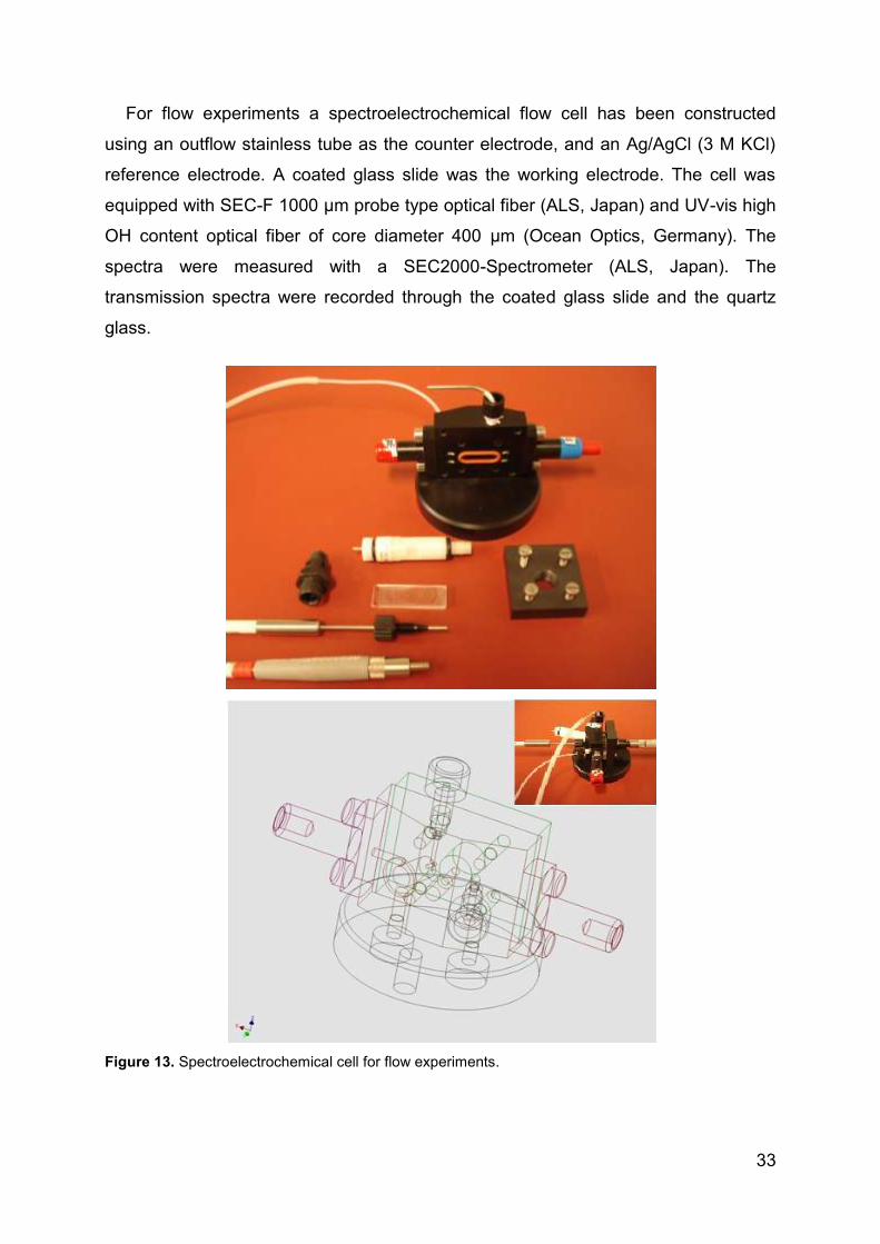
3 Materials and Methods ....................................................................................... 29
3.1 Materials ...................................................................................................... 29
3.1.1 Chemicals ............................................................................................. 29
3.1.2 Instruments ........................................................................................... 30
3.1.3 Buffers .................................................................................................. 35
3.2 Methods ....................................................................................................... 35
3.2.1 Molar extinction coefficients .................................................................. 35
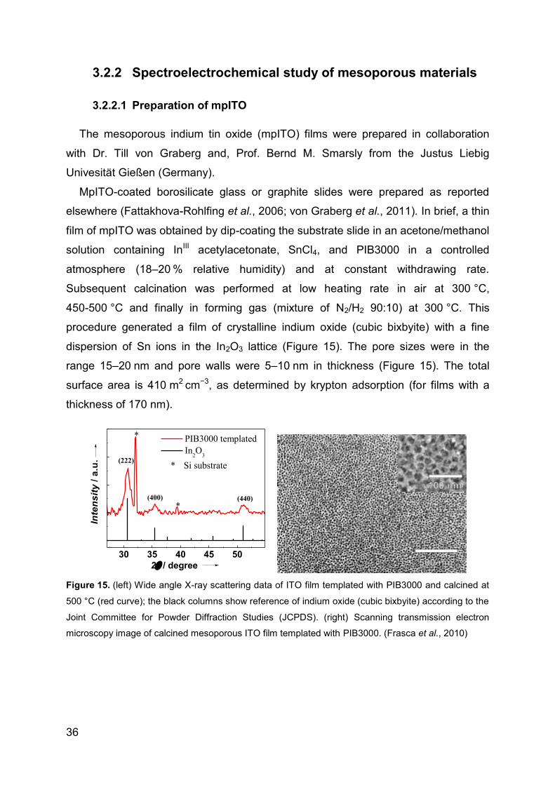
3.2.2 Spectroelectrochemical study of mesoporous materials ....................... 36
3.2.3 Electrochemical study of hSO on AuNPs .............................................. 41
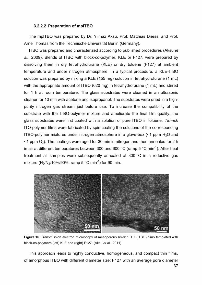
3.2.4 XDH spectroelectrochemical study ....................................................... 44
4 Results and Discussion ...................................................................................... 47
Page 4
4.1 Mesoporous materials ................................................................................. 47
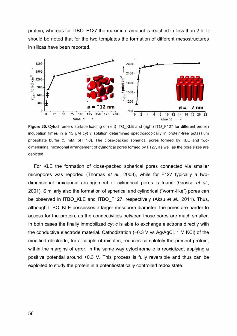
4.1.1 Direct electron transfer of cyt c in mpITO .............................................. 47
4.1.2 UV-Vis spectroelectrochemistry of cyt c in mpITO ................................ 51
4.1.3 Resonance Raman spectroelectrochemistry in mpITO ......................... 53
4.1.4 Spectroelectrochemical studies of cyt c in mpITBO .............................. 54
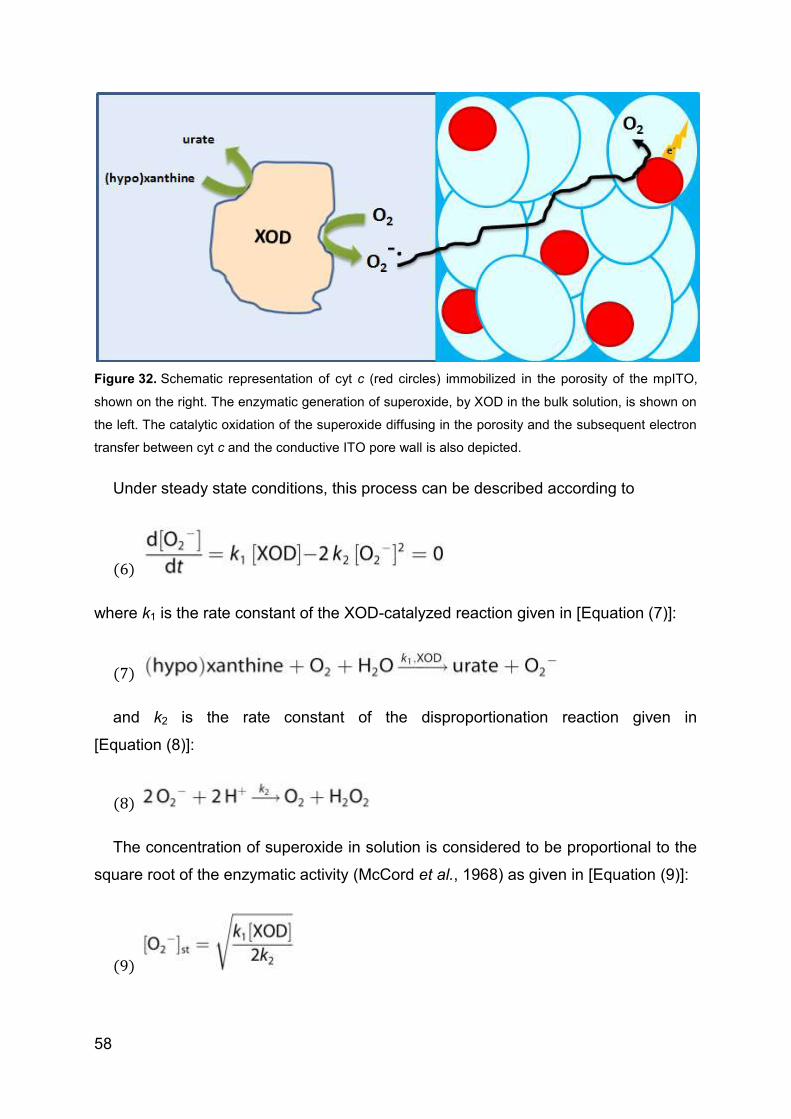
4.1.5 Superoxide biosensor ........................................................................... 57
4.1.6 Reversible electro-system for biochemical switchable optical device ... 60
4.1.7 Spectroelectrochemical studies of hSO-HD in mpITO .......................... 64
4.1.8 Catalytic activity of hSO on planar ATO ................................................ 66
4.2 Direct electrochemistry and catalytic activity of hSO on AuNP .................... 70
4.2.1 Direct Electrochemistry of hSO ............................................................. 70
4.2.2 Surface enhanced resonance Raman spectroscopy ............................. 71
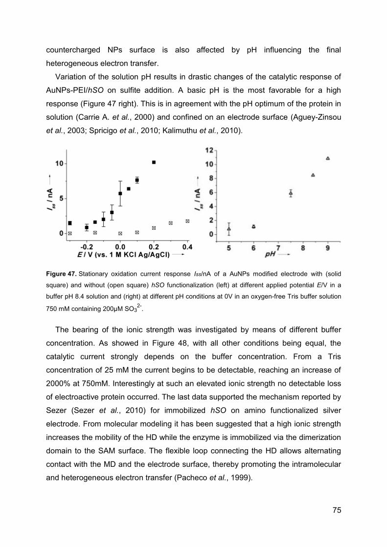
4.2.3 Catalytic activity of hSO ........................................................................ 73
4.2.4 Sulfite biosensor ................................................................................... 76
4.3 Direct electrochemstry of XDH and mAOH1 ................................................ 79
4.3.1 Direct Electrochemistry of immobilized proteins.................................... 79
4.3.2 Mediated spectroelectrochemical titration of XDHwt ............................. 82
5 Summary ............................................................................................................ 85
6 References ......................................................................................................... 93
7 List of publications ........................................................................................... 110
8 List of presentations ......................................................................................... 111
9 Acknowledgements .......................................................................................... 112
Page 5
I
List of Abbreviations
ATO Antimony doped tin oxide Au Gold cSO Chicken sulfite oxidase CV Cyclic Voltammetry CVA Cyclic voltabsorptogram cyt c Cytochrome c DCVA Derivative voltabsorptogram DDAB Didodecyldimethylammonium bromide DET Direct electron transfer DNA Deoxyribonucleic acid E
0 Formal potential
EDC N-Ethyl-N’-(3-dimethylaminopropyl)carbodiimide hydrochloride EP-GC Electrochemically pretreated glassy carbon EPR Electron paramagnetic resonance ET Electron transfer ket heterogeneous electron transfer rate constant Km Michaelis-Menten constant GC Glassy carbon HD Sulfite oxidase heme b5 domain HET Hetero geneous electron transfer hSO Human sulfite oxidase IET Intramolecular electron transfer IEP Isoelectric point ITO Tin doped indium oxide ITBO Tin rich indium tin oxide mAOH1 Mouse Aldehyde oxidase homolog 1 Moco Molybdenum cofactor MD Sulfite oxidase Moco domain MPT Molybdopterin MU Mercapto-undecanol MUA Mercapto-undecanoic acid NP Nanoparticle NHE Normal hydrogen electrode OTTLE Optical transparent thin-layer electrochemical PGE Pyrolytic graphite electrode PEI Polyethylenimine RRS Resonance Raman spectroscopy SAM Self assembled monolayer SDH Sulfite dehydrogenase SERRS Surface enhanced resonance Raman spectroscopy SHE Standard hydrogen electrode SOD Superoxide dismutase SWCNT Single-walled carbon nanotubes TMB 3, 3’, 5, 5’-tetramethylbenzidine TCO Transparent conductive oxide XDH Xanthine dehydrogenase XDHwt Xanthine dehydrogenase wild type XOD Xanthine oxidase
Page 7
III
Abstract
In this thesis, different aspects within the research field of protein spectro- and
electro-chemistry on nanostructured materials are addressed. On the one hand, this
work is related to the investigation of nanostructured transparent and conductive
metal oxides as platform for the immobilization of electroactive enzymes. On the
other hand the second part of this work is related to the immobilization of sulfite
oxidase on gold nanoparticles modified electrode. Finally direct and mediated
spectroelectrochemistry protein with high structure complexity such as the xanthine
dehydrogenase from Rhodobacter capsulatus and its high homologues the mouse
aldehyde oxidase homolog 1.
Stable immobilization and reversible electrochemistry of cytochrome c in a
transparent and conductive tin-doped and tin-rich indium oxide film with a well-
defined mesoporosity is reported. The transparency and good conductivity, in
combination with the large surface area of these materials, allow the incorporation of
a high amount of electroactive biomolecules (between 250 and 2500 pmol cm-2) and
their electrochemical and spectroscopic investigation. Both, the electrochemical
behavior and the immobilization of proteins are influenced by the geometric
parameters of the porous material, such as the structure and pore shape, the surface
chemistry, as well as the protein size and charge. UV-Vis and resonance Raman
spectroscopy, in combination with direct protein voltammetry, are employed for the
characterization of cytochrome c immobilized in the mesoporous indium tin oxide and
reveal no perturbation of the structural integrity of the redox protein. A long term
protein immobilization is reached using these unmodified mesoporous indium oxide
based materials, i.e. more than two weeks even at high ionic strength.
The potential of this modified material as an amperometric biosensor for the
detection of superoxide anions is demonstrated. A sensitivity of about 100 A M-1 m-2,
in a linear measuring range of the superoxide concentration between 0.13 and
0.67 µM, is estimated.
In addition an electrochemical switchable protein-based optical device is designed
with the core part composed of cytochrome c immobilized on a mesoporous indium
tin oxide film. A color developing redox sensitive dye is used as switchable
component of the system. The cytochrome c-catalyzed oxidation of the dye by
Page 8
IV
hydrogen peroxide is spectroscopically investigated. When the dye is co-immobilized
with the protein, its redox state is easily controlled by application of an electrical
potential at the supporting material. This enables to electrochemical reset the system
to the initial state and repetitive signal generation.
The case of negative charged proteins, which does not have a good interaction
with the negative charged indium oxide based films, is also explored. The
modification of an indium tin oxide film with a positive charged polymer and the
employment of a antimony doped tin oxide film were investigated in this work in order
to overcome the repulsion induced by similar charges of the protein and electrode.
Human sulfite oxidase and its separated heme-containing domain are able to direct
exchange electrons with the supporting material.
A study of a new approach for sulfite biosensing, based on enhanced direct
electron transfer of a human sulfite oxidase immobilized on a gold nanoparticles
modified electrode is reported. The spherical gold nanoparticles were prepared via a
novel method by reduction of HAuCl4 with branched poly(ethyleneimine) in an ionic
liquid resulting in particles of about 10 nm in hydrodynamic diameter.
These nanoparticles were covalently attached to a mercaptoundecanoic acid
modified Au-electrode and act as platform where human sulfite oxidase is adsorbed.
An enhanced interfacial electron transfer and electrocatalysis is therefore achieved.
UV-Vis and resonance Raman spectroscopy, in combination with direct protein
voltammetry, were employed for the characterization of the system and reveal no
perturbation of the structural integrity of the redox protein. The proposed biosensor
exhibited a quick steady-state current response, within 2 s and a linear detection
range between 0.5 and 5.4 μM with high sensitivity (1.85 nA μM-1). The investigated
system provides remarkable advantages, since it works at low applied potential and
at very high ionic strength. Therefore these properties could make the proposed
system useful in the development of bioelectronic devices and its application in real
samples.
Finally protein with high structure complexity such as the xanthine dehydrogenase
from Rhodobacter capsulatus and the mouse aldehyde oxidase homolog 1 were
spectroelectrochemically studied. It could be demonstrated that different cofactors
present in the protein structure, like the FAD and the molybdenum cofactor, are able
Page 9
V
to directly exchange electrons with an electrode and are displayed as a single peak
in a square wave voltammogram. Protein mutants bearing a serine substituted to the
cysteines, bounding to the most exposed iron sulfur cluster additionally showed direct
electron transfer which can be attributable to this cluster. On the other hand a
mediated spectroelectrochemical titration of the protein bound FAD cofactor was
performed in presence of transparent iron and cobalt complex mediators. The results
showed the formation of the stable semiquinone and the fully reduced flavin. Two
formal potentials for each single electron exchange step were then determined.
Page 10
VI
Zusammenfassung
In dieser Arbeit werden verschiedenen Aspekte im Forschungsfeld der Protein-
Spekro- und Elektro-Chemie an nanostrukturierte Materialien behandelt. Zum einen
werden in dieser Arbeit nanostrukturierte, transparente und leitfähige Metalloxide als
Basis für die Immobilisierung von elektroaktiven Enzym untersucht.
Des Weiteren behandelt diese Arbeit die Immobilisierung von humaner
Sulfitoxidase auf einer Gold-Nanopartikel-modifizierten Elektrode. Schließlich wird die
direkte und die vermittelte Elektrochemie von Xanthindehydrogenase aus
Rhodobacter capsulatus und Aldehydoxidase Homolog 1, aus Mause, vorgestellt.
Im ersten Teil der Arbeit wird über die stabile Immobilisierung und reversible
Elektrochemie von Cytochrom c in einem transparenten und leitfähigen Zinn-
dotierten und Zinn-reichen Indiumoxid Film mit einer gut definierten Mesoporosität
berichtet. Die Transparenz und gute Leitfähigkeit in Kombination mit der großen
Oberfläche dieser Materialien erlauben die Inkorporation einer große Menge
elektroaktiver Biomoleküle (zwischen 250 und 2500 pmol cm-2) und deren
elektrochemische und spektroskopische Untersuchung. Das elektrochemische
Verhalten und die Proteinimmobilisierung sind durch die geometrischen Parameter
des porösen Materials, wie die Struktur und Porenform, die Oberflächenchemie,
sowie die Größe und Ladung des Proteins beeinflusst. UV-Vis und Resonanz-
Raman-Spektroskopie in Kombination mit direkter Protein-Voltammetrie werden für
die Charakterisierung von Cytochrom c eingesetzt und zeigen keine Störung der
strukturellen Integrität des Redox-Proteins durch die Immobilisierung. Eine
langfristige Immobilisierung des Proteins von mehr als zwei Wochen auch bei hoher
Ionenstärke wurde unter Verwendung dieser unmodifizierten mesoporösen
Indiumoxid-basierten Materialien erreicht.
Das Potential dieses modifizierten Materials für die Verwendung in einem
amperometrischen Biosensor zum Nachweis von Superoxid-Anionen wurde
aufgezeigt. Es wurde eine Empfindlichkeit von etwa 100 A M-1 m-2, in einem linearen
Messbereich der Superoxidkonzentration zwischen 0,13 und 0,67 µM, erreicht.
Außerdem wurde ein elektrochemisch umschaltbares Protein-basiertes optisches
Gerät konzipiert mit Cytochrom c und der mesoporösen Indiumzinnoxidschicht. Ein
redox-sensitiver Farbstoff wurde als schaltbare Komponente des Systems
Page 11
VII
verwendet. Die Cytochrom c Oxidation des Farbstoffs durch Wasserstoffperoxid
wurde spektroskopisch untersucht. Der Redox-Zustand des Farbstoffs, co-
immobilisiert mit dem Protein, ist leicht durch das Anlegen eines elektrischen
Potentials an das Trägermaterial kontrollierbar. Dadurch wird die elektrochemische
Zurücksetzung des Systems auf den Anfangszustand und eine repetitive
Signalerzeugung ermöglicht.
Für negativ geladene Proteine, die keine gute Interaktion mit dem negativ
geladenen Indiumoxid-basierten Film zeigen wurden die Modifikation der
Indiumzinnoxidschicht mit einem positiv geladenen Polymer sowie die Verwendung
eines Antimon-dotierten Zinnoxid Films vorgeschlagen. Dadurch konnte die
Abstoßung induziert durch die ähnliche Ladung des Proteins und der Elektrode
überwunden werden. Es gelang für die humane Sulfit-Oxidase und die separate
Häm-haltige Domäne der Austausch von Elektronen mit dem Trägermaterial.
Im zweiten Teil der Arbeit wird über eine neue Methode für die Biosensorik von
Sulfit berichtet, bei der direkte Elektronentransfer von humaner Sulfitoxidase
immobilisierten auf einer mit Gold-Nanopartikeln modifizierten Elektrode verstärkt
wurde. Die sphärischen Gold-Nanopartikeln, von etwa 10 nm im Durchmesser,
wurden über eine neue Methode durch Reduktion von HAuCl4 mit verzweigtem
Polyethylenimin in einer ionischen Flüssigkeit synthetisiert.
Diese Nanopartikel wurden kovalent an eine mit Mercaptoundecansäure
modifizierten Gold-Elektrode immobilisiert und dienen als Basis für die Adsorption
von Sulfitoxidase adsorbiert wurde. Dadurch wurde ein schneller heterogener
Elektronen-Transfer und verbesserte Elektrokatalyse erreicht. Für die
Charakterisierung des verwendeten Systems eingesetzt wurden UV-Vis und
Resonanz-Raman-Spektroskopie in Kombination mit direkter Protein-Voltammetrie.
Es wurde keine Störung der strukturellen Integrität des Redox-Proteins beobachtet.
Der vorgeschlagene Biosensor zeigte eine schnelle steady-state Stromantwort
innerhalb von 2 s, eine lineare Detektion im Bereich zwischen 0,5 und 5,4 µM Sulfit
mit einer hohen Empfindlichkeit (1,85 nA µM-1). Das untersuchte System bietet
bemerkenswerte Vorteile da es ermöglicht bei niedriger angelegter Spannung und
bei sehr hoher Ionenstärke zu arbeiten. Aufgrund dieser Eigenschaften hat das
vorgeschlagene System großes Potential für die Entwicklung von bioelektronischen
Geräten und der Anwendung in realen Proben.
Page 12
VIII
Schließlich werden im letzten Teil der Arbeit die komplexeren Enzymen
Xanthindehydrogenase aus Rhodobacter capsulatus und Maus Aldehydoxidase
Homolog 1 spektro- und elektrochemisch untersucht. Es konnte gezeigt werden,
dass verschiedene Kofaktoren in der Proteinstruktur, wie FAD und der
Molybdän-Kofaktor direkt Elektronen mit einer Elektrode austauschen können, was
durch einzelne Peaks im Square Wave Voltammogramm angezeigt wird. Es konnte
eine zusätzliche redoxaktive Gruppe mit direktem Elektronen-Transfer nach
Austausch eines Cysteins durch Serin am exponierten Eisen-Schwefel-Cluster
gezeigt werden. Außerdem wurde eine vermittelte spektroelektrochemische Titration
des FAD-Kofaktors in Anwesenheit von Mediatoren der Klasse der Eisen und Kobalt-
Komplexe durchgeführt. Die Ergebnisse zeigen, dass FAD in R. capsulatus XDH zu
einem stabilen Semichinone reduziert werden kann. Es gelang die formalen
Potentiale für die zwei einzigen Elektrontransferprozesse zu bestimmen.
Page 13
IX
Riassunto
In questa tesi sono affrontati diversi aspetti nello studio spettro- ed elettro-chimico
di proteine su materiali nanostrutturati. Nel presente lavoro è inizialmente discussa la
ricerca svolta su ossidi metallici nanostrutturati, trasparenti ed elettricamente
conduttivi usati da piattaforma per l'immobilizzazione di proteine elettroattive. La
seconda parte riguarda l’immobilizzazione della solfito ossidasi su elettrodi modificati
con nanoparticelle di oro. Infine viene presentata l’analisi spettroelettrochimica diretta
e mediata della xantina deidrogenasi del Rhodobacter capsulatus e del suo omologo
l’aldeide ossidasi omologo 1, del topo.
In questa tesi vengono inizialmente discusse la reversibile elettrochimica e la
stabile immobilizzazione del citocromo c su pellicole trasparenti e conduttive, quali
l’ossido d’indio drogato allo stagno e l’ossido d’indio ricco di stagno, dotate di una
ben definita mesoporosità. La loro trasparenza unita alla buona conduttività, in
combinazione con l’estesa superficie di questi materiali, permette l'intrappolamento di
un’elevata quantità di biomolecole elettroattive (tra 250 e 2500 pmol cm-2) e il loro
studio sia elettrochimico che spettroscopico. Il comportamento elettrochimico così
come l'immobilizzazione proteica sono influenzati dai parametri geometrici del
materiale poroso, come ad esempio la struttura e la forma dei pori, dalla chimica di
superficie, così come dalle dimensioni e la carica della proteina in esame. La
spettroscopia UV-Vis e risonanza Raman, in combinazione con la voltammetria
diretta, sono impiegati per la caratterizzazione del citocromo c immobilizzato nella
porosità dei materiali. Nessuna perturbazione nell'integrità strutturale della proteina è
stata rilevata. Un’immobilizzazione della proteina a lungo termine è stata ottenuta
utilizzando questi materiali mesoporosi a base di ossido di indio senza alcuna
modificazione Nello specifico il sistema risulta stabile per più di due settimane anche
ad elevata forza ionica.
Il potenziale di del sistema proposto come biosensore amperometrico viene
mostrato per la rilevazione di anioni superossido. La sensibilità risulta essere di circa
100 A M-1 m-2, in un intervallo lineare di misura per la concentrazione del superossido
compreso tra 0,13 e 0,67 µM.
Inoltre, un dispositivo ottico commutabile elettrochimicamente e basato su
citocromo c immobilizzato su una pellicola mesoporosa di ossido d’indio e stagno è
Page 14
X
stato progettato e sviluppato. Un visibile colorante redox è stato utilizzato come
elemento commutabile nel sistema. Spettroscopicamente si è indagata l’ossidazione
del colorante da parte di perossido d’idrogeno e catalizzata dal citocromo. Quando il
colorante viene co-immobilizzato con la proteina, il suo stato redox è facilmente
controllabile mediante l'applicazione di un potenziale elettrico al materiale di
supporto. Ciò consente di ripristinare il sistema elettrochimico nello stato iniziale e la
generazione ripetitiva di nuovi segnali.
Il caso di proteine cariche negativamente, le quali non presentano una buona
interazione con pellicole basate sull’ossido d’indio, anch’esse cariche negativamente,
è stato anche esplorato. La modifica di una pellicola di ossido d’indio e stagno con un
polimero carico positivamente e l'impiego di una pellicola di ossido di stagno drogato
con antimonio sono stati studiati in questo lavoro al fine di superare la repulsione
indotta dall’analogia di carica tra proteina ed elettrodo. Solfito ossidasi umana e il suo
isolato dominio contenente l’eme sono in grado di scambiare elettroni direttamente
con il materiale di supporto.
Si riporta inoltre lo studio di un nuovo approccio per la biopercezione di solfito,
basato su un migliorato trasferimento elettronico diretto della solfito ossidasi umana
mediante immobilizzazione su un elettrodo modificato con nanoparticelle di oro. Le
nanoparticelle di oro sferiche sono state sintetizzate attraverso un inedito metodo
consistente nella riduzione di HAuCl4 in uno ione liquido per mezzo di polietilenimina
ramificata. Il diametro idrodinamico risultante è stato rilevato di circa 10 nm.
Tali nanoparticelle sono state fissate covalentemente su un elettrodo di oro
modificato con acido mercaptoundecanoico e fungono da piattaforma per
l’assorbimento della solfito ossidasi. Elettrocatalisi, in presenza di substrato, e un
trasferimento di elettroni all’interfaccia migliorati rispetto ad uno stesso elettrodo
senza nanoparticelle è stato così ottenuto. UV-Vis e spettroscopia Raman amplificata
da superfici, in combinazione con la voltammetria diretta, sono stati impiegati per la
caratterizzazione del sistema e non rivelano perturbazione nell'integrità strutturale
dell’enzima. Il biosensore proposto mostra un rapido raggiungimento di una stabile
risposta, in circa 2 s, un intervallo di rilevamento lineare compreso tra 0,5 e 5,4 mM e
una elevata sensibilità (1,85 mM-1 nA). Il sistema studiato fornisce notevoli vantaggi
come la possibilità di lavorare a un’alta forza ionica e applicando un basso
Page 15
XI
potenziale. Queste proprietà potrebbero pertanto rendere il sistema proposto utile
nello sviluppo di dispositivi bioelettronici e il loro utilizzo per campioni reali.
Infine proteine molto complesse come la xantina deidrogenasi del Rhodobacter
capsulatus e l’aldeide ossidasi, omologo 1, del topo, sono state studiate
spettroelettrochimicamente. In questo lavoro viene dimostrato che cofattori diversi
presenti nella struttura delle proteine, come la FAD e il cofattore molibdeno, sono in
grado di scambiare direttamente elettroni con un elettrodo e vengono visualizzati
come un unico picco in voltammetria ad onda quadra. Proteine mutanti, recanti una
serina sostituita a una delle cisteine leganti il più esposto dei centri ferro-zolfo,
mostrano inoltre trasferimento diretto di elettroni attribuibile a tale centro. Un ulteriore
studio del cofattore FAD è stata eseguita per mezzo di titolazione
spettroelettrochimica mediata. Complessi trasparenti di ferro e cobalto sono stati
usati come mediatori. I risultati dimostrano la formazione di un semichinone stabile
del FAD all'interno della struttura proteica possedente due singoli potenziali formali
per ogni scambio di elettroni.
Page 17
1
1 Introduction
1.1 Protein electrochemistry
The field of protein electrochemistry deals with redox proteins which are able to
exchange electrons with an electrode either with the protein free in solution or
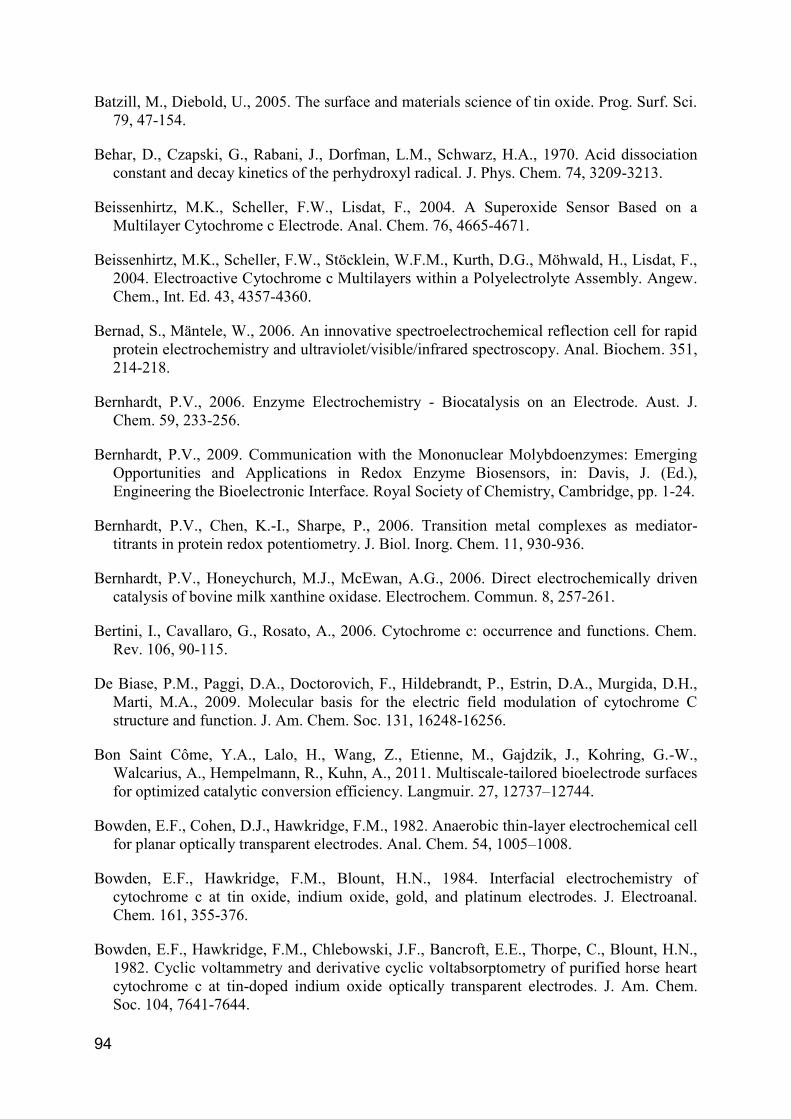
confined on an electrode surface. It can be usually divided into two groups (Figure 1).
In one case an external shuttle molecule is employed as mediator to facilitate the
electron transfer (ET) and it is referred as mediated electron transfer (MET). In any
mediated enzyme catalytic reaction, the mediator must exchange electrons rapidly
with the electrode, since a sustained flow of electrons is required. The electrons are
provided by the electrode via the mediator.
Figure 1. Scheme of electron transfer processes on an electrode surface. (a) Direct Electron Transfer
(DET) and (b) Mediated Electron Transfer (MET) between electrode and protein.
In the other case the electron transfer occurs directly between the protein and the
electrode and is called direct electron transfer (DET). It was thought for a long time to
be virtually impossible. However, from the first publications in the ‘70s (Eddowes et
al., 1977; Yeh et al., 1977) investigations in this field boomed. DET provides rapid
and direct measurements of redox properties and a wide range of electrode
potentials can be applied. Together with the precise redox control afforded by the
Page 18
2
electrode potential it offers an excellent temporal resolution of the activity assay.
Therefore a precise characterization how activity quickly evolves with time following
an instant change in experimental conditions is possible. Furthermore unspecific side
reactions of the mediator, that may cause erroneous results, are prevented.
1.1.1 Direct protein electrochemistry
Direct protein electrochemistry where a protein is confined on an electrode surface
is a powerful tool for investigating the catalytic properties of redox enzymes. From an
operational perspective, direct protein electrochemistry of surface immobilized
molecules also has a number of other advantages, not at least the very small
amounts of the often “priceless” biological material required, down to pmol cm-2, in
comparison with other more classical techniques (Armstrong et al., 1988; Armstrong,
2002; Léger et al., 2008). After immobilization, the same sample can be reused for
the further studies. Precondition for the application of this technique, it is the ability to
connect the active site of the enzyme to the electrode. Basically, two different
strategies can be employed, either protein modification with genetic or chemical
engineering techniques (Campàs et al., 2009; Caruana et al., 2010) or novel
interfacial technologies (Hill et al., 1989; Fedurco, 2000).
The ET is a radiation less electronic rearrangement where an electron moves from
an initial state on an electrode or reductant to a receiving state on another solvated
species or on an electrode of the same energy. The rate is strongly dependent from
the potential difference and the spatial distance between the two redox sites (Marcus
et al., 1985; Marcus, 1993). Direct protein electrochemistry enables to exploit the
naturally high efficiency of biological systems for developing selective biosensors,
energy storage and production systems like biofuel cells, heterogeneous catalysts,
and biomolecular electronic components (Léger et al., 2008).
The most successful electrodes for proteins so far have been noble metals and
carbon due to their elevated conductivity and easy handling. However they often lead
to an irreversible adsorption and denaturation of the proteins onto the electrode
surface and therefore to the impossibility to establish fast ET. A wide used method to
solve this problem is the modification of the electrode by a promoter, which can
prevent the protein denaturation and can lead also to a specific protein-electrode
orientation (Armstrong, 2002). The promoter can reduce the distance between the
active site of the protein and the electrode (Armstrong, 1990). It is not electroactive
Page 19
3
itself and can interact with the enzyme by different kind of forces (electrostatic,
hydrophobic, hydrogen bounds, etc.). By this approach, a DET reaction can be
obtained or in alternative the usage of mediators can solve possible distance
problems and permits the electron shuttling (Figure 1). General promoters are self-
assembly monolayers of amphiphiles or polyelectrolytes (Scheller et al., 2002; Allen
et al., 1984; Fedurco, 2000; Rusling et al., 2008) with several possible functionalities.
A drawback of this approach, however, is that the amount of immobilized protein is
limited to monolayer coverage. Larger amounts of protein, can be obtained by
alternate deposition of proteins and polyelectrolytes (“layer-by-layer” technique)
(Beissenhirtz et al., 2004; Grochol et al., 2007; Spricigo et al., 2009; Dronov et al.,
2007; Ram et al., 2001; Calvo et al., 2004). However this advantage is
counterbalanced by the limited accessibility of the active sites of the proteins in the
inner layers and the low stability.
The advent of nanoscaled materials such as nanotubes, nanoparticles, conductive
and non-conductive metal oxides opens new horizons for the field of bioelectronics
due to the likely deep interactions between the nanomaterials and the proteins
(Armstrong, 2002; Bernhardt, 2006; Chen et al., 2007; Wollenberger et al., 2008).
1.1.2 Protein spectroelectrochemistry
The coupling of electrochemical and optical methods has been used for decades
to study a large range of organic, inorganic and biological redox systems (Kuwana et
al., 1976; Heineman et al., 1984). Althought a large variety of electrochemical
methods are available, they do not render any structural information of the electrode
system besides the detailed knowledge of charge transfer, transport and distribution.
The combination of electrochemical and optical methods to monitor the spectroscopic
variations associated to the potential changes allows a qualified picture of the
chemical structures in electrochemical reactions. The potential of the analyzed
solution may be easily changed by addition of reductants or oxidants. On the other
hand it can be electronically changed by potential imposition at an electrode. A
classical set for such an experiment consist in an optical transparent thin-layer
electrochemical (OTTLE) cell, with a metallic paint ensuring electrical conductivity
and preserving some degree of transparency (Pinkerton et al., 1980; Bowden, et al.,
1982; Heineman et al., 1984; Dai et al., 2011).
Page 20
4
A large variety of spectroscopic methods may be coupled, from UV-Vis, infra-red
(IR) (Arion et al., 2011), resonance Raman (RR) (Kavan et al., 2009) and surface
enhanced resonance Raman (SERR) (Murgida et al., 2006) to electron paramagnetic
resonance (EPR) (Paulsen et al., 1992) and nuclear magnetic resonance (NMR)
(Klod et al., 2009). Nevertheless, there are some spectroscopic methods which are
preferred in spectroelectrochemistry. The choice of the method is often dominated by
not the importance of a spectroscopic method which offers the access of important
structural data of an electron-transfer reaction in experimental studies, but in most
cases, the ease of application. UV-Vis spectroscopy is the most applied method in
spectroelectrochemistry irrespective of the fact that other methods would result in
more detailed structural informations (Dunsch, 2011).
Spectroelectrochemical studies were usually restricted to solution samples, where
a relative concentrated sample is required, attenuated total internal reflection mode
or with reflection cells (Bernad et al., 2006). Only with a signal enhancement, like with
Raman spectroscopy through surface plasmon resonance, the noteworthy reduction
of sample volume and concentration can be obtained.
Absorption UV-Vis spectroelectrochemical investigation of protein boosted in the
last time by the improvements in the field of nanostructured transparent conductive
oxide. The possibility to entrap a large amount of protein in the porous structure of
such materials overcomes the lack of sensitivity (Szamocki et al., 2007). In addition
these materials offer a high transparency in the UV-Vis region and elevated
conductivity over the whole potential range commonly used for proteins investigation
(Topoglidis et al., 2001; Panicco et al., 2008; Renault et al., 2011).
1.1.3 Biosensors
A biosensor is defined as a specific type of chemical sensor comprising a
biological or biologically derived recognition element either integrated within or
intimately associated with a physicochemical transducer. The biological element is
capable of recognizing the presence, activity or concentration of a specific analyte in
solution (Thévenot et al., 1999; Thévenot et al., 2001; Hall, 2002). An analyte is the
compound whose concentration has to be measured. Biosensors basically involve
the quantitative analysis of various substances by converting their biological actions
into measurable signals. Generally the performance of the biosensors is mostly
Page 21
5
dependent on the specificity and sensitivity of the biological reaction, besides the
stability of the biological element.
In general a biosensor comprises three parts: a biological recognition element, a
suitable transducer and an amplification element (Figure 2).
The transducer is an important component of a biosensor through which the
measurement of the target analyte(s) is achieved by selective transformation of a
biomolecule-analyte interaction into a quantifiable output signal. The mode of
transduction may be one of several approaches, including electrochemical, optical,
piezoelectric, magnetic or thermometric transducers.
Every biomolecule from enzyme to an antibody, a nucleic acid, a hormone, an
organelle or whole cell which can selectively interact with other substances, can be
theoretically qualified for biosensor development. Usually, sensors are distinguished
(Thévenot et al., 1999) in sensors using catalytic biorecognition elements (enzymes,
cells, microorganisms) (Gu et al., 2004) and affinity-based recognition elements
(antibodies, antigens, protein receptors, synthetic receptors, nucleic acids) ( ale e ,
2005) (Ge et al., 2008). Catalytic sensors own the concomitant ability to amplify the
signal and regenerate the active site of the biorecognition element (Dryhurst et al.,
1982).
A wide range of enzymes, owing a combination of specificity and amplification
properties, has been successfully used as a recognition element. Since enzymes
allow a wide range of transduction technologies, they have found very wide
applications in the field of biosensors (Schuhmann et al., 2003).
Figure 2. Configuration of a biosensor showing biorecognition, interface, and transduction elements
(Chambers et al., 2008).
Page 22
6
Other kinds of proteins used are for example membrane bound receptors that
change their structural conformation binding specific ligands. This modification
triggers an amplified physiological response, such as ion channel opening or
secreting an enzyme. High affinity and specificity towards the natural targets are the
advantages of such recognition system.
Although the biosensors suffer general problem of stability and complexity
connected to the biological recognition element, they offer many advantage in
respect to classical analytical methods. Biosensors show a very broad range of
detectable analytes, depending on the nature of the recognition element, and high
analyte selectivity with the limitation of interferences in complex samples.
Furthermore, the current tendency in biosensor development is the miniaturization.
This enables and will further expand the integration and parallelization of biosensors
in sophisticated systems.
1.1.3.1 Electrochemical enzyme based biosensors
Biosensors that utilize enzymes as recognition elements represent a wide
extensively studied area, with glucose biosensors dominating the market
(Frost & Sullivan, 2006). Enzymes are favored as recognition elements in biosensors
because they provide a broad range of changes of physical and chemical parameters
during the enzymatic reaction, such as electrons, protons, ions, mass, light and heat.
These changes can be detected using suitable transducer elements. Different
electrochemical methods as potentiometriy, voltammetry and amperometry exist,
where either the potential or the current change depending on the concentration of
the analyte can be measured. Selective and sensitive catalysis of a substrate at
relative low potentials are the great power of enzymes.
Electrochemical enzyme-based sensors are often separated in three different
types or generations. First generation sensors measure the signal via the natural
secondary substrates and products of the enzyme catalyzed reaction. In the second
generation sensors an artificial electron mediator is used instead of the natural
co-substrates. Indeed enzymes in direct electronic contact, based on direct protein
electrochemistry are considered as third generation sensors. Their direct electron
transfer (DET) between the electrode and the protein (Figure 1) may avoid most of
the interferences (Wollenberger, 2005).
Page 23
7
1.2 Electrodes
1.2.1 Nanostructured electrode materials
Nanomaterials have number of features that make them ideally suited for sensor
applications, such as its high surface area, high reactivity, controlled electrode
modification and defined interaction with other partner, e.g. biomolecules. They find
large employ in different fields ranging from biosensors to biofuel cells or more
complex bioelectronic systems. Nanostructured materials include dendrimers,
nanoparticles, nanotubes, nanopores etc (Umasankar et al., 2009).
1.2.1.1 Indium Tin Oxide
Using film of intrinsic stoichiometric materials like metals partial transparency, with
moderate reduction in conductivity, can be obtained. However such materials may
not achieve high transparency and coincidentally elevated conductivity.
A solution is to create electron degeneracy in a material with a wide energy
bandgap (Eg > 3eV or more for visible radiation) by introduction of non-stoichiometry
and/or appropriate dopants. A large number of non-stoichiometric and doped oxide
films (indium, tin, antimony, cadmium, zinc etc.) meet these conditions and exhibit
high transmittance and nearly metallic conductivity (Chopra et al., 1983).
Tin doped indium oxide or indium tin oxide (ITO), with a mean transmittance of
95% and conductivity as high as 104 S-1cm-1, is among the most popular of these thin
films (Granqvist et al., 2002).
ITO is essentially formed by subsititutional doping of In2O3 with Sn which replaces
the In3+ atoms from the cubic bixbyte structure of indium oxide (Fan et al., 1977). Tin,
which exists either as SnO or SnO2, forms an interstitial bond with oxygen. These two
valency states have a direct influence on the ultimate conductivity of ITO. The lower
valence state (+2) results in a net reduction in carrier concentration since the hole
created acts as a trap and reduces the conductivity. On the other hand,
predominance of the SnO2 state (+4) acts as a n-type donor releasing electrons to
the conduction band. The high conductivity of ITO films is due to high carrier
concentration and their mobility increases due to enhanced crystallinity of films
deposited at high temperatures (Balasubramanian et al., 1989).
Page 24
8
The high optical transmittance of this material is a direct consequence of being a
semiconductor with a wide bandgap and therefore the absorption region generally
lies in the ultraviolet part of the electromagnetic spectrum and shifts to shorter
wavelengths with increasing carrier concentration (Gupta et al., 1989). The
transmittance of ITO films is also influenced by a number of minor defects which
include surface roughness and optical inhomogeneity. Opaqueness has been
attributed also to unoxidised tin metal grains on the ITO surface as a result of
instability due to the absence of sufficient oxygen during the deposition (Fan et al.,
1977) or external induction by an applied potential (Kraft et al., 1994; Senthilkumar et
al., 2008).
X-ray photoelectron spectroscopy studies of ITO surfaces showed high
concentrations of In(OH)3-like and InOOH-like surface species, indicating an excess
of negative surface charge (Milliron et al., 2000).
ITO has found an employment in electronic, opto-electronic and mechanical
applications. Uses of ITO have traditionally ranged from transparent heating
elements of aircraft and car windows, heat reflecting mirrors, antireflection and
antistatic coatings, over electronic instrument display panels and even in high
temperature gas sensors. Early electro-optic devices using ITO include
charge-coupled devices, liquid crystal displays and as transparent electrodes for
various display devices like touchscreens. More recently, ITO has been used as a
transparent contact in advanced optoelectronic devices such as solar cells, light
emitting and photo diodes, photo transistors and lasers.
In parallel to the planar films, ITO with a well-defined mesoporous framework
(mpITO) is of considerable interest. With its unique combination of transparency, high
conductivity, well-defined 3D mesoporosity and high surface area, mpITO allows the
incorporation of a high amount of optoelectroactive species, facilitates electron
transport to these centers, and efficiently harvests the electron-induced optical
response or, vice versa, the photon-induced electron flow.
This material thus could open new pathways to novel, highly efficient solar cells
and optoelectronic systems based on transparent electrodes and sensors.
1.2.1.2 Tin rich indium tin oxide
A general problem in the production of ITO is the limited amount of tin, which,
although the cheaper component, is only slightly soluble in the In2O3 phase, typically
Page 25
9
around 1−10 wt %. In case of organized mesostructured ITO, a second problem
appears from the compromise between the conductivity and regular porosity.
To avoid phase segregation of tin-rich ITO, a low-temperature approach with high
control over the In/Sn molar ratio was reported recently, based on the molecular
single-source precursor indium tin tris-tert-butoxide (ITBO; Scheme 1) containing
indium and tin in the molar ratio of 1:1, which facilitates the formation of tin-rich ITO
with an identical stoichiometry in the final product (Aksu et al., 2009).
Most importantly, the resulting tin-rich ITO shows high conductivity and
transparency even in an amorphous state. Using such approach it was possible to
overcome all of the problems connected with the pore
collapse during the crystallization typical for the
template-assisted approach toward mesoporous ITO
(Fattakhova-Rohlfing et al., 2006). As the ITBO
precursor enables the formation of transparent
conducting films without any crystallization step, the
main cause of pore collapse of mesoporous metal
oxides is excluded in these materials. Thus, ITBO
appeared as highly suitable for the preparation of
mesoporous, tin-rich ITO films with reliable high
electrical conductivity and transparency using different templates (Aksu et al., 2011).
1.2.1.3 Antimony doped tin oxide
The research toward the replacing of the rare and expensive indium in transparent
conducting films is of great interest and may in addition provide a different surface
chemistry and energy-level properties.
The most promising materials are the extrinsically doped tin oxides, such as
fluorine- or antimony-doped tin oxide (ATO) (Batzill et al., 2005). Sb is a common
n-type dopant in SnO2. Stjerna (Stjerna et al., 1994) reported a strong increase in the
free electron concentration in the SnO2 band gap when doped with Sb. Therefore it
was concluded that this band could be a half-filled metallic band and that additional
thermal excitation into the Sn-like bands could increase the conductivity. In recent
years, some communications have been published reporting macro and mesoporous
ATO electrodes (Hou et al., 2009; Wang et al., 2009; Urbanová et al., 2010).
Scheme 1 Structure of indium tin tris-tert-butoxide (ITBO).
Page 26
10
1.2.1.4 Gold nanoparticles
An interesting way to build up conductive three dimensional structures on
electrode is offered by metal nanoparticles (NPs). The chemical functionalities
associated with nanoparticles enable the assembly of 2D and 3D NP architectures on
surfaces (Shipway et al., 2000). On the basis of the tremendous success in
supramolecular chemistry, NPs functionalized with various molecular and
biomolecular units were assembled into complex hybrid systems. The electronic
triggering of redox proteins by the incorporation of nanoparticles represents a novel
strategy for the electrical contacting of redox enzymes with their macroscopic
environment.
Colloidal gold nanoparticles (AuNPs) have been around for centuries
predominantly in the work of artists and craftsman because of their intensive visible
colors. However, through research on size, shape, surface chemistry, and optical
properties of gold nanoparticles a door to some very unique and exciting capabilities
has been opened. Gold nanoparticle chemistry and physics has emerged as a broad
new subdiscipline in the domain of colloids and surfaces. NPs with fewer than 300
gold atoms can display distinct optical and electronic properties compared to the bulk
metal. These unusual optical properties of small gold particles, their size-dependent
electrochemistry, and their high chemical stability have made them the model system
of choice for exploring a wide range of phenomena including self-assembly,
biolabeling, catalysis, electron-transfer theories, phase transfer, DNA melting,
DNA assays and crystal growth. They found a large application range from photonic
device fabrications, to sensing of organic and biomolecules, to charge storage
systems (Jennings et al., 2007; Sardar et al., 2009).
The convergence of biotechnology and nanotechnology has led to the
development of hybrid nanomaterials that incorporate the highly selective catalytic
and recognition properties of biomaterials with the unique electronic, photonic, and
catalytic features of nanoparticles. A very interesting property of gold nanoparticles is
to provide a suitable microenvironment for biomolecules immobilization retaining their
biological activity. Their ability to facilitate ET between the immobilized proteins and
electrode surfaces, in addition to the light-scattering properties and extremely large
enhancement ability of the local electromagnetic field led to an intensive use of this
nanomaterial for the construction of biosensors (Li et al., 2010) and electrochemical
biosensors (Pingarrón et al., 2008; Bon Saint Côme et al., 2011).
Page 27
11
Their high surface-to-volume ratio, high surface energy, ability to decrease
proteins–metal particles distance, and the functioning as electron-conducting
pathways between prosthetic groups and the electrode surface, are regarded to be
the general characteristics of gold nanoparticles responsible to facilitate electron
transfer between redox proteins and electrode surfaces (Liu et al., 2003).
Gold nanoparticles are prepared with a wide variety of preparative methods.
These methods are mostly based on precursors containing gold complexes with
tetrachloroauric acid (HAuCl4), being the precursor most commonly used. Various in
situ reactions, such as chemical, photo-induced, thermal decompositions or
controlled solvent evaporation are used for the reduction process (Rao et al., 2000).
Nanoparticles show a relative stabilization in solution towards aggregation and
other modes of decay due to the acquisition of charges either from surface charged
groups or by specific ion adsorption from the bulk solution. Such charges lead to a
repulsive double-layer force between particles. On the other hand nanoparticle
systems adsorbing a polymeric layer can be sterically stabilized due to a steric barrier
which prevente the particles against collision. A much better stabilization is provided
when the adsorbed polymer is a polyelectrolyte. In this case both types of
stabilization can be combined giving rise to electrosterically stabilized systems (Koetz
et al., 2007).
Page 28
12
1.3 Proteins
1.3.1 Cytochrome c
Cytochrome c (cyt c) is a small globular redox protein with a molecular weight of
about 12 kDa. Cyt c is highly soluble, in contrast to other cytochromes, with an
aqueous solubility of about 100 g L-1. It contains a single polypeptide chain of about
100 residues and a mono c-type heme as prosthetic group (protoporphyrin IX, see
Figure 3 left), covalently bound to two cysteine residues and responsible for its
characteristic red color. About 40% of the polypeptide is in α‐helical segments while
the rest consist in turns, irregularly coiled and extended segments (Figure 3 right) (De
Biase et al., 2009). The 18 positively charged lysine residues contribute largely to the
basic isoelectric point in the range of 10.0 – 10.5 (van Gelder et al., 1962).
The real electron carrier of cyt c is the iron coordinated at the center of the
porphyrin ring and to a histidine and a metionine. It can exist in the oxidized (ferric,
Fe3+) or the reduced (ferrous, Fe2+) form and shows a redox potential of around
250 mV vs NHE at pH 7 and 25 °C.
N N
NN
Fe
CH3
CH3
CH3
CH3
O OH OHO
CH3S
CH3
S
Cys
Cys
Figure 3. Structure of the heme group of c‐type cytochromes (left) and structure of the horse heart
cytochrome c (right). Picture made with Pymol using the PDB-file 1HRC (Bushnell et al., 1990).
Cyt c is found in many species ranging from eukaryotes to bacteria and Achaea
(Bertini et al., 2006). In eukaryotes cyt c is a freely diffusing protein of the
mitochondrial intermembrane. Cyt c in the mitochondria is involved in the electron
shuttle between cyt c reductase and cyt c oxidase during oxidative phosphorylation in
Page 29
13
the respiratory chain. In chloroplasts, cyt c transfers electrons from the cytochrome bf
complex to photosystem I. In prokaryotes, cyt c is involved in both aerobic and
anaerobic respiration.
In general it is also involved in early events of apoptosis when released to the
cytoplasm (Ow et al., 2008; Caroppi et al., 2009).
Scavenging superoxide and hydrogen peroxide in mitochondria (Min et al., 2007)
may also be connected to the apoptosis, since these reactive oxygen species are
shown to act as redox signal molecules. However the effort to discover new
functions of cyt c beyond that of an electron carrier in respiration seem still not to be
finished and contribute to the ongoing high interest for this protein.
A quasi-reversible exchange of horse heart cytochrome c was first reported in the
1977 on tin-doped indium oxide electrode (Yeh et al., 1977) and 4, 4'-bipyridine
modified gold electrode (Eddowes et al., 1977) (Figure 4).
Figure 4. (Left) Cyclic voltammetry (a) (scan rate 10, 20, 50, 100, and 200 mV s-1
) and differential
pulse voltammetry (b) (scan rate 2 mV s-1
, pulse height 50 mV, and pulse width 0.5 s) in phosphate
buffer (ionic strength=0.15 M, pH=7.0) at ITO (Yeh et al., 1977) and (right) cyclic voltammetry in
NaCl04 (0.1 M), phosphate buffer (0.02 M) at pH 7 in the presence of 4,4’-bipyridyl (10-2
M) (scan rate
20, 50 and 100 mV s-1
, reference SCE) at bare gold electrode (Eddowes et al., 1979) of a cyt c
solution.
Page 30
14
The extensive studies on cytochrome c suggest that adsorption at the
electrode-solution interface plays a prominent role in the electrode process. It has
been demonstrated that the adsorption of cytochrome c can be accompanied by
noticeable changes in the protein conformation and even the coordination shell of the
heme iron. Non-electrochemical studies such by spectroreflectance (Hinnen et al.,
1983; Niki et al., 1987) and surface-enhanced resonance Raman spectroscopy
(Adelhelm et al., 1989) coupled to electrochemistry confirmed the irreversible
adsorption of cytochrome c at bare gold and silver electrodes.
On the other hand cyt c adsorbs strongly on indium oxide, but not completely
irreversibly, while retaining its native redox potential (El Kasmi et al., 2002). Several
other metal oxide electrodes were used successfully (Bowden et al., 1984; Topoglidis
et al., 2003; Li et al., 2001; Xu et al., 2004; Deng et al., 2009), but the electrode
responses have been found to be very sensitive to the experimental conditions,
especially pretreatment procedures.
In case of self-assembly monolayer (SAM), like with 4,4'-bipyridine, the modifier
acts by adsorbing to the gold surface, thereby providing a suitable interface for
interaction with cytochrome c. Most recently investigations on the electrochemistry of
cyt c were carried out using a large variety of different modifiers (Scheller et al., 1999;
Fedurco, 2000; Wang et al., 2011; Matsui et al., 2011; Hoffmann et al., 2011).
The formal potential (E0) of the absorbed protein is negative shifted when
compared to the respective solution values. Shifts of this type have been well
documented (Jin et al., 1997; Ge et al., 2002) and are still in discussion whether it
should be attributed to the greater relative binding affinity of the ferric form of the
protein (Willit et al., 1990) or to the conversion of a portion of the immobilized cyt c to
a conformational state lacking the axial Met-80 ligand of the heme induced by the
electric field at the binding site (Murgida et al., 2001).
Metal oxides or modified gold surfaces are not the only way for obtaining rapid
electron exchanges between cytochrome c and the electrode. Direct electron transfer
has been observed at a bare glassy carbon electrode (Hagen, 1989) at single-wall
carbon nanotubes (Wang et al., 2002), bio sol–gel matrix (Deriu et al., 2007) and
boron-doped diamond electrodes (Marken et al., 2002).
Another promising way is the use of modified-electrode interfaces obtained
through deposited lipids. These films appear to yield novel opportunities to fashion
'tailor-made" surfaces which can integrate cytochromes and ultimately mimic the
Page 31
15
operating environment of membrane-bound electron-transfer systems (Guerrieri et
al., 1991; Salamona et al., 1991).
The cyt c reduction assay for superoxide‐generating enzyme is widely used since
decades, and was for the first time reported in the 1968 (McCord et al., 1968). Cyt c
oxidizes superoxide to molecular oxygen:
(1) O2-˙ + cyt c3+ → O2 + cyt c2+
High interest is placed in the development of superoxide biosensors employing the
possibility to reoxidize the reduced cyt c2+ to generate current which is correlated to
the superoxide concentration (McNeil et al., 1989; Campanella et al., 1997; Scheller
et al., 1999; Ge et al., 2002; Wegerich et al., 2009).
1.3.2 Mononuclear Molybdoenzymes
Molybdenum is an integral component of diverse groups of enzymes and is
necessary for most living organisms. Molybdenum is widely available to biological
systems due to the solubility of its high-valent oxides in water, being the only second
row transition metal in nature with biological importance.
So far, over 50 molybdoenzymes were discovered to be involved in the catalysis of
reactions at carbon, sulfur and nitrogen cycles and it is found in two basic forms. A
multinuclear MoFe7 cluster is found in nitrogenases (Burgess et al., 1996; Howard et
al., 1996) whereas mononuclear active sites in general catalyze oxygen atom transfer
either to or from a physiological acceptor/donor molecule.
According of the classification proposed by Hille based on a different active site
structure and type of reaction catalyzed (Hille, 1996; Hille, 2002), the monuclear
molybdenum enzymes where subdivided in three families (Figure 5).
In all known cases the enzymes possess a pterin (Figure 5a) and the cofactor is
referred in the literature as molybdopterin or Moco from molybdenum cofactor
(Truglio et al., 2002; Romão, 2009) since it was originally believed to be present only
in molybdenum enzymes. Nevertheless the same form was found later in some
tungsten enzymes (Johnson et al., 1996).
The first family, with the xanthine oxidase from cow’s mil as progenitor,
possesses a MoVIOS(OH) nucleus and one pyranopterin but no covalent bonding of
the metal to the polypeptide chain. These enzymes typically have been found to be
Page 32
16
remarkably similar in their overall size and composition of redox centers, as well as
their ability to oxidize a large variety of purine, pyrimidine, pterin, and aldehyde
substrates (Hille, 1996; Hille et al., 1995).
-
Molybdopterin
-
Sulfite Oxidase Family
Cys
-
Xanthine Oxidase Family
-
-
DMSO Reductase Family
Y = O, S, Se
X = serine, cysteine, selenocysteine
a b
dc
Figure 5. (a) Chemical structure of the MPT and the Moco belonging to three different protein families.
(b) Members of the xanthine oxidase family have Mo centers consisting of a single MPT dithiolene
ligand (L) coordinated to MoOS-OH. (c) Members of the sulfite oxidase family possess a single MPT
dithiolene coordinated to a MoO2 (additional coordination may be taken by water and/or a cysteine
residue that is conserved within the family) (d) Enzymes belonging to the third family are distinguished
by bisdithiolene coordination of the Mo.
The second family includes sulfite oxidase and nitrate reductase (from plants that
assimilate nitrate from the soil). The (oxidized) metal center has a single equivalent of
the MPT with the polypeptide chain coordinates directly to the molybdenum site by a
Page 33
17
cysteinyl residue (LMoVIO2(S–Cys)). Members of this second family catalyze the
oxygen atom transfer reactions either to or from an available ion pair of electrons on
the substrate.
The third family differs either in structure and function, with all members having
two equivalents of the MPT bound to the metal. The Mo coordination sphere is
usually completed by a single Mo=O group and a sixth ligand in an L2MoVIO(X) core.
The reactions catalyzed by members of this last family frequently involve oxygen
atom transfer, but dehydrogenation reactions also occur (Kniemeyer et al., 2001).
In eukaryotes the pyranopterin has a terminal phosphate group on the
pyranopterin side chain. In prokaryotes it is conjugated to nucleosides, usually
cytosine (molybdopterin cytosine dinucleotide e.g. quinoline oxidoreductase from
Pseudomonas putida) or guanosine (molybdopterin guanosine dinucleotide e.g.
DMSO reductase and nitrate reductase from R. sphaeroides, formate dehydrogenase
from Escherichia coli), and occasionally adenosine (e.g. formylmethanofuran
dehydrogenase from Methanococcus thermoautotrophicum) or inosine (Rajagopalan
et al., 1992). The primary role of the pterin cofactor is to position the catalytic metal in
the active site. In addition, it modulates the redox potential at the Mo centre and is
involved, via the pterin ring system, in the transfer of electrons to or from other
prosthetic groups. The pathway by which the pyranopterin core of the cofactor is
synthesized involves a large number of proteins in bacteria and appears to be
universally conserved in biology (Joshi et al., 1996; Schwarz, 2005; Fischer et al.,
2010; Leimkühler et al., 2011).
The mononuclear Mo-enzymes show a large diversity of substrates that they are
capable of oxidizing or reducing and since electron transfer is involved in the catalytic
mechanisms, a large investigation is focused on their application in biosensor and
bioelectronic (Bernhardt, 2009).
1.3.2.1 Sulfite oxidizing enzymes
The group of the sulfite oxidizing enzymes is composed of two subclasses based
on their ability to transfer electrons to molecular oxygen (Hille, 2002): the sulfite
oxidases (SO, found in animals and plants), and the sulfite dehydrogenases (SDH,
found in bacteria).
In animals SO is a dimer and each monomer has three domains. A first domain
(9 kDa) binding a b-type heme (Heme domain, HD), a second containing the Moco
Page 34
18
domain (Moco domain, MD) and a third domain involved in the dimerization between
the two monomers. The HD and the MD are linked by a 10 amino acid flexible loop
(Pacheco et al., 1999).
It catalyzes the oxidation of sulfite to sulfate, with the oxidized cyt c as the
physiological electron acceptor (Hille, 1996; Temple et al., 2000):
(2) SO32− + H2O + 2 cyt c+3 → SO42− + 2 cyt c+2 + 2 H+
The bacterial SDH is a αβ heterodimer, consisting of a subunit containing a Moco
and a cytochrome c552 subunit. The natural electron acceptor for SDH from
Thiobacillus novellus seems to be the cytochrome c550 (Kappler et al., 2000; Kappler
et al., 2005).
Plant sulfite oxidizing enzymes, like the Arabidopsis thaliana SO, consist in a
simple Moco binding domain and lack a heme containing domain (Eilers et al., 2001;
Schrader et al., 2003). It has been shown that oxygen acts as the terminal electron
acceptor for plant SO (Hänsch et al., 2006; Byrne et al., 2009).
1.3.2.1.1 Animal sulfite oxidase structure
The structure of the human SO has not been solved so far, although the structure
of the human SO heme domain is already available (Rudolph et al., 2003).
Nevertheless, the X-ray structure of its high homologues (chicken SO) was reported
(Kisker et al., 1997) (Figure 6). CSO is a homodimeric sulfite oxidase contains two
identical subunits formed from a small N-terminal b5-type cytochrome domain (HD), a
large central Mo-binding domain (MD), and a large C-terminal interface domain.
In each subunit the MD and the HD are linked by a flexible peptide loop of 10
amino acids. It was also observed that the dispositions of the HDs within the dimeric
protein of the unit cell are not in an equivalent position relative to their respective
MDs. This variation in HDs orientation has been interpreted as evidence of domain-
domain flexibility, and supports the hypothesis that conformational change is involved
in the electron transfer between the Moco and heme centers. In the X-ray SO
structure the Mo and Fe centers are ~32 Å apart in a not optimal position and
orientation of the respective redox partners for an IET (Pacheco et al., 1999).
Rearrangement to a more “productive” orientation may occur before IET, which
suggests that fast ET between Mo and Fe centers requires delicate and precise
positioning, orientation, and docking of the two redox partners. The flexible loop
Page 35
19
could provide to the heme domain the necessary mobility to allow its negative
charged exposed edge to interact electrostatically with the positively charged MD.
Figure 6. Crystal structure of chicken liver SO. The dimeric structure is underlined by differently
colored monomers. The MD and HD positions are highlighted only for one subunit for simplicity. The
heme b5 group is indicated in red, whereas the Moco is depicted in green. Picture made with Pymol
using the PDB-file 1SO (Kisker et al., 1997).
Once the IET takes place then the HD moves away from the MD to interact with
the positively charged cyt c, the physiological electron acceptor. This flexibility seems
to guarantee both intra and inter electron transfer reaction. Unlike, in the bacterial
SDH, the heterodimer comprising molybdenum and heme c binding subunits occupy
fixed positions relative to one another during catalysis (Kappler et al., 2000).
In SO the IET reaction rates were found to be affected by various parameters such
as pH, anion concentration and even the nature of the anion; thus, Cl-, SO42-, and
PO43- all have different inhibitory effects (Sullivan Jr. et al., 1993; Pacheco et al.,
1999). An appreciable decrease in the IET rate constant value was observed as well
with an increase in the solvent viscosity (Feng et al., 2002). These results are
consistent with the role of conformational changes electrostatically driven on IET
between Mo and Fe centers. The active site of sulfite oxidase is deeply buried in the
protein and as expected for binding an anionic substrate, the sulfite-binding site is
highly positively charged, and consists of three arginines (R138, R190 and R450)
Page 36
20
and two other residues (W204 and Y322). Wilson and Rajagopalan reported a
comparative study of the reductive half reaction in the truncated MD and the full-
length human SO by stopped flow, which suggested the IET reaction as limiting
reaction step. This study gives rates for the discrete step of SO reduction by sulfite
and the overall reactions (which include the sulfite reduction step followed by IET
between MoIV and FeIII), and thus provides evidence that reduction of the Mo center
is quite fast (~1000 s-1), indicating that this is not the rate-limiting step in the overall
catalytic cycle (~30 s-1) (Wilson et al., 2004).
1.3.2.1.2 Catalytic mechanism of sulfite oxidase
In animals SO catalyzes the physiologically vital oxidation of sulfite to sulfate, the
final step in the oxidative degradation of the sulfur-containing amino acids cysteine
and methionine. The reaction is critical in detoxifying excess of sulfite. SO deficiency
is a fatal genetic disorder that leads to early death, and impaired SO activity is
implicated in sulfite neurotoxicity (Johnson, 2003). The overall mechanism of animal
SO, originally proposed by Hille, has now become generally accepted.
Sulfite is oxidized to sulfate at the Moco center, and the reducing equivalents are
transferred to the heme b5, where the terminal electron carrier cyt c is reduced
(Figure 7) (Brody et al., 1995; Brody et al., 1999; Hille, 1996). The reductive half
reaction of the catalytic sequence involves the reaction of the oxidized enzyme with
sulfite to yield the reduced enzyme and sulfate, whereas the oxidative half reaction
involves the reaction of the SO with cyt c to yield oxidized enzyme and reduced cyt c.
The reductive half reaction starts with the reaction of the MoVI center in the fully
oxidized SO with sulfite to produce sulfate. The transient form of MoIV/FeIII undergoes
intramolecular electron transfer (IET) to generate the MoV/FeII form (Astashkin et al.,
2002). In the oxidative half reaction a one electron reduced form (MoV/FeIII) of the
enzyme is formed. A one-electron transfer to exogenous cyt c accomplishes
reoxidation of the FeII. A second IET step, forming MoVI/FeII followed by reduction of
a second equivalent of cyt c, regenerates the fully oxidized enzyme in the state of
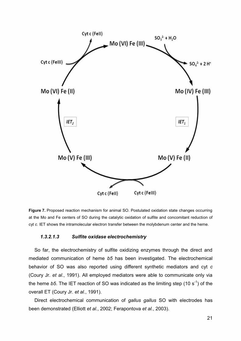
MoVI/FeIII (Figure 7).
Page 37
21
Figure 7. Proposed reaction mechanism for animal SO. Postulated oxidation state changes occurring
at the Mo and Fe centers of SO during the catalytic oxidation of sulfite and concomitant reduction of
cyt c. IET shows the intramolecular electron transfer between the molybdenum center and the heme.
1.3.2.1.3 Sulfite oxidase electrochemistry
So far, the electrochemistry of sulfite oxidizing enzymes through the direct and
mediated communication of heme b5 has been investigated. The electrochemical
behavior of SO was also reported using different synthetic mediators and cyt c
(Coury Jr. et al., 1991). All employed mediators were able to communicate only via
the heme b5. The IET reaction of SO was indicated as the limiting step (10 s-1) of the
overall ET (Coury Jr. et al., 1991).
Direct electrochemical communication of gallus gallus SO with electrodes has
been demonstrated (Elliott et al., 2002; Ferapontova et al., 2003).
Page 38
22
After immobilization on pyrolytic graphite or gold electrode modified with mercapto-
6-hexanol self-assembled monolayer the enzyme can partially retain its activity and a
single pair of peaks were observed with a formal potential of + 90 mV (vs. standard
hydrogen electrode, SHE). Upon addition of sulfite to the cell solution, the
voltammogram changed to an oxidative catalytic wave. However, the immobilized SO
showed a decreased catalytic turnover rate (2-4 s-1) in comparison to SO in solution
with cyt c (100 s-1) (Elliott et al., 2002). This behavior was attributed to the small
fraction of cSO molecules in with an appropriate conformation electrons exchange of
the HD with the electrode surface.
A faster heterogeneous electron transfer of cSO immobilized was reached on
aminated surface mimicking the natural partner of the enzyme (cyt c). Using gold
surface modified with 11-mercapto-1-undecanol and 11-mercapto-1-undecanamine
the ket was in the order of 15 s-1, whereas the catalytic turnover rate was comparable
to the reaction in solution with the natural electron acceptor cyt c (Ferapontova et al.,
2003). The E0 was to −120 mV versus Ag/AgCl (KClsat), correlating fairly well with the
reported redox potential for the heme Fe(III/II) couple in SO obtained through optical
redox titration (Spence et al., 1991; Ferapontova et al., 2004).
The E0 for MoVI/V and MoV/IV interpolated at pH 8 were +6 mV and -184 mV
(vs. NHE), hence the half reduced state of the active site (MoV) is stable (Spence et
al., 1991). These data are in agreement with the IET rates determined at pH 8 using
flash photolysis on the same enzyme (Sullivan Jr. et al., 1993).
CSO has been employed for the constitution of first (Situmorang et al., 1999) and
second (Abass, 2000; Svitel et al., 1998) generation of sulfite biosensors.
Only recently the research is addressing the human variant of the sulfite oxidase
enzyme. Three different approaches for the fabrication of a sulfite biosensing system
have been proposed. In one case the natural partner of the hSO was employed for
the electrochemical mediation with the electrode. Complex protein architectures
containing hSO and cyt c were construct by the layer-by-layer technique (Dronov et
al., 2008) as well as alternated by an anionic polyelectrolyte (Spricigo et al., 2008)
(Spricigo et al., 2009). It resulted in an enhanced sensitivity towards the sulfite
concentration. The system showed appreciable stability only at very low ionic
strength.
Page 39
23
Figure 8. Schematic view of proposed conformational change by electrostatic orientation of
immobilized animal SO on a positive charged electrode during the catalytic oxidation of sulfite surface.
Only a single subunit schematic representation of SO is portrayed. Modified after Sezer et al. 2010.
The direct electrochemical communication of the HD was reported on amino
functionalized silver electrode, with the possibility to work at high ionic strength.
These investigations were complemented by a spectroelectrochemical study using
surface enhanced resonance Raman spectroscopy (SERRS) following the Raman
scattering variation of the heme contained in the hSO. Finally molecular modeling
suggested that a high ionic strength increases the mobility of the HD while the
enzyme is immobilized via the dimerization domain to the SAM surface. The flexible
loop connecting the HD allows alternating contact with the MD and the electrode
surface, thereby promoting the intramolecular and heterogeneous electron transfer
(Sezer et al., 2010).
In the third case either the heme center or the molybdenum center were wired by
mean of an osmium polymer and a direct electrochemical communication of the
isolated MD with electrocatalysis was showed (Spricigo, 2009; Spricigo et al., 2010).
Similarly sulfite dehydrogenase, from Starkeya novella, has been also
characterized electrochemically. Bacterial SDH is a heterodimer of a Moco containing
domain and a heme c containing domain and shows some characteristics of
eukaryotic SO, such as the inhibition by small anions. The voltammetric response of
the Mo center of the bacterial SDH in the presence and absence of substrate was
Page 40
24
published (Aguey-Zinsou et al., 2003; Rapson et al., 2008). For this investigation, an
edge-plane pyrolytic graphite electrode surface was modified with surfactants (DDAB
and polylysine) and signals from the single redox sites were recorded. More recently
SDH has been employed, in combination with cyt c in the construction of a second
generation sulfite biosensor (Kalimuthu et al., 2010) in analogy to the system
reported by Spricigo (Spricigo et al., 2008; Spricigo et al., 2009).
1.3.2.2 Xanthine oxidoreductase enzymes
Enzymes catalyzing the oxidation of hypoxanthine and xanthine, named xanthine
oxidoreductases, are widespread in nature and have been isolated from a wide range
of organisms, from bacteria to man. In terms of their electron acceptor, they fall into
two broad groups by using preferentially molecular oxygen (the oxidases) or NAD+
(the dehydrogenases) (Hille et al., 1995).
Xanthine oxidoreductase (XOR) was described already in 1902 as the substance
in milk which could decolorize methylene blue and it was identified originally as
aldehyde oxidase (Schardinger, 1902). The mammalian enzymes, are synthesized as
the dehydrogenase form xanthine dehydrogenase and exist mostly as such in the cell
but can undergo a dehydrogenase to oxidase conversion (XDH→XOD) by proteolysis
or by oxidation of sulfhydryl residues. One of the major difference between XDH and
XOD is the large formation of the blue neutral semiquinone observed during reductive
titrations of XDH, very little semiquinone is seen during XO reductions (Massey et al.,
1979), indicating a large difference in redox potential between the two enzyme forms.
The enzyme is a target of drugs against gout and hyperuricemia (Elion, 1989), and
the conversion of XDH to XO is of major medical interest as it has been implicated in
diseases characterized by oxygen-radical-induced tissue damage, such as
postischemic reperfusion injury (Richard et al., 1990). XO also might be associated
with blood pressure regulation (Suzuki et al., 1998; Harrison, 2002).
An interesting prokaryotic XDH with similar activity to the mammalian enzymes
was isolated from the phototrophic purple bacterium Rhodobacter capsulatus
(Leimkühler et al., 1998). Despite the similarities to the mammalian XOR enzymes,
the R. capsulatus XDH is isolated with high reactivity toward NAD+ and low reactivity
toward oxygen as electron acceptor and does not undergo the conversion to the
oxidase form.
Page 41
25
1.3.2.2.1 Xanthine dehydrogenas structure
The solved crystal structure of R. capsulatus XDH (Truglio et al., 2002) shows that
the bacterial and bovine XDH (Enroth et al., 2000) have highly similar folds despite
differences in subunit composition. However, the two structures differ in important
details, including the regions necessary for XDH to XOD conversion. Because of the
high structural similarities of the mammalian XOR and R. capsulatus XDH, the
bacterial enzyme is a good model system for studying the mechanism of the enzyme
and can be used for the generation of site-specific mutants.
Figure 9. (Upper) Butterfly structure of R. capsulatus XDH. The Moco, the two [2Fe-2S] and the FAD
cofactor are shown as space-filling models. (down) Alignment of the four cofactors in one single
heterodimer, the catalytic reactions in their respective places, the distances between cofactors and the
cysteines bound at the iron atom are also depicted (Truglio et al., 2002).
The enzyme is a butterfly-shaped (αβ)2 heterotetramer of molecular mass around
300 kDa, with each catalytically independent subunit possessing four prosthetic
groups. One molybdenum active site is located at the bottom of a 15 Å hydrophobic
channel, two spectroscopically different [2Fe-2S] iron-sulfur centers one of which a
type I cluster buried ca. 12 Å below the protein surface and the other a type II
Page 42
26
[2Fe-2S] cluster solvent exposed, and one FAD. The oxidation of xanthine takes
place at the Moco. The two reducing equivalents generated in the course of the
reaction are transferred to an external electron acceptor and the electron flow in and
out of the molybdenum centre is mediated by the pterin cofactor to the closest FeSI
centre and then via the FeSII to the FAD. Reoxidation of the reduced enzyme by
oxidant substrate occurs through FAD (Olson et al., 1974).
1.3.2.2.2 Electrochemistry of xanthine oxidoreductases
XOR from cow milk is the most electrochemical investigated protein of all
mononuclear molybdoenzymes. XOR is widely studied for the detection of xanthine
or hypoxanthine, mainly via detection of the products uric acid, hydrogen peroxide
and superoxide (Sternson, 1976; Kirgöz et al., 2004; Nakatani et al., 2005; Dronov et
al., 2007; Çubukçu et al., 2007; Shan et al., 2009). Only few groups showed a direct
electrochemistry of this enzyme, although often with confusing results. The protein
was studied upon immobilization on a number of electrode materials. First was
adsorbed on glassy carbon (GC) and mercury electrode (Rodrigues et al., 1991),
where evidence of protein denaturation was shown. In recent years XOR was
immobilized on gold (Wang et al., 2004), glassy carbon electrode modified with
single-wall carbon nanotubes (Wu et al., 2007), co-immobilized with nanocrystalline
TiO2 (Zhou et al., 2006) or between two layers of didodecyl-dimethylammonium
bromide (DDAB) (Bernhardt et al., 2006) on pyrolytic graphite (PG) electrode, with
the help of DNA as matrix to embed xanthine oxidase (Liu et al., 2005) and more
recently immobilized on glassy carbon electrode (GCE) by laponite nanoparticles
(Shan et al., 2009). In all cases the DET of the FAD was detectable. Rarely the DET
of the Molybdenum center was reported and only Wu et al. was able to show it for
one of the FeS.
In these publications direct catalytic current of the natural substrates was detected
at potentials 0.6-0.8 V higher than the more positive cofactor potential. In addition
unusual electrocatalytic activity towards nitric oxide and nitrate has been reported.
The discovery of new proteins, belonging to the xanthine oxidase family, led to an
increasing interest to develop methods for the determination of the reduction
potential of the cofactors and to achieve a direct electrochemical communication with
an electrode for a future developing a third generation biosensors exploiting the
selectivity of the enzyme. The XDH from R. capsulatus was studied on freshly
Page 43
27
cleaved PG electrode. It also showed extreme overpotential in the catalytic activity.
This rare phenomenon was first explained as due to an inactive opened pyranopterin
(Aguey-Zinsou et al., 2003) and later as an autocatalytic process associated to the
reaction product uric acid (Kalimuthu et al., 2011). Correira dos Santos published a
direct electrochemistry of an aldehyde oxidoreductase from Desulfovibrio gigas
immobilized on PG and GC electrodes (Correia dos Santos et al., 2004). This
member of the xanthine oxidase family is a FAD-indipendent enzyme and showed
DET of the FeS II at carbon electrodes (PG and GC) and DET of the Moco at gold
electrode in precense of neomycin.
All the published DET potentials are summarized in the table 2 in the chapter 5.
Page 44
28
2 Aim of the work
The aim of this work consists in the electrochemical and optical studies of heme-
and molybdenum-containing enzymes in respect to their interaction with diverse
nanostructured materials. Spectro- and electrochemical methods shall be applied in
order to implement new materials for electrochemical and catalytic studies and for the
development of biosensing systems. With these methods also complex redox
enzymes shall be investigated.
For this purpose, high transparent mesoporous metal oxides films and
nanoparticles shall be employed as support for the enzyme adsorption. Out of this
investigation suitable combinations of different protein and nanostructured materials
should be developed to obtain an electrochemical and optical detection systems for
several small molecules, like superoxide, hydrogen peroxide and sulfite. Finally the
investigation shall be extended to direct and mediated spectroelectrochemical studies
of the behavior of proteins with high structure complexity such as xanthine
dehydrogenase from Rhodobacter capsulatus and its homologous the mouse
aldehyde oxidase homolog 1.
Page 45
29
3 Materials and Methods
3.1 Materials
3.1.1 Chemicals
Horse heart cytochrome c, used without further purification, H2O2 (30% solution in
water), N-ethyl-N’-(3-dimethylaminopropyl)carbodiimide hydrochloride (EDC), poly-L-
lysine, hypoxanthine and 3, 3’, 5, 5’-tetramethylbenzidine (TMB) were purchased
from Sigma (Steinheim, Germany). 11-mercapto-1-undecanoic acid (MUA), 11-
mercapto-1-undecanol (MU) and HAuCl4 were provided by Aldrich (Taufkirchen,
Germany). Acetic acid and toluene (99%) were purchased from Fluka (Schnelldorf,
Germany). Potassium dihydrogen phosphate, dipotassium hydrogen phosphate,
disodium sulfite, sodium sulfate, ferrocene, sodium chloride, sodium phosphate,
phosphoric acid, potassium chloride, 1-butyl-3-methylimidazolium octylsulfate
(bmimOctOSO3) (99%), 1-ethyl-3-methylimidazolium ethylsulfate (emimEtOSO3)
(99%) and ferrocene were purchased from Merck (Darmstadt, Germany). Potassium
hexacyanoferrate (III) and 4-(2-hydroxyethyl)-1-piperazineethanesulfonic acid
(Hepes) were purchased from Roth (Karlsruhe, Germany). Xanthine oxidase from
cow’s mil (XOD, 5.87 U ml-1) and superoxide dismutase (SOD, 4890 U mg-1) were
provided by ICN Biomedicals Inc. (Aurora, USA). Branched poly(ethyleneimine) with
a molecular weight of Mn= 5000 g/mol was obtained from BASF.
[Co(transdiammac)]ClO4)3, [Co(cis-diammac)](ClO4)3, [Co(AMMEsar)]Cl3,
[Co(sep)]Cl3, [Co(AMME-N5S-sar)]Cl3, [Co(CLME-N4S2-sar)]Cl3, [Co((NMe3)2sar)]Cl5,
[Fe(tacn)2]Br3 and Fe(NOTA) (Bernhardt et al., 2006) were a kind gift of Prof. Paul V.
Bernhardt from Queensland University, Brisbane (Australia). XDH an hSO were
expressed and purified in the Prof. Silke Leimkühler’s laboratories as described in
(Leimkühler et al., 2003) and in (Temple et al., 2000) respectively. Other reagents
were of analytical reagent grade and used as received. Goodfellow (Bad Nauheim,
Germany) supplied gold and platinum wires with a diameter of 0.5 mm. All the
solutions were prepared in 18 MΩ Millipore water (Millipore, Eschborn, Germany).
Page 46
30
3.1.2 Instruments
The electrochemical measurements for hSO at AuNP and for XDH at EP-GCE
were performed in a home-made three-electrode electrochemical cell with a total
volume of 1 mL employing a platinum wire as the counter electrode, an Ag/AgCl
(1M KCl) reference electrode. The working electrode can be a gold wire or a glassy
carbon electrode (GCE). Cyclic voltammetric, square wave and amperometric
experiments were performed with PalmSens potentiostat and analyzed with PSLite
1.8 software, or with Gamry Reference 600TM potentiostat (Gamry, USA) and
analyzed with Gamry Echem Analyst 5.50 software.
Figure 10. Electrochemical cell 1 in with (bottom left) a gold wire and (bottom right) glassy carbon
working electrode.
Page 47
31
The electrochemical measurements for the coated glass slide were performed in a
home-made three-electrode electrochemical cell with a total volume of 1 mL
employing a platinum wire as the counter electrode and an Ag/AgCl (1M KCl)
reference electrode. A circular surface area 5.5 mm in diameter of the coated glass
slide was the working electrode. Cyclic voltammetry (CV) experiments were carried
out with a Gamry Reference 600TM potentiostat (Gamry, USA) and analyzed with
Gamry Echem Analyst 5.50 software. All scans were started in the negative potential
range. The potentiostat for the amperometrical experiments was a CH Instrument
Model 750 A (Austin, USA).
Figure 11. Electrochemical cell 2.
Page 48
32
For the stationary spectroelectrochemical experiments a home-made
spectroelectrochemical cell with a total volume of 1 mL employing a platinum wire as
the counter electrode, an Ag/AgCl (1M KCl) reference electrode, and the coated
glass as working electrode with a circular surface area 5.5 mm in diameter were
employed. The electrochemical cell equipped with a quartz window was incorporated
into the sample compartment of a Beckman DU 640 spectrophotometer whereas the
applied potential was controlled by a Gamry Reference 600TM potentiostat (Gamry,
USA). The transmission spectra were recorded through the coated glass slide.
Figure 12. Spectroelectrochemical cell for stationary experiments.
Page 49
33
For flow experiments a spectroelectrochemical flow cell has been constructed
using an outflow stainless tube as the counter electrode, and an Ag/AgCl (3 M KCl)
reference electrode. A coated glass slide was the working electrode. The cell was
equipped with SEC-F 1000 μm probe type optical fiber (ALS, Japan) and UV-vis high
OH content optical fiber of core diameter 400 μm (Ocean Optics, Germany). The
spectra were measured with a SEC2000-Spectrometer (ALS, Japan). The
transmission spectra were recorded through the coated glass slide and the quartz
glass.
Figure 13. Spectroelectrochemical cell for flow experiments.
Page 50
34
The spectroelectrochemical mediated titration was performed under an
atmosphere of N2 at room temperature in a 2 mL cuvette. Potentials were measured
using a combination Pt wire and Ag/AgCl reference electrode calibrated against a
pH 7 quinhydrone solution (E0 = +284 mV vs NHE at pH7). The redox potential was
adjusted by addition of microliter aliquots of TiIII citrate and K2S2O8. The solution was
constantly stirred. The spectra were measured with a USB4000-UV-VIS Miniature
Fiber Optic Spectrometer (Ocean Optics).
Figure 14. Cuvette arrangement for mediated spectroelectrochemical titration.
Page 51
35
3.1.3 Buffers
3.1.3.1 Spectroelectrochemical study of mpITO and mpITBO
5 mM potassium or sodium phosphate pH 7.0 was used for the immobilization and
UV-vis and Raman spectroelectrochemical study of cyt c, TMB and HD with mpITO
and mpITBOs electrodes. 50 mM potassium phosphate pH 7.0 was used for the
electrochemical study of Ru(NH3)62+/3+. 100 mM sodium phosphate pH 7.0, 0.1 M KCl
was employed for the spectroelectrochemical study of free TMB in solution with a
cyt c modified mpITO.
3.1.3.2 Spectroelectrochemical study of XDH and mAOH1
50 mM Tris buffer pH 8.0 was employed for the electrochemical study of the
immobilized XDH variants and mAOH1 on GC and for the spectroelectrochemical
mediated titration of XDHwt.
3.1.3.3 Electrochemical study of hSO using gold nanoparticles
5 mM and 750 mM Tris buffer pH 8.4, with the pH adjustment using acetic acid,
were employed for the electrochemical and surface enhanced Raman studies of hSO
on AuNPs.
3.2 Methods
3.2.1 Molar extinction coefficients
The surface coverage of the mpITO was determined spectroscopically based on
the extinction coefficients (ε) of 106100M-1 cm-1 (410 nm) and 129100m -1 cm-1
(416 nm) of ferric and ferrous cyt c, respectively (Topoglidis et al., 1998; Collinson et
al., 1992).
The concentration of hSO and HD were quantified by using the molar extinction
coefficient of 113000 M-1 cm-1 at 413 nm, while for XDH variant the molar extinction
coefficient was of 31600 M-1 cm-1 at 465 nm.
Page 52
36
3.2.2 Spectroelectrochemical study of mesoporous materials
3.2.2.1 Preparation of mpITO
The mesoporous indium tin oxide (mpITO) films were prepared in collaboration
with Dr. Till von Graberg and, Prof. Bernd M. Smarsly from the Justus Liebig
Univesität Gießen (Germany).
MpITO-coated borosilicate glass or graphite slides were prepared as reported
elsewhere (Fattakhova-Rohlfing et al., 2006; von Graberg et al., 2011). In brief, a thin
film of mpITO was obtained by dip-coating the substrate slide in an acetone/methanol
solution containing InIII acetylacetonate, SnCl4, and PIB3000 in a controlled
atmosphere (18–20 % relative humidity) and at constant withdrawing rate.
Subsequent calcination was performed at low heating rate in air at 300 °C,
450-500 °C and finally in forming gas (mixture of N2/H2 90:10) at 300 °C. This
procedure generated a film of crystalline indium oxide (cubic bixbyite) with a fine
dispersion of Sn ions in the In2O3 lattice (Figure 15). The pore sizes were in the
range 15–20 nm and pore walls were 5–10 nm in thickness (Figure 15). The total
surface area is 410 m2 cm−3, as determined by krypton adsorption (for films with a
thickness of 170 nm).
30 35 40 45 50
*
*
(400) (440)
Inte
ns
ity
/ a
.u.
PIB3000 templated
In2O
3
* Si substrate(222)
2 / degree
Figure 15. (left) Wide angle X-ray scattering data of ITO film templated with PIB3000 and calcined at
500 °C (red curve); the black columns show reference of indium oxide (cubic bixbyite) according to the
Joint Committee for Powder Diffraction Studies (JCPDS). (right) Scanning transmission electron
microscopy image of calcined mesoporous ITO film templated with PIB3000. (Frasca et al., 2010)
Page 53
37
3.2.2.2 Preparation of mpITBO
The mpITBO was prepared by Dr. Yilmaz Aksu, Prof. Matthias Driess, and Prof.
Arne Thomas from the Technische Universität Berlin (Germany).
ITBO was prepared and characterized according to published procedures (Aksu et
al., 2009). Blends of ITBO with block-co-polymer, KLE or F127, were prepared by
dissolving them in dry tetrahydrofurane (KLE) or dry toluene (F127) at ambient
temperature and under nitrogen atmosphere. In a typical procedure, a KLE-ITBO
solution was prepared by mixing a KLE (155 mg) solution in tetrahydrofurane (1 mL)
with the appropriate amount of ITBO (620 mg) in tetrahydrofurane (1 mL) and stirred
for 1 h at room temperature. The glass substrates were cleaned in an ultrasonic
cleaner for 10 min with acetone and isopropanol. The substrates were dried in a high-
purity nitrogen gas stream just before use. To increase the compatibility of the
substrate with the ITBO-polymer mixture and ameliorate the final film quality, the
glass substrates were first coated with a solution of pure ITBO in toluene. Tin-rich
ITO-polymer films were fabricated by spin coating the solutions of the corresponding
ITBO-polymer mixtures under nitrogen atmosphere in a glove-box (<1 ppm H2O and
<1 ppm O2). The coatings were aged for 30 min in nitrogen and then annealed for 2 h
in air at different temperatures between 300 and 600 °C (ramp 5 °C min-1). After heat
treatment all samples were subsequently annealed at 300 °C in a reductive gas
mixture (H2/N2:10%/90%, ramp 5 °C min-1) for 90 min.
Figure 16. Transmission electron microscopy of mesoporous tin-rich ITO (ITBO) films templated with
block-co-polymers (left) KLE and (right) F127. (Aksu et al., 2011)
This approach leads to highly conductive, homogeneous, and compact thin films,
of amorphous ITBO with different diameter size: F127 with an average pore diameter
Page 54
38
of 6.5 nm and KLE with 12.3 nm (Figure 16). The approximate layer thickness,
estimated by SEM, was about 750 nm for the ITBO−KLE and 1.5 μm for ITBO−F127
system on average (Figure 17). Assuming that the thickness of the films is uniform
the resistivity was calculated in the order of 6.390 × 10⁻³ Ω-cm (ITBO−KLE) and
1.725 × 10⁻² Ω-cm (ITBO−F127).
The films show 70−75% optical transmission in the visible range (Figure 18). The
durability of the electronic properties of the mesoporous tin-rich ITO films is
considerably higher in comparison to those prepared from commercially available
ITO. No significant change of the conductivity was observed after 3 months in air at
room temperature.
Figure 17. Cross-sectional SEM images of mesoporous, tin-rich ITO prepared with the molecular
single-source precursor ITBO, using 20 wt % (a, b) F127 and (c, d) KLE as templates, respectively.
(Aksu et al., 2011)
Figure 18. Optical transmission of mesoporous ITBO films in the UV-Vis region. (Aksu et al., 2011)
Page 55
39
3.2.2.3 Preparation of plATO
ATO films were prepared by Dr. Fernando Pérez Caballero and Dr. Anna Fischer
from the Technische Universität Berlin (Germany).
The films were prepared by dip-coating a colloidal suspension of ATO
nanoparticles on ITO-coated glass. Generally, 160 mg of ATO nanoparticles were
dispersed in 2 mL of THF and drops of concentrated HCl solution were added until
the solution turn to transparent. When needed, further drops of THF or Ethanol were
added in order to reach a full transparency of the solution. The speed of coating was
varied between 150 and 300 mm min-1, the relative humidity was set to 25% (± 5%)
and the temperature to 25ºC (± 2ºC). The films were stored at 80 ºC for 24 h and
then calcined to sinter the ATO particles leading to an increase in conductivity. The
films were calcined with an increasing heating (0.6 ºC min-1) and finally kept at
450 ºC for 30 min.
3.2.2.4 Electrode loading
For mpITO-coated borosilicate glass and mpITO-graphite, the slides were
pretreated by immersion for three hours in a stirred solution of 1 % Alconox in water
and in ethanol (96 %), and subsequently, three times immersion in Milli-Q water for
30 min. The system was equilibrated over night with 5 mM potassium phosphate
buffer solution pH 7.0.
In case of mpITBO, the slide pretreatment was avoided and the material was used
as received after 1h equilibration in the measuring buffer.
Immobilization of cyt c was achieved dipping the slides into a protein solution
(10 μM, unless otherwise reported) 5 mM potassium phosphate buffer solution pH 7.0
for 30 min at 4 °C.
For spectroelectrochemical experiments of the hSO heme domain (HD), the
protein immobilization was preceded by an equilibrated overnight of a clean mpITO in
a poly-lysine solution 2 mg/ml in sodium phosphate buffer (5 mM, pH 8.0).
Immobilization of HD was achieved dipping the mpITO slide into a protein solution
(3.2 μM) in sodium phosphate buffer solution (5 mM, pH 7.0) for 3 h at 4 °C.
Prior to all the experiments, the supernatant protein-containing solution was
removed, the cell was rinsed and stirred for 1 min in a fresh buffer solution. The
modified electrode was stored in a low buffer solution at 4 °C until use.
Page 56
40
TMB was adsorbed within the porous film by soaking the electrode in 1 mM TMB
solution and after 30 minutes equilibration the resulting electrode was washed
thoroughly and investigated in a TMB-free phosphate buffer 5mM pH 7.0. The same
procedure was used also when the mpITO was already modified with cyt c.
3.2.2.5 Electrochemistry
Electron transfer rate constant, and the heterogeneous electrochemical transfer
coefficient α were determined from CV pea separation using Laviron’s model
(Laviron, 1979) in case of immobilized species and the method of Nicholson in case
of species free in solution (Nicholson, 1965).
The electroactive surface coverage was determined from the mean value of the
reduction and oxidation peak areas of a background-subtracted cyclic
voltammogram.
For superoxide detection a constant potential of +150 mV against the Ag/AgCl
(1 M KCl) reference electrode was applied to the wor ing electrode, and the analyte
was generated successively by addition of hypoxanthine in a XOD solution.
3.2.2.6 Resonance Raman measurements
The resonance Raman measurements were performed in collaboration with Dr.
Jou-Ju Feng, Dr. Inez M. Weidinger and Prof. Peter Hildebrandt from the Technische
Universität Berlin (Germany).
Potential-dependent resonance Raman (RR) measurements were carried out with
a home-made electrochemical cell using a cylindrical mpITO-covered graphite slide
(12 mm diameter) as the wor ing electrode and an Ag/AgCl (3 M KCl) reference
electrode. RR spectra were recorded with 413 nm excitation using a confocal Raman
microscope (LabRam HR-800, Jobin Yvon). The laser beam was focused onto the
surface of the working electrode with a long working distance objective
(20× magnification; numerical aperture 0.35). The RR spectra were acquired with a
laser power of 3.5 mW, a spectral resolution of 2 cm−1, and an increment per data
point of 0.57 cm−1. The accumulation time for each spectrum was 30 s. Further
details of the setup are given elsewhere (Wackerbarth et al., 1999; Feng et al., 2008).
Page 57
41
3.2.3 Electrochemical study of hSO on AuNPs
3.2.3.1 Synthesis of gold nanoparticles
The gold nanoparticles were prepared in collaboration with Oscar Rojas and
Prof. Joachim Koetz from the Potsdam Universität (Germany).
Polyethyleneimine (0.5 wt. %) and the tetrachloroaurate of 2 mM were mixed with
1:1 ratio (wt/wt) at room temperature in an ionic liquid emim EtSO4 solvent and
heated up to 100 °C for 5 min. The change in colour from yellow to dark red
indicated the formation of gold nanoparticles. They show an UV-vis absorption
maximum at 529 nm, due to the surface plasmon resonance, confirming the
formation of gold particles of small dimension (Figure 19). The AuNPs were dissolved
in Tris buffer 0.5 mM pH 7.0 and the hydrodynamic diameter was determined by
dynamic light scattering (DLS). Measurements were carried out at 25 °C at a fixed
angle of 173° (“bac scattering detection”) by using a Nano Zetasizer (Malvern)
equipped with a He-Ne laser (λ= 633nm; 4 mW) and a digital autocorrelator. The
refractive indices and viscosities of solvents were adapted to the respective
measurements.
Figure 19. UV-vis spectra indicating the surface plasmon resonance of gold nanoparticles grown in 1-
ethyl-3-methylmidazolium ethylsulfate at 100 °C. The grey line indicate the absorption of starting
reaction solution, the black line indicate the same solution after 5 min. at 100 °C.
Transmission electron microscopy (TEM) micrographs of the nanoparticles were
recorded on an EM 902 microscope from Zeiss. The nanoparticles sample was
dissolved in chloroform and prepared by dropping a small amount of the solution, on
copper grids, dried and examined in the transmission electron microscope at an
Page 58
42
acceleration voltage of 90 kV. The zeta potential was determined by means of the
Nano Zetasizer (Malvern) based on the electrophoresis principle which considers the
motion of charged particles by applying an electric field.
Figure 20. (Top) Electron transmission micrograph of gold nanoparticles synthesized in the ionic liquid
1-ethyl-3-methylimidazolium n-ethylsulfate; (bottom left) Histogram showing the particle number
density NAuNPs in the size interval Δd of 1 nm as function of the particle diameter d/nm. The mean
diameter was 6 ± 2 nm; (bottom right) Number distribution NAuNPs/%, as function of the particle
diameter d/nm measured by dynamic light scattering (DLS).
TEM micrograph shows spherical nanoparticles with an average core diameter of
about 6 ± 2 nm whereas the hydrodynamic diameter determined by dynamic light
scattering (DLS) was 9 ± 1 nm (Figure 20). As TEM images show only the gold core,
one can conclude from the hydrodynamic diameter that the gold core is surrounded
Page 59
43
by a coating layer of around 2-3 nm. The AuNPs present a positive zeta potential
around +20 mV as a result of the cationic polymer coating.
3.2.3.2 Electrode modification
A gold wire (Goodfellow, Bad Nauheim, Germany) with a diameter of 0.5 mm was
cleaned by boiling 4 h in 2 M KOH and kept 10 minutes in concentrated HNO3. A
careful rinsing with Millipore water followed every successive step. The electrodes
were stored in concentrated H2SO4 when not in use. The cleaned electrodes were
incubated in an ethanol mixture of 5 mM MUA and 5 mM MU with a volume ratio of
1:3 for at least 24 h at 4 °C. After rinsing with water, the MU/MUA modified
electrodes were incubated in a freshly prepared emim EtSO4 solution of AuNPs for
2 h at room temperature. Further, 5 µL of EDC 0.2 M in H2O was then added into the
solution in order to couple amino functions of the AuNP cap to the carboxylic acid
functionalized SAM gold electrode. 20 minutes were used as coupling time. The
resulted assembled film {Au/MUA-MU/AuNPs-PEI} was characterized by high
resolution scanning electron microscopy (S-4800 microscope, Hitachi, equipped with
a field emission gun).
Figure 21. Scanning electron micrograph of the modified gold electrode Au/MUA-MU/AuNPs-PEI.
The employed immobilization method leads finely to disperse single gold particles
in the size order between 5 and 10 nm, well resolved in the SEM images (Figure 21).
Note that the particles deposited on the electrode surface show no evidence of
aggregation after solvent evaporation. The particles are in the same size order
Page 60
44
measured by dynamic light scattering and by TEM. The AuNPs are clearly separated
from each other forming an accessible surface for protein immobilization.
3.2.3.3 Surface enhanced resonance Raman spectroelectrochemistry
The surface enhanced resonance Raman measurements were performed in
collaboration with Johannes Salewski, Dr. Inez M. Weidinger and Prof. Peter
Hildebrandt from the Technische Universität Berlin (Germany).
For surface enhanced resonance Raman (SERR) spectroscopic measurements
of HD the 413 nm excitation line of a Krypton ion laser and a konfocal Raman
spectrometer (Labram, HR-800, Jobin Yvon) were used. Spectro-electrochemistry
was performed with a homemade electrochemical cell with a volume of ca. 10 mL.
The cell contained an electrochemically roughened and functionalised Ag ring as
working electrode, a 3 M KCl Ag/AgCl reference electrode and a platinum counter
electrode. The laser power on the sample was 1 mW. SERR spectra were
obtained with an accumulation time of 30 s. The working electrode was constantly
rotated to avoid laser induced protein degradation. More details about the setup
are given elsewhere (Wackerbarth et al., 1999).
3.2.4 XDH spectroelectrochemical study
3.2.4.1 Electrode Pretreatment
Electrochemically pretreated glassy carbon (EP-GC) electrode was prepared as
follow. A GC surface was polished to mirror finish using 1 and 0.3 µm alumina slurry
for 3 and 5 minutes respectively. The electrode was sonicated in Milli-Q water, after
every step for the respective polishing time. After rinsed with Milli-Q H2O thoroughly,
a constant positive potential was applied (1.8 V) under stirring in air saturated
potassium phosphate (0.1 M) solution at pH 4. In a fresh solution where then
electrochemically cycled between 0.3 and 1.25 V till a steady state was reached and
stopped in the oxidation region.
3.2.4.2 Electrode modification
4 µl of protein with a concentration between 40 and 90 µM in its own purification
buffer (Tris 50mM pH 8, NaCl 200mM, EDTA 1mM), were dropped onto the freshly
Page 61
45
prepared EP-GC electrode. The electrode was covered to ensure the water to
evaporate slowly, and equilibrated 30 min. at 4 °C.
The protein immobilization was negligibly affected whether the cyclic scan in the
pretreatment was stopped in the oxidation or reduction region.
3.2.4.3 Electrochemical measurement
The electrochemical experiments were carried out in the electrochemical cell 1
(see section 3.1.2), employing a platinum wire as counter electrode, a Ag/AgCl,
1M KCl reference against which all potentials are reported, and a modified glassy
carbon disk electrode. All the experiments where performed at room temperature in a
oxygen-free 2% H2 atmosphere with a Gamry Reference 600TM potentiostat
(Gamry, USA) and analyzed with GamryEchem Analyst 5.50 software.
The working electrode surface area was determined with a reversible system
(K3Fe(CN)6 in 1M KCl aqueous solution), employing the Randles-Sevcik equation:
(3)
2/12/12/351069.2 CADnxip
where D is the diffusion coefficient of Fe(CN)6+3/+2 (0.76 x10⁻⁵ cm2 s-1), n indicates
the exchanged electrons, C the K3Fe(CN)6 concentration (4mM) and ν is the
scanning rate.
3.2.4.4 Spectroelectrochemical mediated titration
All experiments were performed under N2 atmosphere at room temperature.
Potentials were measured using a combination Pt wire and Ag/AgCl reference
electrode calibrated against a pH 7 quinhydrone solution (E0 = +284 mV vs NHE at
pH 7). The redox potential was adjusted by addition of microliter aliquots of TiIII citrate
and K2S2O8. The FAD redox potentials were measured using ca. 15 μM enzyme in
50 mM Tris buffer (pH 8.0) in a 2 ml cuvette under continuous stirring. The solution
potentials were stabilized using the Fe and Co complexes (5 μM each) as mediators:
[Co(trans-diammac)]ClO4)3 (E0= -555 mV vs. NHE), [Co(cis-diammac)](ClO4)3
(-503 mV), [Co(AMMEsar)]Cl3 (-380 mV), [Co(sep)]Cl3 (-296 mV),
[Co(AMMEN5Ssar)]Cl3 (-220 mV), [Co(CLME-N4S2sar)]Cl3 (-134 mV),
[Co((NMe3)2sar)]Cl5 (+10 mV), [Fe(tacn)2]Br3 (+144 mV) and Fe(NOTA) (+195 mV)
(Bernhardt et al., 2006). Changes in the UV-Vis spectrum were monitored
Page 62
46
continuously with an Ocean Optics USB4000 fiber optic spectrometer. Equilibrium
was established when no further change in either the optical absorbance or solution
potential was detectable. The formal potential for the first electron of FAD reduction
(E1) was determined at 465 nm using the equation:
(4)
At the same time the potential of both electron transfer (E1 and E2) were
determined at 465 nm using the equation:
(5)
In the above equations E is the potential, A is the absorption, coxεox and coxεox are
the limiting absorbance values of the XDH at 293 K in its oxidized and reduced forms
respectively.
Page 63
47
4 Results and Discussion
4.1 Mesoporous materials
On planar surfaces only a limited amount of protein can be adsorbed. Therefore
despite the advantage of the high transparency of transparent conductive materials
the amount of the immobilized protein is close to that of a monolayer and requires
high sensitive techniques to be optically detected. For this purpose three
dimensionally structured electrodes are presented in this work. For the investigation
of such materials different kind of static and microfluidic (spectro)electrochemical
cells were designed, constructed and optimized (see section 3.1.2).
4.1.1 Direct electron transfer of cyt c in mpITO
Equilibration of a mpITO electrode in an aqueous solution of cyt c solution results
in a quick and efficient surface interaction of the protein, as revealed by the cyclic
voltammetry (CV), which displayed a reversible response up to scan rates as high as
1 V s−1. Only a minimal decrease was obtained after the cyt c solution has been
exchanged by a protein-free 5 mM phosphate buffer (pH 7.0), demonstrating the
strong binding of cyt c in the mesoporous electrode (Figure 22). The pair of
voltammetric peaks is almost symmetric and the currents increase linearly with the
square root of the scan rate, which gives evidence for a reversible,
diffusion-controlled redox process in the pores (Figure 22, inset) (Laviron, 1979).
The CV shows the characteristic full width at half-maximum for a one-electron-
exchange process of around 90 mV, at a scan rate of 50 mV s−1. However, the peak
broadens at higher scan rates. In the absence of cyt c in the pores no peak current
was observed. The CV signals were stable and unchanged for more than a week
when the cyt c-loaded electrodes were ept in buffer solution at 4 °C. For the cyt c in
the pores, the formal potential (E0) was determined to be 9 ± 1 mV (Figure 22, solid
line), whereas for cyt c in solution E0 was found to be 14 ± 1 mV (Figure 22, dashed
line). These values are very similar to those reported for cyt c immobilized on planar
ITO (El Kasmi et al., 2002) or gold electrodes (Ge et al., 2002; Jin et al., 1997).
Page 64
48
-0.3 -0.2 -0.1 0.0 0.1 0.2 0.3
-5
-4
-3
-2
-1
0
1
2
3
4
0.2 0.4 0.6
-2
0
2
I /
1/2
V-1/2
s-1/2
I / A
E / V
Figure 22. Cyclic voltammograms of a mpITO electrode, obtained in a potassium phosphate buffer (5
mm, pH 7.0) at 50 mVs-1
without addition of cyt c (dotted line), in the presence of 10 mM cyt c in
solution (dashed line), and after immobilization of cyt c (solid line). The inset shows the dependency of
the peak current on the square root of scan rate.
From the CV at different scan rates, the electrochemical transfer coefficient α was
calculated to be 0.55 and the apparent heterogeneous rate constant was estimated
to be 1.20 ± 0.02 s−1 (Laviron, 1979). The rate constant is lower than that determined
for cyt c on planar ITO electrode (18 s−1 (El Kasmi et al., 2002)), but very close to
those measured for cyt c bound in other mesoporous materials (SnO2: 1 ±
0.03 s−1 (Topoglidis et al., 2003)); silica: (1.33 s−1 (Zhang et al., 2007)), on colloidal
Au (1.21 ± 0.08 s−1(Ju et al., 2002)), and in NaY zeolite (0.78 ± 0.04 s−1(Dai et al.,
2004)). The total amount of loaded protein increases with the cyt c concentration in
the initial solution used for incubation. As measured by UV-Vis absorption
spectroscopy, both quantities are directly correlated with the concentration range
under investigation (Figure 23, solid triangles). However, the electrochemical
experiments reveal a drastic decrease of the fraction of electroactive proteins with the
amount of adsorbed cyt c (Figure 23, open triangles). Moreover, broader
voltammetric redox peaks at high cyt c concentration suggest an agglomeration of
the proteins in the pores, which evidently impairs electronic communication with the
electrode.
Page 65
49
0 200 400 600 800
100
200
300
400
500
C
yt-
c /
pm
ol
cm
-2
[Cyt-c] / mol L-1
Figure 23. Adsorbed (solid triangles) and electroactive (open triangles) cyt c surface coverage of
mpITO for different protein loading solutions, in a potassium phosphate buffer (5 mM, pH 7.0),
determined spectroscopically and electrochemically, respectively.
As shown, both experimentally and theoretically, rotational diffusion of immobilized
cyt c is an essential determinant for the dynamics of the interfacial redox process,
since the preferred binding domain exhibits only a relatively poor electronic coupling
with the electrode (Kranich et al., 2008; Paggi et al., 2009). Instead, a high electronic
coupling is associated with a medium-affinity binding site, such that a fast interfacial
electron transfer requires a rotational motion of the immobilized protein. It is very
likely that this mobility is severely restricted for cyt c immobilized inside the narrow
pores as compared to the protein adsorbed on the surface of planar ITO electrodes.
A good compromise between the amount of adsorbed and the fraction of
electroactive protein was achieved with a loading solution of 10 μM cyt c. Taking into
account the geometrical area of the mpITO-coated slide (0.237 cm2), under these
condition a surface concentration of approximately 250 pmol cm−2 was
voltammetrically determined, with nearly 100 % electroactive species. Assuming a
spherical shape for cyt c with a surface area of approximately 7 nm2 (Topoglidis et al.,
1998), the enzyme coverage of 250 pmol cm−2 is approximately 10 times larger than
the theoretical value for a densely packed cyt c monolayer and 15 times larger than
that determined for cyt c immobilized on a planar ITO electrode (El Kasmi et al.,
2002). This high value for the mpITO electrode implies that cyt c effectively
penetrates into the network of pores of the approximately 170 nm-thick ITO layer.
The low ionic strength in the loading condition was essential since it was found
that an attempt to immobilize cyt c directly in a higher ionic strength buffer solution
(100 mM sodium phosphate pH 7.0) led to a 30% smaller protein loading.
Page 66
50
After 1 hour incubation of the cyt c modified mpITO in solutions with high ionic
strength larger than 250 mM, desorption takes place as reflected by a decrease in
the CV peaks. No effect was noted at lower ionic strengths. The main reason of the
cyt c immobilization in the mesoporous structure of the mpITO film is therefore likely
a largely stabilized by electrostatic interactions. A further contribute of the protecting
environment created by the porosity is not to be excluded. In this respect, the
performance of the mpITO electrode is better than that of self-assembled monolayer
(SAM)-coated metal electrodes for which efficient desorption of electrostatically
bound cyt c is already induced at significantly lower ionic strengths.
After removal of the electrostatically bound cyt c, the mpITO electrode gave rise to
a CV that is essentially the same as that for the original material prior to the first
protein loading. However, readsorption of the protein from a cyt c containing solution
did not lead to the same amount of immobilized electroactive protein as a freshly
prepared mpITO electrode. Presumably, a small fraction of electroinactive and minor
contributions of denatured protein could not been removed by incubation with
concentrated electrolyte solutions, thereby blocking adsorption for subsequent
reloading cyt c. However after stringent electrode pretreatment including washing
with detergents and alcohol (see the methods section 3.2.2.4), the original loading
capacity could be restored. The washing sequence was therefore used as standard
pretreatment of the electrode.
Figure 24. Schematic representation of the spectroelectrochemical cell. The cyt c (red circles) is
immobilizer in the porosity of the mpITO. WE indicates the working electrode, CE the counter
electrode and RE the reference electrode. The cell may combine optical and electrochemical
measurement of an analyte in the electrode porosity as well as in the solution.
Page 67
51
The immobilization strategy employed is clearly very facile, with the cyt c adsorption
most probably being based upon electrostatic interactions between negatively
charged groups on the ITO surface and positively charged surface lysine and/or
arginine residues.
4.1.2 UV-Vis spectroelectrochemistry of cyt c in mpITO
The optical properties of ITO allow monitoring of cyt c immobilized on mpITO films
on glass slides simultaneously to UV-Vis spectroscopy. Upon varying the electrode
potential, the oxidation state of the adsorbed cyt c was reversibly switched from the
ferric to the ferrous form, as indicated by the shift of the Soret band from 410 to
416 nm and the formation of the characteristic 520 nm and 550 nm bands in the
Q-band region (Figure 25 left).
400 450 500 550 600 650
0.00
0.01
0.02
0.03
Ab
s
/ nm0 400 800 1200
0.006
0.009
0.012
Ab
s
t / s
Figure 25. (Left) Absorption spectra of cyt c adsorbed on a mpITO electrode at +0.2 V (gray) and at
−0.2 V (black), corresponding to the ferric and ferrous form of cyt c, respectively; (right) absorbance
changes at 416 nm of a mpITO electrode loaded with cyt c in contact with a protein-free buffer,
pH 7.0).
The absorption spectra also allow determining the surface concentration of cyt c,
on the basis of the extinction coefficients for the oxidized (ferric) and reduced
(ferrous) form. Thus, for a loading solution of 10 μM cyt c, surface concentrations of
260 ± 10 and 250 ± 10 pmol cm−2 were obtained for the oxidized and reduced cyt c,
respectively. Since within the error margins the concentrations for the oxidized and
reduced cyt c are the same, these results indicate that the adsorbed protein is fully
redox active. This finding, as well as the value determined for the cyt c surface
concentration, is in very good agreement with the results derived from CV
Page 68
52
measurements. In Figure 25 (right), the absorbance at 416 nm is plotted during the
continuous potential sweep, displaying a cyclic oxidation and reduction of the
enzyme in the porous structure and indicating a reversible process.
-0.2 -0.1 0.0 0.1 0.2
-200
-100
0
100
a)
E / V
I /
nA
-0.2 -0.1 0.0 0.1 0.2
0.008
0.010
0.012
0.014b)
Ab
s
E / V
-0.2 -0.1 0.0 0.1 0.2
-0.04
-0.02
0.00
0.02
0.04c)
E / V
A
bs
/E
Figure 26. a) CV, b) CVA, and c) DCVA obtained for cyt c adsorbed on a mpITO electrode (scan
rate = 5 mV s−1
; 5 mM phosphate buffer, pH 7.0). The absorbance was monitored at 416 nm (see
Figure 25 right). The DCVA was calculated from the CVA after applying a Fourier transform smoothing
algorithm (dashed line in b).
The absorbance at 416 nm following the cyclic variation of electrode potential,
known as cyclic voltabsorptogram (CVA) (Bowden et al., 1982), and the
Page 69
53
corresponding derivative (DCVA) yield a formal reduction potential of 5 ± 4 mV
(Figure 26), very close to that obtained by CV (Figure 22).
4.1.3 Resonance Raman spectroelectrochemistry in mpITO
Resonance Raman (RR) spectroscopy was performed in order to provides
information about the structural integrity of the heme pocket. RR spectroscopy is
based on inelastic scattering, or Raman scattering, of monochromatic light by and
other low-frequency modes in a system. It allows, using the Soret absorption of the
cyt c, to selective study the events at the heme environment. Different RR spectra
are associated to the oxidized and reduced state of the cyt c and besides to the
active and inactive state.
RR spectra of cyt c adsorbed on mpITO electrodes were measured as a function
of the applied potential. The spectra provide no indications for contributions from
non-native states of the protein at relative concentrations of more than 5 % (Oellerich
et al., 2002) and all spectra could be well described by a superposition of the
component spectra of the native ferric and ferrous form of cyt c as well as of the
component spectrum of bare ITO, which has been determined in separate
experiments in the absence of cyt c (Figure 27a). Quantitative analysis of the spectra
afforded the relative concentrations of the ferrous and ferric species, which were then
are plotted as a function of the electrode potential (Figure 27b).
1350 1400 1450 1500 1550 1600 1650
1400 1500 1600
/ cm-1
a)
Raman shift / cm-1
-400 -300 -200 -100 0 100 200
0.0
0.2
0.4
0.6
0.8
1.0
co
x,
cre
d
E / mV
b)
Figure 27. a) RR spectrum of cyt c/mpITO measured at 0.0 V; the component spectra of the ferrous
and ferric form of the native protein are indicated by the dashed and solid lines, respectively. The
dotted line and the inset represents the spectral contribution of ITO; b) Relative concentrations of the
ferric (solid squares) and ferrous forms (open circles) of cyt c as a function of applied potential. The
solid lines refer to the fit of the Nernst equation to the experimental data.
Page 70
54
The results reveal a nearly ideal behavior since the apparent number of
transferred electrons is very close to one (n = 0.97). For cyt c electrostatically
adsorbed on metal electrodes coated with SAMs of carboxyl-terminated thiols,
experimentally determined n values were typically between 0.8 and 0.9, which was
attributed to heterogeneous protein adsorption. Conversely, the present results for
cyt c on mpITO thus suggest a highly homogeneous surface distribution of the
protein. The formal potential derived from RR spectroscopy analysis was found to be
approximately −16 mV, which is slightly more negative than that derived from the
(spectro)electrochemical measurements, but is still within the reported limits.
4.1.4 Spectroelectrochemical studies of cyt c in mpITBO
A general problem in the production of mpITO is the limited amount of cheaper
component, 10 wt % tin, in respect to the expensive component, indium. Furthermore
the elevated production temperature implies a compromise between the conductivity
and regular porosity (von Graberg et al., 2011).
To avoid phase segregation of tin-rich ITO, a low-temperature approach with high
control over the In/Sn molar ratio was reported recently, based on the molecular
single-source precursor indium tin tris-tert-butoxide (ITBO; Scheme 1) containing
indium and tin in the molar ratio of 1:1, which facilitates the formation of tin-rich ITO
with an identical stoichiometry in the final product (Aksu et al., 2009).
-0.6 -0.4 -0.2 0.0 0.2
-400
-200
0
200
0 1 2 3 4 5 60
100
200
300
I [
A]
[Ru(NH4)
+3
3] [mM]
0 mM
1 mM
3 mM
6 mM
I / A
E / V
Figure 28. Cyclic voltammograms of a mpITBO_F127 electrode obtained in a potassium phosphate
buffer solution (50 mM, 500mM, pH 7.0) in absence (black line) and presence of 1mM (red line), 3mM
(cyan line) and 6mM (blue line) of Ru(NH3)6+3
, scan rate 100mV/s. The inset shows the dependency of
the peak current on the Ru(NH3)6+3
concentration.
Due to the recent development of such material, initial electrochemical
experiments with the small mobile redox mediator Ru(NH3)62+/3+ were performed.
Page 71
55
MpITBOs shows a fast reduction and oxidation. The standard rate constants were
calculated to be 4 ± 2 × 10⁻³ cm s−1 and 3 ± 1 × 10⁻³ cm s−1 (Nicholson, 1965) for
mpITBO_F127 and mpITBO_KLE respectively, much higher value than those for a
crystalline mpITO, produced from conventional tin and indium precursors (6 ± 2 ×
10−4 cm s−1) as described in the chapter 3.2.2.1.
This electrochemical efficiency and the optical transparency of the mpITBOs
(Figure 18) lend themselves to application for protein (spectro)electrochemical study.
Equilibration of a mpITBO electrode in an aqueous solution of cyt c solution results in
an efficient protein loading, as revealed by the CVs after substitution of the cell
solution with a cyt c-free buffer solution. They displayed a reversible response at low
scan rate, with a formal potential of −6 ± 5 mV (vs. Ag/AgCl/ 1M KCl). As in the case
of mpITO a small concentration of cyt c in the initial solution was selected for
incubation (see chapter 4.1.1). Assuming a spherical shape for cyt c with a surface
area of ca. 7 nm2 (Topoglidis et al., 1998), the spectroscopic characterization of the
immobilized protein reveals a higher protein loading of the ITBO_KLE (Γcyt c 1900
pmol cm−2, 0.7 nm film thickness) than the ITBO_F127 (Γcyt c 2400 pmol cm−2,
1.5 nm film thickness), taking into account the different film thickness.
-0.2 -0.1 0.0 0.1 0.2 0.3 0.4-2.0
-1.5
-1.0
-0.5
0.0
0.5
1.0
1.5
-0.4 -0.3 -0.2 -0.1 0.0 0.1 0.2 0.3 0.4
-15
-12
-9
-6
-3
0
3
6
9
I / A
E / V
I / A
E / V
-0.2 -0.1 0.0 0.1 0.2 0.3-2.0
-1.5
-1.0
-0.5
0.0
0.5
1.0
1.5
-0.3 -0.2 -0.1 0.0 0.1 0.2 0.3
-15
-12
-9
-6
-3
0
3
6
9
I / A
E / V
I / A
E / V
Figure 29. Background-subtracted cyclic voltammograms of mpITBO electrodes (left mpITBO_KLE
and right mpITBO_F127), obtained in a potassium phosphate buffer (5 mM, pH 7.0) at 5 mVs-1
after
immobilization of cyt c. The insets show the raw CVs at the same condition before (dashed line) and
after (solid line) immobilization of cyt c.
The increase of the protein amount in thicker films may be due to the effective
deep localization of the protein inside the three-dimensional structure. The cyt c
incorporation takes much more time for ITBO_KLE than for ITBO_F127 (Figure 30).
For ITBO_KLE it takes about a week to reach a maximum amount of immobilized
Page 72
56
protein, whereas for ITBO_F127 the maximum amount is reached in less than 2 h. It
should be noted that for the two templates the formation of different mesostructures
in silicas have been reported.
Figure 30. Cytochrome c surface loading of (left) ITO_KLE and (right) ITO_F127 for different protein
incubation times in a 15 μM cyt c solution determined spectroscopically in protein-free potassium
phosphate buffer (5 mM, pH 7.0). The close-packed spherical pores formed by KLE and two-
dimensional hexagonal arrangement of cylindrical pores formed by F127, as well as the pore sizes are
depicted.
For KLE the formation of close-packed spherical pores connected via smaller
micropores was reported (Thomas et al., 2003), while for F127 typically a two-
dimensional hexagonal arrangement of cylindrical pores is found (Grosso et al.,
2001). Similarly also the formation of spherical and cylindrical (“worm-li e”) pores can
be observed in ITBO_KLE and ITBO_F127, respectively (Aksu et al., 2011). Thus,
although ITBO_KLE possesses a larger mesopore diameter, the pores are harder to
access for the protein, as the connectivities between those pores are much smaller.
In both cases the finally immobilized cyt c is able to exchange electrons directly with
the conductive electrode material. Cathodization (−0.3 V vs Ag/AgCl, 1 M KCl) of the
modified electrode, for a couple of minutes, reduces completely the present protein,
within the margins of error. In the same way cytochrome c is reoxidized, applying a
positive potential around +0.3 V. This process is fully reversible and thus can be
exploited to study the protein in a potentiostatically controlled redox state.
Page 73
57
400 450 500 550 600 650 700
0.0
0.1
0.2
0.3
Ab
s
/ nm
Figure 31. Absorption spectra of Cyt c adsorbed on a mpITBO_F127 electrode at +0.2 V (gray line)
and at -0.2 V (black line), corresponding to the ferric and ferrous form of cyt c, respectively.
The optical properties of ITBO allow monitoring of the state of cyt c by UV-Vis
simultaneously with electrochemistry. The oxidation state of the immobilized cyt c
was reversibly switched from the ferric to the ferrous form upon varying the electrode
potential between +0.2 and −0.2 V (vs AgAgCl/1 M KCl). Oxidized and reduced cyt c
display characteristic absorption spectra. As it was observed for mpITO the switch
from oxidized (ferric) to reduced (ferrous) state is indicated by the shift of the Soret
band from 410 to 416 nm and the evolution of the 520 and 550 nm bands in the Q-
band region (Figure 31).
4.1.5 Superoxide biosensor
To assess the performance of mesoporous indium tin oxide based materials as a
support for a biosensor, the reactivity of cyt c adsorbed on mpITO towards O2− was
studied. In nature, cyt c shows an excellent reactivity to many reductants and
oxidants. In the past it has been demonstrated that a biosensor relied on the reaction
of cyt c with O2− [Equation (1)] can be used for superoxide anion radical detection
(Scheller et al., 1999; McNeil et al., 1995; Ge and Lisdat, 2002; Hill et al., 1985;
McNeil et al., 1989; Wollenberger, 2005). Superoxide anion radicals were generated
by enzymatic reaction of xanthine oxidase (XOD) with oxygen and hypoxanthine. A
constant concentration was achieved when superoxide formation and
disproportionation were in steady state [Equation (6)] (McCord et al., 1968).
Page 74
58
Figure 32. Schematic representation of cyt c (red circles) immobilized in the porosity of the mpITO,
shown on the right. The enzymatic generation of superoxide, by XOD in the bulk solution, is shown on
the left. The catalytic oxidation of the superoxide diffusing in the porosity and the subsequent electron
transfer between cyt c and the conductive ITO pore wall is also depicted.
Under steady state conditions, this process can be described according to
(6)
where k1 is the rate constant of the XOD-catalyzed reaction given in [Equation (7)]:
(7)
and k2 is the rate constant of the disproportionation reaction given in
[Equation (8)]:
(8)
The concentration of superoxide in solution is considered to be proportional to the
square root of the enzymatic activity (McCord et al., 1968) as given in [Equation (9)]:
(9)
Page 75
59
At +150 mV, the adsorbed cyt c is oxidized by heterogeneous electron transfer
and reduced by O2−. This reaction gives rise to a catalytic oxidation current (Figure
32). After addition of superoxide dismutase (SOD), as a consequence of the
consumption of superoxide by SOD, the current drops rapidly. This indicates
therefore that only the specific reaction with superoxide in solution is responsible for
the detected signal (Figure 33, inset).
In the proposed case of a cyt c-modified mpITO electrode, a linear dependence of
the catalytic current and the square root of the XOD concentration was obtained in
the range of 1 – 20 mU L−1 with a linear regression of
I (nA) = −0.73 + 30.55 [XOD] (mU L−1) and R2=0.994. From this dependence and
using the spontaneous disproportionation rate of O2− of 2.3×10⁵ M−1 s−1(Behar et al.,
1970; Ge et al., 2002), the linear measuring range of the steady state O2−
concentration of 0.13 to 0.67 μM was estimated with a sensitivity of about 100
A M−1 m−2 (Figure 33). Note that at the blank mpITO electrodes, lacking the
(adsorbed) protein, no current-time traces were generated when superoxide was
generated in solution. Also, addition of hypoxanthine in the absence of XOD did not
show any catalytic response.
0.0 0.1 0.2 0.3 0.4 0.50
1
2
3
4
5
400 600 800
0
2
4
6
SOD
HX
I /
nA
t / s
XOD
I /
nA
[O -2] / M
Figure 33. Linear dependence of the current signal on the superoxide concentration. Amperometric
indication of superoxide using a cyt c/mpITO electrode. The inset shows the current response after
addition of xanthine oxidase, starting the superoxide production with 200 mM hypoxanthine solution
and later scavenging the superoxide radicals by superoxide dismutase (5 mM sodium phosphate
buffer at pH 7.0, applied potential +150 mV vs. Ag/AgCl, 1m KCl).
Page 76
60
After rinsing the electrochemical cell with buffer solution (5 mM phosphate buffer)
to remove all residual reagents, the mpITO electrode was still loaded with cyt c and
displayed the characteristic electrochemical performance of the native immobilized
cyt c, such that it could be used again for detection of superoxide anions. Although
optimization of the cyt c/mpITO electrode for application to real samples is still
necessary, in the present state of development the functionality of the device could
be demonstrated.
4.1.6 Reversible electro-system for biochemical switchable
optical device
The elevated light transmission of the indium tin oxide based materials lends itself
to application in the field of the optical detective systems. Optical adsorption changes
of a protein immobilized in nanostructured materials upon reaction with an analyte
can be utilized for the detection (Hulko et al., 2011). However very tiny changes are
associated to such a detective system. On the other hand the optical detection of a
color developing dye may overcome such problems. The accumulation of a product
with high extinction coefficient, upon enzyme mediated reaction of the dye with the
analyte, may enhance the sensitivity of the method.
The ability of cyt c to oxidize a dye like TMB by hydrogen peroxide in a
mesoporous matrix has been explored in our opto-bioelectronic device cyt c/mpITO
(Figure 34).
Figure 34. Side-view, by scanning electron microscopy of a mpITO film and a schematic
representation of a nanoreactor consisting of a cytochrome c molecule entrapped in an ITO pore. The
catalytic oxidation of TMB, by cyt c, in presence of hydrogen peroxide is also depicted as well as the
electrochemical regeneration of the dye by electron exchange with the conductive ITO pore wall.
Page 77
61
The easily accessible cyt c immobilized in mpITO is able to catalyze the oxidation
of the chromogenic electron donor TMB by H2O2 in the bulk solution. In Figure 35 the
increase of the absorption at the wavelengths of the intermediate (370 and 650 nm)
and the very slow formation of the fully oxidized product (450 nm) are shown. In less
than half an hour a constant value is reached. Cyt c is firmly entrapped in the
mesopores and for several days maintains a stable output signal as a result of the
contact with a solution containing hydrogen peroxide and TMB (Figure 35 inset b).
400 500 600 700
0.0
0.5
1.0
1.5
b)
TMB+
TMB+
TMB2+
Ab
s
/ nm
a)
0 10 20 30 40 50 600.0
0.5
1.0
1.5
Ab
s
t / min
450 nm
650 nmA
bs
t / min
370 nm
0 10 20 30 40 50 600.0
0.5
1.0
1.5
Figure 35. Absorption spectra of 1 mL TMB solution (1 mM) in contact with a cyt c modified mpITO
film after 0, 30 s, 7, 10, 15, 30, 40, 50 minutes from the addition of 100 μM H2O2 (100 mM sodium
phosphate pH 7.0). The dashed line shows the same experiment with a cyt c-free mpITO film. The
inset a) shows the trend of the different wavelengths associated to the TMB process, and b) shows the
trend at 370 nm for the first (bold line), second (dotted line) and third (dashed line) day.
As indicated in the scheme in Figure 36, the colorless TMB in a mild acidic
solution (pH 4–7) can undergo two successive one-electron electro-oxidation
processes. The first step yields an intermediate product TMB-free radical in the first
step, which forms a colored charge-transfer complex with another TMB radical (λ=
370, 652 nm) (Misono et al., 1997). A second step forms slowly the completely
oxidized product quinonediimine (λ= 450 nm) (Josephy et al., 1982; Jiao et al., 2004;
Yang et al., 2005). The formation of a radical cation and the two-step mechanism
were demonstrated for TMB using spectrophotometric and EPR data (Marquez et al.,
1997; Josephy et al., 1982). TMB reacts directly very slowly with H2O2, as depicted
by the dashed line in Figure 35.
The optimum pH for the formation of the blue intermediate is displayed in mild
acidic conditions. The lower activity at high pH values is in analogy to the catalytic
Page 78
62
cycle of horseradish peroxidase and also reflects the influence of hydroxyl ions on
the TMB charge-transfer complex formation.
H+
- e-
Charge transfer complex
TMB
1/2
TMB+·
C
- e-
TMB2+
Figure 36. The scheme of 3, 3’, 5, 5’-tetramethylbenzidine (TMB) undergoing two successive
one-electron reduction–oxidation processes. The intermediate product TMB-free radical is in
equilibrium with the colored charge transfer complex which is detected in at 370 nm.
In an attempt to develop a more compact system, avoiding problems connected to
TMB diffusion and side reactions in the bulk, TMB was first immobilized into the
mpITO. In addition to the enzymatic reduction, TMB can also be electrochemically
oxidized and reduced. After TMB was assembled on the mpITO film electrode, two
pairs of redox waves, centered at 0.24 and 0.50 V, can be obtained at low scan rate
Page 79
63
(Figure 37b). The current waves obviously result from the two consecutive one
electron exchanges of the immobilized TMB mediator. Scanning at higher rate, the
variation of the adsorbed TMB+ calculated by mean of the optical absorption at
370 nm shows a cyclic oxidation and reduction of TMB (Figure 37a), with a negligible
formation of TMB2+. The repetitive oxidation and reduction cycles for an elevated
number of cycles over a period of 20 minutes indicate a stable reversible process
(Figure 37a inset).
0.0 0.5 1.0 1.5 2.0 2.5 3.00.0
0.5
1.0
Re
lati
ve
co
nc
en
tra
tio
n
t / min
0 5 10 15 200
1
t / min
a)
-0.1 0.0 0.1 0.2 0.3 0.4 0.5 0.6 0.7
-1.0
-0.5
0.0
0.5
1.0b)
I /
nA
E / V
Figure 37. a) Absorption measurements at 370 nm for the relative concentration of oxidized dye in a
TMB modified mpITO electrode. The inset shows the same process over a period of 20 minutes (5 mM
sodium phosphate buffer, pH 7.0. Applied potential range (scan rate = 50 mV s-1
, potential
limits = -0.4 V and +0.7 V) and in parallel. b) Cyclic voltammogram of a mpITO electrode after
modification with TMB, obtained in a sodium phosphate buffer (100 mM, pH 7.0) at 10 mVs-1
.
Finally TMB was sequentially immobilized after the cyt c modification of mpITO. A
spectroelectrochemical flow through cell was constructed to provide a holder for the
mpITO coated glass slide, an optical window, an electrical contact as well as solution
in and outlets (Figure 13). The adsorption of TMB at 370 nm was recorded versus the
time for subsequent three injections of H2O2 (Figure 38). A stable adsorption could be
observed when the buffer is flowing in the cell. Immediately after the injection of the
sample solution, the adsorption increases rapidly due to the cyt c-catalyzed reaction
of hydrogen peroxide and the dye. Once the higher level of colored intermediate is
reached, the signal shows no significant changes, within the limits of the noise, also
after injection of fresh H2O2-free buffer. Upon application of a reducing potential at
the mpITO a sharp drop of the adsorption to initial level is displayed because of the
Page 80
64
reduction of TMB back to the transparent form. The potential was therefore switched
on in order to reset the system and to prepare it to the next injection.
0 5 10 15
0.000
0.005
0.010
H2O
2H
2O
2
A
bs
t / min
H2O
2
Eon
Eon
Eoff
Eon
Eon
B
Eon
Eon
Eoff
Eon
Eon
Eon
Eon
Eoff
Eon
Eon
B B
Figure 38. Absorbance changes at 370 nm of a TMB/cyt c modified mpITO. The moment of H2O2
addition and the potential apply are marked. The experiment is performed in 5 mM potassium
phosphate buffer, pH 7.0; the added H2O2 solution was 10 μM; the moments when a potential of -0.5 V
vs. Ag/AgCl, 1 M KCl was switched on and off is indicated with “Eon” and “Eoff”; B indicates the moment
when a fresh H2O2-free buffer was flowed.
This work is the first reported study of the immobilization of catalytically active
proteins in a mesoporous transparent and conducting film together with a color
developing dye for the optical read out in response of an external signal. The
peroxidase activity of the protein was employed to oxidize a dye to its optical
detectable form. The dye co-immobilized with cyt c could also be reset to the earlier
state by application of reducing potential.
4.1.7 Spectroelectrochemical studies of hSO-HD in mpITO
The negative charged surface of ITO, IEP 3.0 (Koreeda et al., 2004) due to the
surplus of In(OH)3-like and InOOH-like surface species (Milliron et al., 2000), which
promote the interaction with cyt c, creates an unfavorable environment for negatively
charged proteins.
Therefore in order to show spectroelectrochemical evidence for the direct electron
exchange of an isolated HD from hSO, the mpITO was modified by a cationic
polymer. In such a way a situation comparable to the surface condition of gold
nanoparticles investigated with hSO and reported in the next chapter (4.2) is
obtained. In recent SERR spectroelectrochemical experiments (Sezer et al., 2010)
Page 81
65
HD has also identified as the electron transfer mediator between the surface
absorbed hSO and an aminated electrode.
Figure 39. Absorption spectra of HD adsorbed on a poly-L-lysine modified mpITO electrode at +0.1 V
(gray) and at −0.4 V (blac ), corresponding to the ferric and ferrous form of HD, respectively.
Protein load was not observed when a clean mpITO was used, due to incorrect
surface charge. On the other hand a high protein loading of 330 pmol cm-2,
calculated from the absorption spectra is reached when a poly-L-Lysine modified
mpITO was used (Figure 39). The high conductivity and the optical transparency of
ITO allow monitoring redox transformation and UV-Vis spectroscopy of immobilized
HD simultaneously.
Figure 40. (Left) Cyclic voltabsorptogram (CVA) and (right) derivative cyclic voltabsorptogram (DCVA)
obtained for HD adsorbed on a poly-L-Lysine modified mpITO electrode (scan rate=5 mV s−1; 5 mM
phosphate buffer, pH 7.0). The absorbance was monitored at 416 nm. The dashed line was calculated
from the CVA applying a Fourier transform smoothing algorithm.
Page 82
66
Upon varying the electrode potential, the oxidation state of the adsorbed HD was
reversibly switched from the ferric to the ferrous form, as indicated by the red shift of
the Soret band and the formation of two bands in the Q-band region. The derivative
cyclic voltabsorptogram (DCVA), calculated from the cyclic voltabsorptogram (CVA)
at 421 nm, shows a formal potential of the HD of -146 ± 3 mV (Figure 40).
Since within the error margins the formal potential calculated
spectroelectrochemically is identical to the results derived from CV measurements
(see section 4.2.1), these results suggest that the HD is the protein part of the hSO
involved in the electron exchange with the electrode surface modified with positive
charged polymers.
4.1.8 Catalytic activity of hSO on planar ATO
The research toward the replacing of the rare and expensive indium in transparent
conducting films and the possibility to work with negative charged proteins is of great
interest. A promising material is the antimony-doped tin oxide (ATO) (Batzill et al.,
2005). Sb is a common n-type dopant, for the enhancement of the conductivity, in tin
oxide (Stjerna et al., 1994).
Since so far protein electrochemistry on such transparent conductive oxide is very
poor previous electrochemical experiments were performed. At planar ATO (plATO) a
fast and quasi-reversible reduction and oxidation of the small mobile positive charged
(Ru(NH3)62+/3+) and negative charge (Fe(CN)6
3-/4-) redox mediators is shown (Figure
41a and b). The standard rate constants were calculated to be 3 ± 1 x 10⁻³ cm s−1 for
both mediators (Nicholson, 1965). The formal potential was -287 ± 1 mV and
262 ± 2 mV for Ru(NH3)62+/3+ and for Fe(CN)6
3-/4- respectively. The plATO films
display a comparable rate constants as amorphous mpITBO films and higher than
those for a crystalline mpITO with Ru(NH3)62+/3+ (see chapter 4.1.4). But unlikely to
indium based tin oxides, surprisingly the ATO film in addition to positive charged
mediators display electron exchange also with a negative charged redox couple
ferri/ferrocyanide. The electrochemical parameters of all the studied transparent
conductive oxides are summarized in the table 1 in the chapter 5.
Furthermore the electrochemistry of an uncharged molecule, dopamine, was
tested at plATO. Dopamine shows a non reversible oxidation and a reduction peak
centered at around 160 mV (Figure 41c).
Page 83
67
The possibility to work with negative charged molecules opens new doors to use
proteins which fail to communicate with indium based tin oxide films without surface
modification as shown in chapter 4.1.7.
-0.6 -0.4 -0.2 0.0 0.2
-300
-200
-100
0
100
200
I /
nA
E / V
a)
-0.2 0.0 0.2 0.4 0.6-150
-100
-50
0
50
100
b)
I /
nA
E / V
-0.4 -0.2 0.0 0.2 0.4
-30
-20
-10
0
10
c)
I /
nA
E / V
Figure 41. Cyclic voltammograms of a plATO electrode obtained in a potassium phosphate buffer
solution a) 50 mM, 0.1 M KCl, pH 7.0 in absence (blue dotted line) and presence of 2 mM of
Ru(NH3)6+3
(blue dashed line) at 0.6 V s-1
, b) 50 mM, 1 M KCl, pH 7.0 in absence (red dotted line) and
presence of 2mM of Fe(CN)63-/4-
at 0.6 V s-1
(red dashed line) and c) 50 mM, 1 M KCl, pH 7.0 in
absence (purple dotted line) and presence of 10mM of dopamine at 0.05 V s-1
(purple dashed line).
The black curves show the background-subtracted cyclic voltammogramms of each substance at
plATO.
In order to investigate the behavior of anionic proteins, human sulfite oxidase
(hSO) has been adsorbed on the plATO. Likely the film, lacking a tridimensional
structure and high surface area, is able to absorb only few amount of protein.
However the presence of the protein on the surface was indicated by its
electrocatalytic activity with the substrate. Catalytic oxidation current was showed in
presence of sulfite (Figure 42a). Unfortunately no cyclic voltammetrical peaks could
Page 84
68
be seen, even at low scan rate as well as no change were detected in the absorption
spectra through the plATO film.
Nevertheless the protein immobilized on plATO displayed a high long time stability
and is able to work in elevated ionic strength. It showed no change in electroactivity
after a period of 12 days stored in 5mM Tris buffer at 4 °C and it worked even in
750mM Tris buffer. It has to be mentioned that the stationary oxidation current reach
a steady state at applied potential of about 0V (Figure 42b) and no direct sulfite
oxidation is shown until the applied potential exceed the 0.2 V.
-0.2 0.0 0.2
-0.1
0.0
0.1
0.2
-0.2 0.0 0.2-2
-1
0
a)
Tris 750mM pH8,4
+SO3 200µM Tris 750mM pH8,4
&hSOx Tris 750mM
&hSOx+SO3 200µM Tris 750mM
&hSOx+SO3 200µM Tris 5mM
&hSOx+SO3 200µM Tris 750mM
&hSOx+SO3 200µM Tris 5mM
% (8)
% (9)
% (10)
I /
nA
E / V
I / n
A
E / V
-0.1 0.0 0.1 0.2 0.3
0.2
0.3
0.4
b)
I /
nA
E / V
Figure 42. a) CV of a plATO modified with hSO with (brown curve) and without (black curve) 200 µM
sulfite, in a 750 mM Tris buffer solution pH 8.4 at 2 mV s-1
. The inset shows the full CV. b) Stationary
oxidation current response of the same electrode at different applied potential in a buffer Tris buffer
pH 8.4 solution 750 mM containing 200μM SO32-
.
0 2000 4000 6000
0
40
80
a)
I /
nA
t / s
10001200
800
500300
250200
150
100
70
50
40
30
20
15
10
5
2M
0 3 6 9 12 15 18 21
10
20
30
40
0 200 400 600 800 1000 12000
20
40
60
80
100
[SO-2
3] / M
I /
nA
b)
[SO-2
3] / M
I /
nA
Figure 43. a) Oxidation current detected with a plATO modified with hSO at different SO32-
concentrations. b) Linear dependence of the current signal on the sulfite concentration. The inset
shows the current response peaks obtained using the same modified electrode in a flow system after
addiction of different sulfite concentration. In 750 mM Tris buffer solution at pH 8.4 and applied
potential 0 V.
Page 85
69
As shown in the Figure 43 for plATO/hSO inserted in a fluidic system the detected
oxidation current increases as the concentration of sulfite increases. The current
follow a Michaelis-Menten relationship to the sulfite concentration with a Km, app. of
43 ± 3 µM and a Imax of 103 ± 2 nA (Figure 43). At the same time a linear range,
between 1 and 20 µM sulfite concentration, is displayed by oxidation current and
sulfite concentration. The linearity is defined by the equation Ip = a + b [SO32-] where a
is 6 ± 1 μM and b is 1.8 ± 0.1 μA/μM. The R2 was equal to 0.98.
Page 86
70
4.2 Direct electrochemistry and catalytic activity of
hSO on AuNP
4.2.1 Direct Electrochemistry of hSO
The incubation of an AuNP-modified electrode in an aqueous solution of hSO
results in the formation of a protein film on the surface. After exchange of the protein
solution by a protein-free buffer solution cyclic voltammetry was carried out, either in
low and high ionic strength, to study the direct (unmediated) electron exchange
between the protein and the electrode. An oxidation and a reduction peak were
observed, centered at -145 ± 5 mV, demonstrating the interaction of hSO with the
positive charged AuNPs (Figure 3a and b) comparable with previous reported data
(Sezer et al., 2010).
Figure 44. Cyclic voltammogram of hSO on an AuNP-modified gold electrode in a a) 5mM and b)
750mM Tris buffer pH 8.4 for various scan rates (25, 60, 80, 120, 200 mV s-1
). In c) is showed the
dependency of the oxidation (square) and reduction (circle) peak current on the scan rate.
Page 87
71
The formal potential derived from CV measurements is similar to that calculated
spectroelectrochemically for an isolated HD from hSO on mpITO (see chapter 4.1.7).
These results indicate that the HD in the protein is involved in the electron exchange
with the electrode surface. From the peak area of the anodic wave an electroactive
protein surface concentration of 30 fmol cm-2 was calculated. In absence of hSO no
peak appeared. The peak separation and the peak current increase with increasing
scan rate. The nonlinear dependency of the peak current from the scan rate indicates
that a weakly adsorbed species contribute to the redox process at the electrode
surface (Figure 44c). Presumably the mobility of the HD, which shuttle electrons from
the MD to the electrode, is responsible for this behavior in contrast of the simple
Laviron’s model on diffusionless electrochemical system (Laviron, 1979). By
increasing the buffer concentration an increase of peak separation is observed,
which indicates a slower heterogeneous electron transfer between protein and
electrode. A better orientation for the intra molecular electron transfer may be the
reason for this phenomenon supported also by the higher catalytic activity at elevated
ionic strength which will be discussed in the next chapters (see paragraph 4.2.3).
4.2.2 Surface enhanced resonance Raman spectroscopy
Surface enhanced resonance Raman (SERR) spectroscopy can be used to
selectively monitor the HD structural state of immobilized hSO if the excitation line
matches with the Soret band adsorption of the heme cofactor and the surface
plasmon resonance of the metallic support. To achieve this two-fold resonance violet
light excitation and nanostructured Ag supports are necessary.
SERR spectro-electrochemical measurements of hSO were thus performed using
electrochemically roughened Ag electrodes as bulk support. SERR spectra were
recorded for hSO adsorbed on Ag-MUA/MU-AuNP-PEI surfaces and compared to the
spectra in the absence of nanoparticles on Ag-MUA/MU-PEI surfaces (Figure 45a). In
both systems the spectra were identical to the RR spectra in solution and to the ones
on Ag electrodes coated with amino-terminated SAMs, measured in a previous work
(Sezer et al., 2010). Hence is possible to conclude that no denaturation of the heme
environment occurs upon enzyme adsorption. The SERR intensities of HD on
Ag-MUA/MU-PEI are 3 times lower than in the case of HD on Ag-C8(NH2)/C6(OH)
SAM coated electrodes, which can be rationalised by the larger distance of the heme
Page 88
72
from the Ag surface due to the different coating thickness (ca. 1.5 nm for C8(NH2)
and 4 nm for MUA-PEI).
Figure 45. a) SERR spectrum of hSO at open circuit. The dashed and dotted lines represent the
component spectra of the reduced and oxidized species respectively. b) Molar fraction of reduced Xred
(circles) and oxidized Xox (squares) HD adsorbed on Ag-MUA/MU-PEI (open symbols) and Ag-
MUA/MU-AuNP-PEI (solid symbols) electrodes, with an experimental error in the order of 0.01, as a
function of potential E/V.
Interestingly comparable SERR intensities were measured for Ag-MUA/MU-PEI and
Ag-MUA/MU-AuNP-PEI systems although in the latter case the protein is distinctly
further away from the electrode surface. An explanation of this effect can be a
plasmon coupling of the AuNPs with the surface plasmon resonances of the Ag
support observed also previously for Au island films on rough Ag supports (Feng et
al., 2010). As a result a similar surface enhancement is achieved at the AuNP
surface.
Page 89
73
The molar fractions of reduced and oxidised HD were in the following determined
with SERRS in the potential range from -0.4 to +0.2 V vs. Ag/AgCl, 3M KCl. For the
Ag-MUA/MU-PEI-hSO system at 750 mM Tris buffer concentration a sharp redox
transition with a midpoint potential of -114 mV was determined (Figure 45b), which is
close to the value of -110 mV obtained for hSO on C8(NH2)/C6(OH) SAMs at the
same ionic strength (Sezer et al., 2010). The main fraction of the enzyme on the
surface remained redox active as only 10% of HD could not be reduced at very
negative potentials. The apparent number of electrons n, derived from a Nernstian
analysis, was determined to be 0.65, which is lower than in the case of SAM coated
electrodes where under these conditions n = 0.9 was measured (Sezer et al., 2010).
However, this lower value is in agreement with previous measurements of redox
proteins on polyelectrolyte coated electrodes (Weidinger et al., 2006) and can be
explained by the much more heterogeneous arrangement of NH2 groups in the case
of PEI in comparison to the highly ordered SAM coatings.
For the Ag-MUA/MU-AuNP-PEI system the same midpoint potential (-115 mV)
with a slightly lower n value (n = 0.6) was observed (Figure 45). Also the amount of
redox active HD remains the same indicating a good electrical communication
between the AuNPs and the electrode.
4.2.3 Catalytic activity of hSO
When the AuNP-PEI/hSO modified gold electrode was immersed in a sulfite
solution the peaks of the CV disappeared and a drastic increase in the oxidation
current is observed for potentials higher than about -150 mV (Figure 46). Since sulfite
does not directly oxidize at bare and AuNP-modified gold electrode in this potential
region the observed electrocatalytic oxidation can be attributed to the presence of
hSO. For comparison the gold electrode modified with PEI, but without NPs has also
been loaded with hSO. As for SERR experiment an electroactive amount comparable
to the AuNPs modified electrode is obtained. Nevertheless the electrocatalytic sulfite
oxidation current was significantly smaller without AuNPs (dashed black curve in
Figure 46). The reduction current visible, when the AuNPs were present, in the more
negative potential region is to be assigned to the higher catalytic activity of the
AuNPs confined on the electrode surface towards reduction of oxygen (El-Deab et
al., 2002; El-Deab et al., 2003) in respect to the gold electrode (Haruta, 1998; Zhang
et al., 2003) which is in this case additionally blocked by the SAM layer.
Page 90
74
Figure 46. Cyclic voltammogram of a hSO/AuNP-functionalized (solid black line), hSO/PEI-
functionalized (dashed black line), AuNP-functionalized (dashed grey line) and bare (solid grey line)
SAM-modified gold electrode in a 750mM Tris buffer pH 8.4, 200µM SO32-
solution at 2 mV s-1
.
To avoid the influence of oxygen the following hydrodynamic voltammograms were
recorded in inert atmosphere. The hydrodynamic voltammograms were recorded in
order to obtain a substrate diffusion limited experiment at different applied potential
(Figure 47 left). A high response to sulfite was detected starting from -0.1 V vs.
Ag/AgCl, 1M KCl corresponding to the potential range where roughly all the
electroactive HD of the protein are oxidized by the electrodes (see Figure 44a). The
stationary oxidation current increases with increasing applied potential. Due to the
considerable low electron transfer rate no leveling-off is obtained, but rather a
continuous increase with the increasing potential. Above 0.1 V however a direct
sulfite oxidation may also occur (Figure 47 left, open squares). Therefore in order to
avoid direct sulfite oxidation, as side reaction, a potential of 0 V was used for the
following amperometric experiments. This potential further ensures minimization of
unspecific oxidization from other interfering compounds at high potentials.
The pH plays an important role in the overall mechanism and interactions of this
system. The formal potential of the molybdenum center has a strong pH dependency
since two protons are involved in the catalytic cycle whereas the heme has no
virtually pH dependence (Sullivan Jr. et al., 1993; Elliott et al., 2002). At the same
time the interaction of the MD and the HD depending on the respective charge is
expected to be pH influenced. Likely the interaction of negative charged hSO on the
Page 91
75
countercharged NPs surface is also affected by pH influencing the final
heterogeneous electron transfer.
Variation of the solution pH results in drastic changes of the catalytic response of
AuNPs-PEI/hSO on sulfite addition. A basic pH is the most favorable for a high
response (Figure 47 right). This is in agreement with the pH optimum of the protein in
solution (Carrie A. et al., 2000) and confined on an electrode surface (Aguey-Zinsou
et al., 2003; Spricigo et al., 2010; Kalimuthu et al., 2010).
Figure 47. Stationary oxidation current response Iss/nA of a AuNPs modified electrode with (solid
square) and without (open square) hSO functionalization (left) at different applied potential E/V in a
buffer pH 8.4 solution and (right) at different pH conditions at 0V in an oxygen-free Tris buffer solution
750 mM containing 200μM SO32-
.
The bearing of the ionic strength was investigated by means of different buffer
concentration. As showed in Figure 48, with all other conditions being equal, the
catalytic current strongly depends on the buffer concentration. From a Tris
concentration of 25 mM the current begins to be detectable, reaching an increase of
2000% at 750mM. Interestingly at such an elevated ionic strength no detectable loss
of electroactive protein occurred. The last data supported the mechanism reported by
Sezer (Sezer et al., 2010) for immobilized hSO on amino functionalized silver
electrode. From molecular modeling it has been suggested that a high ionic strength
increases the mobility of the HD while the enzyme is immobilized via the dimerization
domain to the SAM surface. The flexible loop connecting the HD allows alternating
contact with the MD and the electrode surface, thereby promoting the intramolecular
and heterogeneous electron transfer (Pacheco et al., 1999).
Page 92
76
Figure 48. Current response at different Tris buffer pH 8.4 concentration. The inset shows
amperometric curves obtained at 0V in a 200µM SO32-
oxygen-free solution containing 25, 100, 200,
500 and 750mM Tris.
4.2.4 Sulfite biosensor
The AuNP-PEI/hSO electrode was applied for sulfite detection at high ionic
strength condition applying a constant low potential. Figure 49 shows the
amperometric response to subsequent addition of sulfite to the constantly stirred
750 mM Tris pH 8.4 buffer solution at an applied potential of 0 V versus Ag/AgCl, 1M
KCl. After each sulfite injection a rapid increase of current is observed that reached a
steady state within 2 s (zoom in Figure 49). The current increased linearly with sulfite
concentration with a sensitivity of 1.85 nA µM-1. This is in a similar order of magnitude
of other enzyme-based sulfite biosensors, such as the cytochrome c mediated hSO
(Spricigo et al., 2009) and bacterial sulfite dehydrogenase (Kalimuthu et al., 2010) or
chicken SO−polytyramine-modified glassy carbon electrode (Situmorang et al.,
1999), but higher than other mediated SO-based biosensors (Svitel et al., 1998;
Spricigo et al., 2010).
The high sensitivity allowed measurement down to 0.5 µM sulfite. A linear
dependence of the current as a function of sulfite concentration could be seen in a
range between 0.5 and 5.4 μM (inset in Figure 49). The short response time of the
AuNPs/hSO system enabled nearly immediate detection. The possibility to work at
Page 93
77
elevated ionic strength promises its employs in real samples as a sulfite biosensor or
as component of a biofuel cell and more complex bioelectronic systems.
Figure 49. a) Current response I/nA after subsequent additions of SO32-
in stirring 750 mM Tris buffer
solution at pH 8.4 and applied potential 0 V. One of the current steps is zoomed to emphasize the
quick increase of current. b) Linear dependency of the stationary current signal Iss/nA on the sulfite
concentration [SO32-
]/μM defined by the equation Ip = a + b [SO32-
] where a is 0.2 ± 0.2 μM and b is 1.8
± 0.1 μA/μM.
In addiction the possibility that oxygen may act as electron acceptor and as
competitor to the electrode was investigated since this enzyme is also capable to
transmit electrons, extracted from sulfite, to different artificial mediators (ferricyanide,
2,6-dichlorophenol indophenol and methylene blue) besides the natural electron
acceptor cytochrome c (Cohen et al., 1971a; Cohen et al., 1971b).
Figure 50. Stationary oxidation current Iss/nA at different sulfite concentration [SO32-]/μM in presence
(open circles) and absence (solid square) of oxygen in stirred 750 mM Tris buffer solution at pH 8.4
and applied potential 0 V.
Page 94
78
As shown in figure 9, for different sulfite concentrations, a decrease of catalytic
current is visible for sulfite. A Km, app. of 101 ± 13 µM and 72 ± 14 µM with a Imax of 60
± 2 nA and 70 ± 4 nA were estimated for measurement in air and in nitrogen
atmosphere, respectively.
The Km, app. values were comparable to that calculated for hSO on antimony-doped
tin oxide electrodes (see chapter 4.1.8), but much higher than the Michaelis-Menten
constant for native protein in solution (17 µM (Garret et al. 1998)).
Since the assembly of hSO does not involve cross-linking or protecting
membranes the stability of the described sensor is restricted to a few days. Further
effort should be focused towards the improvement of the long term stability.
Page 95
79
4.3 Electrochemistry of XDH and mAOH1
4.3.1 Direct Electrochemistry of immobilized proteins
Equilibration of a drop of XDH from R. Capsulatus on freshly prepared EP-GC,
results in a protein immobilization displaying a durable and stable electrical signal.
On the contrary, its equilibration on a freshly cleaned glassy carbon shows no
detectable direct electron transfer between the protein cofactors and the electrode.
The phenomenon may be attributed to the coupling of the enzyme with the oxygen
functionality created during the cathodization process. The carbon surface area
seems to be unchanged upon pretreatment and a similar surface area for the
pretreated and untreated GC electrode was calculate, (6.8 ± 0.5) * 10-2 cm2 and
(6.4 ± 0.8) * 10-2 cm2 respectively.
-1.2 -1.0 -0.8 -0.6 -0.4
-6
-4
-2
0
2
4
I / A
E / mV
-1.0 -0.8 -0.6 -0.4
-0.5
0.0
0.5
Figure 51. Cyclic voltammogram of XDH immobilized on an EP-GCE (solid line) and of bare EP-GCE
(dotted line) in a 50mM Tris buffer pH 8.0 at scan rate of 50mV/s. The inset shows the background-
subtracted CV.
Figure 51 shows the cyclic voltammogram (CVs) of a XDH modified EP-GC, it
exhibits a couple of reversible and symmetric peaks at scan rate of 50 mV s-1, with a
peak current ratio (Iox/Ired) of 0.9. The anodic and cathodic peaks are centered at
-535 mV. The half-peak width (i.e. the width of the peak at half its height) for these
peaks are around 50 mV which is close to the theoretical value (~90 mV / n) for a
Page 96
80
reversible 2 electrons exchange process. At more negative potentials, the
immobilized enzyme shows a broadened and small peak centered around -800 mV.
No voltammetric response was displayed at electrochemically pretreated electrode
modified with the same procedure but equilibrated with a drop of buffer without the
enzyme.
In order to have more defined peak, square wave voltammetry (SWV) was
employed to ensure a higher sensitivity and less capacitive charge current effects. At
pH 8.0 several SWVs (Figure 52 left) result in a peak at -532 ± 5 mV and second
smaller one at -781 ± 9 mV.
Upon the immobilization of two recombinant XDH variant, bearing one of the
cysteines bounding to the FeSII (Cys39 and Cys63) substituted by a serine, an
additional peak appears in the SWVs. This third peak is shown at -633 mV and
-622 mV, for the XDHC39S and the XDHC63S respectively (Figure 52 right). The
less negative response can be assigned to the FAD cofactor, the more negative one
to the Mo cofactor whereas the additional peak appearing in the SWV
voltammogramms of the variants is attributed to the second and more exposed of the
two iron sulfur cluster.
-1.0 -0.9 -0.8 -0.7 -0.6 -0.5 -0.4 -0.3
-20
-19
-18
-17
-16
I / A
E / mV -1.0 -0.9 -0.8 -0.7 -0.6 -0.5 -0.4 -0.3
-15
-14
-13
(a)
I / A
E / mV
(b)
Figure 52. (Left) Square wave voltammetry of XDHwt immobilized on an EP-GCE (solid line) and of
bare EP-GCE (dotted line). (Right) Square wave voltammetry of XDH C39S (a) and C63S (b)
immobilized on an EP-GCE. Experimental condition were 50mM Tris buffer solution pH 8.0, potential
step 4mV, square wave amplitude 25 mV, and square wave frequency 25 Hz. The arrow indicates the
scanning direction.
For comparison another member of the xanthine oxidase family and high
homologues to the XDH was immobilized on the EP-GCE. The mAOH1 (mouse
aldehyde oxidase homolog 1) has an elevated degree of similarity and presumably a
relative identical structure with the XDH. As expected for such a similarity, the protein
Page 97
81
shows two different peaks which for analogy may be associated to the Moco and
FAD cofactor.
-1.0 -0.8 -0.6 -0.4-1
0
1
2
3
4
5
6
7
pH 4
pH 5
pH 6
pH 8
pH 7
I / A
E / mV
Figure 53. (Left) Square wave voltammetry of mAOH1 immobilized on an EP-GCE at different pH
condition in 50mM Tris buffer solution pH 8.0, potential step 4mV, square wave amplitude 25 mV, and
square wave frequency 25 Hz. The arrow indicates the scanning direction.
Figure 54 describes the peak potentials trend with different pH solution within the
pH studied range 4.0 - 8.0. For XDHwt The peak potential shifts linearly to cathodic
direction with a slope of -51 ± 1 mV pH-1 and -49 ± 4 mV pH-1 for the FAD and the
Moco respectively. A similar slope is displayed from the corresponding peaks in the
case of XDHC63S and the FeSII shows a slope of -51.4 ± 0.7 mV pH-1, whereas
mAOH1 shows a slope of -45 ± 3 mV pH-1 and -40 ± 3 mV pH-1 for the FAD and the
Moco respectively. Although the mAOH1 depart partially from the XDHs variations, all
these potential slopes are close to the theoretical value of 59 mV pH-1 expected for
the process proton/electron ratio of 1.0. FAD center undergoes clearly a one-electron
reduction to the semiquinone forms accompanying one-proton addition to the group;
for the molybdenum center the pH dependency is associated to ionizable groups in
the vicinity, perhaps within the molybdenum coordination sphere itself, as was also
reported by optical absorption changes (Ryan et al., 1995); in the case of the iron
sulfur center it is likely that the proton maybe bound to the nearby groups which can
interact electrostatically with the cluster as in was found in the xanthine oxidase
(Porras et al., 1982; Wu et al., 2007).
Page 98
82
4 5 6 7 8
-500
-450
-400
-350
-300(a)
E /
mV
pH
4 5 6 7 8
-750
-700
-650
-600
-550(b)
E /
mV
pHEquation y = a + b*x
Weight Instrumental
Residual Sum of Squares
9.5655
Adj. R-Square 0.97791
Value
mAOH1 Intercept -464.29835
mAOH1 Slope -38.78184
4 5 6 7 8
-600
-550
-500
-450
-400(c)
E /
mV
pH
Figure 54. pH dependency of the FAD (a), Moco (b) and FeSII (c) potentials for the XDH wild type
(black line), XDH C63S (green dashed line) and mAOH1 (blue line).
4.3.2 Mediated spectroelectrochemical titration of XDHwt
R. capsulatus XDH in its fully oxidized state has an absorption maximum at
465 nm associated to the absorption of the oxidized FAD, slightly red-shifted in
comparison with the bovine XO, probably due to different environments of FAD in the
two proteins (Leimkühler et al., 2003).
The flavin and FeS centers dominate the UV-Vis absorption spectrum of the
oxidized enzyme. The absorption attributable to the molybdenum center is quite small
compared to the changes associated with reduction of the FAD and iron−sulfur
centers of the enzyme (Hille, 1996; Ryan et al., 1995).
FAD cofactor undergoes a two successive electron transfer (Cable et al., 2005):
(10) FAD + H+ + e-→ FADH
(11) FADH + H+ + e-→ F DH2
Page 99
83
It is known that the FAD-semiquinone, FADH, shows an absorption maximum
around 630 nm (Hunt et al., 1993).
350 400 450 500 550 600 650 7000.0
0.1
0.2
0.3
0.4
Buffer
-698 mV
-643 mV-587 mV-564 mV
-473 mV
-495 mV
-461 mV
-407 mV
-373 mV-168 mV
Ab
s
/ nm
Figure 55. XDH adsorption at different solution potential in presence of 5 µM mediators (see
experimental part 3.2.4.4).
Therefore to measure the potential of the FAD/FADH● and FADH●/FADH2 couples
in XDH, an electrometrical titration in presence of several cobalt and iron complexes
as mediators was performed. The solution was made anaerobic and reduced
stepwise by addition of TiIII followed by measurement of the absorption spectrum and
the redox potential.
One series of absorption spectra for XDHwt measured at various redox potentials
is shown in Figure 55. Moving to more negative potentials the absorption maximum
at 465 nm decreases, meanwhile a shoulder with a maximum at 623 nm appears, but
decreasing back later. This latter absorption indicates the formation of the blue
neutral flavin semiquinone (Hunt et al., 1993).
Figure 56a shows the titration curves (plots of optical changes vs. redox potential)
for XDHwt at 465 nm and figure 5c at 623 nm. In each case two sets of data are
shown: circles mark the values measured during the reduction, whereas the stars
correspond to the data taken while the system was reoxidized by dithionite addiction.
The formation of a stable semiquinone could be clearly observed. Modeling the
absorption data at 465 nm by equation (4) a midpoint potential for the first half
reaction EFAD/FADH = - 479 ± 20 mV was obtained.
This result is expected to have a large error due to the optical contribution of the
two iron-sulfur centers in the region around 460 nm.
Page 100
84
At the same time analyzing the trend of the adsorption at 623 nm by equation (5) a
formal potential of -453 ± 16 and -608 ± 27 for the first and the second half reaction,
respectively, was calculate (Figure 56b). Both potential result ca. 80mV higher than
the value, corrected for the theoretical shift due to the different solution pH, reported
by Hunt (Hunt et al., 1993) for milk xanthine dehydrogenase, but in good agreement
in respect to the separation between flavin half-potentials.
-700 -600 -500 -400 -300 -200 -100
0.2
0.3
0.4
Ab
s
E / mV
(a)
-700 -600 -500 -400 -300 -200 -100
0.02
0.04
0.06
Ab
s
E / mV
(b)
Figure 56. (a) Adsorption changes at 465 nm and (b) at 623 nm after reduction (circles) and
reoxidation (stars) upon addition of Ti(III) and S2O82-
respectivelly.
Page 101
85
5 Summary
This work contributed to different aspects within the research field of protein
spectro- and electro-chemistry on nanostructured materials. In this respect novel
optical transparent materials were exploited for the investigation of redox proteins
and biosensing systems. For this purpose several measuring set up were designed,
constructed and optimized.
One aspect was related to the investigation of mesoporous transparent and
conductive metal oxides, as supporting materials, in combination with cytochrome c
and human sulfite oxidase for the development of biosensing systems. A further
aspect was the investigation of electrode surfaces modified with spherical gold
nanoparticles for the enhancement of the catalytic electroactivity of human sulfite
oxidase with the purpose of sulfite biosensing. Finally, the direct electronic
communication of protein with high structure complexity such as xanthine
dehydrogenase from Rhodobacter capsulatus and its high homologues mouse
aldehyde oxidase homolog 1 and the two formal potential of the FAD cofactor were
spectroelectrochemically determined. An overall view of the methods and strategy
employed in this thesis for protein spectroelectrochemical investigation are
summarized in Figure 57.
Figure 57. General representation of the different strategies and methods employed in this work. The
different possibilities of electron transfer reactions via mediators, porous materials and nanoparticles
are illustrated. Further the opportunity of optical detection by absorption UV-Vis spectroscopy,
resonance Raman (RR) and surface enhanced resonance Raman (SERR) are also depicted. DET
stay for direct electron transfer, MET for mediated electron transfer, while Mred and Mox for the reduced
and oxidized form of a mediator molecule, respectively.
Page 102
86
The first part of the work is dedicated to the spectroelectrochemical
characterization of the redox protein cytochrome c (cyt c) immobilized on a recently
developed mesoporous tin-doped (mpITO) and tin-rich (mpITBO) indium oxide and
antimony-doped oxide (ATO). Due to the high conductivity and optical transparency,
electrochemical methods could be combined with spectroscopic approaches.
Equilibration of mpITO and tin-rich mpITBO in a cyt c solution resulted in stable
protein adsorption. Immobilization of the native protein into the porous structure was
quickly achieved with long-term stability and no detectable denaturation. The
structural integrity of the heme pocket was demonstrated by resonance Raman (RR)
spectra, which displayed a remarkable signal-to-noise ratio. This good spectral
quality is noteworthy since RR spectroscopic characterization of immobilized proteins
usually requires the additional signal amplification of the surface-enhanced Raman
effects provided by rough Ag surfaces. These materials evidently overcome this
restriction due to the substantially increased surface concentration. Film thickness
was found to be proportional to the protein loading which is likely due to the deep
localization of the immobilized cyt c. The immobilization time was highly influenced by
the pore shape and size. The surface charge instead plays an important role in the
ability of the material to selectively immobilize charged proteins.
Due to the high loading and durable immobilization of cyt c, even at high ionic
strength, and the accessibility of the protein for small molecules, the cyt c/mpITO
device represents a system appropriate for biosensor development. As shown for the
detection of superoxide in solution upon enzymatic generation of superoxide in the
electrochemical cell, a steady-state amperometric response was observed. The
sensitivity was much higher than for recently reported sensors employing cyt c
covalently bound to alkanethiol-modified gold electrodes (Chen et al., 2008), but
lower than for sensors whereupon cyt c has been assembled in multilayers using
electrostatic interaction (Beissenhirtz et al., 2004). Although this electrostatic
immobilization resulted in increased sensitivity, in contrast to the system proposed in
this work, the biosensors already showed a serious loss of protein from the electrode
within two days of storage or a few measurements (Beissenhirtz et al., 2004).
The particular properties showed by the mpITO in combination with cyt c open the
possibility to develop several more complex catalytic systems. An electrochemical
switchable protein-based optical device was designed with the core part composed of
Page 103
87
cyt c immobilized on a mesoporous indium tin oxide film. Although it could be
possible the direct optical observation of the redox changes at the catalytic centers,
in this proposed switchable system a color developing redox sensitive dye was used.
Optical changes of the protein are generally very tiny due to the restriction of the
protein amount, which can be immobilized (Hulko et al., 2011; Panicco et al., 2008).
In contrast a dye allows enhancing the optical output due to its larger extinction
coefficient and to the possibility of product accumulation. The cyt c-catalyzed
oxidation of the dye by hydrogen peroxide was spectroscopically investigated. When
the dye is co-immobilized with the protein, its redox state is easily controlled by
application of an electrical potential at the supporting material. This enables to
electrochemically reset the system to the initial state and repetitive signal generation.
The present work contributes to the development of reusable sensing systems with a
visual evaluation of the detection process related to an analyte.
Further investigation was extended to negative charged proteins which do not
have good interaction with the negative charged indium tin oxide based films. A
strategy based on the charge inversion, by a positive charged polymer, was
implemented. It allows direct spectroelectrochemical investigation of the isolated
domain containing the b5-type heme cofactor from human sulfite oxidase (hSO).
In case of more complex negative enzymes a novel transparent conductive
material, the antimony-doped tin oxide (ATO), was considered. This transparent and
conductive metal oxide shows quick direct electron exchange with small redox
molecules (Table 1). Unlike to the indium tin oxide based materials the negative
charged redox mediator showed a reversible electrochemistry on ATO films. In
addition ATO films resemble the indium tin oxide based materials in the reversible
electrochemistry of a positive charged mediator. The charge seems in the case of
ATO films to be not relevant in the electrochemical processes. Similar behaviors are
showed either with a planar and a mesoporous ATO film.
It should be pointed out that the negative charged hSO, which show no adsorption
on the other metal oxides, could be adsorbed on ATO film. An increase of the
oxidation current when sulfite was in contact with an ATO modified with hSO was
detected at potential lower than that associated to a direct oxidation of the substrate.
Page 104
88
Table 1. Formal potential (E0) of two mediators, corresponding to the mean value of the oxidation and
reduction peak potential, and heterogeneous electron transfer constant (K), calculated by the method
of Nicholson (Nicholson 1965), at different metal oxide electrodes. For mesoporous materials the block
co-polymer used in the synthesis are reported in parenthesis. The working solution were 2 mM
Ru(NH3)6Cl2 in potassium phosphate buffer solution 50 mM, 0.1 M KCl, pH 7.0 and 2 mM K3Fe(CN)6 in
potassium phosphate buffer solution 50 mM, 1 M KCl, pH 7.0. Reference electrode Ag/AgCl, 1M KCl.
(n.d. = non determinable, n/a = not applicable).
A bioelectrocatalytic sulfite conversion was likely displayed and the oxidation
current could be correlated to the substrate concentration. This combined system has
interesting features, e.g. for biosensing and for integration to novel opto- and electro-
chemical biodevises. These features are the possibility to work at elevated ionic
strength and the long term stability combined to the high conductivity and
transparency of the supporting material. Furthermore the low working potential
employed (0 V) could ensures reduced interferences in case of biosensing
application and a high driving force in biofuel cell devices. Although the
electrocatalysis of an enzyme immobilized on ATO surface was shown for the first
time, further development of the three dimensional structure in the supporting metal
oxide is required. Porosity of appropriate size will enable the entrapment higher
protein amount permitting then to increase the conversion capability and to extend
the investigation to optical methods.
The present work contributes to the extension of opto- and electrochemical studies
of proteins on different supporting materials, included novel materials with low or no
content of the rare and expensive indium. The differentiation of the supporting
material enables to control the surface chemistry, the geometry of the porosity and
Ru(NH3)62+/3+ Fe(CN)6
3-/4-
E0 / mV K /cm s-1 E0 / mV K /cm s-1
mpITO (PIB3000) -226 ± 6 (6 ± 2) · 10-4 n/a n/a
planar ITO -223 ± 13 n.d. n/a n/a
mpITBO (F127) -282 ± 14 (4 ± 2) · 10-3 n/a n/a
mpITBO (KLE) -261 ± 14 (3 ± 1) · 10-3 n/a n/a
planar ATO -287 ± 1 (3 ± 1) · 10-3 262 ± 2 (3.2 ± 0.9) · 10-3
mpATO (PBD-10k) -309 ± 8 (3 ± 1) · 10-3 242 ± 8 (2.1 ± 0.1) · 10-3
Page 105
89
the working potential window. These characteristics are of high interest for the
combination of these metal oxides with a large variety of proteins and enzymes.
Thereby fundamental studies of the biomolecules but also the construction of new
devices will be therefore likely.
The second part of this thesis pertains to the development of a sulfite biosensing
system, based on direct electron transfer of an immobilized human sulfite oxidase
(hSO) to a gold nanoparticles modified electrode.
The small mobile protein domain containing the b5-type heme cofactor was
spectroelectrochemically found to be responsible for the electron shuttle between the
catalytic active molybdenum center and the electrode. The structural integrity of the
heme pocket was demonstrated by surface enhanced resonance Raman
spectroscopy. The spectroscopic studies further displayed that almost all the
immobilized enzyme molecules are reachable for a direct electron transfer with the
electrode. The hSO direct electron transfer was possible also at a very high ionic
strength, where other systems based on mediated electron transfer or on
layer-by-layer technique need a membrane protection. Gold nanoparticles (AuNPs)
enhance the electrocatalytic activity of the immobilized protein in respect to the
AuNPs-free electrode. Basic pH condition, elevated ionic strength, oxygen-free
solution and very low applied potential (0 V) were the optimum conditions for sulfite
biosensing. At the current status the detection of sulfite was possible in a range
between 0.5 and 5.4 μM with a high sensitivity (1.85 nA µM-1). This system based on
hSO and gold nanoparticles shows remarkable advantages such as the possibility to
work at low applied potential, avoiding sulfite direct oxidation, and to work at very
high ionic strength without leaking of protein. In addition the short response time
enables nearly immediate detection. Therefore these properties could make the
system proposed in this work useful for the development of bioelectronic devices and
its application in real samples.
Although the above mentioned ATO film and the AuNP-modified electrode show
comparable properties with respect to immobilization stability of hSO and small
overpotential for electrocatalytic sulfite oxidation, the two supporting material could
accomplish different tasks. ATO offers the possibility to couple optical and
electrochemical methods which are of considerable interest in the basic investigation
of the enzyme. On contrary the AuNPs provide features like the wide opportunity to
Page 106
90
build several three dimensional architectures, high handiness and device
miniaturization possibilities which address the requirement in the biosensor and
biofuel cells.
Finally in the third part protein with high structure complexity such as the xanthine
dehydrogenase from R. capsulatus (XDH) and its high homologues the mouse
aldehyde oxidase homolog 1 (mAOH1) were electrochemically and
spectroelectrochemically studied.
An electrochemical pretreatment was employed to generate oxygen functionality
on a glassy carbon electrode. The oxygen functionality created in this way promoted
the protein immobilization on the electrode surface. It could be demonstrated that the
FAD as well as the molybdenum cofactor (Moco), as terminal cofactors of the
electron pathway inside the proteins, are able to directly exchange electrons with the
electrode. Both cofactors showed the typical potential shift usually associated to one
electron exchange per each proton exchanged. Although FAD should show two
different formal potentials for each electron exchange, it displayed via direct
electrochemistry only one peak which contains both electron exchanges. This
behavior of the cofactors is also reported here for the first time with mAOH1, which is
a member of the xanthine oxidase family and structurally a XDH-like protein.
Only in the case of XDH mutants bearing a serine substituted to the cysteines
bound to the second iron sulfur cluster in the electron internal pathway (FeSII) a
further electrochemical signal appeared. Based on previous investigation and EPR
measurement this third signal could be assigned to this most exposed FeSII
(Aguey-Zinsou et al., 2003). The other iron sulfur cluster, which is the first in the
electron internal pathway (FeSI), was probably too buried for any direct electron
transfer. For comparison the data from the present work are listed in Table 2 with an
overview of the published potentials, determined by direct electrochemistry at
different electron surfaces. In every investigation the FAD signal was displayed, when
possible. These signals match with the potential measured at the EP-GC electrode,
within negligible discrepancy. The signal of the molybdenum center was reported
only in some cases and is located in the far negative region. Furthermore this work
showed that it is possible to observe the potential of the second iron sulfur cluster,
which was until now rarely reported. It has to be remarked that the lack of a clear
electro-oxidation of the substrate in the present work, as well as in the previous
Page 107
91
published electrochemical studied, raises doubts about the actual native
conformation of the enzyme on the electrode surfaces.
Enzyme Working electrode
FAD FeSII Moco
(Rodrigues et al., 1991) XOD GC or Hg -463
(Wang et al., 2004) XOD Au + SWNT -532
(Liu et al., 2005) XOD PG + DNA -508
-612
(Bernhardt et al., 2006) XOD EPPG + DDAB -479
(Zhou et al., 2006) XOD PG + TiO2 -519
-605
(Wu et al., 2007) XOD GC + SWCNT -520 -600 -820
(Shan et al., 2009) XOD GC + Laponite -533
(Correia dos Santos et al., 2004) AOR GC or PG n/a -507 -767
(Aguey-Zinsou et al., 2003) XDH EPPG -537 -637 -747
This work XDH EP-GC -532 -622 -781
This work mAOH1 EP-GC -549
-794
Table 2. Up dated list of redox potentials electrochemically determined for protein members of the
xanthine oxidase family. The working electrodes are also reported. For clarity all the potentials are
reported at pH 8, against a Ag/AgCl, 1M KCl reference. (n/a = not applicable).
In order to separate the two successive electron transfer steps of the FAD cofactor
in the XDH structure a mediated spectroelectrochemical titration was performed. The
changes in the solution potential, by chemical mean, showed an evolution of the
adsorption spectra of the XDH. The fully oxidized state has an UV-Vis absorption
maximum at 465 nm dominated by the flavin and FeS. In contrast the
FAD-semiquinone shows an absorption maximum around 630 nm. A stable
semiquinone was detected and is a characteristic for the XDH. In xanthine oxidase, in
contrast, no stable semiquinone was found (Hunt et al., 1993). The absorption
changes around 630 nm were used for the calculation of the two distinct potentials of
the FAD/FADH● and FADH●/FADH2 couples in XDH. Several transparent iron and
cobalt complexes were used as mediators in solution in order to avoid disturbances
from their UV-vis absorption. Interestingly the arithmetic mean of the two potential is
identical, within the error margins, with the potential measured for FAD by direct
electrochemistry.
This work provides insights into the fundamental enzymatic electron pathway of
proteins from the xanthine oxidase family by combination of the direct
electrochemical and the mediated spectroelectrochemical investigations. Knowledge
Page 108
92
about these processes may not only be relevant for further studies of XDH from
R. capsulatus and mouse AOH1 but also for a deep understanding of the different
members of the molybdo-containing enzymes. Due to the dimensional restriction of
the porous material currently available these complex enzymes could not be
immobilized in mesoporous transparent and conductive oxides. Further improvement
of porosity in such material will lead to the possibility to entrap high amount of
enzymes. Then direct or mediated electrochemical investigation combined with
optical methods described in this work, like UV-Vis, resonance Raman as well as
surface enhanced resonance Raman could be achieved employing very tiny amount
of these enzymes. Therefore the results reported in this work could provide a base
for the future research on spectroelectrochemestry of more complex proteins and
enzymes confined on surfaces and likely more new fascinating biodevices.
Page 109
93
6 References
Abass, A., 2000. Development of an amperometric sulfite biosensor based on sulfite oxidase
with cytochrome c, as electron acceptor, and a screen-printed transducer. Sens. Actuators,
B 62, 148-153.
Adelhelm, P., Stockburger, M., 1989. Cytochrome c at charged interfaces. 1. Conformational
and redox equilibria at the electrode/electrolyte interface probed by surface-enhanced
resonance Raman spectroscopy. Biochemistry 28, 6710-6721.
Aguey-Zinsou, K.-F., Bernhardt, P.V., Kappler, U., McEwan, A.G., 2003. Direct
electrochemistry of a bacterial sulfite dehydrogenase. J. Am. Chem. Soc. 125, 530-535.
Aguey-Zinsou, K.F., Bernhardt, P.V., Leimkühler, S., 2003. Protein film voltammetry of
Rhodobacter capsulatus xanthine dehydrogenase. J. Am. Chem. Soc. 125, 15352-8.
Aksu, Y., Driess, M., 2009. A Low-Temperature Molecular Approach to Highly Conductive
Tin-rich Indium Tin Oxide Thin Films with Durable Electro-Optical Performance. Angew.
Chem., Int. Ed. 48, 7778-7782.
Aksu, Y., Frasca, S., Wollenberger, U., Driess, M., Thomas, A., 2011. A Molecular Precursor
Approach to Tunable Porous Tin-rich Indium Tin Oxide with Durable High Electrical
Conductivity for Bioelectronic Devices. Chem. Mater. 23, 1798-1804.
Allen, P.M., Allen, H., Hill, O., Walton, N.J., 1984. Surface modifiers for the promotion of
direct electrochemistry of cytochrome c. J. Electroanal. Chem. 178, 69-86.
Arion, V.B., Rapta, P., Telser, J., Shova, S.S., Breza, M., Luspai, K., Kozisek, J., 2011.
Syntheses, electronic structures, and EPR/UV-vis-NIR spectroelectrochemistry of
nickel(II), copper(II), and zinc(II) complexes with a tetradentate ligand based on S-
methylisothiosemicarbazide. Inorg. Chem. 50, 2918-2931.
Armstrong, F.A., Hill, H.A.O., Walton, N.J., 1988. Direct electrochemistry of redox proteins.
Acc. Chem. Res. 21, 407-413.
Armstrong, F.A., 1990. Probing metalloproteins by voltammetry, in: Bioinorganic Chemistry.
eds. Springer-Verlag, Berlin/Heidelberg.
Armstrong, F.A., 2002. Voltammetry of proteins, in: Encyclopedia of Electrochemistry. eds.
Wiley VCH Weinheim, pp. 13-29.
Astashkin, A.V., Raitsimring, A.M., Feng, C., Johnson, J.L., Rajagopalan, K.V., Enemark,
J.H., 2002. Pulsed EPR studies of nonexchangeable protons near the Mo(V) center of
sulfite oxidase: direct detection of the alpha-proton of the coordinated cysteinyl residue
and structural implications for the active site. J. Am. Chem. Soc 124, 6109-6118.
Balasubramanian, N., Subrahmanyam, A., 1989. Electrical and optical properties of reactively
evaporated indium tin oxide (ITO) films-dependence on substrate temperature and tin
concentration. J. Phys. D: Appl. Phys. 22, 206-209.
Page 110
94
Batzill, M., Diebold, U., 2005. The surface and materials science of tin oxide. Prog. Surf. Sci.
79, 47-154.
Behar, D., Czapski, G., Rabani, J., Dorfman, L.M., Schwarz, H.A., 1970. Acid dissociation
constant and decay kinetics of the perhydroxyl radical. J. Phys. Chem. 74, 3209-3213.
Beissenhirtz, M.K., Scheller, F.W., Lisdat, F., 2004. A Superoxide Sensor Based on a
Multilayer Cytochrome c Electrode. Anal. Chem. 76, 4665-4671.
Beissenhirtz, M.K., Scheller, F.W., Stöcklein, W.F.M., Kurth, D.G., Möhwald, H., Lisdat, F.,
2004. Electroactive Cytochrome c Multilayers within a Polyelectrolyte Assembly. Angew.
Chem., Int. Ed. 43, 4357-4360.
Bernad, S., Mäntele, W., 2006. An innovative spectroelectrochemical reflection cell for rapid
protein electrochemistry and ultraviolet/visible/infrared spectroscopy. Anal. Biochem. 351,
214-218.
Bernhardt, P.V., 2006. Enzyme Electrochemistry - Biocatalysis on an Electrode. Aust. J.
Chem. 59, 233-256.
Bernhardt, P.V., 2009. Communication with the Mononuclear Molybdoenzymes: Emerging
Opportunities and Applications in Redox Enzyme Biosensors, in: Davis, J. (Ed.),
Engineering the Bioelectronic Interface. Royal Society of Chemistry, Cambridge, pp. 1-24.
Bernhardt, P.V., Chen, K.-I., Sharpe, P., 2006. Transition metal complexes as mediator-
titrants in protein redox potentiometry. J. Biol. Inorg. Chem. 11, 930-936.
Bernhardt, P.V., Honeychurch, M.J., McEwan, A.G., 2006. Direct electrochemically driven
catalysis of bovine milk xanthine oxidase. Electrochem. Commun. 8, 257-261.
Bertini, I., Cavallaro, G., Rosato, A., 2006. Cytochrome c: occurrence and functions. Chem.
Rev. 106, 90-115.
De Biase, P.M., Paggi, D.A., Doctorovich, F., Hildebrandt, P., Estrin, D.A., Murgida, D.H.,
Marti, M.A., 2009. Molecular basis for the electric field modulation of cytochrome C
structure and function. J. Am. Chem. Soc. 131, 16248-16256.
Bon Saint Côme, Y.A., Lalo, H., Wang, Z., Etienne, M., Gajdzik, J., Kohring, G.-W.,
Walcarius, A., Hempelmann, R., Kuhn, A., 2011. Multiscale-tailored bioelectrode surfaces
for optimized catalytic conversion efficiency. Langmuir. 27, 12737–12744.
Bowden, E.F., Cohen, D.J., Hawkridge, F.M., 1982. Anaerobic thin-layer electrochemical cell
for planar optically transparent electrodes. Anal. Chem. 54, 1005–1008.
Bowden, E.F., Hawkridge, F.M., Blount, H.N., 1984. Interfacial electrochemistry of
cytochrome c at tin oxide, indium oxide, gold, and platinum electrodes. J. Electroanal.
Chem. 161, 355-376.
Bowden, E.F., Hawkridge, F.M., Chlebowski, J.F., Bancroft, E.E., Thorpe, C., Blount, H.N.,
1982. Cyclic voltammetry and derivative cyclic voltabsorptometry of purified horse heart
cytochrome c at tin-doped indium oxide optically transparent electrodes. J. Am. Chem.
Soc. 104, 7641-7644.
Page 111
95
Brody, M.S., Hille, R., 1995. The reaction of chicken liver sulfite oxidase with
dimethylsulfite. Biochim. Biophys. Acta 1253, 133-135.
Brody, M.S., Hille, R., 1999. The kinetic behavior of chicken liver sulfite oxidase.
Biochemistry 38, 6668-6677.
Burgess, B.K., Lowe, D.J., 1996. Mechanism of Molybdenum Nitrogenase. Chem. Rev 96,
2983-3012.
Bushnell, G.W., Louie, G.V., Brayer, G.D., 1990. High-resolution three-dimensional structure
of horse heart cytochrome c. J. Mol. Biol. 214, 585-595.
Byrne, R.S., Hänsch, R., Mendel, R.R., Hille, R., 2009. Oxidative half-reaction of arabidopsis
thaliana sulfite oxidase: generation of superoxide by a peroxisomal enzyme. J. Biol. Chem.
284, 35479-35484.
Cable, M., Smith, E.T., 2005. Identifying the n = 2 reaction mechanism of FAD through
voltammetric simulations. Anal. Chim. Acta 537, 299-306.
Calvo, E.J., Wolosiuk, A., 2004. Supramolecular Architectures of Electrostatic Self-
Assembled Glucose Oxidase Enzyme Electrodes. ChemPhysChem 5, 235-239.
Campanella, L., Favero, G., Tomassetti, M., 1997. A modified amperometric electrode for the
determination of free radicals. Sens. Actuators, B 44, 559-565.
Campàs, M., Prieto-Simón, B., Marty, J.-L., 2009. A review of the use of genetically
engineered enzymes in electrochemical biosensors. Semin. Cell Dev. Biol 20, 3-9.
Caroppi, P., Sinibaldi, F., Fiorucci, L., Santucci, R., 2009. Apoptosis and human diseases:
mitochondrion damage and lethal role of released cytochrome C as proapoptotic protein.
Curr. Med. Chem. 16, 4058-4065.
Carrie A., T., Tyler N., G., Rajagopalan, K.V., 2000. Optimization of Expression of Human
Sulfite Oxidase and Its Molybdenum Domain. Arch. Biochem. Biophys. 338, 281-287.
Caruana, D.J., Howorka, S., 2010. Biosensors and biofuel cells with engineered proteins. Mol.
Biosyst. 6, 1548-1556.
Chambers, J.P., Arulanandam, B.P., Matta, L.L., Weis, A., Valdes, J.J., 2008. Biosensor
recognition elements. Curr. Issues Mol. Biol. 10, 1-12.
Chen, D., Wang, G., Li, J., 2007. Interfacial Bioelectrochemistry: Fabrication, Properties and
Applications of Functional Nanostructured Biointerfaces. J. Phys. Chem. B 111, 2351-
2367.
Chen, X.J., West, A.C., Cropek, D.M., Banta, S., 2008. Detection of the superoxide radical
anion using various alkanethiol monolayers and immobilized cytochrome c. Anal. Chem.
80, 9622-9629.
Chopra, K.L., Major, S., Pandya, D.K., 1983. Transparent conductors—A status review. Thin
Solid Films 102, 1-46.
Page 112
96
Cohen, H.J., Fridovich, I., 1971a. Hepatic sulfite oxidase. Purification and properties. J. Biol.
Chem. 246, 359-366.
Cohen, H.J., Fridovich, I., 1971b. Hepatic sulfite oxidase. The nature and function of the
heme prosthetic groups. J. Biol. Chem. 246, 367-373.
Collinson, M., Bowden, E.F., 1992. UV-visible spectroscopy of adsorbed cytochrome c on tin
oxide electrodes. Anal. Chem. 64, 1470-1476.
Correia dos Santos, M.M., Sousa, P.M.P., Gonçalves, M.L.S., Romão, M.J., Moura, I.,
Moura, J.J.G., 2004. Direct electrochemistry of the Desulfovibrio gigas aldehyde
oxidoreductase. Eur. J. Biochem. 271, 1329-38.
Coury Jr., L.A., Murray, R.W., Johnson, J.L., Rajagopalan, K.V., 1991. Electrochemical study
of kinetics of electron transfer between synthetic electron acceptors and reduced
molybdoheme protein sulfite oxidase. J. Phys. Chem. 95, 6034-6040.
Çubukçu, M., Timur, S., Anik, Ü., 2007. Examination of performance of glassy carbon paste
electrode modified with gold nanoparticle and xanthine oxidase for xanthine and
hypoxanthine detection. Talanta 74, 434-439.
Dai, Y., Zheng, Y., Swain, G.M., Proshlyakov, D.A., 2011. Equilibrium and kinetic behavior
of Fe(CN)6(3-/4-) and cytochrome c in direct electrochemistry using a film electrode thin-
layer transmission cell. Anal. Chem. 83, 542-548.
Dai, Z., Liu, S., Ju, H., 2004. Direct electron transfer of cytochrome c immobilized on a NaY
zeolite matrix and its application in biosensing. Electrochim. Acta 49, 2139-2144.
Deng, Z., Gong, Y., Luo, Y., Tian, Y., 2009. WO3 nanostructures facilitate electron transfer
of enzyme: application to detection of H2O2 with high selectivity. Biosens. Bioelectron.
24, 2465-2469.
Deriu, D., Pagnotta, S.E., Santucci, R., Rosato, N., 2007. Spectroscopic and electrochemical
characterization of cytochrome c encapsulated in a bio sol–gel matrix. Biometals 21, 417-
423.
Dronov, R., Kurth, D.G., Möhwald, H., Scheller, F.W., Lisdat, F., 2007. A self-assembled
cytochrome c/xanthine oxidase multilayer arrangement on gold. Electrochim. Acta 53,
1107-1113.
Dronov, R., Kurth, D.G., Möhwald, H., Spricigo, R., Leimkühler, S., Wollenberger, U.,
Rajagopalan, K.V., Scheller, F.W., Lisdat, F., 2008. Layer-by-layer arrangement by
protein-protein interaction of sulfite oxidase and cytochrome c catalyzing oxidation of
sulfite. J. Am. Chem. Soc. 130, 1122-1123.
Dryhurst, G., Kadish, K.M., Scheller, F.W., Renneberg, R., 1982. Biological
Electrochemistry. Academic Press, New York.
Dunsch, L., 2011. Recent Advances in in situ multi-spectroelectrochemistry. J. Solid State
Electrochem. 15, 1631-1646.
Eddowes, M.J., Hill, H.A.O., 1977. Novel method for the investigation of the
electrochemistry of metalloproteins: cytochrome c. J. Chem. Soc., Chem. Commun. 771b.
Page 113
97
Eddowes, M.J., Hill, H.A.O., 1979. Electrochemistry of horse heart cytochrome c. J. Am.
Chem. Soc. 101, 4461-4464.
Eilers, T., Schwarz, G., Brinkmann, H., Witt, C., Richter, T., Nieder, J., Koch, B., Hille, R.,
Hänsch, R., Mendel, R.R., 2001. Identification and biochemical characterization of
Arabidopsis thaliana sulfite oxidase. A new player in plant sulfur metabolism. J. Biol.
Chem. 276, 46989-46994.
El Kasmi, A., Leopold, M.C., Galligan, R., Robertson, R.T., Saavedra, S.S., El Kacemi, K.,
Bowden, E.F., 2002. Adsorptive immobilization of cytochrome c on indium/tin oxide
(ITO): electrochemical evidence for electron transfer-induced conformational changes.
Electrochem. Commun. 4, 177-181.
Elion, G., 1989. The purine path to chemotherapy. Science 244, 41-47.
Elliott, S.J., McElhaney, A.E., Feng, C., Enemark, J.H., Armstrong, F.A., 2002. A
Voltammetric Study of Interdomain Electron Transfer within Sulfite Oxidase. J. Am.
Chem. Soc. 124, 11612-11613.
El-Deab, M. S., Ohsaka T., 2002. An extraordinary electrocatalytic reduction of oxygen on
gold nanoparticles-electrodeposited gold electrodes. Electrochem. Commun. 4, 288-292.
El-Deab, M. S., Ohsaka T., 2003. Electrocatalysis by nanoparticles: oxygen reduction on gold
nanoparticles-electrodeposited platinum electrodes. J. Electroanal. Chem. 553, 107-115.
Enroth, C., Eger, B.T., Okamoto, K., Nishino, T., Nishino, T., Pai, E.F., 2000. Crystal
structures of bovine milk xanthine dehydrogenase and xanthine oxidase: Structure-based
mechanism of conversion. Proc. Natl. Acad. Sci. U. S. A. 97, 10723–10728.
Fan, J.C.C., Goodenough, J.B., 1977. X-ray photoemission spectroscopy studies of Sn-doped
indium-oxide films. J. Appl. Phys. 48, 3524-3531.
Fattakhova-Rohlfing, D., Brezesinski, T., Rathouský, J., Feldhoff, A., Oekermann, T., Wark,
M., Smarsly, B.M., 2006. Transparent Conducting Films of Indium Tin Oxide with 3D
Mesopore Architecture. Adv. Mater. 18, 2980-2983.
Fedurco, M., 2000. Redox reactions of heme-containing metalloproteins: dynamic effects of
self-assembled monolayers on thermodynamics and kinetics of cytochrome c electron-
transfer reactions. Coord. Chem. Rev. 209, 263-331.
Feng, C., Kedia, R.V., Hazzard, J.T., Hurley, J.K., Tollin, G., Enemark, J.H., 2002. Effect of
solution viscosity on intramolecular electron transfer in sulfite oxidase. Biochemistry 41,
5816-5821.
Feng, J.-J., Gernert, U., Hildebrandt, P., Weidinger, I.M., 2010. Induced SER‐Activity in
Nanostructured Ag–Silica–Au Supports via Long‐Range Plasmon Coupling. Adv. Funct.
Mat. 20, 1954-1961.
Feng, J.-J., Hildebrandt, P., Murgida, D.H., 2008. Silver nanocoral structures on electrodes: a
suitable platform for protein-based bioelectronic devices. Langmuir 24, 1583-6.
Page 114
98
Ferapontova, E.E., Christenson, A., Hellmark, A., Ruzgas, T., 2004. Spectroelectrochemical
study of heme- and molybdopterin cofactor-containing chicken liver sulphite oxidase.
Bioelectrochemistry 63, 49-53.
Ferapontova, E.E., Ruzgas, T., Gorton, L., 2003. Direct electron transfer of heme- and
molybdopterin cofactor-containing chicken liver sulfite oxidase on alkanethiol-modified
gold electrodes. Anal. Chem. 75, 4841-4850.
Fischer, M., Thöny, B., Leimkühler, S., 2010. The biosynthesis of folate and pterins and their
enzymology, in: Comprehensive Natural Products II: Chemistry and Biology, 7. pp. 599-
648.
Frasca, S., von Graberg, T., Feng, J.-J., Thomas, A., Smarsly, B.M., Weidinger, I.M.,
Scheller, F.W., Hildebrandt, P., Wollenberger, U., 2010. Mesoporous Indium Tin Oxide as
a Novel Platform for Bioelectronics. ChemCatChem 2, 839-845.
Frost & Sullivan, 2006. Biosensors in Medical Diagnostics (Technical Insights).
Garret, R. M., Johnson, J. L., Graf, T. N., Feigenbaum, A., Rajagopalan, K.V. 1998. Human
sulfite oxidase R160Q: Identification of the mutation in a sulfite oxidase-deficient patient
and expression and characterization of the mutant enzyme. Proc. Natl. Acad. Sci. U.S.A.
95, 6394–6398.
Ge, B., Lisdat, F., 2002. Superoxide sensor based on cytochrome c immobilized on mixed-
thiol SAM with a new calibration method. Anal. Chim. Acta 454, 53-64.
Ge, Y., Turner, A.P.F., 2008. Too large to fit? Recent developments in macromolecular
imprinting. Trends Biotechnol. 26, 218-224.
van Gelder, B., Slater, E.C., 1962. The extinction coefficient of cytochrome c. Biochim.
Biophys. Acta 58, 593-595.
von Graberg, T., Hartmann, P., Rein, A., Gross, S., Seelandt, B., Röger, C., Zieba, R., Traut,
A., Wark, M., Janek, J., Smarsly, B.M., 2011. Mesoporous tin-doped indium oxide thin
films: effect of mesostructure on electrical conductivity. Sci. Technol. Adv. Mater. 12,
025005.
Granqvist, C.G., Hultåker, A., 2002. Transparent and conducting ITO films: new
developments and applications. Thin Solid Films 411, 1-5.
Grochol, J., Dronov, R., Lisdat, F., Hildebrandt, P., Murgida, D.H., 2007. Electron transfer in
SAM/cytochrome/polyelectrolyte hybrid systems on electrodes: a time-resolved surface-
enhanced resonance Raman study. Langmuir 23, 11289-11294.
Grosso, D., Balkenende, A.R., Albouy, P.A., Ayral, A., Amenitsch, H., Babonneau, F., 2001.
Two-Dimensional Hexagonal Mesoporous Silica Thin Films Prepared from Block
Copolymers: Detailed Characterization and Formation Mechanism. Chem. Mater. 13,
1848-1856.
Gu, M.B., Mitchell, R.J., Kim, B.C., 2004. Whole-cell-based biosensors for environmental
biomonitoring and application. Adv. Biochem. Eng. Biotechnol. 87, 269-305.
Page 115
99
Guerrieri, A., Cataldi, T.R.I., Hill, H.A.O., 1991. Direct electrical communication of
cytochrome c and cytochrome b5 at basal plane graphite electrodes modified with lauric
acid or laurylamine. J. Electroanal. Chem. 297, 541-547.
Gupta, L., Mansingh, A., Srivastava, K., 1989. Band gap narrowing and the band structure of
tin-doped indium oxide films. Thin Solid Films 176, 33-44.
Hagen, W.R., 1989. Direct electron transfer of redox proteins at the bare glassy carbon
electrode. Eur. J. Biochem. 182, 523-530.
Hall, R.H., 2002. Biosensor technologies for detecting microbiological foodborne hazards.
Microbes Infect. 4, 425-432.
Hänsch, R., Lang, C., Riebeseel, E., Lindigkeit, R., Gessler, A., Rennenberg, H., Mendel,
R.R., 2006. Plant sulfite oxidase as novel producer of H2O2: combination of enzyme
catalysis with a subsequent non-enzymatic reaction step. J. Biol. Chem. 281, 6884-6888.
Harrison, R., 2002. Structure and function of xanthine oxidoreductase: where are we now?
Free Radical Biol. Med. 33, 774-797.
Haruta, M., 1998. Size- and support-dependency in the catalysis of gold. Catal. Today 36,
153-166.
Heineman, W.R., Hawkridge, F.M., Blount, H.N., 1984. Spectroelectrochemistry at Optically
Transparent Electrodes. II. Electrodes Under Thin-Layer and Semi-infinite Diffusion
Conditions and Indirect Coulometric Titrations, in: Electroanalytical Chemistry. A. J.
Bard, Dekker, New York.
Hill, H.A.O., Lawrance, G.A., 1989. Some consequences of mixed and dilute surface
modification of gold electrodes for protein electrochemistry. J. Electroanal. Chem. 270,
309-318.
Hill, H.A.O., Tew, D.G., Walton, N.J., 1985. An opsonised microelectrode: Observation of
the respiratory burst of a single human neutrophil. FEBS Lett. 191, 257-263.
Hille, R., 1996. The Mononuclear Molybdenum Enzymes. Chem. Rev. 96, 2757-2816.
Hille, R., 2002. Molybdenum enzymes containing the pyranopterin cofactor: an overview.
Met. Ions Biol. Syst. 39, 187-226.
Hille, R., Nishino, T., 1995. Flavoprotein structure and mechanism. 4. Xanthine oxidase and
xanthine dehydrogenase. FASEB J. 9, 995-1003.
Hinnen, C., Parsons, R., Niki, K., 1983. Electrochemical and spectroreflectance studies of the
adsorbed horse heart cytochrome c and cytochrome c3 from D. Vulgaris, miyazaki strain,
at gold electrode. J. Electroanal. Chem. 147, 329-337.
Hoffmann, R., Kriele, A., Obloh, H., Tokuda, N., Smirnov, W., Yang, N., Nebel, C.E., 2011.
The creation of a biomimetic interface between boron-doped diamond and immobilized
proteins. Biomaterials 32, 7325-7332.
Page 116
100
Hou, K., Puzzo, D., Helander, M.G., Lo, S.S., Bonifacio, L.D., Wang, W., Lu, Z., Scholes,
G.D., Ozin, G.A., 2009. Dye‐Anchored Mesoporous Antimony‐Doped Tin Oxide
Electrochemiluminescence Cell. Advanced Materials 21, 2492-2496.
Howard, J.B., Rees, D.C., 1996. Structural Basis of Biological Nitrogen Fixation. Chem. Rev.
96, 2965-2982.
Hulko, M., Hospach, I., Krasteva, N., Nelles, G., 2011. Cytochrome C Biosensor—A Model
for Gas Sensing. Sensors 11, 5968-5980.
Hunt, J., Massey, V., Dunham, W.R., Sands, R.H., 1993. Redox potentials of milk xanthine
dehydrogenase. Room temperature measurement of the FAD and 2Fe/2S center potentials.
J. Biol. Chem. 268, 18685-18691.
Jennings, T., Strouse, G., 2007. Past, present, and future of gold nanoparticles. Adv. Exp.
Med. Biol. 620, 34-47.
Jiao, K., Yang, T., Wang, Z.J., 2004. Thin-layer Spectroelectrochemistry of 3, 3´,5, 5´-
Tetramethyl- benzidine on Pt Minigrid Optically Transparent Electrode. Chin. Chem. Lett.
16, 655-658.
Jin, W., Wollenberger, U., Kärgel, E., Schunck, W.-H., Scheller, F.W., 1997. Electrochemical
investigations of the intermolecular electron transfer between cytochrome c and NADPH-
cytochrome P450-reductase. J. Electroanal. Chem. 433, 135-139.
Johnson, J.L., 2003. Prenatal diagnosis of molybdenum cofactor deficiency and isolated
sulfite oxidase deficiency. Prenat. Diagn. 23, 6-8.
Johnson, M.K., Rees, D.C., Adams, M.W.W., 1996. Tungstoenzymes. Chem. Rev. 96, 2817-
2840.
Josephy, P.D., Eling, T., Mason, R.P., 1982. The horseradish peroxidase-catalyzed oxidation
of 3,5,3’,5’-tetramethylbenzidine. Free radical and charge-transfer complex intermediates.
J. Biol. Chem. 257, 3669 -3675.
Josephy, P.D., Mason, R.P., Eling, T., 1982. Cooxidation of the clinical reagent 3,5,3’5’-
tetramethylbenzidine by prostaglandin synthase. Cancer Res. 42, 2567-2570.
Joshi, M.S., Johnson, J.L., Rajagopalan, K.V., 1996. Molybdenum cofactor biosynthesis in
Escherichia coli mod and mog mutants. J. Bacteriol. 178, 4310-4312.
Ju, H., Liu, S., Ge, B., Lisdat, F., Scheller, F.W., 2002. Electrochemistry of Cytochrome c
Immobilized on Colloidal Gold Modified Carbon Paste Electrodes and Its Electrocatalytic
Activity. Electroanalysis 14, 141-147.
Kalimuthu, P., Leimkühler, S., Bernhardt, P.V., 2011. Xanthine dehydrogenase
electrocatalysis: autocatalysis and novel activity. J. Phys. Chem. B 115, 2655-2662.
Kalimuthu, P., Tkac, J., Kappler, U., Davis, J.J., Bernhardt, P.V., 2010. Highly sensitive and
stable electrochemical sulfite biosensor incorporating a bacterial sulfite dehydrogenase.
Anal. Chem. 82, 7374-7379.
Page 117
101
Kappler, U., Bailey, S., 2005. Molecular Basis of Intramolecular Electron Transfer in Sulfite-
oxidizing Enzymes Is Revealed by High Resolution Structure of a Heterodimeric Complex
of the Catalytic Molybdopterin Subunit and a c-Type Cytochrome Subunit. J. Biol. Chem.
280, 24999 -25007.
Kappler, U., Bennett, B., Rethmeier, J., Schwarz, G., Deutzmann, R., McEwan, A.G., Dahl,
C., 2000. Sulfite: Cytochrome c oxidoreductase from Thiobacillus novellus. Purification,
characterization, and molecular biology of a heterodimeric member of the sulfite oxidase
family. J. Biol. Chem. 275, 13202-13212.
Kavan, L., Janda, P., Krause, M., Ziegs, F., Dunsch, L., 2009. Rotating cell for in situ Raman
spectroelectrochemical studies of photosensitive redox systems. Anal. Chem. 81, 2017-
2021.
Kirgöz, Ü.A., Timur, S., Wang, J., Telefoncu, A., 2004. Xanthine oxidase modified glassy
carbon paste electrode. Electrochem. Commun. 6, 913-916.
Kisker, C., Schindelin, H., Pacheco, A., Wehbi, W.A., Garrett, R.M., Rajagopalan, K.V.,
Enemark, J.H., Rees, D.C., 1997. Molecular basis of sulfite oxidase deficiency from the
structure of sulfite oxidase. Cell 91, 973-983.
Klod, S., Ziegs, F., Dunsch, L., 2009. In situ NMR spectroelectrochemistry of higher
sensitivity by large scale electrodes. Anal. Chem. 81, 10262-10267.
Kniemeyer, O., Heider, J., 2001. Ethylbenzene dehydrogenase, a novel hydrocarbon-oxidizing
molybdenum/iron-sulfur/heme enzyme. J. Biol. Chem 276, 21381-21386.
Koetz, J., Sabine, K., 2007. Polyelectrolytes and Nanoparticles. Springer Berlin Heidelberg,
Berlin, Heidelberg.
Koreeda, Y., Hirata, Y., Sameshima, S., 2004. Analysis of particle connection in a two-
component powder compact by electrical conductivity measurements. J. Ceram. Process.
Res. 5, 337-342.
Kraft, A., Hennig, H., Herbst, A., Heckner, K.H., 1994. Changes in electrochemical and
photoelectrochemical properties of tin-doped indium oxide layers after strong anodic
polarization. J. Electroanal. Chem. 365, 191–196.
Kranich, A., Ly, H.K., Hildebrandt, P., Murgida, D.H., 2008. Direct observation of the gating
step in protein electron transfer: electric-field-controlled protein dynamics. J. Am. Chem.
Soc 130, 9844-9848.
Kuwana, T., Heineman, W.R., 1976. Study of electrogenerated reactants using optically
transparent electrodes. Acc. Chem. Res. 9, 241–248.
Laviron, E., 1979. General Expresion of the Linear Potential Sweep Voltammogramm in the
Case of Diffusionless Electrochemical System. J. Electroanal. Chem. 101, 19-28.
Léger, C., Bertrand, P., 2008. Direct Electrochemistry of Redox Enzymes as a Tool for
Mechanistic Studies. Chem. Rev. 108, 2379-2438.
Page 118
102
Leimkühler, S., Hodson, R., George, G.N., Rajagopalan, K.V., 2003. Recombinant
Rhodobacter capsulatus Xanthine Dehydrogenase, a Useful Model System for the
Characterization of Protein Variants Leading to Xanthinuria I in Humans. J. Biol. Chem.
278, 20802-20811.
Leimkühler, S., Kern, M., Solomon, P.S., McEwan, A.G., Schwarz, G., Mendel, R.R., Klipp,
W., 1998. Xanthine dehydrogenase from the phototrophic purple bacterium Rhodobacter
capsulatus is more similar to its eukaryotic counterparts than to prokaryotic molybdenum
enzymes. Mol. Microbiol. 27, 853-869.
Leimkühler, S., Wuebbens, M.M., Rajagopalan, K.V., 2011. The History of the Discovery of
the Molybdenum Cofactor and Novel Aspects of its Biosynthesis in Bacteria. Coord.
Chem. Rev. 255, 1129-1144.
Li, Q., Luo, G., Feng, J., 2001. Direct Electron Transfer for Heme Proteins Assembled on
Nanocrystalline TiO2 Film. Electroanalysis 13, 359-363.
Li, Y., Schluesener, H.J., Xu, S., 2010. Gold nanoparticle-based biosensors. Gold Bull. 43,
29-41.
Liu, S., Leech, D., Ju, H., 2003. Application of Colloidal Gold in Protein Immobilization,
Electron Transfer, and Biosensing. Anal. Lett. 36, 1-19.
Liu, X., Peng, W., Xiao, H., Li, G., 2005. DNA facilitating electron transfer reaction of
xanthine oxidase. Electrochem. Commun. 7, 562-566.
Marcus, R.A., 1993. Electron transfer reactions in chemistry. Theory and experiment. Rev.
Mod. Phys. 65, 599-610.
Marcus, R.A., Sutin, N., 1985. Electron transfers in chemistry and biology. Biochim.
Biophys. Acta 811, 265-322.
Marken, F., Paddon, C.A., Asogan, D., 2002. Direct cytochrome c electrochemistry at boron-
doped diamond electrodes. Electrochem. Commun. 4, 62-66.
Marquez, L.A., Dunford, H.B., 1997. Mechanism of the Oxidation of 3,5,3‘,5‘-
Tetramethylbenzidine by Myeloperoxidase Determined by Transient- and Steady-State
Kinetics. Biochemistry 36, 9349-9355.
Massey, V., Ghisla, S., Moore, E.G., 1979. 8-Mercaptoflavins as active site probes of
flavoenzymes. J. Biol. Chem. 254, 9640 -9650.
Matsui, M., Takahashi, N., Ozaki, J.-ichi, 2011. Adsorption of cytochrome c on nanoshell
carbon. Carbon 13, 4505-4510.
McCord, J.M., Fridovich, I., 1968. The reduction of cytochrome c by milk xanthine oxidase.
J. Biol. Chem. 243, 5753-60.
McNeil, C.J., Athey, D., Ho, W.O., 1995. Direct electron transfer bioelectronic interfaces:
application to clinical analysis. Biosens. Bioelectron. 10, 75-83.
Page 119
103
McNeil, C.J., Smith, K.A., Bellavite, P., Bannister, J.V., 1989. Application of the
electrochemistry of cytochrome c to the measurement of superoxide radical production.
Free Radical Res. 7, 89-96.
Milliron, D.J., Hill, I.G., Shen, C., Kahn, A., Schwartz, J., 2000. Surface oxidation activates
indium tin oxide for hole injection. J. Appl. Phys. 87, 572-576.
Min, L., Jian-xing, X., 2007. Detoxifying function of cytochrome c against oxygen toxicity.
Mitochondrion 7, 13-16.
Misono, Y., Ohkata, Y., Morikawa, T., Itoh, K., 1997. Resonance raman and absorption
spectroscopic studies on the electrochemical oxidation processes of 3,3’,5,5’-
tetramethylbenzidine. J. Electroanal. Chem. 436, 203-212.
Murgida, D., Hildebrandt, P., 2006. Surface-Enhanced Vibrational Spectroelectrochemistry:
Electric-Field Effects on Redox and Redox-Coupled Processes of Heme Proteins, in:
Kneipp, K., Moskovits, M., Kneipp, H. (Eds.), Surface-Enhanced Raman Scattering.
Springer Berlin Heidelberg, pp. 313-334.
Murgida, D.H., Hildebrandt, P., 2001. Heterogeneous Electron Transfer of Cytochrome c on
Coated Silver Electrodes. Electric Field Effects on Structure and Redox Potential. J. Phys.
Chem. B 105, 1578-1586.
Nakatani, H.S., dos Santos, L.V., Pelegrine, C.P., Terezinha, S., Gomes, M., Matsushita, M.,
de Souza, N.E., Visentainer, J.V., 2005. Biosensor Based on Xanthine Oxidase for
Monitoring Hypoxanthine in Fish Meat. Am. J. Biochem. Biotechnol. 1, 85-89.
Nicholson, R.S., 1965. Theory and Application of Cyclic Voltammetry for Measurement of
Electrode Reaction Kinetics. Anal. Chem. 37, 1351-1355.
Niki, K., Kawasaki, Y., Kimura, Y., Higuchi, Y., Yasuoka, N., 1987. Surface-enhanced
Raman scattering of cytochromes c3 adsorbed on silver electrode and their redox behavior.
Langmuir 3, 982–986.
Oellerich, S., Wackerbarth, H., Hildebrandt, P., 2002. Spectroscopic Characterization of
Nonnative Conformational States of Cytochrome c. J. Phys. Chem. B 106, 6566-6580.
Olson, J.S., Ballou, D.P., Palmer, G., Massey, V., 1974. The Mechanism of Action of
Xanthine Oxidase. J. Biol. Chem. 249, 4363-4382.
Ow, Y.-L.P., Green, D.R., Hao, Z., Mak, T.W., 2008. Cytochrome c: functions beyond
respiration. Nat. Rev. Mol. Cell Biol. 9, 532-542.
Pacheco, A., Hazzard, J.T., Tollin, G., Enemark, J.H., 1999. The pH dependence of
intramolecular electron transfer rates in sulfite oxidase at high and low anion
concentrations. J. Biol. Inorg. Chem. 4, 390-401.
Paggi, D.A., Martín, D.F., Kranich, A., Hildebrandt, P., Martí, M.A., Murgida, D.H., 2009.
Computer simulation and SERR detection of cytochrome c dynamics at SAM-coated
electrodes. Electrochim. Acta 54, 4963-4970.
ale e , E., 2005. Electrochemistry of nucleic acids and proteins : towards electrochemical
sensors for genomics and proteomics, 1st ed. Elsevier, Amsterdam ; Boston.
Page 120
104
Panicco, P., Astuti, Y., Fantuzzi, A., Durrant, J.R., Gilardi, G., 2008. P450 versus P420:
Correlation between Cyclic Voltammetry and Visible Absorption Spectroscopy of the
Immobilized Heme Domain of Cyt P450 BM3. J. Phys. Chem. B 112, 14063-14068.
Paulsen, K.E., Orville, A.M., Frerman, F.E., Stankovich, M.T., Lipscomb, J.D., 1992. EPR-
spectroelectrochemistry of mammalian electron-transfer flavoprotein-ubiquinone
oxidoreductase. Prog. Clin. Biol. Res. 375, 69-73.
Pingarrón, J.M., Yáñez-Sedeño, P., González-Cortés, A., 2008. Gold nanoparticle-based
electrochemical biosensors. Electrochim. Acta 53, 5848-5866.
Pinkerton, T.C., Hajizadeh, K., Deutsch, E., Heineman, W.R., 1980. Optically transparent
thin-layer electrochemical flow cell for liquid chromatography. Anal. Chem. 52, 1542–
1544.
Porras, A.G., Palmer, G., 1982. The room temperature potentiometry of xanthine oxidase. pH-
dependent redox behavior of the flavin, molybdenum, and iron-sulfur centers. J. Biol.
Chem. 257, 11617 -11626.
Rajagopalan, K.V., Johnson, J.L., 1992. The pterin molybdenum cofactors. J. Biol. Chem.
267, 10199-10202.
Ram, M.K., Bertoncello, P., Ding, H., Paddeu, S., Nicolini, C., 2001. Cholesterol biosensors
prepared by layer-by-layer technique. Biosens. Bioelectron. 16, 849-856.
Rao, C.N.R., Kulkarni, G.U., Thomas, P.J., Edwards, P.P., 2000. Metal nanoparticles and
their assemblies. Chem. Soc. Rev. 29, 27-35.
Rapson, T.D., Kappler, U., Bernhardt, P.V., 2008. Direct catalytic electrochemistry of sulfite
dehydrogenase: mechanistic insights and contrasts with related Mo enzymes. Biochim.
Biophys. Acta 1777, 1319-1325.
Renault, C., Harris, K.D., Brett, M.J., Balland, V., Limoges, B., 2011. Time-resolved UV-
visible spectroelectrochemistry using transparent 3D-mesoporous nanocrystalline ITO
electrodes. Chem. Commun. 47, 1863.
Richard, V., Murry, C., Jennings, R., Reimer, K., 1990. Oxygen-derived free radicals and
postischemic myocardial reperfusion: therapeutic implications. Fundam. Clin. Pharmacol.
4, 85-103.
Rodrigues, C.G., Wedd, A.G., Bond, A.M., 1991. Electrochemistry of xanthine oxidase at
glassy carbon and mercury electrodes. J. Electroanal. Chem. 312, 131-140.
Romão, M.J., 2009. Molybdenum and tungsten enzymes: a crystallographic and mechanistic
overview. Dalton Trans. 4053-4068.
Rudolph, M.J., Johnson, J.L., Rajagopalan, K.V., Kisker, C., 2003. The 1.2 A structure of the
human sulfite oxidase cytochrome b(5) domain. Acta Crystallogr. D Biol. Crystallogr 59,
1183-1191.
Rusling, J.F., Wang, B., Yun, S., 2008. Electrochemistry of Redox Enzymes, in:
Bioelectrochemistry: Fundamentals, Experimental Techniques and Applications. P. N.
Bartlett, Wiley, Chichester, pp. 39-85.
Page 121
105
Ryan, M.G., Ratnam, K., Hille, R., 1995. The Molybdenum Centers of Xanthine Oxidase and
Xanthine Dehydrogenase. J. Biol. Chem. 270, 19209 -19212.
Salamona, Z., Tollin, G., 1991. Bioelectrochemistry and Bioenergetics : Interfacial
electrochemistry of cytochrome c at a lipid bilayer modified electrode: Effect of
incorporation of negative charges into the bilayer on cyclic voltammetric parameters.
Bioelectr. Bioenerg. 26, 321-334.
Sardar, R., Funston, A.M., Mulvaney, P., Murray, R.W., 2009. Gold nanoparticles: past,
present, and future. Langmuir 25, 13840-13851.
Schardinger, F., 1902. Über das Verhalten der Kuhmilch gegen Methylenblau und seine
Verwendung zur Unterscheidung von ungekochter und gekochter Milch. Z. Untersuch
Nahrungs Genussmittel. 5, 1113-1121.
Scheller, F.W., Wollenberger, U., Lei, C., Jin, W., Ge, B., Lehmann, C., Lisdat, F., Fridman,
V., 2002. Bioelectrocatalysis by redox enzymes at modified electrodes. Rev. Mol.
Biotechn. 82, 411-424.
Scheller, W., Jin, W., Ehrentreich-Förster, E., Ge, B., Lisdat, F., Büttemeier, R.,
Wollenberger, U., Scheller, F.W., 1999. Cytochrome C Based Superoxide Sensor for In
Vivo Application. Electroanalysis 11, 703-706.
Schrader, N., Fischer, K., Theis, K., Mendel, R.R., Schwarz, G., Kisker, C., 2003. The crystal
structure of plant sulfite oxidase provides insights into sulfite oxidation in plants and
animals. Structure 11, 1251-1263.
Schuhmann, W., Bonsen, E.M., 2003. Biosensors, in: Bard, A.J. (Ed.), Encyclopedia of
Electrochemistry. Wiley-VCH Verlag GmbH & Co. KGaA., Weinheim, Germany, pp.
350-384.
Schwarz, G., 2005. Molybdenum cofactor biosynthesis and deficiency. Cell. Mol. Life Sci 62,
2792-2810.
Senthilkumar, M., Mathiyarasu, J., Joseph, J., Phani, K.L.N., Yegnaraman, V., 2008.
Electrochemical instability of indium tin oxide (ITO) glass in acidic pH range during
cathodic polarization. Mater. Chem. Phys. 108, 403–407.
Sezer, M., Spricigo, R., Utesch, T., Millo, D., Leimkühler, S., Mroginski, M.A.,
Wollenberger, U., Hildebrandt, P., Weidinger, I.M., 2010. Redox properties and catalytic
activity of surface-bound human sulfite oxidase studied by a combined surface enhanced
resonance Raman spectroscopic and electrochemical approach. Phys. Chem. Chem. Phys.
12, 7894-7903.
Shan, D., Wang, Y., Zhu, M., Xue, H., Cosnier, S., Wang, C., 2009. Development of a high
analytical performance-xanthine biosensor based on layered double hydroxides modified-
electrode and investigation of the inhibitory effect by allopurinol. Biosens. Bioelectron. 24,
1171-1176.
Shan, D., Wang, Y.-N., Xue, H.-G., Cosnier, S., Ding, S.-N., 2009. Xanthine oxidase/laponite
nanoparticles immobilized on glassy carbon electrode: Direct electron transfer and
multielectrocatalysis. Biosens. Bioelectron. 24, 3556-3561.
Page 122
106
Shipway, A.N., Katz, E., Willner, I., 2000. Nanoparticle Arrays on Surfaces for Electronic,
Optical, and Sensor Applications. ChemPhysChem 1, 18-52.
Situmorang, M., Hibbert, D.B., Gooding, J.J., Barnett, D., 1999. A sulfite biosensor fabricated
using electrodeposited polytyramine: application to wine analysis. Analyst 124, 1775-
1779.
Spence, J.T., Kipke, C.A., Enemark, J.H., Sunde, R.A., 1991. Stoichiometry of electron
uptake and the effect of anions and pH on the molybdenum and heme reduction potentials
of sulfite oxidase. Inorg. Chem. 30, 3011-3015.
Spricigo, R., 2009. Investigations of Sulfite Oxidase and the Molybdenum Cofactor at
Surfaces. PhD Thesis, Universität Potsdam.
Spricigo, R., Dronov, R., Lisdat, F., Leimkühler, S., Scheller, F., Wollenberger, U., 2009.
Electrocatalytic sulfite biosensor with human sulfite oxidase co-immobilized with
cytochrome c in a polyelectrolyte-containing multilayer. Anal. Bioanal. Chem. 393, 225-
233.
Spricigo, R., Dronov, R., Rajagopalan, K.V., Lisdat, F., Leimkühler, S., Scheller, F.W.,
Wollenberger, U., 2008. Electrocatalytically functional multilayer assembly of sulfite
oxidase and cytochrome c. Soft Matter 4, 972.
Spricigo, R., Richter, C., Leimkühler, S., Gorton, L., Scheller, F.W., Wollenberger, U., 2010.
Sulfite biosensor based on osmium redox polymer wired sulfite oxidase. Colloids Surf., A
354, 314-319.
Sternson, L.A., 1976. Electrochemical Determination of Xanthine Oxidase-Catalyzed
Oxidation of Xanthine. Anal. Lett. 9, 641.
Stjerna, B., Olsson, E., Granqvist, C.G., 1994. Optical and electrical properties of radio
frequency sputtered tin oxide films doped with oxygen vacancies, F, Sb, or Mo. J. Appl.
Phys. 76, 3797.
Sullivan Jr., E.P., Hazzard, J.T., Tollin, G., Enemark, J.H., 1993. Electron transfer in sulfite
oxidase: effects of pH and anions on transient kinetics. Biochemistry 32, 12465-12470.
Suzuki, H., DeLano, F.A., Parks, D.A., Jamshidi, N., Granger, D.N., Ishii, H., Suematsu, M.,
Zweifach, B.W., Schmid-Schönbein, G.W., 1998. Xanthine oxidase activity associated
with arterial blood pressure in spontaneously hypertensive rats. Proc. Natl. Acad. Sci. U. S.
A. 95, 4754-4759.
Svitel, J., Stredansky, M., Pizzariello, A., Miertus, S., 1998. Composite Biosensor for Sulfite
Assay: Use of Water-Insoluble Hexacyanoferrate(III) Salts as Electron-Transfer Mediators.
Electroanalysis 10, 591-596.
Szamocki, R., Velichko, A., Mücklich, F., Reculusa, S., Ravaine, S., Neugebauer, S.,
Schuhmann, W., Hempelmann, R., Kuhn, A., 2007. Improved enzyme immobilization for
enhanced bioelectrocatalytic activity of porous electrodes. Electrochem. Commun. 9,
2121-2127.
Temple, C.A., Graf, T.N., Rajagopalan, K.V., 2000. Optimization of expression of human
sulfite oxidase and its molybdenum domain. Arch. Biochem. Biophys. 383, 281-287.
Page 123
107
Thévenot, D.R., Toth, K., Durst, R.A., Wilson, G.S., 1999. Electrochemical Biosensors:
Recomended Definitions and Classification. Pure Appl. Chem. 71, 2333-2348.
Thévenot, D.R., Toth, K., Durst, R.A., Wilson, G.S., 2001. Electrochemical Biosensors:
Recomended Definitions and Classification. Anal. Lett. 34, 635.
Thomas, A., Schlaad, H., Smarsly, B., Antonietti, M., 2003. Replication of Lyotropic Block
Copolymer Mesophases into Porous Silica by Nanocasting: Learning about Finer Details
of Polymer Self-Assembly. Langmuir 19, 4455-4459.
Topoglidis, E., Astuti, Y., Duriaux, F., Grätzel, M., Durrant, J.R., 2003. Direct
Electrochemistry and Nitric Oxide Interaction of Heme Proteins Adsorbed on
Nanocrystalline Tin Oxide Electrodes. Langmuir 19, 6894-6900.
Topoglidis, E., Cass, A.E.G., Gilardi, G., Sadeghi, S., Beaumont, N., Durrant, J.R., 1998.
Protein Adsorption on Nanocrystalline TiO2 Films: An Immobilization Strategy for
Bioanalytical Devices. Anal. Chem. 70, 5111-5113.
Topoglidis, E., Cass, A.E.G., O’Regan, B., Durrant, J.R., 2001. Immobilisation and
bioelectrochemistry of proteins on nanoporous TiO2 and ZnO films. J. Electroanal. Chem.
517, 20-27.
Truglio, J.J., Theis, K., Leimkühler, S., Rappa, R., Rajagopalan, K.V., Kisker, C., 2002.
Crystal Structures of the Active and Alloxanthine-Inhibited Forms of Xanthine
Dehydrogenase from Rhodobacter capsulatus. Structure 10, 115-125.
Umasankar, Y., Kumar, S.A., Chen, S.-ming, 2009. Nanostructured Materials for
Electrochemical Biosensors. Nova Science Pub Inc.
Urbanová, V., Vytras, K., Kuhn, A., 2010. Macroporous antimony film electrodes for
stripping analysis of trace heavy metals. Electrochem. Commun. 12, 114–117.
Wackerbarth, H., Klar, U., Gunther, W., Hildebrandt, P., 1999. Novel Time-Resolved
Surface-Enhanced (Resonance) Raman Spectroscopic Technique for Studying the
Dynamics of Interfacial Processes: Application to the Electron Transfer Reaction of
Cytochrome c at a Silver Electrode. Appl. Spectrosc. 53, 283-291.
Wang, J., Li, M., Shi, Z., Li, N., Gu, Z., 2002. Direct electrochemistry of cyt c at a glassy
carbon electrode modified with single-wall carbon nanotubes. Anal. Chem. 74, 1993-1997.
Wang, L., Yuan, Z., 2004. Direct Electrochemistry of Xanthine Oxidase at a Gold Electrode
Modified with Single-Wall Carbon Nanotubes. Anal. Sci. 20, 635-638.
Wang, S., Guo, Z., Zhang, H., 2011. Direct electrochemistry of cytochrome c entrapped in
agarose hydrogel in room temperature ionic liquids. Bioelectrochemistry 82, 55-62.
Wang, Y., Brezesinski, T., Antonietti, M., Smarsly, B., 2009. Ordered mesoporous Sb-, Nb-,
and Ta-doped SnO2 thin films with adjustable doping levels and high electrical
conductivity. ACS Nano 3, 1373-1378.
Wegerich, F., Turano, P., Allegrozzi, M., Möhwald, H., Lisdat, F., 2009. Cytochrome C
mutants for superoxide biosensors. Anal. Chem. 81, 2976-2984.
Page 124
108
Weidinger, I.M., Murgida, D.H., Dong, W.-F., Möhwald, H., Hildebrandt, P., 2006. Redox
processes of cytochrome c immobilized on solid supported polyelectrolyte multilayers. J.
Phys. Chem. B 110, 522-529.
Willit, J.L., Bowden, E.F., 1990. Adsorption and redox thermodynamics of strongly adsorbed
cytochrome c on tin oxide electrodes. J. Phys. Chem. 94, 8241-8246.
Wilson, H.L., Rajagopalan, K.V., 2004. The role of tyrosine 343 in substrate binding and
catalysis by human sulfite oxidase. J. Biol. Chem. 279, 15105-15113.
Wollenberger, U., 2005. Third generation biosensors — integrating recognition and
transduction in electrochemical sensors, in: L. Gorton (Eds.) Biosensors and Modern
Biospecific Analytical Techniques, in D. Barcelo (Eds.) Comprehensive Analytical
Chemistry Vol. XLIV, Bioanalytic, 44. Elsevier, Amsterdam, pp. 65-130.
Wollenberger, U., Spricigo, R., Leimkühler, S., Schröder, K., 2008. Protein Electrodes with
Direct Electrochemical Communication, in: Biosensing for the 21st Century, Advances in
Biochemical Engineering/Biotechnology. pp. 19-64.
Wu, Y., Hu, S., 2007. Direct electron transfer of xanthine oxidase and its catalytic reduction
to nitrate. Anal. Chim. Acta 602, 181-186.
Xu, X., Tian, B., Zhang, S., Kong, J., Zhao, D., Liu, B., 2004. Electrochemistry and
biosensing reactivity of heme proteins adsorbed on the structure-tailored mesoporous
Nb2O5 matrix. Analytica Chimica Acta 519, 31-38.
Yang, T., Jiao, K., Wang, Z.-J., 2005. A Thin-layer Spectroelectrochemical Study of 3, 3’, 5,
5’-Tetramethylbenzidine at Optically Transparent Pt Minigrid Electrode. Chin. J. Appl.
Chem. 22, 355-360.
Yeh, P., Kuwana, T., 1977. Reversible electrode reaction of cyt c. Chem. Lett. 1145-1148.
Zhang, X., Wang, J., Wu, W., Qian, S., Man, Y., 2007. Immobilization and electrochemistry
of cytochrome c on amino-functionalized mesoporous silica thin films. Electrochem.
Commun. 9, 2098-2104.
Zhang, Y., Asahina, S., Yoshihara, S., Shirakashi, T., 2003. Oxygen reduction on Au
nanoparticle deposited boron-doped diamond films. Electrochim. Acta 48, 741-747.
Zhou, H., Xu, Y., Chen, T., Suzuki, I., Li, G., 2006. Electrochemistry of xanthine oxidase and
its interaction with nitric oxide. Anal. Sci. 22, 337-340.
Page 126
110
7 List of publications
I. Mesoporous Indium Tin Oxide as a Novel Platform for Bioelectronics Stefano Frasca, Till von Graberg, Jiu-Ju Feng, Arne Thomas, Bernd M. Smarsly, Inez M. Weidinger, Frieder W. Scheller, Peter Hildebrandt, Ulla Wollenberger ChemCatChem 2 (2010) 839-845.
II. A new type of microemulsion consisting of two halogen-free ionic liquids and one oil component
Oscar Rojas, Brigitte Tiersch, Stefano Frasca, Ulla Wollenberger, Joachim Koetz Colloids and Surfaces A: Physicochemical and Engineering Aspects 369 (2010) 82-87.
III. A Molecular Precursor Approach to Tunable Porous Tin-rich Indium Tin Oxide with Durable High Electrical Conductivity for Bioelectronic Devices
Yilmaz Aksu, Stefano Frasca, Ulla Wollenberger, Matthias Driess, Arne Thomas Chemistry of Materials 23 (2011) 1798-1804.
IV. Electrochemical switchable protein-based optical device Stefano Frasca, Claudia Richter, Till von Graberg, Bernd M. Smarsly, Ulla Wollenberger Engineering in Life Sciences 6 (2011) 554-558.
V. Human sulfite oxidase electrochemistry on gold nanoparticles modified electrode
Stefano Frasca, Oscar Rojas, Johannes Salewski, Bettina Neumann, Konstanze Stiba, Inez M. Weidinger, Brigitte Tiersch, Silke Leimkühler, Joachim Koetz, Ulla Wollenberger. Bielectrochemistry (2012) doi:10.1016/j.bioelechem.2011.11.012.
VI. Biosensing using optical transparent conductive electrode materials
Stefano Frasca, Anna Fischer, Ulla Wollenberger In preparation.
VII. Characterization of XDH variants from Rhodobacter capsulatus using
direct electron transfer and spectroelectrochemistry Stefano Frasca, Silke Leimkühler, Ulla Wollenberger In preparation.
Page 127
111
8 List of presentations
I. Poster presentations:
22.-25.03.2009 7th Spring Meeting of the International Society of Electrochemistry, Szczyrk (Poland):
“Direct rotein Electrochemistry in 3D-Mesoporous ITO Electrodes” 5.-10.07.2009 6th Gordon Research Conference on Mo & W Enzymes, Barga (Italy):
“The role of the two [2Fe2S] clusters in Rhodobacter capsulatus xanthine dehydrogenase”
26.-28.05.2010 Biosensors 2010, 20th Anniversary World Congress on Biosensors, Glasgow (UK):
“Cytochrome c loaded mesoporous indium tin oxide (mpITO) superoxide biosensor”
“Electrochemical studies and application of Molybdo-enzymes” 13.-15.09.2010 Biochemistry 2010, Bochum (Germany):
“Electrochemical behavior of Mammalian Xanthine Oxidase Homologues” 26.09-01.10.2010 61th Annual meeting of ISE, Nizza (France):
“Mesoporous ITO and Tin-rich ITO as a Novel latform for Bioelectronics” 06.-10.03-2011 Hybrid Materials 2011, Strasbourg (France):
“Gold Nanoparticles with Defined Size as Support for the Direct Electron Transfer and Catalysis of the human Sulfite Oxidase”
II. Oral presentations:
29.03.-01.04.2009 6. Deutschen BioSensor Symposium, Freiburg (Germany):
“Immobilization and Bioelectrochemical Behaviour of Cytochrome c in Mesoporous ITO Electrodes“
03.-06.04.2011 7. Deutschen BioSensor Symposium, Heiligenstadt (Germany):
“Protein-modifizierte mesoporöse Materialen für elektrochemische und optische Detektion“
08.12.-05.2011 XXI International Symposium on Bioelectrochemistry and Bioenergetics of the BES,
Kraków (Poland):
“Biosensing System based on the Electrocatalytic Activity of human Sulfite Oxidase supported by Ultrafine Au nanoparticles with Defined Size”
Page 128
112
9 Acknowledgements
Zuerst möchte ich mich vor allem bei meiner Doktormutter Prof. Dr. Ulla Wollenberger für
die Einführung in die interessanten und interdisziplinären Projekte, sowie in die
elektrochemische Welt bedanken. Des Weiteren bedanke ich mich bei Prof. Dr. Silke
Leimkühler für ihre Hilfe in der Protein- und Moco-Welt. Außerdem wurde mir, durch die
Doktorarbeit, eine sehr interessante und lehrreiche Erfahrung in Deutschland ermöglicht,
wofür ich beiden zu großem Dank verpflichtet bin.
Weiterhin bedanke ich mich für die angenehme Arbeitsatmosphäre, die produktive
Zusammenarbeit und die erfolgreichen Diskussionen im Labor bei Arto Badalyan, Miriam
Adamovski, Clement Comminges, Thomas Dietz, Andrea Kühn, Lei Peng, Roberto Spricigo,
C. Merlin Tientcheu, Aysu Yarman, Cigdem Yildirim und bei allen aktuellen und ehemaligen
Mitarbeitern der AG Leimkühler.
Besonderer Dank gilt meinen beiden hilfreichen und netten Studentinnen Claudia Richter
und Bettina Neumann. Sie haben viel Zeit und Engagement darin verwendet, meine, nicht
immer schlauen Ideen auszuprobieren und umzusetzen.
Großer Danke gebührt Patrick Schulz, der aus meinen verrückten Ideen echte und
funktionierende Zellen gebaut hat, ohne diese meine Experimente unmöglich
gewesen wären.
Außerdem bedanke ich mich ganz herzlich bei den vielen Kollaborationspartnern, wie
Till von Graberg und Prof. Dr. Bernd M. Smarsly von der JLU Gießen, sowie Prof. Dr. Peter
Hildebrandt, Dr. Inez M. Weidinger, Johannes Salewski, Dr. Jiu-Ju Feng, Dr. Anna Fischer,
Dr. Fernando Pérez Caballero und Dr. Yilmaz Aksu von der TU Berlin.
Weiterer Dank gilt Prof. Dr. Frieder W. Scheller und Dr. habil. Axel Warsinke für die vielen
hilfreichen Diskussionen und Ratschläge.
I acknowledge also Prof. P. V. Bernhardt from University of Queensland (Brisbane,
Australia) for the scientific experience and for the terrific possibility to visit the Down Under.
¿Y come no agradecer el Tico de las nanopartículas Oscar Rojas? Sobre todo por su
entusiasmo contagioso en el trabajo, la comida, en la cerveza y sin duda en el fútbol.
Un grande grazie va agli ex-compagni di casa Jörn, Mario, Viola e Manuela per
l’introduzione alla Deutsche Vita e a Franziska, Valentina, Francesco per non farmi mancare
mai un pezzo di casa vicino anche a 1546 km di distanza.
Per concludere ringrazio tutti gli amici Colleferrini, Romani, Fiorentini, Universitari,
CERMiani, Calcistici, Fantacalcistici, PerMnganini, Capoeristi che si ricordano sempre di me.
Infine mi piacerebbe ringraziare mio padre Carlo, mia madre Luisa e mio fratello Daniele
per l’affetto e il supporto morale in tutti questi anni distanti.
An alle GRAZIE!
Page 129
113
Lebenslauf
Persönliche Angaben
Name: Stefano Frasca
Geburtsdatum: 12. Januar 1981
Geburtsort: Colleferro (Italien)
Telefon: +49 15201787561
E-Mail: [email protected]
[email protected]
Hochschulausbildung
02/2008 – heute Promotionsstudium Analytische Biochemie, Universität Potsdam im Rahmen des Exzellenzclusters "Unifying Concepts in Catalysis" (UniCat). Dissertationsthema:
„Biocatalysis on Nanostructured Surfaces: Investigation and Application of Redox Proteins using Spectro-Electrochemical Methods“
02/2006 – 08/2006 Diplomarbeit an der Università di Roma „La Sapienza“ Thema: „Synthese von Berberin-basierten Alkaloiden mittels Suzuki-Kupplungsreaktion“
09/2000 – 07/2006 Diplomstudium Chemie, Spezialisierung Organische Chemie, Università di Roma „La Sapienza“ (Italien) Abschluss: Dottore in Chimica (Gesamtprädikat: 110 von 110 Punkten)
Praktische Tätigkeiten
02/2008 – heute Wissenschaftliche Mitarbeiter / Doktorand an der Universität Potsdam (Deutschland)
12/2010 Examens-Prüfer für iSQI GmbH International Software
Quality Institute, Potsdam (Deutschland)
10/2006 – 01/2008 Wissenschaftliche Mitarbeiter am CERM (Centre of
Magnetic Resonance), Università di Firenze (Italien) 09/2004 – 08/2005 Mitarbeiter im Erasmusbüro der Università di Roma „La
Sapienza“ (Italien)
Auslandsaufenthalte
10/2010 – 11/2010 Forschungsaufenthalt an der University of Queensland
(Australien)
08/2006 – 09/2006 Forschungsaufenthalt an der Université de Liège (Belgien)
09/2004 – 08/2005 Studienaufenthalt an der Universidad de Navarra (Spanien)
Page 130
114
Schulausbildung
09/1995 – 06/2000 ITIS S. Cannizzaro in Colleferro (Italien), Schwerpunkt
Industrielle Chemie, Abschluss: Abitur (Note: 92 von 100
Punkten)
Sprachkenntnisse
Deutsch sehr gute Kenntnisse
Italienisch Muttersprache
Englisch verhandlungssicher
Spanisch verhandlungssicher
Französisch Gute Kenntnisse
Promotionsbegleitende Aktivitäten
01/2012 Führungskompetenzen Workshop
07/2010 Entrepreneurial Postgraduate Education (EPE, Science goes Market)
02/2010 Intercultural Communication Workshop
Interessen
Mitglied im Fußballverein „FC Britische Löwen“, Salsatanzen, Reisen, Geschichte und
Geopolitik.