Acta Bioquím Clín Latinoam 2013; 47 (1): 17-24 Acta Bioquímica Clínica Latinoamericana Incorporada al Chemical Abstract Service. Código bibliográfico: ABCLDL. ISSN 0325-2957 ISSN 1851-6114 en línea ISSN 1852-396X (CD-ROM) Summary T�� ����������� �� �������� ������ �� � ��������� ����� �� ������������ �� ����������� �� �������� ������ �� � ��������� ����� �� ������������ ��� ������� �� ��� ������� ���� ��� ����� ������ �� ���g� q��������� �� ��� 17 �D� ���g����� ���� ������ fi������� �����g� ��� g��������, ����� �v�� � �������g ��� ��p����� �� ��� p��x���� ������ ��� �������p����. A� � ������, ������fi������ �� ���g����� �� ��� ����� ������� �� ��������� ���� �� ����� ���p������ ����� ����������� p��������� �� ��� ������� ���g�����. U���g � ����������� �� p������ �������p������� �� �g����� g��� ��� ������fix�� ���� ���� �p���fi� ����������, �� p��v��� ������ �v������ ��� ��� p������� �� ������ ���g����� �� ��� ����� �� � p������ ���� ����� ������ ��j��� ��� �� �������������� ���gg���� �� ����������� ���� ������� �� �p���fi� ������� �����. T�� �������p�������/�������������� ���� ��� ������� ������������ �� ����� ���� ��q�����, W������ ����, ��� ���� �p���������� ��������. T�� ���p�� ����������� �� �������p������� ��� ������fix����� p�������� p��v���� � fl�x���� �pp����� ���� ��� �� �x������ �� ��� ������fi������ �� ��� v������ p������� ����� �� �� ��v��v�� �� �v��fl�� p����������. Key words: protein electrophoresis * overflow proteinuria * myoglobinuria* rhabdomyolysis Resumen La destruccion del músculo esquelético en la condición patológica cono- cida como rabdomiolisis resulta en la liberación al torrente sanguíneo de elevadas concentraciones de la proteína mioglobina de 17 kDa, la cual filtra libremente a través del glomérulo sobrepasando frecuentemente la capaci- dad de reabsorción del túbulo proximal. Por lo tanto, la identificación de mioglobina en orina es una herramienta esencial que complementa otros parámetros bioquímicos en el diagnóstico de la enfermedad. En el presente Clinical Biochemistry In recognition of Prof. Dr. Marco A. Pizzolato’s professional career Biochemical analysis of myoglobinuria associated with rhabdomyolysis A������� ���q�í���� �� ���g��������� �������� ��� ������������� A�á���� ���q�í���� �� ���g�����ú��� ��������� ��� �������ó���� ` Agueda Rostagno 1a , Jorge Ghiso 1a,b 1 Ph.D. a Department of Pathology, New York Universi- ty School of Medicine, New York, NY, 10016 USA. b Department of Psychiatry, New York Universi- ty School of Medicine, New York, NY, 10016 USA.
Transcript
Acta Bioquím Clín Latinoam 2013; 47 (1): 17-24
Acta Bioquímica Clínica LatinoamericanaIncorporada al Chemical Abstract Service. Código bibliográfico: ABCLDL.
ISSN 0325-2957ISSN 1851-6114 en líneaISSN 1852-396X (CD-ROM)
Key words: protein electrophoresis * overflow proteinuria * myoglobinuria* rhabdomyolysis
Resumen
La destruccion del músculo esquelético en la condición patológica cono-cida como rabdomiolisis resulta en la liberación al torrente sanguíneo de elevadas concentraciones de la proteína mioglobina de 17 kDa, la cual filtra libremente a través del glomérulo sobrepasando frecuentemente la capaci-dad de reabsorción del túbulo proximal. Por lo tanto, la identificación de mioglobina en orina es una herramienta esencial que complementa otros parámetros bioquímicos en el diagnóstico de la enfermedad. En el presente
Clinical Biochemistry
In recognition of Prof. Dr. Marco A. Pizzolato’s professional career
Biochemical analysis of myoglobinuria associated with rhabdomyolysisA������� ���q�í���� �� ���g��������� �������� ��� �������������A�á���� ���q�í���� �� ���g�����ú��� ��������� ��� �������ó����
` Agueda Rostagno1a, Jorge Ghiso1a,b
1 Ph.D.a Department of Pathology, New York Universi-
ty School of Medicine, New York, NY, 10016 USA.
b Department of Psychiatry, New York Universi-ty School of Medicine, New York, NY, 10016 USA.
18 Rostagno A y Ghiso J
Acta Bioquím Clín Latinoam 2013; 47 (1): 17-24
Introduction
Electrophoresis is a versatile technique in the clinical laboratory that is routinely utilized to analyze complex mixtures of proteins in biological fluids (e.g. serum, CSF, urine) based on the differential charge exhibited by their different components at a given pH. In general terms, the electrophoretic pattern of plasma proteins is delin-eated by the differential distribution of 14 major compo-nents (1). Under normal conditions, this electrophoretic profile remains relatively constant whereas changes in the number of components and/or their concentration are either associated with specific pathogenic processes or simply reflect genetic differences without pathologic connotations. It is perhaps in cases in which the presence of extra-components is suspected that this technique is most frequently utilized, generally in combination with additional identification approaches requiring the use of specific antibodies (e.g. immunoelectrophoresis, immu-nofixation, immunoblot, ELISA). The classic example is the detection and identification in serum and/or urine of monoclonal components –intact or fragmented im-munoglobulins and/or free light chains– associated with B cell dyscrasias (2).
The methodology becomes more relevant in situa-tions in which the homogeneous spikes are not reac-tive with antibodies against immunoglobulin heavy or
light chains. It is precisely in those cases where the clini-cal data plays a key role in the selection of the proper panel of antibodies, which may include those recogniz-ing tumor-associated proteins including α-fetoprotein, lysozyme or β2-microglobulin, or molecules involved in tissue damage, like amylase, hemoglobin or myoglobin. The present report describes the biochemical approach to the identification of myoglobin in the urine of a re-versible case of rhabdomyolysis triggered by undesir-able secondary effects of lipid-lowering drugs.
Materials and Methods
PATIENT INFORMATION
A 65 year-old male patient with a history of hyperten-sion and paroxysmal atrial fibrillation –medicated with anti-hypertensives and Coumadin– attended a routine physical examination and, after general laboratory re-view, was started on a regimen of statins in combination with fibrates for hypercholesterolemia and triglyceri-demia. Three months later he returned to the hospital with severe symptoms of bilateral lower extremity weak-ness. The initial concern was spinal cord compression due to paraspinal hematoma as a result of the chronic anti-coagulant treatment. However, on admission labs, the patient showed high levels of serum transaminases,
trabajo, mediante la combinación de electroforesis en geles de agarosa e inmunofijación emplean-do anticuerpos específicos, se provee evidencia directa de la presencia de mioglobina intacta en la orina de un paciente con insuficiencia renal aguda asociada a rabdomiolisis desencadenada por efecto secundario de una terapia reductora de lípidos. Los datos electroforéticos e inmunoquímicos fueron corroborados mediante secuencia N-terminal de aminoácidos, ���������� y espectrometría de masa. La simple combinación de electroforesis e inmunofijación provee una estrategia flexible que puede extenderse a la identificación de diversas proteínas involucradas en proteinurias de sobrecarga.
Biochemical analysis of myoglobinuria associated with rhabdomyolysis 19
Acta Bioquím Clín Latinoam 2013; 47 (1): 17-24
BUN and creatinine, proteinuria and dark tea-colored urine. Initial values of CK were highly elevated (74,153 U/L) and continued to rise, peaking three days later (139,980 U/L); serum, CSF and urinary specimens were collected for electrophoresis.
SERUM ELECTROPHORESIS AND IMMUNOFIXATION
Semi-automated agarose electrophoresis and immu-nofixation were performed with the Hydrasys electro-phoresis system (Sebia, Inc., Norcross, USA) accord-ing to the manufacturer’s instructions (3). For protein electrophoresis, 10 μL of either serum, 6-fold concen-trated CSF (original protein content: 30 mg/dL) or 30-fold concentrated urine (original protein content: 900 mg/24 h; urine volume in 24 h: 600 mL) were manu-ally applied to the sample template and allowed to dif-fuse for 5 min. Electrophoresis (pH= 8.6, 20 W, 20 oC, 7 min), drying (65 oC, 10 min), staining with amido black (4 min), destaining, and final drying (75 oC, 8 min) steps were performed automatically by the system. The gels were finally scanned with the Hyrys (Sebia, Inc., Norcross, USA) densitometer.
For immunofixation, six comparable aliquots of se-rum, concentrated CSF, and concentrated urine sam-ples were separated by electrophoresis (pH= 8.6, 20W, 20 oC, 9 min) and subjected to individual immunoreac-tions with monospecific antisera to the human immu-noglobulin light chains (κ, λ) and the major heavy (γ, α, μ) chains. This process was followed by the automatic steps of staining with violet blue, distaining, and drying. In a parallel experiment, concentrated urine was sub-jected to the immunofixation protocol using polyclonal anti-myoglobin antibody (Dako) using identical experi-mental conditions to those described above.
AMINO ACID SEQUENCE ANALYSIS
N-terminal sequence analysis of the homogeneous urinary band was carried out via automatic Edman deg-radation on a 494 Procise Protein Sequencer (Applied Biosystems, Foster City, USA). A 10 μL sample of the concentrated urine was separated on agarose gel using the same conditions as above, transferred by contact blot to polyvinylidene difluoride membrane (Immobi-lon-P, Millipore Corp., Bedford, USA) and stained with 0.1% Coomassie Blue R-250 in 50% methanol, as de-scribed (4). The homogeneous band in the β-region was excised from the membrane and subjected to N-terminal sequencing.
WESTERN BLOT ANALYSIS
Five microliters sample of the concentrated urine were separated by 12% SDS-PAGE and electro-trans-ferred to Immobilon-P membrane (1h, 400 mA) using 10 mM 3-cyclohexylamino-1-propanesulphonic acid
(CAPS) buffer, pH=11.0, containing 10% (v/v) metha-nol. After transference, the membrane was blocked for 1h at 37 oC with 3% non-fat dry milk in Tris-buffered saline containing 0.1% Tween 20 and allowed to react with polyclonal anti-myoglobin antibody (Dako, Carpin-teria, CA; 1:1000) followed by horseradish peroxidase (HRP)-labeled anti-rabbit IgG (Biosource/Invitrogen; 1:5000). Fluorograms were developed by enhanced chemiluminescence (ECL) with ECL Western blotting detection reagent (GE Healthcare) and exposed to Hy-perfilm ECL (GE Healthcare), as described (4).
MASS SPECTROMETRY ANALYSIS
The molecular mass of the homogeneous urinary band was assessed via matrix-assisted laser desorption time of flight ionization mass spectrometry (MALDI-ToF-MS) at the New York University Protein Analysis Facility. A 30-fold concentrated urine sample (1 μL) was mixed with 1μl of 10 mg/mL α-cyano-4-hydroxycinnamic acid (Sigma-Aldrich, St. Louis, MO) in 50% acetonitrile and 0.1% trifluoroacetic acid, and 1 μL of the mixture spot-ted on an aluminum plate, air-dried, and analyzed on a Micromass TofSpec-2E mass spectrometer (Bruker Dal-tonics, Bremen, Germany) in linear mode using stan-dard instrument settings, as previously described (5). Internal and external calibration were carried out using trypsinogen (average mass = 23981.9) and horse myo-globin (average mass = 16952.5).
Results
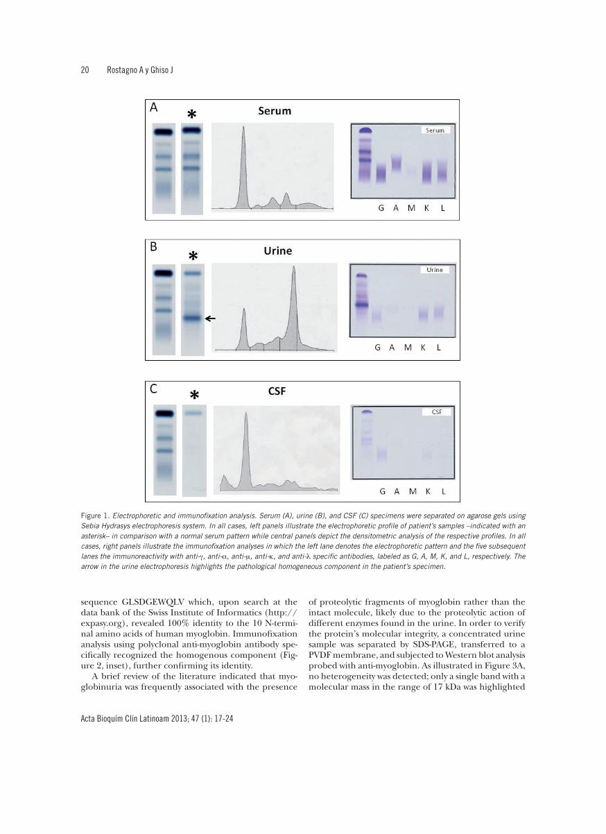
The electrophoretic analysis of the serum, urine, and CSF proteins of the patient being studied are illustrated in Figure 1. Total protein values for serum (7.5 g/dL) and CSF (30 mg/L) were within normal levels and the electrophoretic patterns were unremarkable. This was not the case of the urine specimen; a moderate protein-uria (0.9 g / 600 mL) was recorded in tea-colored 24 h urine and a homogeneous component that accounted for 52% of the urinary protein was clearly visible in the β-region after electrophoresis. Standard immuno-fixation protocols designed to identify the presence of monoclonal immunoglobulins and/or free light chains in biological samples rendered negative results in all three biological fluids, leaving the homogeneous com-ponent present in the urine still unidentified.
The biochemical identification of the unknown urinary homogeneous component was carried out through N-terminal amino acid sequence analysis. For this purpose, concentrated urine was separated by elec-trophoresis in agarose gel, transferred to PVDF, stained, and the band of interest –illustrated in Figure 2– ex-cised from the membrane, and subjected to automatic Edman degradation. The first 10 cycles rendered the
20 Rostagno A y Ghiso J
Acta Bioquím Clín Latinoam 2013; 47 (1): 17-24
sequence GLSDGEWQLV which, upon search at the data bank of the Swiss Institute of Informatics (http://expasy.org), revealed 100% identity to the 10 N-termi-nal amino acids of human myoglobin. Immunofixation analysis using polyclonal anti-myoglobin antibody spe-cifically recognized the homogenous component (Fig-ure 2, inset), further confirming its identity.
A brief review of the literature indicated that myo-globinuria was frequently associated with the presence
of proteolytic fragments of myoglobin rather than the intact molecule, likely due to the proteolytic action of different enzymes found in the urine. In order to verify the protein’s molecular integrity, a concentrated urine sample was separated by SDS-PAGE, transferred to a PVDF membrane, and subjected to Western blot analysis probed with anti-myoglobin. As illustrated in Figure 3A, no heterogeneity was detected; only a single band with a molecular mass in the range of 17 kDa was highlighted
Biochemical analysis of myoglobinuria associated with rhabdomyolysis 21
Acta Bioquím Clín Latinoam 2013; 47 (1): 17-24
by the antibody, a mass compatible with the intact mol-ecule. Figure 3B shows the MALDI-TOF mass spectrom-etry analysis of the homogeneous component, revealing a molecular mass of 17053.10 Da (M+H) in agreement with the theoretical mass of 17052.63 (M+H), and con-firming that the urinary myoglobin molecule was not degraded.
Discussion
The term proteinuria relates to the presence of pro-teins in the urine that exceeds the amount of 150 mg in 24h (6). Passage of molecules across the glomerular barrier is a function of size, charge and configuration. Molecules with an effective radius < 2.0 nm readily fil-ter while those with higher size are partly or completely retained. As a consequence, proteins with a relative mo-lecular mass >40 kDa are almost completely retained whereas smaller components easily enter the glo-merular filtrate. However, size is not the only element restricting glomerular filtration. Charge plays also a critical role and negatively charged components of the glomerular basement membrane favor the retention of anionic molecules.
Most of the proteins in the glomerular filtrate are reabsorbed and metabolized at the tubular level. Under certain pathologic circumstances, increased synthesis or release to the circulation of specific low molecular
mass proteins results in an elevation of their plasma concentration, saturating the tubular re-absorption mechanism and translating in their appearance in the urine, a process known as “overflow proteinuria” (7). Table I summarizes the most frequent pathologic condi-tions and their corresponding protein components as-sociated with overload proteinuria. Within this group, the most common finding is related to the presence of monoclonal immunoglobulins, in particular free light chains (Bence Jones proteins), typically associated with overproduction in cases of lymphoproliferative diseases and multiple myeloma. Due to the low molecular mass of these light chains (25 kDa for the intact molecule and variable smaller size when proteolytically degrad-ed) they massively filtrate through the glomeruli, sur-passing the capability of the tubular reabsorption and making them clearly visible in the urine. Although less frequent among the molecules associated with over-load proteinurias (8), lysozyme is found in the urine in cases of acute granulocytic and monoblastic leuke-mias since this 14 kDa protein is highly concentrated in granulocytes and monocytes. In these leukemias there is an abnormal elevation of lysozyme in plasma that overwhelms the re-absorption machinery resulting in its presence in the urine (9-11). Another molecule that may be identified in overload proteinurias is β2-microglobulin (12), typically elevated in lymphopro-liferative processes in which high serum levels of the protein (above 6 mg/L) are usually indicative of poor disease prognosis (13). It should be noted that lysozyme and β2-microglobulin have similar molecular mass, comparable serum concentration, and are both reab-sorbed at the tubular level; therefore, their presence in the urine may not only be indicative of overload pro-teinuria –due to increased serum concentration with preserved renal function– but may reflect an alteration of the renal tubular re-absorption, as in the case of tu-bular proteinurias (14). In this sense, β2-microglobulin is a well known indicator of renal tubular dysfunction, widely used in the clinical setup (15). Among other mol-
ecules associated with overload proteinurias it is perti-nent to cite amylase and hemoglobin (12). Amylase, a 45 kDa pancreatic protease, can be transiently found in the urine of patients with acute pancreatitis (12) whereas the presence of hemoglobin in urine is primar-ily associated with hemolytic anemia of different origins –including autoimmune conditions, blood transfusion reactions, and paroxistic hemoglobinuria – as well as with multi-trauma situations in which the destruction of red blood cells occurs massively and in a short period of time transiently overwhelming the tubular reabsorption mechanisms. Finally, the presence of myoglobin in the urine –as in the case of the present report– is another example of overload proteinuria. Visible myoglobinuria occurs when urinary myoglobin exceeds 250 mg/L, which corresponds to the destruction of more than 100 g of muscle tissue (16). Both, myoglobinuria and hemo-globinuria are quite common in hospitalized patients and occur in association with a variety of diseases. Myo-globinuria can be inferred if urinary dipstick testing shows a positive result for blood in the absence of red cells in the sediment. This false positive result for blood occurs because the dipstick test –with a sensitivity of 80% for the detection of rhabdomyolysis– is unable to distinguish between myo-globin and hemoglobin (17).
In normal subjects, urinary myoglobin is usually be-low the detection limits of most methods and is prob-ably <0.4 mg/L. Under conditions of severe exercise, myoglobin concentration in serum can increase up to 40-fold with a concomitant high increase in the urinary concentration without the risk of acute renal failure. Thus, it can be inferred that values <15 mg/L can be tol-erated by the kidney without risks. The symptoms and history of myoglobinuria are obvious in certain clini-cal situations such as in crush injuries or severe burns. In about one-fourth of the cases, particularly in non-traumatic rhabdomyolysis, the symptoms are vague and biochemical analysis is necessary for diagnosis. Milder forms of rhabdomyolysis may not cause any muscle symptoms while more severe types exhibit intense my-algia, tenderness, weakness and swelling of the affected muscles (18). If the swelling is very rapid, as it may hap-pen when someone is released from under a collapsed building, the movement of fluid into the blood stream may cause hypotension and shock. Other symptoms are non specific and result either from the consequences of muscle breakdown or from the condition that originat-ed it (18-20). Release of muscle components into the bloodstream causes disturbances of electrolytes, which can lead to nausea, vomiting, confusion and even coma in the most severe cases. Non-traumatic myoglobinuria with acute renal failure is a relatively common disease in patients with alcohol overdose, a history of drug ad-diction, or experiencing side effects to specific medica-tions. In adults, drug-induced muscle damage account for approximately half of all rhabdomyolysis cases. In
Biochemical analysis of myoglobinuria associated with rhabdomyolysis 23
Acta Bioquím Clín Latinoam 2013; 47 (1): 17-24
addition to alcohol abuse and consumption of illegal substances including cocaine, amphetamines, Ectasy and LSD, there are close to 200 medications that have been implicated in the development of rhabdomyolysis (21-23). Antipsychotic drugs, corticosteroids and HIV medications have all been reported to cause rhabdo-myolysis but undoubtedly one of the most relevant are statins, used to lower cholesterol levels by inhibiting the enzyme HMG-CoA reductase, which plays a central role in the production of cholesterol in the liver (23). Al-though the risk of serious myopathy from statins is less than 0.1%, the wide use of these drugs makes screening patients for muscle damage a very important follow up criteria. Statins differ among themselves in several ways including in their ability to reduce cholesterol with cur-rent data indicating that atorvastatin –the best selling of the group– and rosuvastatin are the most potent, and fluvastatin is the least potent (24). Statins also differ in how strongly they interact with other drugs, an impor-tant element in patients receiving combined therapeu-tic regimes. Most critical for the current studies, statins differ also in the frequency with which they cause severe muscle damage. A recent retrospective review of records from over 250,000 patients revealed that the incidence of rhabdomyolysis was 0.44 per 10,000 patients treated with statins other than cerivastatin (25) (26). Howev-er, the risk was over tenfold greater if cerivastatin was used, which resulted in the drug’s withdrawal from the market by its manufacturer in 2001. The risk of rhab-domyolisis was also notably enhanced when standard statins (atorvastatin, fluvastatin, lovastatin, pravastatin, simvastatin) were combined with fibrate (fenofibrate or gemfibrozil) treatment, as in the case reported herein.
Rhabdomyolysis accounts for 5% to 7% of all cases with acute renal failure and myoglobinuria is, there-fore, a frequent finding in patients requiring dialysis (27). It is unclear why heme pigmenturia is associated with acute renal tubular necrosis. The difficulty with which acute renal failure is induced with pure myoglo-bin or hemoglobin solutions in experimental animals, as well as the absence of renal pathology in about half the patients with muscle phosphorylase deficiency that show spontaneous myoglobinuria suggests that coinci-dent factors may be required to enhance the toxicity of hemoproteins to the kidney, among them dehydration and hypotension (27-28).
In addition to the presence of myoglobin in urine, rhabdomyolysis is usually accompanied by other bio-chemical parameters, including greater than 40-fold increase of serum myoglobin and elevated levels of sev-eral serum enzymes such as CK (>40-fold increase) as well as aspartate aminotransferase (>4-fold increase) and LDH (>2-fold increase) (27). Urine myoglobin is the first test analyte to increase; it subsides rapidly with-in the first few days, reflecting its small molecular mass and its half-life of about 2-3 hours. CK increases within
a few hours after muscle damage but remains elevated a few days longer than myoglobin. It is important to re-member that urine is a hostile environment for proteins in general. It contains several proteolytic enzymes and various salts that can denature or hydrolyze proteins. As it occurs with other proteins, myoglobin most fre-quently brakes down in urine although it is stable for a few days in the fridge. Thus, the sooner the sample is processed, better chances for the successful identifica-tion of the components that are inducing the overload proteinuria.
The case illustrated herein presented with a combi-nation of renal failure, liver compromise, and muscle weakness that oriented the diagnosis towards rhabdo-myolysis, which was confirmed by the biochemical iden-tification of myoglobin in the urine. In this particular situation, rhabdomyolysis was linked to the undesirable side effect of statins. The medication was halted and af-ter approximately 2 weeks of hemodialysis for the renal failure the patient fully recovered his renal function and his enzymatic profile normalized. Due to remaining se-vere bilateral lower extremity weakness that precluded him to ambulate, he was transferred to a sub-acute reha-bilitation facility for further treatment. In summary, this report highlights the relevance of the clinical labora-tory as a key player in diagnosis emphasizing its role in cases of overflow proteinurias. Although homogeneous components appearing in protein electrophoresis stud-ies most frequently consist of intact or truncated mono-clonal immunoglobulins, additional unrelated protein spikes should be taken into consideration in relation to different pathological entities. As illustrated herein, simple modifications of routine methodologies avail-able to the clinical laboratory allows the identification of uncommon proteins in biological fluids.
CORRESPONDENCE
JORGE GHISO, PH.D.550 First Avenue, MSB 556NEW YORK, NY, 10016Tel: 212-263-7997Fax: [email protected]
References
1. Laurell CB. Composition and variation of the gel elec-trophoretic fractions of plasma, cerebrospinal fluid, and urine. Scand J Clin Lab Invest 1972; 29(Suppl 124): 71-82.
3. Bossuyt X, Bogaerts A, Schiettekatte G, Blanckaert N. Serum protein electrophoresis and immunofixation by a semiautomated electrophoresis system. Clin Chem 1998; 4: 944-9.
4. Tomidokoro Y, Lashley T, Rostagno A, Neubert TA, Bo-jsen-Moller M, Braendgaard H, �� ��. Familial Danish dementia: Co-existence of ADan and Aβ amyloid sub-units in the absence of compact plaques. J Biol Chem 2005; 280:36883-94.
5. Tomidokoro Y, Rostagno A, Neubert TA, Lu Y, Rebeck GW, Frangione B, �� ��. Iowa variant of familial Al-zheimer’s Disease: accumulation of posttranslationally modified AβD23N in parenchymal and cerebrovascular amyloid deposits. Am J Pathol 2010; 176: 1841-54.
6. Wailer KV, Ward KM, Mahan JD, Wlsmatt DK. Current concepts in proteinuria. Clin Chem 1989; 35: 755-65.
7. Carroll MF, Temte JL. Proteinuria in adults: a diagnostic approach. Am. Fam. Physician 2000; 62: 1333-40.
8. Keren DF. Examination of urine for proteinuria. In: Odder A, editor. Protein electrophoresis in clinical di-agnosis. Oxford, UK: Oxford University Press; 2003. p. 217-258.
9. Levinson SS, Elin RJ, Yam L. Light chain proteinuria and lysozymuria in a patient with acute monocytic leu-kemia. Clin Chem 2002; 48:1131-2.
10. Tardy F, Bulle C, Prin L, Cordier J-F, Deviller P. High concentrations of eosinophil-derived neurotoxin in pa-tient’s urine mimic lysozyme fa-cathodic bands in aga-rose gel electrophoresis. Clin Chem 1993; 39: 919-20.
11. Abuelo JG. Proteinuria: Diagnostic principles and pro-cedures. Ann Int Med 1983; 98: 186-91.
12. Beckett G, Walker S, Rae P, Ashby P. Renal Disease. In: Lecture Notes: Clinical Biochemistry. 8th ed. Chiches-ter: John Wiley & Sons; 2010. p. 51-70.
13. Boccadoro M, Pileri A. Diagnosis, prognosis, and stan-dard treatment of multiple myeloma. Hematol Oncol Clin North Am 1997; 11: 111-31.
14. Handy BC. Urinary β2-microglobulin masquerading as a Bence Jones proteins. Arch Pathol Lab Med 2001; 125: 555-7.
15. Bethea M, Forman DT. Beta 2-microglobulin: its signifi-cance and clinical usefulness. Ann Clin Lab Sci 1990; 20: 163-8.
16. Beetham R. Biochemical investigation of suspected rhabdomyolysis. Ann Clin Biochem 2000; 37: 581-7.
17. Melli G, Chaudhry V, Cornblath DR. Rhabdomyolysis: an evaluation of 475 hospitalized patients. Medicine (Bal-timore) 2005; 84: 377-85.
18. Bosch X, Poch E, Grau JM. Rhabdomyolysis and acute kidney injury. New England J Med 2009; 361: 62-72.
19. Huerta-Alardín AL, Varon J, Marik PE. Bench-to-bed-side review: rhabdomyolysis -an overview for clinicians. Critical Care 2005; 9: 158-69.
20. Sauret JM, Marinides G, Wang GK. Rhabdomyolysis. Am Family Physician 2002; 65: 907-12.
21. Deighan CJ, Wong KM, McLaughlin KJ, Harden P. Rhabdomyolysis and acute renal failure resulting from alcohol and drug abuse. QJM 2000; 93: 29-33.
22. Singhal PC, Rubin RB, Peters A, Santiago A, Neugarten J. Rhabdomyolysis and acute renal failure associated with cocaine abuse. J Toxicol Clin Toxicol 1990; 28: 321–30.
23. Elsayed EF, Reilly RF. Rhabdomyolysis: a review with emphasis on the pediatric population. Pediatr Nephrol 2010; 25: 7-18.
24. Mills EJ, Wu P, Chong G, Ghement I, Singh S, Akl EA, �� ��. Efficacy and safety of statin treatment for cardiovas-cular disease: a network meta-analysis of 170 255 pa-tients from 76 randomized trials. Q J Med 2011; 104: 109-24.
25. Graham DJ, Staffa JA, Shatin D, Andrade SE, Schech SD, La Grenade L, �� ��. Incidence of hospitalized rhab-domyolysis in patients treated with lipid-lowering drugs. JAMA 2004; 292: 2585-90.
26. Golomb BA, Evans MA. Statin adverse effects: a re-view of the literature and evidence for a mitochon-drial mechanism. Am J Cardiovasc Drugs 2008; 8: 373-418.
27. Hamilton RW, Hopkins MBr, Shihabi ZK. Myoglobinuria, hemoglobinuria, and acute renal failure. Clin Chem 1989; 35: 1713-20.
28. Blacher Y, Fong JSC, DeChadarevian JP, Drumond KN. Muscle extract infusion in rabbits:a new experimental model for crush syndrome. Circ Res 1981; 49: 114-24.