Review Biochemical and medical importance of vanadium compounds Jan Korbecki 1 , Irena Baranowska-Bosiacka 1 , Izabela Gutowska 2 and Dariusz Chlubek 1 1 Department of Biochemistry and Medical Chemistry, Pomeranian Medical University, Szczecin, Poland; 2 Department of Biochemistry and Hu- man Nutrition, Pomeranian Medical University, Szczecin, Poland Vanadium belongs to the group of transition metals and is present in the air and soil contaminants in large urban agglomerations due to combustion of fossil fu- els. It forms numerous inorganic compounds (vanadyl sulfate, sodium metavanadate, sodium orthovanadate, vanadium pentoxide) as well as complexes with organic compounds (BMOV, BEOV, METVAN). Depending on the research model, vanadium compounds exhibit antitu- mor or carcinogenic properties. Vanadium compounds generate ROS as a result of Fenton’s reaction or of the reaction with atmospheric oxygen. They inactivate the Cdc25B 2 phosphatase and lead to degradation of Cd- c25C, which induces G 2 /M phase arrest. In cells, vana- dium compounds activate numerous signaling pathways and transcription factors, including PI3K-PKB/Akt-mTOR, NF-κB, MEK1/2-ERK, that cause cell survival or increased expression and release of VEGF. Vanadium compounds inhibit p53-dependent apoptosis and promote entry into the S phase of cells containing functional p53 protein. In addition, vanadium compounds, in particular organic de- rivatives, have insulin-mimetic and antidiabetic proper- ties. Vanadium compounds lower blood glucose levels in animals and in clinical trials. They also inhibit the activ- ity of protein tyrosine phosphatase 1B. By activating the PI3K-PKB/Akt pathway, vanadium compaunds increase the cellular uptake of glucose by the GLUT4 transporter. The PKB/Akt pathway is also used to inactivate glycogen synthase kinase-3. The impact of vanadium compounds on inflammatory reactions has not been fully studied. Vanadium pentoxide causes expression of COX-2 and the release of proinflammatory cytokines in a human lung fibroblast model. Other vanadium compounds activate NF-κB in macrophages by activating IKKβ. Key words: vanadium, pollution, cancer, diabetes, insulin-mimetic action, inflammation Received: 12 February, 2012; revised: 19 April, 2012; accepted: 08 May, 2012; available on-line: 11 June, 2012 NATURAL OCCURRENCE OF VANADIUM Vanadium is a transition metal, owing its name to Vanadís – Norse goddess of beauty and fertility. It is estimated that more than 60 thousand tons of this ele- ment are emitted into the atmosphere each year as the result of human activities (mostly combustion of fos- sil fuels) (Aragón & Altamirano-Lozano, 2001). This is due to high vanadium concentrations in both crude oil (3–260 μg/g) and hard coal (14–56 μg/g). Atmospheric pollution with vanadium of natural origin is relatively low and estimated at several tons annually. The conse- quence of emission of large amounts of vanadium into the atmosphere is the relatively high concentration (20– 300 ng/m 3 ) of this element in the air of big cities, with values reaching up to 10 mg/m 3 observed in the New York City and other large urban agglomerations (Aragón & Altamirano-Lozano, 2001; Lin et al., 2004). Soils in ar- eas not subject to anthropogenic changes contain small amounts of vanadium, originating mostly from volcanic rocks (Połedniok & Buhl, 2003; Nadal et al., 2004). In- dustrial activities result in a significant increase in these levels, reaching 19.3 μg/g of soil in the vicinity of a crude oil refinery in Catalonia (Nadal et al., 2004). Va- nadium present in soil is accumulated in plants (Nadal et al., 2004; Marcano et al., 2006). Contamination with va- nadium is also observed in water reservoirs: rivers, lakes and seas. Bottom sediments of the Persian Gulf contain vanadium at concentrations as high as 100 μg/g of dry sediment (Pourang et al., 2005). About 10% of ground- water samples from California and some other states of the USA contain vanadium in amounts exceeding 25 μg/ dm 3 (Wright & Belitz, 2010). This is due to vanadium being washed out of water-bearing rocks (Wright & Be- litz, 2010). As evidenced by studies of vanadium levels in the hair of residents of different countries, Poland’s popula- tion as a whole is not significantly exposed to high lev- els of vanadium. The measured value is of the order of 0.055 μg/g, being three times lower than the value of 0.171 μg/g for residents of the U.S., Canada or India (Stefańska et al., 2005). Hair vanadium content in stu- dents in Białystok is even lower (0.038 μg/g) due to a non-polluted environment (Stefańska et al., 2005). On the other hand, vanadium pollution is observed in the Upper Silesia region (Połedniok & Buhl, 2003). Indus- trial pollution of the Silesian regions combined with au- tomobile exhaust fumes is transported by rivers into the sea and are deposited in bottom sediments of the rivers. Thus, the sediments in the Bay of Szczecin are highly polluted with vanadium and other elements originating * e-mail: [email protected]Abbreviations: BEOV, bis(ethylmaltolato)oxovanadium(IV); BKOV, bis(kojato)oxovanadium(IV); BMOV, bis(maltolato)oxovanadium(IV); Cdc25B 2 , cell division control/cycle 25 homolog B 2 ; Cdc25C, cell di- vision control/cycle 25 homolog C; CksHs1, human cyclin depend- ent kinase subunit type 1; COX-2, cyclooxygenase 2; CXCL10, C-X- C motif chemokine 10; EGFR, epidermal growth factor receptor; ERK, extracellular signal-regulated kinase; E2F, Transcription factor E2F; GLUT4, Glucose transporter type 4; GSK3, glycogen synthase kinase-3; HIF-1α, hypoxia inducible factor 1α; IC 50 , half maximal inhibitory concentration; IκBα, inhibitor of κB activity α; IKKβ, IκB kinase subunit β; IL-6 Interleukin-6; IL-8, Interleukin-8; MAPK, mi- togen-activated protein kinase; MEK1/2, MAPK/ERK kinase 1 and 2; MIP-2, macrophage inflammatory protein-2; mTOR, mammalian target of rapamycin; NF-κB, nuclear factor κB; NF-AT, nuclear factor of activated T-cells; p38, protein 38; p53, protein 53; PI3K, phos- phatidylinositol 3-kinase; PKB/Akt, protein kinase B; PTP-1B, protein tyrosine phosphatase 1B; pRb, retinoblastoma protein; ROS, reac- tive oxygen species; SSB, single-strand break; TNFα, Tumor necro- sis factor α; VEGF, vascular endothelial growth factor. Vol. 59, No 2/2012 195–200 on-line at: www.actabp.pl
Transcript
Review
Biochemical and medical importance of vanadium compoundsJan Korbecki1, Irena Baranowska-Bosiacka1, Izabela Gutowska2 and Dariusz Chlubek1
1Department of Biochemistry and Medical Chemistry, Pomeranian Medical University, Szczecin, Poland; 2Department of Biochemistry and Hu-man Nutrition, Pomeranian Medical University, Szczecin, Poland
Vanadium belongs to the group of transition metals and is present in the air and soil contaminants in large urban agglomerations due to combustion of fossil fu-els. It forms numerous inorganic compounds (vanadyl sulfate, sodium metavanadate, sodium orthovanadate, vanadium pentoxide) as well as complexes with organic compounds (BMOV, BEOV, METVAN). Depending on the research model, vanadium compounds exhibit antitu-mor or carcinogenic properties. Vanadium compounds generate ROS as a result of Fenton’s reaction or of the reaction with atmospheric oxygen. They inactivate the Cdc25B2 phosphatase and lead to degradation of Cd-c25C, which induces G2/M phase arrest. In cells, vana-dium compounds activate numerous signaling pathways and transcription factors, including PI3K-PKB/Akt-mTOR, NF-κB, MEK1/2-ERK, that cause cell survival or increased expression and release of VEGF. Vanadium compounds inhibit p53-dependent apoptosis and promote entry into the S phase of cells containing functional p53 protein. In addition, vanadium compounds, in particular organic de-rivatives, have insulin-mimetic and antidiabetic proper-ties. Vanadium compounds lower blood glucose levels in animals and in clinical trials. They also inhibit the activ-ity of protein tyrosine phosphatase 1B. By activating the PI3K-PKB/Akt pathway, vanadium compaunds increase the cellular uptake of glucose by the GLUT4 transporter. The PKB/Akt pathway is also used to inactivate glycogen synthase kinase-3. The impact of vanadium compounds on inflammatory reactions has not been fully studied. Vanadium pentoxide causes expression of COX-2 and the release of proinflammatory cytokines in a human lung fibroblast model. Other vanadium compounds activate NF-κB in macrophages by activating IKKβ.
Received: 12 February, 2012; revised: 19 April, 2012; accepted: 08 May, 2012; available on-line: 11 June, 2012
NATURAL OCCURRENCE OF VANADIUM
Vanadium is a transition metal, owing its name to Vanadís – Norse goddess of beauty and fertility. It is estimated that more than 60 thousand tons of this ele-ment are emitted into the atmosphere each year as the result of human activities (mostly combustion of fos-sil fuels) (Aragón & Altamirano-Lozano, 2001). This is due to high vanadium concentrations in both crude oil (3–260 μg/g) and hard coal (14–56 μg/g). Atmospheric pollution with vanadium of natural origin is relatively low and estimated at several tons annually. The conse-quence of emission of large amounts of vanadium into the atmosphere is the relatively high concentration (20–
300 ng/m3) of this element in the air of big cities, with values reaching up to 10 mg/m3 observed in the New York City and other large urban agglomerations (Aragón & Altamirano-Lozano, 2001; Lin et al., 2004). Soils in ar-eas not subject to anthropogenic changes contain small amounts of vanadium, originating mostly from volcanic rocks (Połedniok & Buhl, 2003; Nadal et al., 2004). In-dustrial activities result in a significant increase in these levels, reaching 19.3 μg/g of soil in the vicinity of a crude oil refinery in Catalonia (Nadal et al., 2004). Va-nadium present in soil is accumulated in plants (Nadal et al., 2004; Marcano et al., 2006). Contamination with va-nadium is also observed in water reservoirs: rivers, lakes and seas. Bottom sediments of the Persian Gulf contain vanadium at concentrations as high as 100 μg/g of dry sediment (Pourang et al., 2005). About 10% of ground-water samples from California and some other states of the USA contain vanadium in amounts exceeding 25 μg/dm3 (Wright & Belitz, 2010). This is due to vanadium being washed out of water-bearing rocks (Wright & Be-litz, 2010).
As evidenced by studies of vanadium levels in the hair of residents of different countries, Poland’s popula-tion as a whole is not significantly exposed to high lev-els of vanadium. The measured value is of the order of 0.055 μg/g, being three times lower than the value of 0.171 μg/g for residents of the U.S., Canada or India (Stefańska et al., 2005). Hair vanadium content in stu-dents in Białystok is even lower (0.038 μg/g) due to a non-polluted environment (Stefańska et al., 2005). On the other hand, vanadium pollution is observed in the Upper Silesia region (Połedniok & Buhl, 2003). Indus-trial pollution of the Silesian regions combined with au-tomobile exhaust fumes is transported by rivers into the sea and are deposited in bottom sediments of the rivers. Thus, the sediments in the Bay of Szczecin are highly polluted with vanadium and other elements originating
from distant regions (Glasby et al., 2004). Due to the riv-er runoff, vanadium pollution of the Bay of Szczecin is comparable to the pollution of the Persian Gulf oilfields (Glasby et al., 2004; Pourang et al., 2005).
VANADIUM IN LIVING ORGANISMS
After entering the circulatory system via the gastro-intestinal or respiratory tract, vanadium compounds are transported by transferrin or, less commonly, by albumin or low-molecular components of plasma, such as citrates and, to a lesser extent, lactates or phosphates (Kiss et al., 2000). Next, vanadium compounds are accumulated in kidneys and, to a smaller degree, in spleen, bones and liver (Hansen et al., 1982). Human body contains ca. 100 μg of vanadium, with equilibrium between the amount of vanadium excreted from the body and the amount of vanadium absorbed from the outside environment (up to several dozen micrograms daily) (Byrne & Kosta, 1978; Kordowiak & Holko, 2009). For certain mammals, such as rats, vanadium is a necessary microelement; however, due to the omnipresence of this element at low concen-trations, no necessity of nutritional intake of vanadium was determined in humans (Lin et al., 2004; Kordowiak & Holko, 2009).
Aquatic organisms, such as ascidians, accumulate va-nadium in circulatory system cells known as vanadocytes (Kawakami et al., 2006; Kawakami et al., 2009). Blood vanadium levels in these organisms exceed 10 mM, while the sea concentration of vanadium is about 35 nM (Kawakami et al., 2009). Vanadium compounds are trans-ported into the cytoplasm of vanadocytes, bound and reduced to the +4 oxidation state by the binding pro-teins – vanabins, and finally deposited in the acidic envi-ronment of vacuoles as vanadium compounds in the +3 oxidation state (Kawakami et al., 2006).In human body, vanadium has an oxidation state of
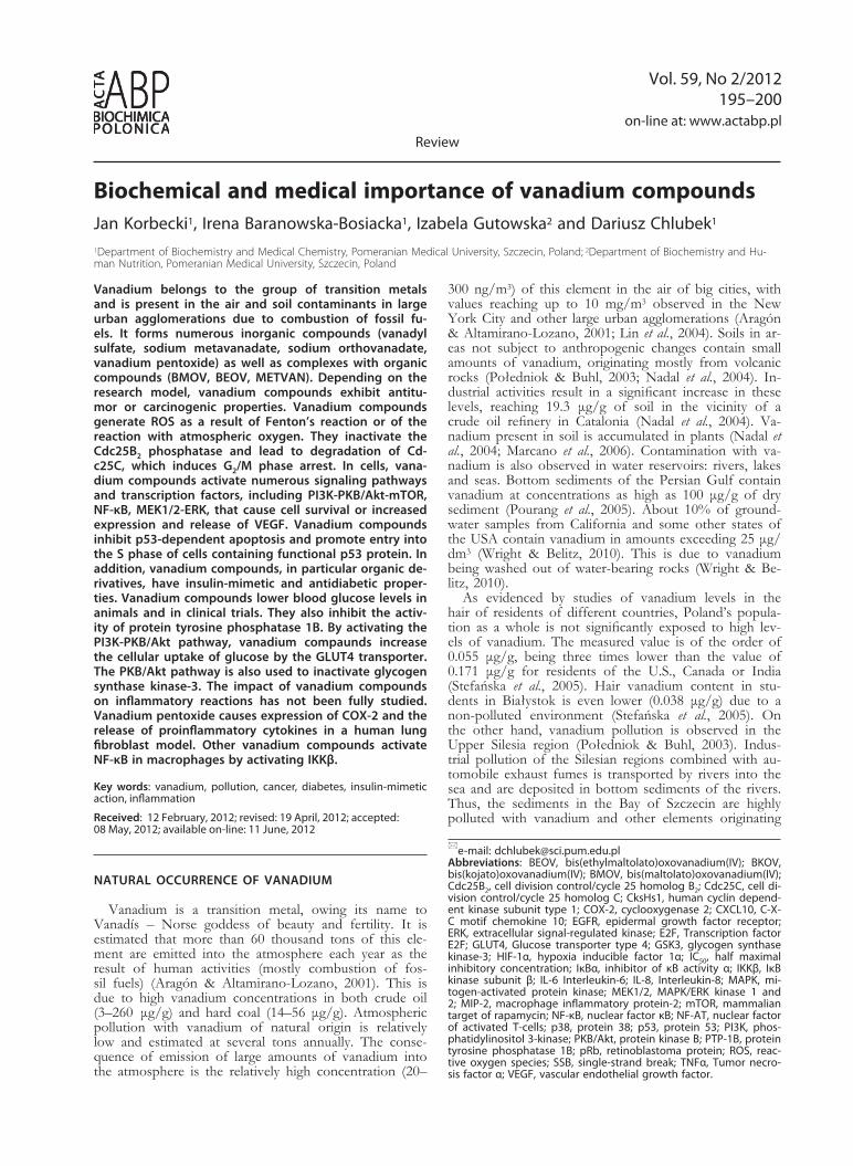
+4 or +5 (Kordowiak & Holko, 2009). Vanadium com-pounds in the +5 oxidation state (metavanadates or or-thovanadates, forming oligomers) enter cells via anionic channels, while vanadium compounds in the +4 oxida-tion state (vanadyl cations) permeate the cellular mem-brane by diffusion (Fig. 1) (Aureliano & Gândara, 2005; Kordowiak & Holko, 2009). Vanadium forms numerous derivatives with low-molecular organic compounds. Va-nadium organic derivatives have been synthesized since 1990s (Thomson et al., 2009). Examples of such com-pounds include maltol complexes such as BMOV or
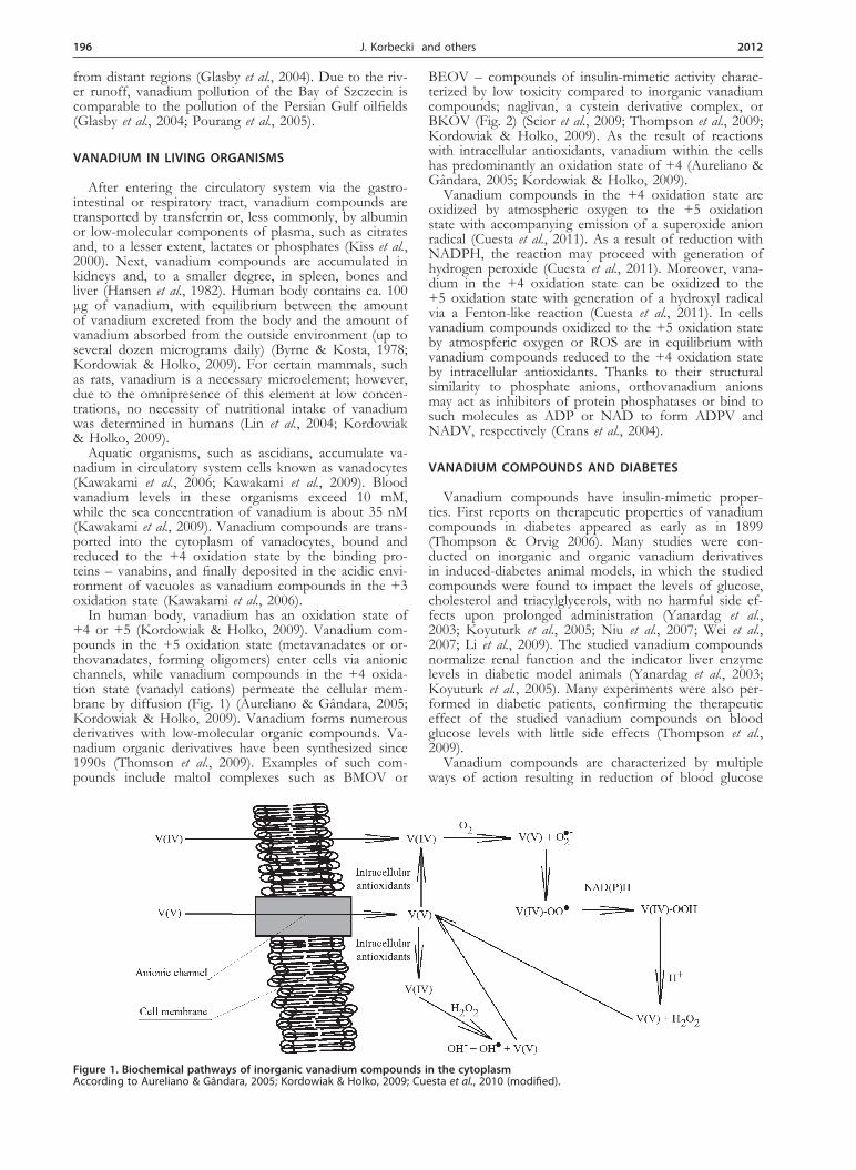
BEOV – compounds of insulin-mimetic activity charac-terized by low toxicity compared to inorganic vanadium compounds; naglivan, a cystein derivative complex, or BKOV (Fig. 2) (Scior et al., 2009; Thompson et al., 2009; Kordowiak & Holko, 2009). As the result of reactions with intracellular antioxidants, vanadium within the cells has predominantly an oxidation state of +4 (Aureliano & Gândara, 2005; Kordowiak & Holko, 2009).Vanadium compounds in the +4 oxidation state are
oxidized by atmospheric oxygen to the +5 oxidation state with accompanying emission of a superoxide anion radical (Cuesta et al., 2011). As a result of reduction with NADPH, the reaction may proceed with generation of hydrogen peroxide (Cuesta et al., 2011). Moreover, vana-dium in the +4 oxidation state can be oxidized to the +5 oxidation state with generation of a hydroxyl radical via a Fenton-like reaction (Cuesta et al., 2011). In cells vanadium compounds oxidized to the +5 oxidation state by atmospferic oxygen or ROS are in equilibrium with vanadium compounds reduced to the +4 oxidation state by intracellular antioxidants. Thanks to their structural similarity to phosphate anions, orthovanadium anions may act as inhibitors of protein phosphatases or bind to such molecules as ADP or NAD to form ADPV and NADV, respectively (Crans et al., 2004).
VANADIUM COMPOUNDS AND DIABETES
Vanadium compounds have insulin-mimetic proper-ties. First reports on therapeutic properties of vanadium compounds in diabetes appeared as early as in 1899 (Thompson & Orvig 2006). Many studies were con-ducted on inorganic and organic vanadium derivatives in induced-diabetes animal models, in which the studied compounds were found to impact the levels of glucose, cholesterol and triacylglycerols, with no harmful side ef-fects upon prolonged administration (Yanardag et al., 2003; Koyuturk et al., 2005; Niu et al., 2007; Wei et al., 2007; Li et al., 2009). The studied vanadium compounds normalize renal function and the indicator liver enzyme levels in diabetic model animals (Yanardag et al., 2003; Koyuturk et al., 2005). Many experiments were also per-formed in diabetic patients, confirming the therapeutic effect of the studied vanadium compounds on blood glucose levels with little side effects (Thompson et al., 2009).
Vanadium compounds are characterized by multiple ways of action resulting in reduction of blood glucose
Figure 1. Biochemical pathways of inorganic vanadium compounds in the cytoplasmAccording to Aureliano & Gândara, 2005; Kordowiak & Holko, 2009; Cuesta et al., 2010 (modified).
Vol. 59 197Biochemical and medical importance of vanadium compounds
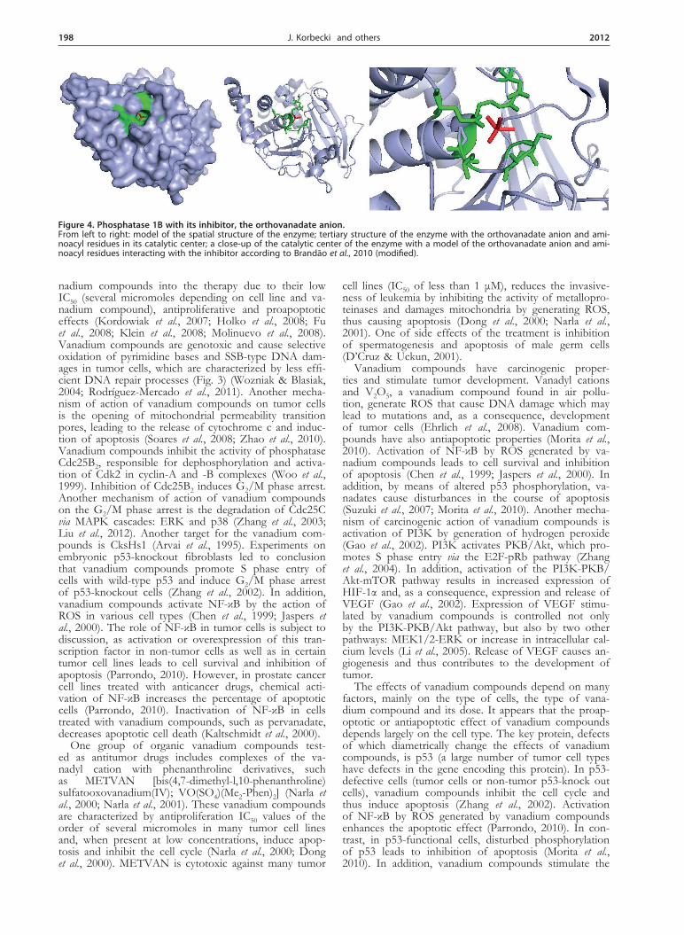
levels (Vardatsikos et al., 2009). Thanks to their structural similarity to orthophosphate anions, the orthovanadate anion and vanadium organic derivatives are inhibitors of protein phosphotyrosine phosphatases (Fig. 4). (Crans et al., 2004). They may inhibit the activity of PTP-1B,
which is an enzyme responsi-ble for dephosphorylation of insulin receptors, causing insu-lin resistance (Scior et al., 2009; Scior et al., 2010). However, the mechanism of phosphotyros-ine phosphatase inactivation is not yet fully understood, as it appears that this process may also be caused by free radi-cals (Bartosz, 2003). One may assume that vanadium com-pounds cause PTP-1B inhibi-tion via ROS (Kaltschmidt et al., 2000). Another mechanism of reduction of blood glucose levels by vanadium compounds is the activation of PKB/Akt leading to increased uptake of glucose by the GLUT4 trans-porter (Vardatsikos et al., 2009). Activation of PKB/Akt results also in phosphorylation and in-activation of GSK3, leading to stimulation of the synthesis of glycogen from glucose (Vardat-sikos et al., 2009).
VANADIUM AND TUMOR CELLS
In chemically-induced tu-mor models in experimental animals, vanadium compounds show chemopreventive proper-ties by means of optimization of phase I and phase II xeno-biotic transformation enzymes
(Bishayee et al., 2000; Ray et al., 2007; Chakraborty et al., 2007). Inorganic and organic vanadium compounds were tested in human tumor cell line models. The re-sults were promising with respect to introduction of va-
Figure 2. Examples of organic vanadium derivatives of medical importance According to Dong et al., 2000; D’Cruz & Uckun, 2001; Scior et al., 2008; Thompson et al., 2009, (modified).
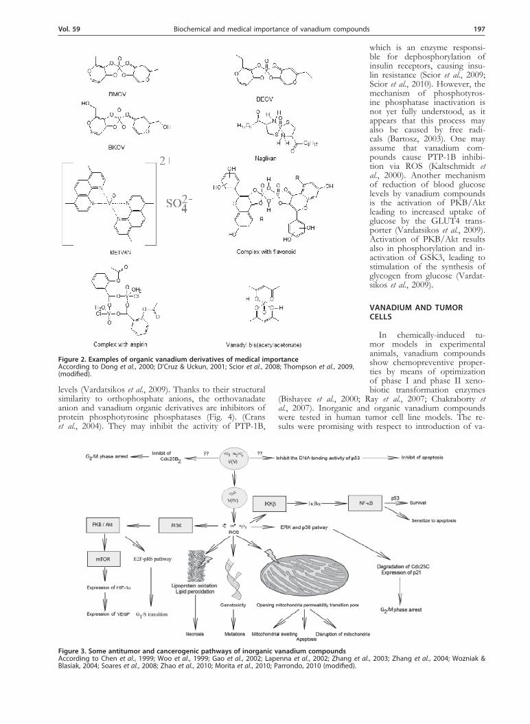
Figure 3. Some antitumor and cancerogenic pathways of inorganic vanadium compoundsAccording to Chen et al., 1999; Woo et al., 1999; Gao et al., 2002; Lapenna et al., 2002; Zhang et al., 2003; Zhang et al., 2004; Wozniak & Blasiak, 2004; Soares et al., 2008; Zhao et al., 2010; Morita et al., 2010; Parrondo, 2010 (modified).
198 2012J. Korbecki and others
nadium compounds into the therapy due to their low IC50 (several micromoles depending on cell line and va-nadium compound), antiproliferative and proapoptotic effects (Kordowiak et al., 2007; Holko et al., 2008; Fu et al., 2008; Klein et al., 2008; Molinuevo et al., 2008). Vanadium compounds are genotoxic and cause selective oxidation of pyrimidine bases and SSB-type DNA dam-ages in tumor cells, which are characterized by less effi-cient DNA repair processes (Fig. 3) (Wozniak & Blasiak, 2004; Rodríguez-Mercado et al., 2011). Another mecha-nism of action of vanadium compounds on tumor cells is the opening of mitochondrial permeability transition pores, leading to the release of cytochrome c and induc-tion of apoptosis (Soares et al., 2008; Zhao et al., 2010). Vanadium compounds inhibit the activity of phosphatase Cdc25B2, responsible for dephosphorylation and activa-tion of Cdk2 in cyclin-A and -B complexes (Woo et al., 1999). Inhibition of Cdc25B2 induces G2/M phase arrest. Another mechanism of action of vanadium compounds on the G2/M phase arrest is the degradation of Cdc25C via MAPK cascades: ERK and p38 (Zhang et al., 2003; Liu et al., 2012). Another target for the vanadium com-pounds is CksHs1 (Arvai et al., 1995). Experiments on embryonic p53-knockout fibroblasts led to conclusion that vanadium compounds promote S phase entry of cells with wild-type p53 and induce G2/M phase arrest of p53-knockout cells (Zhang et al., 2002). In addition, vanadium compounds activate NF-κB by the action of ROS in various cell types (Chen et al., 1999; Jaspers et al., 2000). The role of NF-κB in tumor cells is subject to discussion, as activation or overexpression of this tran-scription factor in non-tumor cells as well as in certain tumor cell lines leads to cell survival and inhibition of apoptosis (Parrondo, 2010). However, in prostate cancer cell lines treated with anticancer drugs, chemical acti-vation of NF-κB increases the percentage of apoptotic cells (Parrondo, 2010). Inactivation of NF-κB in cells treated with vanadium compounds, such as pervanadate, decreases apoptotic cell death (Kaltschmidt et al., 2000).One group of organic vanadium compounds test-
ed as antitumor drugs includes complexes of the va-nadyl cation with phenanthroline derivatives, such as METVAN [bis(4,7-dimethyl-l,10-phenanthroline)sulfatooxovanadium(IV); VO(SO4)(Me2-Phen)2] (Narla et al., 2000; Narla et al., 2001). These vanadium compounds are characterized by antiproliferation IC50 values of the order of several micromoles in many tumor cell lines and, when present at low concentrations, induce apop-tosis and inhibit the cell cycle (Narla et al., 2000; Dong et al., 2000). METVAN is cytotoxic against many tumor
cell lines (IC50 of less than 1 μM), reduces the invasive-ness of leukemia by inhibiting the activity of metallopro-teinases and damages mitochondria by generating ROS, thus causing apoptosis (Dong et al., 2000; Narla et al., 2001). One of side effects of the treatment is inhibition of spermatogenesis and apoptosis of male germ cells (D’Cruz & Uckun, 2001).
Vanadium compounds have carcinogenic proper-ties and stimulate tumor development. Vanadyl cations and V2O5, a vanadium compound found in air pollu-tion, generate ROS that cause DNA damage which may lead to mutations and, as a consequence, development of tumor cells (Ehrlich et al., 2008). Vanadium com-pounds have also antiapoptotic properties (Morita et al., 2010). Activation of NF-κB by ROS generated by va-nadium compounds leads to cell survival and inhibition of apoptosis (Chen et al., 1999; Jaspers et al., 2000). In addition, by means of altered p53 phosphorylation, va-nadates cause disturbances in the course of apoptosis (Suzuki et al., 2007; Morita et al., 2010). Another mecha-nism of carcinogenic action of vanadium compounds is activation of PI3K by generation of hydrogen peroxide (Gao et al., 2002). PI3K activates PKB/Akt, which pro-motes S phase entry via the E2F-pRb pathway (Zhang et al., 2004). In addition, activation of the PI3K-PKB/Akt-mTOR pathway results in increased expression of HIF-1α and, as a consequence, expression and release of VEGF (Gao et al., 2002). Expression of VEGF stimu-lated by vanadium compounds is controlled not only by the PI3K-PKB/Akt pathway, but also by two other pathways: MEK1/2-ERK or increase in intracellular cal-cium levels (Li et al., 2005). Release of VEGF causes an-giogenesis and thus contributes to the development of tumor.
The effects of vanadium compounds depend on many factors, mainly on the type of cells, the type of vana-dium compound and its dose. It appears that the proap-optotic or antiapoptotic effect of vanadium compounds depends largely on the cell type. The key protein, defects of which diametrically change the effects of vanadium compounds, is p53 (a large number of tumor cell types have defects in the gene encoding this protein). In p53-defective cells (tumor cells or non-tumor p53-knock out cells), vanadium compounds inhibit the cell cycle and thus induce apoptosis (Zhang et al., 2002). Activation of NF-κB by ROS generated by vanadium compounds enhances the apoptotic effect (Parrondo, 2010). In con-trast, in p53-functional cells, disturbed phosphorylation of p53 leads to inhibition of apoptosis (Morita et al., 2010). In addition, vanadium compounds stimulate the
Figure 4. Phosphatase 1B with its inhibitor, the orthovanadate anion. From left to right: model of the spatial structure of the enzyme; tertiary structure of the enzyme with the orthovanadate anion and ami-noacyl residues in its catalytic center; a close-up of the catalytic center of the enzyme with a model of the orthovanadate anion and ami-noacyl residues interacting with the inhibitor according to Brandão et al., 2010 (modified).
Vol. 59 199Biochemical and medical importance of vanadium compounds
cell cycle, thus inhibiting apoptosis, as both processes are mutually connected (Zhang et al., 2002). Moreover, NF-κB activation inhibits apoptosis of tumor cells. An-other important fact is that vanadium compounds cause much more DNA damage in tumor cells compared to non-tumor cells when present at the same levels (Wozni-ak & Blasiak, 2004). Extensive DNA damage leads to apoptosis of tumor cells while a less intensive damage evoked by vanadium compounds in non-tumor cells may stimulate synthesis and activation of repair enzymes, thus protecting those cells from apoptosis. The above pro-cesses promote tumor cell growth at early stages of the disease and have an antitumor effect in the advanced stages of cancer. Studies in animals treated with carcino-gens suggest that vanadium compounds used at low lev-els have selective effects on the tumor cells (Ray et al., 2007; Chakraborty et al., 2007).
VANADIUM COMPOUNDS AND INFLAMMATORy REACTIONS
The impact of vanadium compounds on inflammatory reactions has not been fully studied. Experiments con-ducted to date suggest that vanadates activate NF-κB, a transcription factor of key importance in inflammatory reactions (Chen et al., 1999; Ye et al., 1999). Studies con-ducted on RAW 264.7 macrophages showed that this was due to activation of IKKβ and degradation of IκBα (Chen et al., 1999; Ye et al., 1999). Activation of NF-κB leads to changes in expression of numerous genes, including TNFα and MIP-2, which belong to the CXC chemokine family (Ye et al., 1999; Chong et al., 2000). Another vanadium compound prevalent in air pollution and causing inflammatory reactions is vanadium pentox-ide. Exposure to vanadium pentoxide-containing dust causes inflammatory reactions in lungs, leading to expres-sion of, among others, COX-2, IL-6, IL-8 and CXCL10 (Ingram et al., 2007; Rondini et al., 2010). Vanadium pen-toxide causes COX-2 expression in epithelial bronchial Beas-2B cells via the NF-AT pathway (Tang et al., 2007). Another pathway for the increase in COX-2 expression, encompassing EGFR and the p38 cascade, was observed in A249 lung cancer cells (Chen et al., 2006).
CONCLUSIONS AND FUTURE DIRECTIONS
Due to the ability to generate ROS, which exert non-specific effects on different cell structures, vanadium compounds have many routes of action, sometimes dia-metrically opposite. They may have both antitumor and carcinogenic properties. The mechanisms of action of the vanadium compounds can be understood thanks to rapid advances in the knowledge of free radicals and the signaling pathways involving them. However, still little is known regarding the effect of vanadium compounds on the immune system and inflammatory reactions. New findings in this area may shed new light on the biochem-ical processes taking place in organisms treated with vanadium compounds. Currently, promising clinical tri-als of organic vanadium derivatives in the treatment of diabetes are under way. Soon they should be of com-mon use. However, the effects of a long-term adminis-tration of low doses of vanadium as a potential carcino-gen and correlation between the use of vanadium com-pounds and disorders of a free-radical background have not been fully studied yet. One of such diseases having a free-radical background is Parkinson’s disease. Vana-dium compounds induce ROS generation in the brain,
which may contribute to degeneration of dopaminergic neuronal cells of the substantia nigra, which in turn leads to Parkinson’s disease (Afeseh Ngwa et al., 2009; Cuesta et al., 2011).
It is possible that an antitumor therapy using vana-dium compounds will be developed in the near future. However, due to the carcinogenic effect of vanadium, such treatment should be combined with numerous oth-er drugs (such as anti-VEGF antibodies) to enhance the therapeutic effect of vanadium.
REFERENCES
Afeseh Ngwa H, Kanthasamy A, Anantharam V, Song C, Witte T, Houk R, Kanthasamy AG (2009) Vanadium induces dopaminergic neurotoxicity via protein kinase Cdelta dependent oxidative signal-ing mechanisms: relevance to etiopathogenesis of Parkinson’s dis-ease. Toxicol Appl Pharmacol 240: 273–285.
Aragón AM, Altamirano-Lozano M (2001) Sperm and testicular modi-fications induced by subchronic treatments with vanadium (IV) in CD-1 mice. Reprod Toxicol 15: 145–151.
Arvai AS, Bourne Y, Hickey MJ, Tainer JA (1995) Crystal structure of the human cell cycle protein CksHs1: single domain fold with simi-larity to kinase N-lobe domain. J Mol Biol 249: 835–842.
Aureliano M, Gândara RM (2005) Decavanadate effects in biological systems. J Inorg Biochem 99: 979–985.
Bartosz G (2003) Obrazki z pola bitwy. In Druga twarz tlenu. pp 324–325. PWN, Warszawa (in Polish).
Bishayee A, Oinam S, Basu M, Chatterjee M (2000) Vanadium chemo-prevention of 7,12-dimethylbenz(a)anthracene-induced rat mam-mary carcinogenesis: probable involvement of representative hepatic phase I and II xenobiotic metabolizing enzymes. Breast Cancer Res Treat 63: 133–145.
Brandão TA, Hengge AC, Johnson SJ (2010) Insights into the reaction of protein-tyrosine phosphatase 1B: crystal structures for transition state analogs of both catalytic steps. J Biol Chem 285: 15874–15883.
Byrne AR, Kosta L (1978) Vanadium in foods and in human body fluids and tissues. Sci Total Environ 10: 17–30.
Chakraborty T, Swamy AH, Chatterjee A, Rana B, Shyamsundar A, Chatterjee M (2007) Molecular basis of vanadium-mediated inhibi-tion of hepatocellular preneoplasia during experimental hepatocar-cinogenesis in rats. J Cell Biochem 101: 244–258.
Chen F, Demers LM, Vallyathan V, Ding M, Lu Y, Castranova V, Shi X (1999) Vanadate induction of NF-kappaB involves IkappaB ki-nase beta and SAPK/ERK kinase 1 in macrophages. J Biol Chem 274: 20307–20312.
Chien PS, Mak OT, Huang HJ (2006) Induction of COX-2 protein expression by vanadate in A549 human lung carcinoma cell line through EGF receptor and p38 MAPK-mediated pathway. Biochem Biophys Res Commun 339: 562–568.
Chong IW, Lin SR, Hwang JJ, Huang MS, Wang TH, Tsai MS, Hou JJ, Paulauskis JD (2000) Expression and regulation of macrophage inflammatory protein-2 gene by vanadium in mouse macrophages. Inflammation 24: 127–139.
Crans DC, Smee JJ, Gaidamauskas E, Yang L (2004) The chemistry and biochemistry of vanadium and the biological activities exerted by vanadium compounds. Chem Rev 104: 849–902.
Cuesta S, Francés D, García GB (2011) ROS formation and antioxi-dant status in brain areas of rats exposed to sodium metavanadate. Neurotoxicol Teratol 3: 1–6.
D’Cruz OJ, Uckun FM (2001) Bis(4,7-dimethyl and 5-dinitro-1,10-phenanthroline) sulfato-oxovanadium(IV)-mediated in vivo male germ cell apoptosis. J Appl Toxicol 21: 331–339.
Dong Y, Narla RK, Sudbeck E, Uckun FM (2000) Synthesis, X-ray structure, and anti-leukemic activity of oxovanadium(IV) complexes. J Inorg Biochem 78: 321–330.
Ehrlich VA, Nersesyan AK, Atefie K, Hoelzl C, Ferk F, Bichler J, Valic E, Schaffer A, Schulte-Hermann R, Fenech M, Wagner KH, Knasmüller S (2008) Inhalative exposure to vanadium pentoxide causes DNA damage in workers: results of a multiple end point study. Environ Health Perspect 116: 1689–1693.
Fu Y, Wang Q, Yang XG, Yang XD, Wang K (2008) Vanadyl bisa-cetylacetonate induced G1/S cell cycle arrest via high-intensity ERK phosphorylation in HepG2 cells. J Biol Inorg Chem 13: 1001–1009.
Gao N, Ding M, Zheng JZ, Zhang Z, Leonard SS, Liu KJ, Shi X, Jiang BH (2002) Vanadate-induced expression of hypoxia-inducible factor 1 alpha and vascular endothelial growth factor through phos-phatidylinositol 3-kinase/Akt pathway and reactive oxygen species. J Biol Chem 277: 31963–31971.
Glasby GP, Szefer P, Geldon J, Warzocha J (2004) Heavy-metal pol-lution of sediments from Szczecin Lagoon and the Gdansk Basin, Poland. Sci Total Environ 330: 249–69.
200 2012J. Korbecki and others
Hansen TV, Aaseth J, Alexander J (1982) The effect of chelating agents on vanadium distribution in the rat body and on uptake by human erythrocytes. Arch Toxicol 50: 195–202.
Holko P, Ligeza J, Kisielewska J, Kordowiak AM, Klein A (2008) The effect of vanadyl sulphate (VOSO4) on autocrine growth of human epithelial cancer cell lines. Pol J Pathol 59: 3–8.
Ingram JL, Antao-Menezes A, Turpin EA, Wallace DG, Mangum JB, Pluta LJ, Thomas RS, Bonner JC (2007) Genomic analysis of hu-man lung fibroblasts exposed to vanadium pentoxide to identify candidate genes for occupational bronchitis. Respir Res 8: 34.
Jaspers I, Samet JM, Erzurum S, Reed W (2000) Vanadium-induced kappaB-dependent transcription depends upon peroxide-induced ac-tivation of the p38 mitogen-activated protein kinase. Am J Respir Cell Mol Biol 23: 95–102.
Kaltschmidt B, Kaltschmidt C, Hofmann TG, Hehner SP, Dröge W, Schmitz ML (2000) The pro- or anti-apoptotic function of NF-kap-paB is determined by the nature of the apoptotic stimulus. Eur J Biochem 267: 3828–3835.
Kawakami N, Ueki T, Amata Y, Kanamori K, Matsuo K, Gekko K, Michibata H (2009) A novel vanadium reductase, Vanabin2, forms a possible cascade involved in electron transfer. Biochim Biophys Acta 1794: 674–679.
Kawakami N, Ueki T, Matsuo K, Gekko K, Michibata H (2006) Selec-tive metal binding by Vanabin2 from the vanadium-rich ascidian, Ascidia sydneiensis samea. Biochim Biophys Acta 1760: 1096–1101.
Kiss T, Kiss E, Garribba E, Sakurai H (2000) Speciation of insulin-mimetic VO(IV)-containing drugs in blood serum. J Inorg Biochem 80: 65–73.
Klein A, Holko P, Ligeza J, Kordowiak AM (2008) Sodium orthova-nadate affects growth of some human epithelial cancer cells (A549, HTB44, DU145). Folia Biol (Krakow) 56: 115–121.
Kordowiak AM, Holko P (2009) Pochodne wanadu jako związki o istotnym znaczeniu biologicznym. Część I. Działanie przeciwcukrzy-cowe. Post Biol Kom 36: 361–376 (in Polish).
Kordowiak AM, Klein A, Goc A, Dabroś W (2007) Comparison of the effect of VOSO4, Na3VO4 and NaVO3 on proliferation, viability and morphology of H35-19 rat hepatoma cell line. Pol J Pathol 58: 51–57.
Koyuturk M, Tunali S, Bolkent S, Yanardag R (2005) Effects of vana-dyl sulfate on liver of streptozotocin-induced diabetic rats. Biol Trace Elem Res 104: 233–247.
Lapenna D, Ciofani G, Bruno C, Pierdomenico SD, Giuliani L, Giam-berardino MA, Cuccurullo F (2002) Vanadyl as a catalyst of human lipoprotein oxidation. Biochem Pharmacol 63: 375–380.
Li J, Tong Q, Shi X, Costa M, Huang C (2005) ERKs activation and calcium signaling are both required for VEGF induction by vana-dium in mouse epidermal Cl41 cells. Mol Cell Biochem 279: 25–33.
Li M, Smee JJ, Ding W, Crans DC (2009) Anti-diabetic effects of so-dium 4-amino-2,6-dipicolinatodioxovanadium(V) dihydrate in strep-tozotocin-induced diabetic rats. J Inorg Biochem 103: 585–589.
Lin TS, Chang CL, Shen FM (2004) Whole blood vanadium in Tai-wanese college students. Bull Environ Contam Toxicol 73: 781–786.
Liu TT, Liu YJ, Wang Q, Yang XG, Wang K (2012) Reactive-oxygen-species-mediated Cdc25C degradation results in differential anti-proliferative activities of vanadate, tungstate, and molybdate in the PC-3 human prostate cancer cell line. J Biol Inorg Chem 17: 311–320.
Marcano L, Carruyo I, Fernández Y, Montiel X, Torrealba Z (2006) Determination of vanadium accumulation in onion root cells (Al-lium cepa L.) and its correlation with toxicity. Biocell 30: 259–267.
Molinuevo MS, Cortizo AM, Etcheverry SB (2008) Vanadium(IV) complexes inhibit adhesion, migration and colony formation of UMR106 osteosarcoma cells. Cancer Chemother Pharmacol 61: 767–773.
Morita A, Yamamoto S, Wang B, Tanaka K, Suzuki N, Aoki S, Ito A, Nanao T, Ohya S, Yoshino M, Zhu J, Enomoto A, Matsumoto Y, Funatsu O, Hosoi Y, Ikekita M (2010) Sodium orthovanadate inhib-its p53-mediated apoptosis. Cancer Res 70: 257–265.
Nadal M, Schuhmacher M, Domingo JL (2004) Metal pollution of soils and vegetation in an area with petrochemical industry. Sci Total En-viron 321: 59–69.
Narla RK, Chen CL, Dong Y, Uckun FM (2001) In vivo antitumor ac-tivity of bis(4,7-dimethyl-1,10-phenanthroline) sulfatooxovanadium (IV) (METVAN [VO(SO4)(Me2-Phen)2]). Clin Cancer Res 7: 2124–2133.
Narla RK, Dong Y, D’Cruz OJ, Navara C, Uckun FM (2000) Bis(4,7-dimethyl-1,10-phenanthroline) sulfatooxovanadium (IV) as a novel apoptosis-inducing anticancer agent. Clin Cancer Res 6: 1546–1556.
Narla RK, Dong Y, Klis D, Uckun FM (2001) Bis(4,7-dimethyl-1,10-phenanthroline) sulfatooxovanadium (IV) as a novel antileukemic agent with matrix metalloproteinase inhibitory activity. Clin Cancer Res 7: 1094–1101.
Niu Y, Liu W, Tian C, Xie M, Gao L, Chen Z, Chen X, Li L (2007) Effects of bis(alpha-furancarboxylato)oxovanadium(IV) on glucose
metabolism in fat-fed/streptozotocin-diabetic rats. Eur J Pharmacol 572: 213–219.
Parrondo R, de las Pozas A, Reiner T, Rai P, Perez-Stable C (2010) NF-kappaB activation enhances cell death by antimitotic drugs in human prostate cancer cells. Mol Cancer 9: 182–195.
Połedniok J, Buhl F (2003) Speciation of vanadium in soil. Talanta 59: 1–8.
Pourang N, Nikouyan A, Dennis JH (2005) Trace element concentra-tions in fish, surficial sediments and water from northern part of the Persian Gulf. Environ Monit Assess 109: 293–316.
Ray RS, Ghosh B, Rana A, Chatterjee M (2007) Suppression of cell proliferation, induction of apoptosis and cell cycle arrest: chemo-preventive activity of vanadium in vivo and in vitro. Int J Cancer 120: 13–23.
Rodríguez-Mercado JJ, Mateos-Nava RA, Altamirano-Lozano MA (2011) DNA damage induction in human cells exposed to vanadium oxides in vitro. Toxicol In Vitro 25: 1996–2002.
Rondini EA, Walters DM, Bauer AK (2010) Vanadium pentoxide in-duces pulmonary inflammation and tumor promotion in a strain-dependent manner. Part Fibre Toxicol 7: 9.
Scior T, Guevara-García JA, Melendez FJ, Abdallah HH, Do QT, Ber-nard P (2010) Chimeric design, synthesis, and biological assays of a new nonpeptide insulin-mimetic vanadium compound to inhibit protein tyrosine phosphatase 1B. Drug Des Devel Ther 4: 231–242.
Scior T, Mack HG, García JA, Koch W (2009) Antidiabetic Bis-Malto-lato-OxoVanadium(IV): conversion of inactive trans- to bioactive cis-BMOV for possible binding to target PTP-1B. Drug Des Devel Ther 2: 221–231.
Soares SS, Henao F, Aureliano M, Gutiérrez-Merino C (2008) Vana-date induces necrotic death in neonatal rat cardiomyocytes through mitochondrial membrane depolarization. Chem Res Toxicol 21: 607–618.
Stefańska E, Ostrowska L, Czapska D, Karczewski J, Borawska M (2005) Hair vanadium content and nutritional status of students of the Medical University of Białystok. Rocz Panstw Zakl Hig 56: 157–163.
Suzuki K, Inageda K, Nishitai G, Matsuoka M (2007) Phosphorylation of p53 at serine 15 in A549 pulmonary epithelial cells exposed to vanadate: involvement of ATM pathway. Toxicol Appl Pharmacol 220: 83–91.
Tang H, Sun Y, Xiu Q, Lu H, Han H (2007) Cyclooxygenase-2 in-duction requires activation of nuclear factor of activated T-cells in Beas-2B cells after vanadium exposure and plays an anti-apoptotic role. Arch Biochem Biophys 468: 92–99.
Thompson KH, Lichter J, LeBel C, Scaife MC, McNeill JH, Orvig C (2009) Vanadium treatment of type 2 diabetes: a view to the future. J Inorg Biochem 103: 554–558.
Thompson KH, Orvig C (2006) Vanadium in diabetes: 100 years from Phase 0 to Phase I. J Inorg Biochem 100: 1925–1935.
Vardatsikos G, Mehdi MZ, Srivastava AK (2009) Bis(maltolato)-oxova-nadium (IV)-induced phosphorylation of PKB, GSK-3 and FOXO1 contributes to its glucoregulatory responses (review). Int J Mol Med 24: 303–309.
Wei D, Li M, Ding W (2007) Effect of vanadate on gene expression of the insulin signaling pathway in skeletal muscle of streptozotocin-induced diabetic rats. J Biol Inorg Chem 12: 1265–1273.
Wozniak K, Blasiak J (2004) Vanadyl sulfate can differentially damage DNA in human lymphocytes and HeLa cells. Arch Toxicol 78: 7–15.
Wright MT, Belitz K (2010) Factors controlling the regional distribu-tion of vanadium in groundwater. Ground Water 48: 515–525.
Yanardag R, Bolkent S, Karabulut-Bulan O, Tunali S (2003) Effects of vanadyl sulfate on kidney in experimental diabetes. Biol Trace Elem Res 95: 73–85.
Ye J, Ding M, Zhang X, Rojanasakul Y, Nedospasov S, Vallyathan V, Castranova V, Shi X (1999) Induction of TNFalpha in macrophages by vanadate is dependent on activation of transcription factor NF-kappaB and free radical reactions. Mol Cell Biochem 198: 193–200.
Zhang Z, Chen F, Huang C, Shi X (2002) Vanadate induces G2/M phase arrest in p53-deficient mouse embryo fibroblasts. J Environ Pathol Toxicol Oncol 21: 223–231.
Zhang Z, Gao N, He H, Huang C, Luo J, Shi X (2004) Vanadate activated Akt and promoted S phase entry. Mol Cell Biochem 255: 227–237.
Zhang Z, Leonard SS, Huang C, Vallyathan V, Castranova V, Shi X (2003) Role of reactive oxygen species and MAPKs in vanadate-induced G2/M phase arrest. Free Radic Biol Med 34: 1333–1342.
Zhao Y, Ye L, Liu H, Xia Q, Zhang Y, Yang X, Wang K (2010) Va-nadium compounds induced mitochondria permeability transition pore (PTP) opening related to oxidative stress. J Inorg Biochem 104: 371–378.