Biochemical Characterization of P4-ATPase MutationsIdentified in Patients with Progressive Familial IntrahepaticCholestasis*
Received for publication, August 23, 2012, and in revised form, October 5, 2012 Published, JBC Papers in Press, October 11, 2012, DOI 10.1074/jbc.M112.413039
Alex Stone, Christopher Chau, Christian Eaton, Emily Foran, Mridu Kapur, Edward Prevatt, Nathan Belkin,David Kerr, Torvald Kohlin, and Patrick Williamson1
From the Department of Biology, Amherst College, Amherst, Massachusetts 01002
Background:Mutations in the P4-ATPase ATP8B1 cause progressive familial intrahepatic cholestasis (PFIC).Results: Homologous mutations in yeast P4-ATPase Dnf2p alter enzyme activity and subunit interaction phenotypes.Conclusion: This approach provides a method for characterizing the pathological basis of PFIC mutations.Significance: This approach identifies residues involved in substrate binding and a potential path for phospholipid movement.
Mutations in the P4-ATPase ATP8B1 cause the inheritedliver disease progressive familial intrahepatic cholestasis. Sev-eral of these mutations are located in conserved regions of thetransmembrane domain associated with substrate binding andtransport. Assays for P4-ATPase-mediated transport in livingyeast cells were developed and used to characterize the specific-ity and kinetic parameters of this transport. Progressive familialintrahepatic cholestasis mutations were introduced into theyeast plasma membrane P4-ATPase Dnf2p, and the effect ofthese mutations on its catalysis of phospholipid transport weredetermined. The results of these measurements have implica-tions for the basis of the disease and for themechanism of phos-pholipid transit through the enzyme during the reaction cycle.
P4-ATPases are a subfamily of the ATP-dependent P-typeATPases; members of this subfamily have been shown to cata-lyze transbilayer transport of phospholipids (1–3). P4-ATPasescouple ATP hydrolysis to the transport of phospholipids fromthe outer or lumenal leaflet to the cytoplasmic leaflet of mem-branes and are found in eukaryotes but not prokaryotes (4).Most of the members of this subfamily of transporters operateas heterodimers in combination with subunits from the Cdc50family of membrane proteins (5); both proper localization andtransport activity require transporter association with the sub-unit (6–8).In humans, there are 14 genes for P4-ATPases. Mutations in
one of them, ATP8B1 or FIC1, give rise to the rare autosomalrecessive diseases progressive familial intrahepatic cholestasis(PFIC12 or Byler disease) and the related but less severe benign
recurrent intrahepatic cholestasis (BRIC1) (9). The diseaseresults in defects in bile salt secretion in the liver canaliculi,leading to episodes of jaundice and severe pruritus, togetherwith nonhepatic symptoms that can include growth defects,diarrhea, and hearing loss; expressivity of themutations is oftenvariable (10). The protein product of this gene appears in theapical membranes of a variety of epithelia and notably in thecanalicularmembrane of the liver (11) and the stereociliamem-brane in hair cells (12).Analysis of PFIC and BRIC patients have identified a signifi-
cant range of ATP8B1 mutations that give rise to the disease(10). As might be expected, PFIC patients harbor more severechanges in the gene, such as frameshift and nonsense muta-tions, whereas BRIC is more generally associated with singlemissense mutations that may result in residual enzyme activity(10). Missense mutations occur throughout the proteinsequence but can be roughly assigned to two locations, cyto-plasm and membrane, based on their position in the sequenceand the overall conservation of this sequence in the P-typeATPase family. The structure of P-typeATPases (13) includes alarge, highly conserved cytoplasmic region organized into thephosphorylation (P), nucleotide binding (N), and actuator (A)domains (see Fig. 3). These domains cooperate to bind ATP,phosphorylate the critical Asp in the phosphorylation domain,release ADP, and then hydrolyze the aspartylphosphate. Manyof the PFIC and BRICmutations occur in one of these cytoplas-mic domains and can therefore be expected to interfere withthis well understood sequence of steps that makes up the ATPhydrolysis reaction. More problematical (and more specific tothe P4 subfamily) are mutations that alter amino acids in themembrane region, because this region determines the specific-ity of substrate transport and is also a potential locus of inter-action with the subunit, itself a transmembrane protein. It istherefore likely that study of the phenotype of these mutantscan shed light on these aspects of P4-ATPase transport.However interesting these phenotypesmight be, they cannot
be easily studied in ATP8B1 for several reasons. First, it is notclear what phospholipids this mammalian enzyme transports.Based on the phospholipid composition of bile (14) and onaccumulation of fluorescent phospholipids in transfected cells
* This work was supported in part by National Science Foundation Grant0443858 (to P. W.), by a Howard Hughes Medical Institute Grant (toAmherst College), and by a Faculty Research Grant from Amherst College.
1 To whom correspondence should be addressed: Dept. of Biology, AmherstCollege, McGuire Life Science Bldg., Amherst, MA 01002. Tel.: 413-542-2143; E-mail: [email protected].
2 The abbreviations used are: PFIC, progressive familial intrahepatic choles-tasis; BRIC, benign recurrent intrahepatic cholestasis; NBD, 1-acyl-2-(6-((7-nitro-2–1,3-benzoxadiazol-4-yl)amino)hexanoyl)-sn-glycero-3-phospho;PS, phosphatidylserine; PC, phosphatidylcholine; HBS, Hanks’ buffered sa-line; Cub, C-terminal fragment of ubiquitin; Nub, N-terminal fragment ofubiquitin.
(15), it has been suggested that the enzyme transports phos-phatidylserine (PS) to the cytoplasmic leaflet from the cell sur-face. However, small RNAi inhibition of ATP8B1 expressionhas been reported to have no effect on PS transport in Caco-2cells (16). Second, it is not clear where the transport takes place;ATP8B1 is related to the ATP8A1/Drs2p class of P4-ATPasesthat play an important role in intracellular vesicle trafficking(17, 18). Third, it has been suggested that ATP8B1 interactswith two different subunits, CDC50A and CDC50B, (15, 19),but the latter may not be entirely functional (19).Fortunately, understanding the biochemical phenotype of
these mutations is not necessarily limited by these issues withATP8B1 because the mutations themselves are largely in resi-dues that are conserved across the P4-ATPase subfamily. It istherefore possible to consider measuring the impact of thesemutations in a biochemically more tractable system. Ideally,such a system should have 1) a P4-ATPase with well character-ized transporter activity and specificity, 2) well characterizedsubunit interactions, and 3) robust assays for measuringchanges in these characteristics. For this purpose, we turned toSaccharomyces cerevisiae, which has five P4-ATPases. Two ofthese, Dnf1p and Dnf2p, are plasma membrane enzymes (20)that internalize phosphatidylcholine (PC) from the outer leafletof the plasma membrane. The subunit from the Cdc50 familythat interacts with these enzymes (Lem3p) is well known, andthere is already a sensitive assay based on the split ubiquitinsystem for assessing this interaction (6). Low resolution assaysfor measuring phospholipid transport in yeast have beenreported (20–23), but a higher resolution assay capable ofmeasuring enzyme kinetic parameters was developed here tocomplement the subunit interaction assay. These assaysmake itpossible to assess the biochemical consequences of introducingthe mutations identified in PFIC/BRIC patients into a yeasttransporter. The development and application of the transportassay and the results of the characterization of suchmutants arereported here.
EXPERIMENTAL PROCEDURES
Reagents—Unlabeled and NBD labeled ((1-acyl-2-(6-((7-nitro-2-1,3-benzoxydiazo-4-yl)amino)-hexanoyl)-sn-glycero-3-phosphocholine were purchased from Avanti Polar Lipids. Zy-molase (20,000 units/g) was obtained from MP BiochemicalsLLC. �-Glucuronidase (112,400 units/ml) was obtained fromSigma-Aldrich. AlexaFluor 488-labeled monoclonal antibodyHA-11 to the HA tag was obtained from Covance (cataloguenumber A488-101L). Pierce Y-PER yeast permeabilization rea-
gent was obtained from Thermo Scientific. O-Nitrophenyl-�-D-galactopyranoside was obtained from Sigma-Aldrich.Plasmids and Yeast Strains—The dnf2 gene cloned into
pRS426 as described previously for drs2 (6) was a gift of JoostHolthuis (University of Utrecht). pMetYC-gate, pNubWT-Xgate, and pNXgate33–3HA were provided by ChristopherGrefen (University of Tubingen).PFY3275 and SEY6210 were provided by Ryan Baldridge and
Todd Graham (Vanderbilt University). TPY051, TPY053,TPY055, TPY058, and TPY066 were provided by ThomasPomorski and Joost Holthuis (University of Utrecht). THYAP4and THY AP5 were provided by Christopher Grefen (Univer-sity of Tubingen). A complete list of yeast strains used is pro-vided in Table 1.Mutant Generation—To mutagenize dnf2, fragments har-
boring the desired base changes were generated by PCR andinserted into gapped parental pRS426-dnf2 (24) by in vivorecombination in the �dnf1/�dnf2 strain PFY3275 (25). Prim-ers for the mutagenesis are listed in Table 2. For analysis by themating-based split ubiquitin method, mutated dnf2 genes weretagged with B1 and B2 sequences by PCR and introduced intothe pMetYCgate vector (26, 27) in ThyAP4 yeast. Primers forthe tagging of B1 and B2 to dnf2were identical to those used inprevious studies (6). Successful mutant construction was con-firmed by sequencing.NBD Phospholipid Transport—The rate of NBD-labeled
phospholipid uptake was measured in a Beckman Coulter XLEPICS flow cytometer. Yeast was taken from cultures in logphase growth after dilution of at least 1:100 from overnight intoselective medium. For measuring the effects of methionine lev-els on the activity of Dnf1p expressed from a methionine pro-moter, yeast grown overnight in the absence of methionine wasdiluted intomedium containing 0.01 or 0.3mMmethionine andgrown until they reached log phase. In all cases, cells were col-lected by centrifugation and diluted in Hanks’ buffered saline(HBS) to a concentration yielding about 500 events/s in thecytometer. Propidium iodide (Sigma-Aldrich)was added to thissuspension to label dead cells. An aliquot of NBD-labeled phos-pholipid dissolved at 1 mg/ml in chloroform was dried under astream of argon and resuspended at a concentration of 40�g/ml inHBS.Aliquots of thismicellar suspensionwere dilutedinto the yeast suspension to the desired final concentration, andthe mixture was introduced into the cytometer. The time fromthe addition of probe to the onset of data collection was 15 �2 s; this “dead time” limits the resolution of measurements of
very fast fluorescence changes. Substrate specificity experi-ments were conducted using saturating probe concentrations.Data were analyzed using WinMDI2.9 (Joe Trotter, The
Scripps Research Institute). Cells with high binding of pro-pidium iodide were gated out, and cells of a restricted size class(corresponding roughly to gate 3 in Fig. 1A) were selected. Inthe data for the cells transformed with pRS426/dnf2 and itsmutants, a variable fraction of the cells were not transporting atall; the kinetic data presented here are only for those cells thatwere engaged in transport. The green (FITC) fluorescence fromthe gated cells actively engaged in transport was collected inlogarithmicmode, and the resulting data set was analyzed usingSigmaPlot (Systat Software, Inc.). After averaging the fluores-cence intensity of all of the cells in each individual time bin(usually about 0.6 s), the datawere smoothed by averaging 5–10s of data, and the rate of fluorescence increase was determinedby linear regression. Kinetic parameters were determined fromsuch rates by nonlinear regression to the Michaelis-Mentenequation. Apparent Vmax values for mutants were determinedin comparison with values for the wild type determined in par-allel on each day to control for cytometer variation. Averagesfor kinetic parametersweremade frommeasurements taken ondifferent cultures on different days. Inhibition constants wereestimated by non-linear regression to the control Vmax at vari-able inhibitor concentrations (28).Protein Expression—Yeast was inoculated from an overnight
into 5ml of selective medium and allowed to grow to log phase.After testing for transport, 2 ml of cells were spun down gently,washed with HBS, and then fixed with 2% paraformaldehyde inHBS at room temperature for 10 min. The paraformaldehydewas quenched and removed by washing with HBS containing 1M glycine, and the cells were then resuspended in 1 ml of HBSplus 1 M sorbitol and converted to spheroplasts by adding 10units of zymolase and 50 units of �-glucuronidase and incubat-ing for 15 min at room temperature. Fifty �l of this suspensionof spheroplasts was centrifuged and resuspended in 15 �l of
HBS plus 1 M sorbitol containing 1% Triton X-100. Five �l of a1:200 dilution in HBS of AlexaFluor-labeled anti HA antibodywas added, and the mixture was incubated for 30 min at roomtemperature. The mixture was then diluted 50-fold with HBSplus 1 M sorbitol, propidium iodide was added, and the cellswere introduced into the cytometer. Fluorescence was meas-ured in the green (FITC) channel for those cells that were pro-pidium iodide-positive. There was no green fluorescence abovethat of the negative control under these conditions for cells inthe population that could still exclude propidium iodide.Transporter-Subunit Interaction—Interaction of mutated
Dnf2p transporters with Lem3pwas assessed using themating-based split ubiquitin assay as described previously (6, 29).Briefly, the transporter was tagged at the C terminus with theC-terminal fragment of ubiquitin (Cub) followed by the LexAreporter and expressed in the ThyAP4 MATa strain of yeast.Lem3p was tagged at the N terminus with the N-terminal frag-ment of ubiquitin (Nub) and expressed in the ThyAP5 MAT�strain of yeast. ThyAP5 cells transformedwith pNubWT-Xgateand overexpressing a Nub fragment capable of spontaneousinteractionwith theCub tag provided a positive control for Cubexpression, whereas ThyAP5 cells containing an empty Nubvector, pNXgate33-3HA, provided a negative control for back-ground Cub activity. To assay interaction, dnf2-Cub and Nubstrains were cocultured on richmedium plates to allowmating,and diploids were then selected by replica plating on selectivemedium lacking leucine and tryptophan and containingmethi-onine. Interaction was assessed by a preliminary growth assaycarried out by replicating these plates onto similar plates lack-ing adenine and histidine. For the quantitative measurementsof interaction, 1 ml of log phase cultures of diploids in liquidmedium lacking adenine, tryptophan, and methionine werecollected, washed in water, pelleted, and permeabilized in 200�l of Y-PER reagent for 20 min at room temperature. Aliquotsof this suspension were diluted into 1 ml of Z-buffer (60 mM
Na2HPO4, 40 mM NaH2PO4, 10 mM KCl, 1 mM MgSO4) con-taining 400 �g/ml O-nitrophenyl-�-D-galactopyranoside andincubated for 10 min at 37 °C. The reaction was stopped by theaddition of 0.5 ml of 1 M sodium carbonate, the cell bodies wereremoved by centrifugation, and the absorbance of the superna-tant was measured at 410 nm. Activity was normalized to theA600 of the initial culture to correct for differences in initial cellnumber.Sequence Alignments—Approximately 250 fungal P4-ATPase
protein sequences and 100 fungal proton transporter sequenceswere aligned using the Promals3D server and default settings.The Arabidopsis thaliana proton transporter (30) (ProteinData Bank entry 3B8C) was used as the input structure (31).The resulting alignment was trimmed of proton transportersequences, and the conservation at each position in the P4-ATPase sequences was calculated using the Consurf server,again with default settings (32).
RESULTS
Transport of NBD-labeled Phospholipids by Living YeastCells—Although NBD-labeled phospholipids have often beenused tomeasure phospholipid transport in yeast, the kinetics ofthis process are not well characterized. We therefore used flow
TABLE 2Primers used for site-directed mutagenesis of dnf2
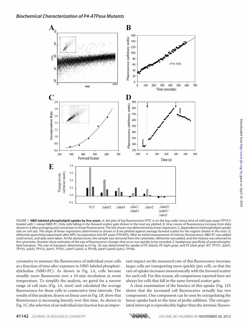
cytometry to measure the fluorescence of individual yeast cellsas a function of time after exposure to NBD-labeled phosphati-dylcholine (NBD-PC). As shown in Fig. 1A, cells becomesteadily more fluorescent over a 10-min incubation at roomtemperature. To simplify the analysis, we gated for a narrowrange of cell sizes (Fig. 1A, inset) and calculated the averagefluorescence for those cells in consecutive time intervals. Theresults of this analysis, drawnon linear axes in Fig. 1B, show thatfluorescence is increasing linearly over this time. As shown inFig. 1C, the selection of an individual size fraction has an impor-
tant impact on the measured rate of this fluorescence increase;larger cells are transporting more quickly (per cell), so that therate of uptake increasesmonotonically with the forward scatterfor each cell. For this reason, all comparisons reported here arealways for cells that fall in the same forward scatter gate.A close examination of the kinetics of this uptake (Fig. 1D)
shows that the increased cell fluorescence actually has twocomponents. One component can be seen by extrapolating thelinear uptake back to the time of probe addition. The extrapo-lated intercept is reproducibly higher than the intrinsic fluores-
FIGURE 1. NBD-labeled phospholipid uptake by live yeast. A, dot plot of log fluorescence (FITC is on the log scale) versus time of wild type yeast (TPY51)treated with 1-oleoyl-NBD-PC. Only cells falling in the forward scatter gate shown in the inset are plotted. B, time course of fluorescence increase from datashown in A after averaging and conversion to linear fluorescence. The line shown was determined by linear regression. C, dependence of phospholipid uptakerate on cell size. The slope of linear regressions determined as shown in B are plotted against average forward scatter for the regions shown in the inset. D,dithionite quenching experiment after MPC incorporation into WT yeast (THYAP5). After an initial measurement of intrinsic fluorescence, NBD-PC was added(solid arrow), and data were taken. At the dashed arrow, the sample was removed from the cytometer, dithionite was added, and the mixture was returned tothe cytometer. Brackets show estimates of the size of fluorescence changes that occur too quickly to be recorded. E, headgroup specificity of yeast phospho-lipid transport. The rate of transport, determined as in Fig. 1B, was determined for uptake of PC (black), PE (light gray), and PS (dark gray). WT, TPY51; �dnf1,TPY55; �dnf2, TPY53; �dnf1, TPY61; �dnf1/�dnf2, is TPY58; �dnf1/�dnf2/�drs2, TPY66.
Biochemical Characterization of P4-ATPase Mutants
41142 JOURNAL OF BIOLOGICAL CHEMISTRY VOLUME 287 • NUMBER 49 • NOVEMBER 30, 2012
cence of the cells, indicating that there is a small increase influorescence, which is complete within the 15 s between theaddition of the probe and the onset of data collection by thecytometer. The rate of this increase is thus too fast to measure.This rapid initial increase in fluorescence is then followed bythe slower linear increase in fluorescence. To determinewhether either of these components corresponds to probereaching the inside of the cell, we used the membrane-imper-meant reducing agent sodium dithionite. This reagent convertsthe nitro moiety of the NBD group to an amine and therebyreduces its fluorescence by about 100-fold; because it is animpermeant anion, it only affects probe that is not in the cellinterior (33). As shown in Fig. 1D, the addition of this reagentafter a period of incorporation produces a fast reduction influorescence, followed by a much slower decay. The first ofthese corresponds roughly in magnitude to the initial rapid risein fluorescence observed at time 0. Its rate is dependent on thedithionite concentration and thus corresponds to reduction ofprobe that is cell-associated but not inside the cell. The rate ofthe second decay phase does not depend on dithionite concen-tration (data not shown), implying that the slower componentcorresponds to inaccessible, internalized probe reaching theexternal surface either by flip from the internal leaflet, by exo-cytosis, or by degradation to amembrane-permeable form. Thepossibility that these results were an artifact of dithionite treat-ment was tested by carrying out similar experiments withoutdithionite, using BSA added to the medium to extract probethat was not internal. The results observed were virtually iden-tical to those shown here for dithionite (data not shown), indi-cating that these results are not peculiar to the use of dithionite.Together, these data suggest that 1) NBD phospholipidbecomes associated with cells at a rate that is too fast to meas-ure; 2) the size of this external pool of probe is very small andchanges very little during the first minutes of incubation; 3) atany given time, the total cell-associated fluorescence includesboth internal and external probe; and 4) the linearly increasingamount of probe corresponds to transport of the phospholipidanalog into the cell interior.In mammalian cells, phospholipid transport is restricted to
internalization of PE and PS, with the rate of the latter predom-inating, and PC is not transported at all (34). Application of theassay described above with NBD-labeled PC, PE, and PSshowed that phospholipid transport in yeast does not conformto this pattern (Fig. 1E). In particular, PE is generally the mostrapidly transported phospholipid, whereas PC andPS are trans-ported well and at about equal rates. In contrast to previousreports (20), we found no evidence for internalization of sphin-gomyelin (data not shown). Single deletions of dnf1, dnf2, ordrs2 all reduce but do not eliminate transport (Fig. 1E). PCtransport is completely abrogated in yeast harboring deletionsof both dnf1 and dnf2, showing clearly that these two transport-ers are responsible for PC transport at the plasma membraneobserved inwild type cells aswell as formuch of the transport ofPE. In the absence of the protein products of these two genes,the remaining transport of the aminophospholipids PS and PEresembles that seen in mammalian cells. Finally, the data showthat further deletion of drs2 eliminates transport of all phos-pholipid types, implying that the other two P4-ATPases, Neo1p
and Dnf3p, play no role at all in phospholipid transport at theyeast plasmamembrane. Single deletion of dnf3has no effect ontransport, confirming this conclusion. The effect of deletion ofneo1 on transport cannot be tested, because deletions of thisP4-ATPase are lethal.These observations suggest that, properly applied, these
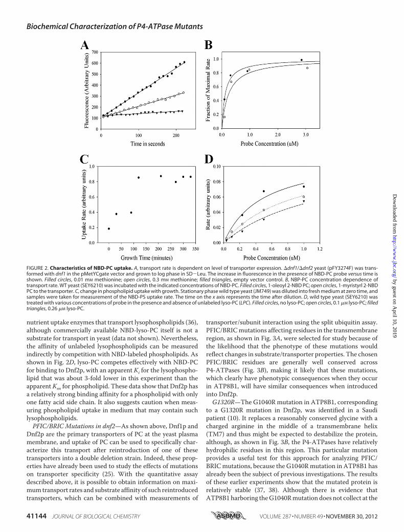
measurements could provide a quantitative assay for enzyme-catalyzed active transport of phospholipids. To testwhether therate of transport is dependent on the amount of transporterpresent in the membrane, we took advantage of the fact that, inthe absence of Dnf2p, internalization of NBD-PC is completelydependent on Dnf1p. We therefore introduced a plasmid con-taining the dnf1 gene under the control of a methionine pro-moter into a �dnf1/�dnf2 double deletion strain. As shown inFig. 2A, the rate of transport for NBD-PC in this system is nowregulated by the level of methionine in the medium, implyingthat the transport rate is a function of the amount of transporterin the cell. Second, we tested whether the rate of transport isdependent on substrate concentration in a saturable manner.To this end, we measured the rate of transport with NBD-la-beled phospholipid presented to the cells in the absence of car-rier vesicles or detergent micelles that present nonsaturable,competing binding sites for the probe. As shown in Fig. 2B,Under these circumstances, the rate of transport is substrateconcentration-dependent and is saturable. One important con-sideration is that the free probe concentration is limited by thecritical micelle concentration of the probe, and we thereforecompared the concentration dependence using NBD-PC witheither oleoyl or myristoyl side chains in the C1 position of theglycerol backbone; the critical micelle concentration of thesetwo versions of the probe should differ by about a factor of 40(35). As shown in Fig. 2B, the dependence of the rate of trans-port on probe concentration is the same in both cases, showingthat 1) transport rate is dependent on saturable binding ofphospholipid to an external binding site, 2) this dependence isnot an artifact of probe critical micelle concentration, and 3)the affinity of the transporter for the probe is not very sensitiveto small changes in fatty acid side chain length.The ability to make quantitative estimates of transporter
activity in yeast facilitated investigation of two basic cell biolog-ical questions about transport. The first question is whetherphospholipid transport rates change depending onwhether thecell is dividing. To examine this question, we inoculated yeastfrom a stationary culture into fresh medium and measuredtransport rates at intervals after inoculation. As shown in Fig.2C, stationary phase yeast barely transports at all. As the yeastenters into logarithmic phase growth, transport rates increase,reaching a plateau by about 3 h after inoculation. Comparisonof transport rates with probes containing different headgroupsand yeast containing different deletions showed a similar pat-tern for all major phospholipid groups and all three transport-ers (data not shown). Because of this finding, care was taken toensure that cells were well into log phase growth when theywere taken for transport measurements.A quantitative assay for phospholipid internalization rate in
yeast also provides a platform for investigating interactions withsubstrates forwhich suitable fluorescent versions arenot available.For example, it has been suggested that the P4-ATPases are
Biochemical Characterization of P4-ATPase Mutants
NOVEMBER 30, 2012 • VOLUME 287 • NUMBER 49 JOURNAL OF BIOLOGICAL CHEMISTRY 41143
nutrient uptake enzymes that transport lysophospholipids (36),although commercially available NBD-lyso-PC itself is not asubstrate for transport in yeast (data not shown). Nevertheless,the affinity of unlabeled lysophospholipids can be measuredindirectly by competition with NBD-labeled phospholipids. Asshown in Fig. 2D, lyso-PC competes effectively with NBD-PCfor binding to Dnf2p, with an apparent Ki for the lysophospho-lipid that was about 3-fold lower in this experiment than theapparentKm for phospholipid. These data show that Dnf2p hasa relatively strong binding affinity for a phospholipid with onlyone fatty acid side chain. It also suggests caution when meas-uring phospholipid uptake in medium that may contain suchlysophospholipids.PFIC/BRIC Mutations in dnf2—As shown above, Dnf1p and
Dnf2p are the primary transporters of PC at the yeast plasmamembrane, and uptake of PC can be used to specifically char-acterize this transport after reintroduction of one of thesetransporters into a double deletion strain. Indeed, these prop-erties have already been used to study the effects of mutationson transporter specificity (25). With the quantitative assaydescribed above, it is possible to obtain information on maxi-mumtransport rates and substrate affinity of such reintroducedtransporters, which can be combined with measurements of
transporter/subunit interaction using the split ubiquitin assay.PFIC/BRICmutations affecting residues in the transmembraneregion, as shown in Fig. 3A, were selected for study because ofthe likelihood that the phenotype of these mutations wouldreflect changes in substrate/transporter properties. The chosenPFIC/BRIC residues are generally well conserved acrossP4-ATPases (Fig. 3B), making it likely that these mutations,which clearly have phenotypic consequences when they occurin ATP8B1, will have similar consequences when introducedinto Dnf2p.G1320R—The G1040R mutation in ATP8B1, corresponding
to a G1320R mutation in Dnf2p, was identified in a Saudipatient (10). It replaces a reasonably conserved glycine with acharged arginine in the middle of a transmembrane helix(TM7) and thus might be expected to destabilize the protein,although, as shown in Fig. 3B, the P4-ATPases have relativelyhydrophilic residues in this region. This particular mutationprovides a useful test for this approach for analyzing PFIC/BRICmutations, because the G1040Rmutation in ATP8B1 hasalready been the subject of previous investigations. The resultsof these earlier experiments show that the mutated protein isrelatively stable (37, 38). Although there is evidence thatATP8B1harboring theG1040Rmutation does not collect at the
FIGURE 2. Characteristics of NBD-PC uptake. A, transport rate is dependent on level of transporter expression. �dnf1/�dnf2 yeast (pFY3274F) was trans-formed with dnf1 in the pMetYCgate vector and grown to log phase in SD�Leu. The increase in fluorescence in the presence of NBD-PC probe versus time isshown. Filled circles, 0.01 mM methionine; open circles, 0.3 mM methionine; filled triangles, empty vector control. B, NBP-PC concentration dependence oftransport rate. WT yeast (SEY6210) was incubated with the indicated concentrations of NBD-PC. Filled circles, 1-oleoyl 2-NBD PC; open circles, 1-myristyrl 2-NBDPC to the transporter. C, change in phospholipid uptake with growth. Stationary phase wild type yeast (JM749) was diluted into fresh medium at zero time, andsamples were taken for measurement of the NBD-PS uptake rate. The time on the x axis represents the time after dilution. D, wild type yeast (SEY6210) wastreated with various concentrations of probe in the presence and absence of unlabeled lyso-PC (LPC). Filled circles, no lyso-PC; open circles, 0.1 �M lyso-PC; filledtriangles, 0.26 �M lyso-PC.
Biochemical Characterization of P4-ATPase Mutants
41144 JOURNAL OF BIOLOGICAL CHEMISTRY VOLUME 287 • NUMBER 49 • NOVEMBER 30, 2012
canalicularmembrane (37), it does reach the plasmamembrane(38), although perhaps at reduced levels (37). It has beenreported that this mutant protein may interact with its subunitfrom the Cdc50 family (38), although the interaction may beweak (37). Because there is no assay for transport by ATP8B1,these studies could not provide evidence one way or another onthe crucial question of whether the mutation affects enzymeactivity.With this background, we investigated the properties of the
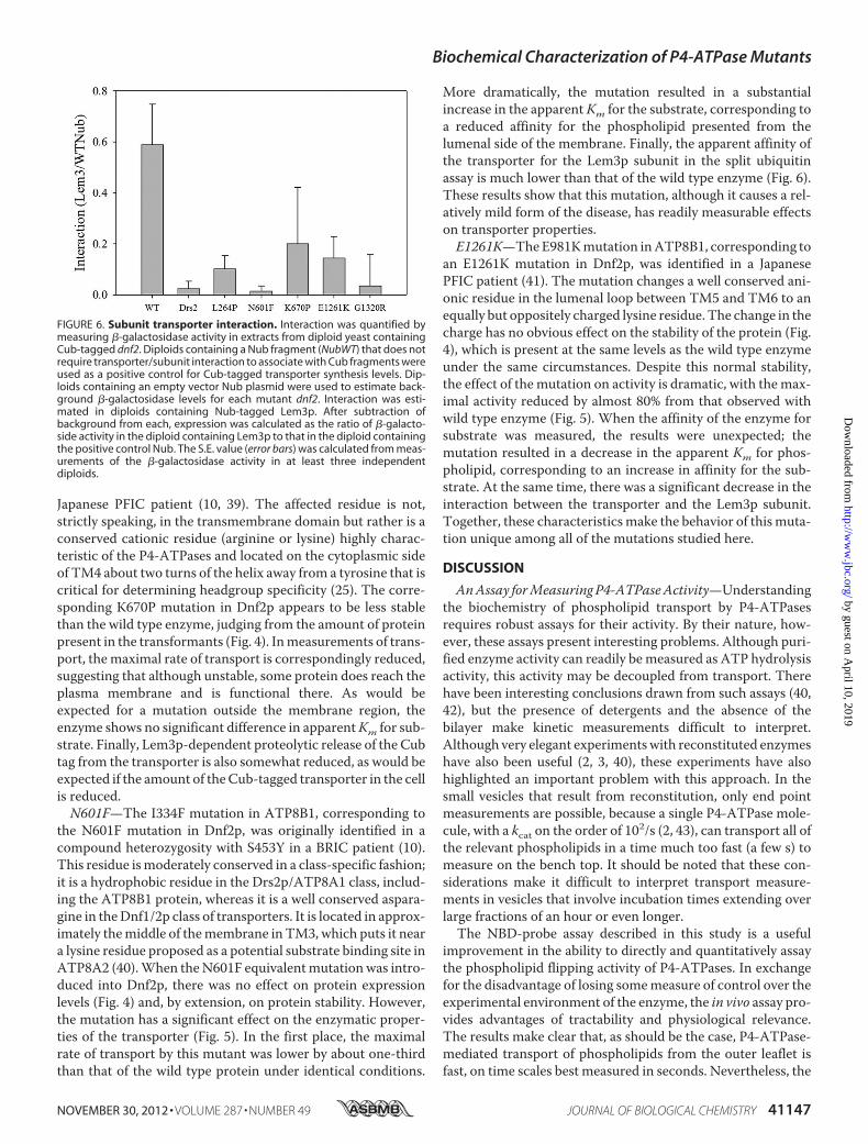
corresponding Dnf2p mutant, G1320R. As in the case of theATP8B1 mutant, we found that the mutated yeast protein isreasonably stable and accumulates in cells to concentrationscomparable with those of the wild type protein expressed fromthe same promoter under the same conditions (Fig. 4). How-ever, the mutated protein is completely incapable of transport-ing phospholipids (Fig. 5A). In addition, the ability of theG1320Rmutant to interact with Lem3p, the normal subunit forthis transporter, is also largely abrogated (Fig. 6). Together,these data show that, although this mutation does not havedramatic effects on protein stability, its disease phenotype isreadily explained by its effect on protein activity.
L264P—The PFICmutant L127P, corresponding to L264P inDnf2p, was identified in a Japanese patient (10, 39) and substi-tutes a hydrophobic but helix-breaking proline for amoderatelyconserved leucine in the first transmembrane domain near thelumenal side of the membrane. This mutated ATP8B1 has alsobeen the subject of experiments that suggested that the proteinwas stable, properly localized in the cell, and able to interactwith its cognate Cdc50 family subunit (38), raising the questionof why it results in disease. We therefore analyzed the homolo-gous L264P mutant of Dnf2p. As in the case of the ATP8B1homolog, the mutant is as stable as wild type enzyme (Fig. 4);moreover, as shown in Fig. 5A, it transports at the same maxi-mal rate, and with the same Km, as wild type enzyme, showingthat the mutated enzyme is both active and properly localized.However, the interaction of the mutant Dnf2p with Lem3p ismeasurably weaker than that observedwith the wild type trans-porter (Fig. 6), which may finally provide a clue as to why thecorresponding ATP8B1 mutation has an abnormal phenotypein humans.K670P—The R412P mutation in ATP8B1, corresponding to
K670P in Dnf2p, was identified as a homozygous mutation in a
FIGURE 3. A, locations of mutations tested here on a schematic of P4ATPase organization (phosphorylation (P), nucleotide binding (N), and actuator (A) domainsare shown). Dark boxes show mutation in ATP8B1. B, local alignment of ATP8B1 and Dnf2p sequence around mutations. the bar graph below shows theconservation scores based on an alignment with �250 P4-ATPase sequences. The conservation scores as determined by Consurf have been multiplied by �1so that more positive values correspond to more conserved residues. *, mutated residue. C, corresponding residues in ATP8B1, Dnf2p, and the proton pumpAHA2 structure (Protein Data Bank entry 3B8C).
Biochemical Characterization of P4-ATPase Mutants
NOVEMBER 30, 2012 • VOLUME 287 • NUMBER 49 JOURNAL OF BIOLOGICAL CHEMISTRY 41145
FIGURE 4. Protein expression levels. Binding of Alexa-tagged anti-HA antibody to a C-terminal HA-tagged Dnf2p was measured by cytometry. Curves are FITCfluorescence histograms for permeabilized cells, as judged by the ability to bind PI. . . . . . . ., no dnf2; ——, wild type dnf2; – – – -, L264P; - - - - -, N601F; –�–�–,K670P; – – – –, E1261K; –��–��, G1320R. Inset, total fluorescence over background. Total fluorescence was calculated by multiplying the fluorescence of each binby the fraction of total cells in that bin, total fluorescence in the absence of dnf2 was subtracted, and the remainder was shown to provide an estimate of theoverall protein level.
FIGURE 5. Effect of PFIC BRIC mutations in dnf2 on PC transport at the plasma membrane. A, maximal transport rate for each of the PFIC/BRIC mutationsin dnf2 as a fraction of the rate observed in parallel with wild type dnf2 control. B, apparent Km measurement. The uptake rate as a function of NBD-PCconcentration was measured for wild type (open circles), N601F (filled triangles), and E1261K (filled circles). The fraction of the maximal rate observed for eachmutant (see Fig. 5A) was plotted against different concentrations of probe and fitted to the equation, f � Vmax/(1 � (Km/x)). At least two separate clones of eachmutation were tested, and the results were averaged. C, summary of apparent Km and Vmax with S.E. values for each mutant dnf2. Error bars, S.E.
Biochemical Characterization of P4-ATPase Mutants
41146 JOURNAL OF BIOLOGICAL CHEMISTRY VOLUME 287 • NUMBER 49 • NOVEMBER 30, 2012
Japanese PFIC patient (10, 39). The affected residue is not,strictly speaking, in the transmembrane domain but rather is aconserved cationic residue (arginine or lysine) highly charac-teristic of the P4-ATPases and located on the cytoplasmic sideof TM4 about two turns of the helix away from a tyrosine that iscritical for determining headgroup specificity (25). The corre-sponding K670P mutation in Dnf2p appears to be less stablethan the wild type enzyme, judging from the amount of proteinpresent in the transformants (Fig. 4). Inmeasurements of trans-port, themaximal rate of transport is correspondingly reduced,suggesting that although unstable, some protein does reach theplasma membrane and is functional there. As would beexpected for a mutation outside the membrane region, theenzyme shows no significant difference in apparentKm for sub-strate. Finally, Lem3p-dependent proteolytic release of the Cubtag from the transporter is also somewhat reduced, as would beexpected if the amount of theCub-tagged transporter in the cellis reduced.N601F—The I334F mutation in ATP8B1, corresponding to
the N601F mutation in Dnf2p, was originally identified in acompound heterozygosity with S453Y in a BRIC patient (10).This residue ismoderately conserved in a class-specific fashion;it is a hydrophobic residue in the Drs2p/ATP8A1 class, includ-ing the ATP8B1 protein, whereas it is a well conserved aspara-gine in theDnf1/2p class of transporters. It is located in approx-imately themiddle of themembrane in TM3, which puts it neara lysine residue proposed as a potential substrate binding site inATP8A2 (40).When theN601F equivalentmutationwas intro-duced into Dnf2p, there was no effect on protein expressionlevels (Fig. 4) and, by extension, on protein stability. However,the mutation has a significant effect on the enzymatic proper-ties of the transporter (Fig. 5). In the first place, the maximalrate of transport by this mutant was lower by about one-thirdthan that of the wild type protein under identical conditions.
More dramatically, the mutation resulted in a substantialincrease in the apparent Km for the substrate, corresponding toa reduced affinity for the phospholipid presented from thelumenal side of the membrane. Finally, the apparent affinity ofthe transporter for the Lem3p subunit in the split ubiquitinassay is much lower than that of the wild type enzyme (Fig. 6).These results show that this mutation, although it causes a rel-atively mild form of the disease, has readily measurable effectson transporter properties.E1261K—TheE981Kmutation inATP8B1, corresponding to
an E1261K mutation in Dnf2p, was identified in a JapanesePFIC patient (41). The mutation changes a well conserved ani-onic residue in the lumenal loop between TM5 and TM6 to anequally but oppositely charged lysine residue. The change in thecharge has no obvious effect on the stability of the protein (Fig.4), which is present at the same levels as the wild type enzymeunder the same circumstances. Despite this normal stability,the effect of the mutation on activity is dramatic, with themax-imal activity reduced by almost 80% from that observed withwild type enzyme (Fig. 5). When the affinity of the enzyme forsubstrate was measured, the results were unexpected; themutation resulted in a decrease in the apparent Km for phos-pholipid, corresponding to an increase in affinity for the sub-strate. At the same time, there was a significant decrease in theinteraction between the transporter and the Lem3p subunit.Together, these characteristicsmake the behavior of this muta-tion unique among all of the mutations studied here.
DISCUSSION
AnAssay forMeasuring P4-ATPaseActivity—Understandingthe biochemistry of phospholipid transport by P4-ATPasesrequires robust assays for their activity. By their nature, how-ever, these assays present interesting problems. Although puri-fied enzyme activity can readily bemeasured as ATP hydrolysisactivity, this activity may be decoupled from transport. Therehave been interesting conclusions drawn from such assays (40,42), but the presence of detergents and the absence of thebilayer make kinetic measurements difficult to interpret.Although very elegant experimentswith reconstituted enzymeshave also been useful (2, 3, 40), these experiments have alsohighlighted an important problem with this approach. In thesmall vesicles that result from reconstitution, only end pointmeasurements are possible, because a single P4-ATPase mole-cule, with a kcat on the order of 102/s (2, 43), can transport all ofthe relevant phospholipids in a time much too fast (a few s) tomeasure on the bench top. It should be noted that these con-siderations make it difficult to interpret transport measure-ments in vesicles that involve incubation times extending overlarge fractions of an hour or even longer.The NBD-probe assay described in this study is a useful
improvement in the ability to directly and quantitatively assaythe phospholipid flipping activity of P4-ATPases. In exchangefor the disadvantage of losing somemeasure of control over theexperimental environment of the enzyme, the in vivo assay pro-vides advantages of tractability and physiological relevance.The results make clear that, as should be the case, P4-ATPase-mediated transport of phospholipids from the outer leaflet isfast, on time scales best measured in seconds. Nevertheless, the
FIGURE 6. Subunit transporter interaction. Interaction was quantified bymeasuring �-galactosidase activity in extracts from diploid yeast containingCub-tagged dnf2. Diploids containing a Nub fragment (NubWT) that does notrequire transporter/subunit interaction to associate with Cub fragments wereused as a positive control for Cub-tagged transporter synthesis levels. Dip-loids containing an empty vector Nub plasmid were used to estimate back-ground �-galactosidase levels for each mutant dnf2. Interaction was esti-mated in diploids containing Nub-tagged Lem3p. After subtraction ofbackground from each, expression was calculated as the ratio of �-galacto-side activity in the diploid containing Lem3p to that in the diploid containingthe positive control Nub. The S.E. value (error bars) was calculated from meas-urements of the �-galactosidase activity in at least three independentdiploids.
Biochemical Characterization of P4-ATPase Mutants
NOVEMBER 30, 2012 • VOLUME 287 • NUMBER 49 JOURNAL OF BIOLOGICAL CHEMISTRY 41147
data also show that it is the transport step that is rate-limiting;the probe saturates external binding sites at a rate too fast forthe assay to measure. The low level of PC transport observedwhen dnf1 and dnf2 are deleted shows that endocytic uptake ofphospholipids is negligible under the conditions of the assay, aresult that is not unexpected given the short duration of theuptake measurements and the low level of steady-state probeincorporation into the membrane (Fig. 1D). Although notexploited extensively in these studies, the data from the lyso-PCinhibition experiment show that a competition protocol can beused to obtain information about transported substrates whoseuptake cannot be directly measured. Even this preliminaryexperiment, however, suggests that the P4-ATPases will act aslysophospholipid uptake enzymes when the concentrations ofsuch potential substrates in the medium reach the micromolarlevel. Although in this case the affinity for the lysophospholipidis higher than for the corresponding phospholipid probe, theseresults do not show that uptake of such lysophospholipids is theprimary function of these transporters, because it is known thatNBD phospholipids may not be as efficiently taken up byP4-ATPases as natural phospholipids (44).Insights about PFIC and BRIC—Several of the findings
reported here tend to validate the strategy of analyzing PFICmutants by studying the properties of the correspondingmuta-tions in Dnf2p. The most useful of the evidence comes fromthose mutations that have already been subject to study in thehuman transporter. The unexpected stability of the proteinmolecule harboring theG1040Rmutation (the reduced but stillsubstantial protein levels), combined with a compromisedinteraction with subunit, are reflected faithfully in the pheno-type of the G1320Rmutant of Dnf2p. Similarly, the robust nor-mality of the L127P mutant in ATP8B1 is reproduced in theL264P formofDnf2p. So far as direct comparisons can bemade,these data suggest that the evolutionary conservation of resi-dues is consistent with a similarity in the phenotypes of thecorresponding mutations in the two proteins.The development of disease symptoms when ATP8B1 is
mutated is strong evidence that the mutation has a significantimpact on transporter function, even if the affected function isnot known. In the absence of functional assays, the ability tomeasure protein synthesis has concentrated attention on pos-sible indirect effects of such mutations (e.g. on protein folding)(16, 38). Indeed, the data presented here support just such amechanism for the effect of the PFIC mutation R412P, wherethe corresponding K670P mutant shows parallel reductions inprotein level, in maximal transport activity, and in transporteravailable for interaction with subunit, whereas the activity thatremains seems normal in its affinity for phospholipid substrate.In the mutations studied here, the phenotype of the R412P/
K670Pmutation is an outlier in that it has an impact on proteinstability but little effect on the enzymatic properties of thetransporter. That said, there is considerable diversity in thenature of the effects on phenotypes. The simplest of these isthe phenotype of G1040R/G1320R, where the disruption ofTM7 results in no transport activity at the plasma membraneand little interaction with subunit. Although less dramatic,both I334F/N601F and E981K/E1261K are also mutations thatdo not reduce protein levels but do reduce transport activity at
the plasma membrane as well as interaction with subunit.Remarkably, the effects of these mutations on Km are completeopposites of one another, a matter discussed in more detailbelow. The lack of effect of the L127P/L264P mutation on pro-tein levels confirmed here is complemented by the lack of effectof this mutation on either maximal transport activity or sub-strate affinity, raising the question of why a patient harboringthis mutation has PFIC at all. The observation of a reducedaffinity for subunit observed with the L264P mutation maypoint to an explanation, especially in light of an important dif-ference between the human and yeast systems. In yeast, each ofthe P4-ATPases, with the exception of Neo1p, is associatedwith its own subunit; in the case of Dnf2p, that subunit isLem3p, a subunit that is shared with Dnf1p in normal yeast butis not shared in our experimental system because Dnf1p is notpresent. In humans, it is likely that all of the P4-ATPases inter-act with a common pool of subunits, because there are onlythree subunit genes in mammals, one of which is probably notfunctional (45), and 12 transporters other than the two ATP9relatives of neo1. Under physiological conditions, the L127Pmutant of ATP8B1 may thus be at a disadvantage in competi-tion with other transporters for this common pool of subunits.Because association with a subunit is a functional necessity(6–8, 19), this disadvantage may be the source of the diseasephenotype.Together, these data show that each of the mutations origi-
nally identified in ATP8B1 results in a measurable defect whenrecreated in Dnf2p and, in most cases, in a defect in the level oftransport activity at the physiologically relevant location.Because so much less is known with certainty about the prop-erties of ATP8B1, these results are probably best treated asqualitative indications of the nature of the induced defectsrather than quantitative predictions of the phenotypes of thecorresponding ATP8B1 forms. For example, the results ofBaldridge and Graham (25) suggest that the same residue isimportant in two transporters (Drs2p andDnf1p)with differenttransport specificities. However, the collection of residues thatinteract with substrate is undoubtedly larger than the list that isalready known. As a result, until the specificity and kineticparameters of phospholipid binding and transport are directlycharacterized and quantitated for ATP8B1 itself, the changes inPC transport measured here can only provide an indication ofthe kind of defect that these mutations induce. Nevertheless,even this information clearly differentiates between the conse-quences of different mutations, making these results a poten-tially useful guide to howdifferent forms of the diseasemight beunderstood and treated.In thinking about the more general implications of the phe-
notypes of these mutations, and particularly their effects oninteraction between subunit and transporter, it is important toremember that the latter is not static; quite to the contrary, it isa dynamic part of the reaction cycle of transport (6). In partic-ular, the subunit interacts most strongly with the lumenal-fac-ing E2P conformation of the enzyme, the pointwhere the trans-porter binds the phospholipid substrate. Mutations can affectsubunit-transporter interaction, not by directly altering theprotein-protein interaction between the two peptides, but byaltering the rates of individual steps in the reaction cycle, and
Biochemical Characterization of P4-ATPase Mutants
41148 JOURNAL OF BIOLOGICAL CHEMISTRY VOLUME 287 • NUMBER 49 • NOVEMBER 30, 2012
thereby how the protein is distributed between different con-formational stages of the reaction cycle. One consequence ofthis complex dynamic is that the relationship between interac-tion and transport activity is not readily predictable, as has beenobserved in the study of subunits with altered disulfides in thelumenal domain (46).Recently, a conserved tyrosine on the cytoplasmic surface of
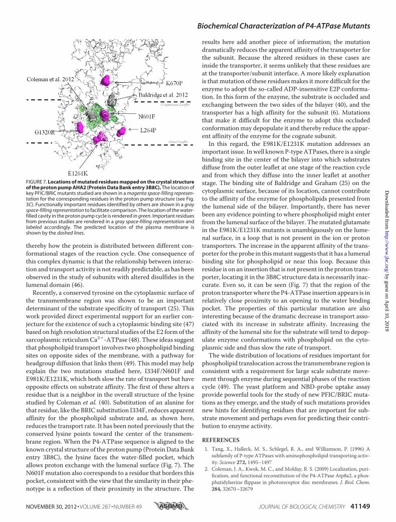
the transmembrane region was shown to be an importantdeterminant of the substrate specificity of transport (25). Thiswork provided direct experimental support for an earlier con-jecture for the existence of such a cytoplasmic binding site (47)based on high resolution structural studies of the E2 formof thesarcoplasmic reticulumCa2�-ATPase (48). These ideas suggestthat phospholipid transport involves two phospholipid bindingsites on opposite sides of the membrane, with a pathway forheadgroup diffusion that links them (49). This model may helpexplain the two mutations studied here, I334F/N601F andE981K/E1231K, which both slow the rate of transport but haveopposite effects on substrate affinity. The first of these alters aresidue that is a neighbor in the overall structure of the lysinestudied by Coleman et al. (40). Substitution of an alanine forthat residue, like the BRIC substitution I334F, reduces apparentaffinity for the phospholipid substrate and, as shown here,reduces the transport rate. It has been noted previously that theconserved lysine points toward the center of the transmem-brane region. When the P4-ATPase sequence is aligned to theknown crystal structure of the proton pump (ProteinData Bankentry 3B8C), the lysine faces the water-filled pocket, whichallows proton exchange with the lumenal surface (Fig. 7). TheN601Fmutation also corresponds to a residue that borders thispocket, consistent with the view that the similarity in their phe-notype is a reflection of their proximity in the structure. The
results here add another piece of information; the mutationdramatically reduces the apparent affinity of the transporter forthe subunit. Because the altered residues in these cases areinside the transporter, it seems unlikely that these residues areat the transporter/subunit interface. A more likely explanationis thatmutation of these residuesmakes it more difficult for theenzyme to adopt the so-called ADP-insensitive E2P conforma-tion. In this form of the enzyme, the substrate is occluded andexchanging between the two sides of the bilayer (40), and thetransporter has a high affinity for the subunit (6). Mutationsthat make it difficult for the enzyme to adopt this occludedconformationmay depopulate it and thereby reduce the appar-ent affinity of the enzyme for the cognate subunit.In this regard, the E981K/E1231K mutation addresses an
important issue. Inwell knownP-typeATPases, there is a singlebinding site in the center of the bilayer into which substratesdiffuse from the outer leaflet at one stage of the reaction cycleand from which they diffuse into the inner leaflet at anotherstage. The binding site of Baldridge and Graham (25) on thecytoplasmic surface, because of its location, cannot contributeto the affinity of the enzyme for phospholipids presented fromthe lumenal side of the bilayer. Importantly, there has neverbeen any evidence pointing to where phospholipid might enterfrom the lumenal surface of the bilayer. Themutated glutamatein the E981K/E1231K mutants is unambiguously on the lume-nal surface, in a loop that is not present in the ion or protontransporters. The increase in the apparent affinity of the trans-porter for the probe in thismutant suggests that it has a lumenalbinding site for phospholipid or near this loop. Because thisresidue is on an insertion that is not present in the proton trans-porter, locating it in the 3B8C structure data is necessarily inac-curate. Even so, it can be seen (Fig. 7) that the region of theproton transporterwhere the P4-ATPase insertion appears is inrelatively close proximity to an opening to the water bindingpocket. The properties of this particular mutation are alsointeresting because of the dramatic decrease in transport asso-ciated with its increase in substrate affinity. Increasing theaffinity of the lumenal site for the substrate will tend to depop-ulate enzyme conformations with phospholipid on the cyto-plasmic side and thus slow the rate of transport.The wide distribution of locations of residues important for
phospholipid translocation across the transmembrane region isconsistent with a requirement for large scale substrate move-ment through enzyme during sequential phases of the reactioncycle (49). The yeast platform and NBD-probe uptake assayprovide powerful tools for the study of new PFIC/BRIC muta-tions as they emerge, and the study of such mutations providesnew hints for identifying residues that are important for sub-strate movement and perhaps even for predicting their contri-bution to enzyme activity.
REFERENCES1. Tang, X., Halleck, M. S., Schlegel, R. A., and Williamson, P. (1996) A
subfamily of P-type ATPases with aminophospholipid transporting activ-ity. Science 272, 1495–1497
2. Coleman, J. A., Kwok, M. C., and Molday, R. S. (2009) Localization, puri-fication, and functional reconstitution of the P4-ATPase Atp8a2, a phos-phatidylserine flippase in photoreceptor disc membranes. J. Biol. Chem.284, 32670–32679
FIGURE 7. Locations of mutated residues mapped on the crystal structureof the proton pump AHA2 (Protein Data Bank entry 3B8C). The location ofkey PFIC/BRIC mutants studied are shown in a magenta space-filling represen-tation for the corresponding residues in the proton pump structure (see Fig.3C). Functionally important residues identified by others are shown in a grayspace-filling representation to facilitate comparison. The location of the water-filled cavity in the proton pump cycle is rendered in green. Important residuesfrom previous studies are rendered in a gray space-filling representation andlabeled accordingly. The predicted location of the plasma membrane isshown by the dashed lines.
Biochemical Characterization of P4-ATPase Mutants
NOVEMBER 30, 2012 • VOLUME 287 • NUMBER 49 JOURNAL OF BIOLOGICAL CHEMISTRY 41149
3. Zhou, X., and Graham, T. R. (2009) Reconstitution of phospholipid trans-locase activitywith purifiedDrs2p, a type-IVP-typeATPase frombuddingyeast. Proc. Natl. Acad. Sci. U.S.A. 106, 16586–16591
4. Axelsen, K. B., and Palmgren,M. G. (2001) Inventory of the superfamily ofP-type ion pumps in Arabidopsis. Plant Physiol. 126, 696–706
5. Saito, K., Fujimura-Kamada, K., Furuta, N., Kato, U., Umeda, M., andTanaka, K. (2004) Cdc50p, a protein required for polarized growth, asso-ciates with the Drs2p P-type ATPase implicated in phospholipid translo-cation in Saccharomyces cerevisiae.Mol. Biol. Cell 15, 3418–3432
6. Lenoir, G., Williamson, P., Puts, C. F., and Holthuis, J. C. (2009) Cdc50pplays a vital role in the ATPase reaction cycle of the putative aminophos-pholipid transporter Drs2p. J. Biol. Chem. 284, 17956–17967
7. Bryde, S., Hennrich, H., Verhulst, P. M., Devaux, P. F., Lenoir, G., andHolthuis, J. C. (2010) CDC50 proteins are critical components of the hu-man class-1 P4-ATPase transport machinery. J. Biol. Chem. 285,40562–40572
8. Coleman, J. A., and Molday, R. S. (2011) Critical role of the �-subunitCDC50A in the stable expression, assembly, subcellular localization, andlipid transport activity of the P4-ATPase ATP8A2. J. Biol. Chem. 286,17205–17216
9. Bull, L. N., van Eijk,M. J., Pawlikowska, L., DeYoung, J. A., Juijn, J. A., Liao,M., Klomp, L.W., Lomri, N., Berger, R., Scharschmidt, B. F., Knisely, A. S.,Houwen, R. H., and Freimer, N. B. (1998) A gene encoding a P-type AT-Pase mutated in two forms of hereditary cholestasis. Nat. Genet. 18,219–224
10. Klomp, L. W., Vargas, J. C., van Mil, S. W., Pawlikowska, L., Strautnieks,S. S., van Eijk,M. J., Juijn, J. A., Pabon-Pena, C., Smith, L. B., DeYoung, J. A.,Byrne, J. A., Gombert, J., van der Brugge, G., Berger, R., Jankowska, I.,Pawlowska, J., Villa, E., Knisely, A. S., Thompson, R. J., Freimer, N. B.,Houwen, R. H., and Bull, L. N. (2004) Characterization of mutations inATP8B1 associated with hereditary cholestasis. Hepatology 40, 27–38
11. Eppens, E. F., vanMil, S.W., de Vree, J. M., Mok, K. S., Juijn, J. A., Elferink,R., Berger, R., Houwen, R. H., and Klomp, L. W. (2001) FIC1, the proteinaffected in two forms of hereditary cholestasis, is localized in the cholan-giocyte and the canalicular membrane of the hepatocyte. J. Hepatol. 35,436–443
12. Stapelbroek, J. M., Peters, T. A., van Beurden, D. H., Curfs, J. H., Joosten,A., Beynon, A. J., van Leeuwen, B.M., van der Velden, L.M., Bull, L., OudeElferink, R. P., van Zanten, B. A., Klomp, L. W., and Houwen, R. H. (2009)ATP8B1 is essential for maintaining normal hearing. Proc. Natl. Acad. Sci.U.S.A. 106, 9709–9714
13. Palmgren, M. G., and Nissen, P. (2011) P-type ATPases. Annu. Rev. Bio-phys. 40, 243–266
14. Paulusma, C. C., Groen, A., Kunne, C., Ho-Mok, K. S., Spijkerboer, A. L.,Rudi de Waart, D., Hoek, F. J., Vreeling, H., Hoeben, K. A., van Marle, J.,Pawlikowska, L., Bull, L. N., Hofmann, A. F., Knisely, A. S., and OudeElferink, R. P. (2006) Atp8b1 deficiency in mice reduces resistance of thecanalicular membrane to hydrophobic bile salts and impairs bile salttransport. Hepatology 44, 195–204
15. Paulusma, C. C., Folmer, D. E., Ho-Mok, K. S., de Waart, D. R., Hilarius,P. M., Verhoeven, A. J., and Oude Elferink, R. P. (2008) ATP8B1 requiresan accessory protein for endoplasmic reticulum exit and plasma mem-brane lipid flippase activity. Hepatology 47, 268–278
16. Verhulst, P. M., van der Velden, L. M., Oorschot, V., van Faassen, E. E.,Klumperman, J., Houwen, R. H., Pomorski, T. G., Holthuis, J. C., andKlomp, L. W. (2010) A flippase-independent function of ATP8B1, theprotein affected in familial intrahepatic cholestasis type 1, is required forapical protein expression andmicrovillus formation in polarized epithelialcells. Hepatology 51, 2049–2060
17. Chen, C. Y., Ingram, M. F., Rosal, P. H., and Graham, T. R. (1999) Role forDrs2p, a P-type ATPase and potential aminophospholipid translocase, inyeast late Golgi function. J. Cell Biol. 147, 1223–1236
18. Hua, Z., Fatheddin, P., and Graham, T. R. (2002) An essential subfamily ofDrs2p-related P-type ATPases is required for protein trafficking betweenGolgi complex and endosomal/vacuolar system. Mol. Biol. Cell 13,3162–3177
19. van der Velden, L.M.,Wichers, C. G., van Breevoort, A. E., Coleman, J. A.,Molday, R. S., Berger, R., Klomp, L. W., and van de Graaf, S. F. (2010)
Heteromeric interactions required for abundance and subcellular local-ization of human CDC50 proteins and class 1 P4-ATPases. J. Biol. Chem.285, 40088–40096
20. Pomorski, T., Lombardi, R., Riezman, H., Devaux, P. F., vanMeer, G., andHolthuis, J. C. (2003) Drs2p-related P-type ATPases Dnf1p and Dnf2p arerequired for phospholipid translocation across the yeast plasma mem-brane and serve a role in endocytosis.Mol. Biol. Cell 14, 1240–1254
21. Natarajan, P., Wang, J., Hua, Z., and Graham, T. R. (2004) Drs2p-coupledaminophospholipid translocase activity in yeast Golgi membranes andrelationship to in vivo function. Proc. Natl. Acad. Sci. U.S.A. 101,10614–10619
22. Alder-Baerens, N., Lisman, Q., Luong, L., Pomorski, T., andHolthuis, J. C.(2006) Loss of P4 ATPases Drs2p and Dnf3p disrupts aminophospholipidtransport and asymmetry in yeast post-Golgi secretory vesicles.Mol. Biol.Cell 17, 1632–1642
23. Chen, S., Wang, J., Muthusamy, B. P., Liu, K., Zare, S., Andersen, R. J., andGraham, T. R. (2006) Roles for the Drs2p-Cdc50p complex in proteintransport and phosphatidylserine asymmetry of the yeast plasma mem-brane. Traffic 7, 1503–1517
25. Baldridge, R. D., and Graham, T. R. (2012) Identification of residues de-fining phospholipid flippase substrate specificity of type IV P-type AT-Pases. Proc. Natl. Acad. Sci. U.S.A. 109, E290–E298
26. Obrdlik, P., El-Bakkoury, M., Hamacher, T., Cappellaro, C., Vilarino, C.,Fleischer, C., Ellerbrok, H., Kamuzinzi, R., Ledent, V., Blaudez, D., Sand-ers, D., Revuelta, J. L., Boles, E., Andre, B., and Frommer, W. B. (2004) K�
channel interactions detected by a genetic system optimized for system-atic studies ofmembrane protein interactions. Proc. Natl. Acad. Sci. U.S.A.101, 12242–12247
27. Grefen, C., Obrdlik, P., and Harter, K. (2009) The determination of pro-tein-protein interactions by themating-based split-ubiquitin system (mb-SUS).Methods Mol. Biol. 479, 217–233
28. Kakkar, T., Boxenbaum, H., andMayersohn,M. (1999) Estimation ofKi ina competitive enzyme-inhibitionmodel. Comparisons among threemeth-ods of data analysis. Drug Metab. Dispos. 27, 756–762
29. Grefen, C., Lalonde, S., and Obrdlik, P. (2007) Split-ubiquitin system foridentifying protein-protein interactions inmembrane and full-length pro-teins. Curr. Protoc. Neurosci. Chapter 5, Unit 5.27
30. Pedersen, B. P., Buch-Pedersen, M. J., Morth, J. P., Palmgren, M. G., andNissen, P. (2007) Crystal structure of the plasmamembrane proton pump.Nature 450, 1111–1114
31. Pei, J., Kim, B. H., and Grishin, N. V. (2008) PROMALS3D. A tool formultiple protein sequence and structure alignments. Nucleic Acids Res.36, 2295–2300
32. Ashkenazy, H., Erez, E., Martz, E., Pupko, T., and Ben-Tal, N. (2010) Con-Surf 2010. Calculating evolutionary conservation in sequence and struc-ture of proteins and nucleic acids. Nucleic Acids Res. 38,W529–W533
33. McIntyre, J. C., and Sleight, R. G. (1991) Fluorescence assay for phospho-lipid membrane asymmetry. Biochemistry 30, 11819–11827
34. Seigneuret, M., and Devaux, P. F. (1984) ATP-dependent asymmetric dis-tribution of spin-labeled phospholipids in the erythrocyte membrane. Re-lation to shape changes. Proc. Natl. Acad. Sci. U.S.A. 81, 3751–3755
35. Mukerjee, P., and Mysels, K. J. (1971) Critical Micelle Concentrations ofAqueous Surfactant Systems, United States National Bureau of Standards,Washington, D. C.
36. Riekhof, W. R., and Voelker, D. R. (2009) The yeast plasma membraneP4-ATPases are major transporters for lysophospholipids. Biochim. Bio-phys. Acta 1791, 620–627
37. Folmer, D. E., van derMark, V. A., Ho-Mok, K. S., Oude Elferink, R. P., andPaulusma, C. C. (2009) Differential effects of progressive familial intrahe-patic cholestasis type 1 and benign recurrent intrahepatic cholestasis type1 mutations on canalicular localization of ATP8B1. Hepatology 50,1597–1605
38. van der Velden, L.M., Stapelbroek, J.M., Krieger, E., van den Berghe, P. V.,Berger, R., Verhulst, P. M., Holthuis, J. C., Houwen, R. H., Klomp, L. W.,and van de Graaf, S. F. (2010) Folding defects in P-type ATP 8B1 associ-ated with hereditary cholestasis are ameliorated by 4-phenylbutyrate.
Biochemical Characterization of P4-ATPase Mutants
41150 JOURNAL OF BIOLOGICAL CHEMISTRY VOLUME 287 • NUMBER 49 • NOVEMBER 30, 2012
Hepatology 51, 286–29639. Egawa, H., Yorifuji, T., Sumazaki, R., Kimura, A., Hasegawa, M., and
Tanaka, K. (2002) Intractable diarrhea after liver transplantation for By-ler’s disease: successful treatmentwith bile adsorptive resin. Liver Transpl.8, 714–716
40. Coleman, J. A., Vestergaard, A. L., Molday, R. S., Vilsen, B., and PeterAndersen, J. (2012) Critical role of a transmembrane lysine in aminophos-pholipid transport by mammalian photoreceptor P4-ATPase ATP8A2.Proc. Natl. Acad. Sci. U.S.A. 109, 1449–1454
41. Numakura, C., Abukawa, D., Kimura, T., Tanabe, S., and Hayasaka, K.(2011) A case of progressive familial intrahepatic cholestasis type 1 withcompound heterozygous mutations of ATP8B1. Pediatr. Int. 53, 107–110
42. Paterson, J. K., Renkema, K., Burden, L., Halleck, M. S., Schlegel, R. A.,Williamson, P., and Daleke, D. L. (2006) Lipid specific activation of themurine P4-ATPase Atp8a1 (ATPase II). Biochemistry. 45, 5367–5376
43. Moriyama, Y., and Nelson, N. (1988) Purification and properties of a van-adate- and N-ethylmaleimide-sensitive ATPase from chromaffin granulemembranes. J. Biol. Chem. 263, 8521–8527
44. Pomorski, T., Muller, P., Zimmermann, B., Burger, K., Devaux, P. F., andHerrmann, A. (1996) Transbilayer movement of fluorescent and spin-labeled phospholipids in the plasma membrane of human fibroblasts. Aquantitative approach. J. Cell Sci. 109, 687–698
45. Katoh, Y., and Katoh, M. (2004) Identification and characterization of
CDC50A, CDC50B and CDC50C genes in silico.Oncol. Rep. 12, 939–94346. Puts, C. F., Panatala, R., Hennrich, H., Tsareva, A., Williamson, P., and
Holthuis, J. C. (2012) Mapping functional interactions in a heterodimericphospholipid pump. J. Biol. Chem. 287, 30529–30540
47. Lenoir, G.,Williamson, P., and Holthuis, J. C. (2007) On the origin of lipidasymmetry. The flip side of ion transport. Curr. Opin. Chem. Biol. 11,654–661
48. Obara, K., Miyashita, N., Xu, C., Toyoshima, I., Sugita, Y., Inesi, G., andToyoshima, C. (2005) Structural role of countertransport revealed inCa2�
pump crystal structure in the absence of Ca2�. Proc. Natl. Acad. Sci. U.S.A.102, 14489–14496
49. Stone, A., and Williamson, P. (2012) Outside of the box: Recent Newsabout Phospholipid translocation by P4 ATPases. J. Chem. Biol. 5,131–136
50. Robinson, J. S., Klionsky, D. J., Banta, L. M., and Emr, S. D. (1988) Proteinsorting in Saccharomyces cerevisiae. Isolation of mutants defective in thedelivery and processing of multiple vacuolar hydrolases.Mol. Cell Biol. 8,4936–4948
51. Maddock, J. R., Weidenhammer, E. M., Adams, C. C., Lunz, R. L., andWoolford, J. L., Jr. (1994) Extragenic suppressors of Saccharomyces cerevi-siae prp4 mutations identify a negative regulator of PRP genes. Genetics136, 833–847
Biochemical Characterization of P4-ATPase Mutants
NOVEMBER 30, 2012 • VOLUME 287 • NUMBER 49 JOURNAL OF BIOLOGICAL CHEMISTRY 41151
Prevatt, Nathan Belkin, David Kerr, Torvald Kohlin and Patrick WilliamsonAlex Stone, Christopher Chau, Christian Eaton, Emily Foran, Mridu Kapur, Edward
Progressive Familial Intrahepatic CholestasisBiochemical Characterization of P4-ATPase Mutations Identified in Patients with
doi: 10.1074/jbc.M112.413039 originally published online October 11, 20122012, 287:41139-41151.J. Biol. Chem.
10.1074/jbc.M112.413039Access the most updated version of this article at doi:
Alerts:
When a correction for this article is posted•
When this article is cited•
to choose from all of JBC's e-mail alertsClick here


![V-ATPase · From Wiki: Vacuolar-type H+ -ATPase (V-ATPase) is a highly conserved evolutionarily ancient enzyme with remarkably diverse functions in eukaryotic organisms.[1] membranes](https://static.documents.pub/doc/80x56/5fa3fb056ad5ca477269e2ce/v-atpase-from-wiki-vacuolar-type-h-atpase-v-atpase-is-a-highly-conserved-evolutionarily.jpg)


