Page 1
1
Biochemical properties of Neisseria gonorrhoeae LgtE
Andrzej Piekarowicz1 and Daniel C. Stein2
1Institute of Microbiology, University of Warsaw, 02-096 Warsaw, Poland and 2Department of Cell Biology and
Molecular Genetics, University of Maryland, College Park, MD 20742
Corresponding Author: Dr. Daniel C. Stein
Phone: 301-405-5448
Fax: 301-314-9489
Email: [email protected]
Running Title: Biochemical properties of Neisseria gonorrhoeae LgtE
Page 2
2
Summary
A fragment of chromosomal DNA encoding the lgtE gene of Neisseria gonorrhoeae strain F62 was
amplified by the polymerase chain reaction and cloned into the expression vector pET15b. Functional LgtE was
purified and its biochemical properties determined. The purified enzyme was maximally active in buffer
containing manganese; minimal activity was obtained in buffer containing other divalent cations. LgtE was only
able to mediate the addition of Uridine diphosphate-galactose into Neisserial lipooligosaccharides (LOSs). We
used a variety of genetically defined and chemically verified LOS structures to determine acceptor specificity.
LgtE was able to mediate the addition of galactose into a variety of LOS structures, indicating the this enzyme
possesses broad acceptor specificity. Furthermore, it was able to add multiple galactose residues onto LOS. We
also determined that this enzyme was capable of adding galactose onto both the ? and ? chains of Neisserial
LOS.
Page 3
3
Introduction
Lipooligosaccharide (LOS) is an important virulence determinant of the pathogenic Neisseria (12, 16).
It consists of an oligosaccharide component that is attached to lipid A via a KDO linkage. The genes involved in
the synthesis of the oligosaccharide portion of this molecule have been identified and characterized from a
variety of species (1, 2, 11, 13, 17, 28). A common feature of LOS expression in all of these species is the
expression of multiple phase-variable LOS structures. Most of this variability is attributed to changes in the
carbohydrate composition of the molecule. The genetic basis for this variation has been well characterized. Key
genes in the biosynthetic pathway contain homopolymeric runs of guanine (2, 11). Changes in the number of
guanines result in reading frame shifts, with the end result being the truncation or elongation of a particular LOS
molecule, depending on the nature of the starting reading frame (4, 5, 37).
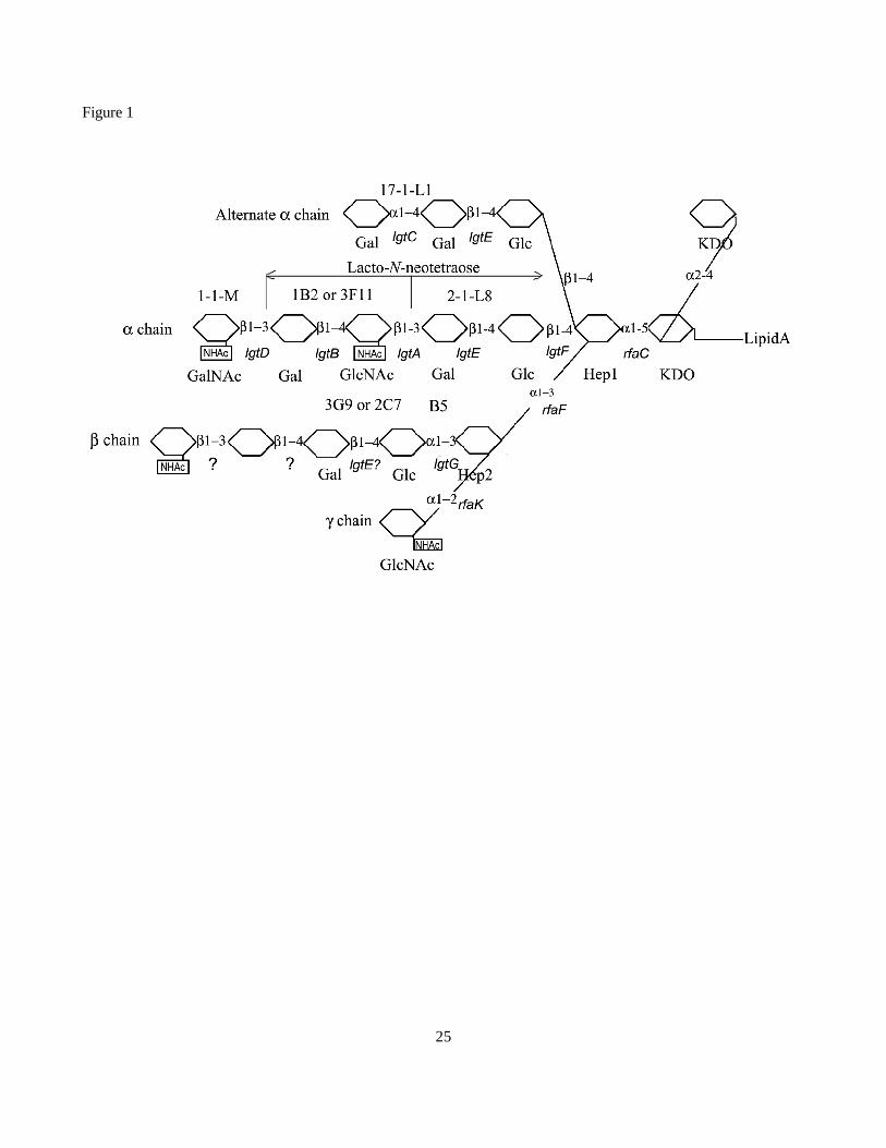
The data presented in Figure 1 summarizes the genetic potential and reported carbohydrate structures
that have been identified in Neisseria gonorrhoeae (7, 9, 10, 15, 18, 34-36). The genes responsible for the
addition of most of these sugars have been defined genetically; loss of gene function results in the truncation of
an LOS structure. Biochemical characterization of several of these gene products has been performed by
measuring the incorporation of sugars from various UDP-sugars to a variety of synthetic carbohydrates. LgtA
possessed broad substrate specificity towards ? and ? galactosides. Depending on the acceptor, this enzyme
could mediate the transfer of GlcNAc from UDP-GlcNAc and GalNAc from UDP-GalNAc (3). However, this
broad specificity was not seen in vivo (27). The biochemical properties of LgtB and LgtC have similarly been
examined (21, 30, 31). Both enzyme possessed the predicted galactosyltransferase activities.
Genetic evidence supporting the function of lgtE as encoding a glycosyl transferase responsible for the
addition of galactose ? -1, 4 to glucose has been reported in a variety of publications (6, 11, 14, 26). However,
purified LgtE was unable to mediate the transfer of galactose to synthetic LOS biosynthetic intermediates (30).
Erwin et al. (6) showed that when lgtE was nonfunctional, galactose was not added onto the ? chain. However,
Page 4
4
no direct biochemical evidence was presented to implicate LgtE directly in this addition. We purified functional
LgtE from Escherichia coli strains containing recombinant plasmids expressing LgtE, and then used this
recombinant enzyme to demonstrate its ability to modify various Neisserial LOSs that possessed defined
structures.
Experimental Procedures
Bacterial strains, plasmids, oligonucleotides and culture conditions. N. gonorrhoeae strain F62 was obtained
from Dr. P. F. Sparling, University of North Carolina, Chapel Hill. N. subflava 44 and F62? lgtA? lgtFG+ have
been previously characterized in this laboratory (24, 28). Escherichia coli strain DH5? MCR was obtained from
Life Technologies (Rockville, MD); Strain ER2566 (F- ? - fhuA2 [lon] ompT lacZ::T7 geneI gal sulA11 ? (mcrC-
mrr)114::IS10 R(mcr-73::miniTN10)2 R(zgb-210::TN10)1(tet s)endA1 [Dcm]) was obtained from New
England Biolabs, (Beverly, MA). Plasmid pET15b was obtained from Novagen (Madison, WI). Neisseria strains
were grown in standard gonococcal medium {designated GCP if broth, GCK if agar} (Difco laboratories) plus
growth supplements (33) and 0.042% sodium bicarbonate if in broth or in a 37 oC CO2 incubator. E. coli strains
were grown on LB plates (23). Ampicillin was used at 50 ? g/ml, spectinomycin at 50 ? g/ml and X-gal at 35
? g/ml when the selection or colorimetric detection were applied.
N. gonorrhoeae strain F62? lgtA? lgtF? rfaKlgtG+ was constructed by PCR amplification of rfaK region
using primers RFAK147 and RFAK3780, and cloning this fragment in the SmaI-HindIII sites of pK18up (25),
giving pRFAK. A 458 bp DraI fragment, located within the coding sequence of rfaK, was deleted from this
plasmid, giving pRFAK? 2-1. This deletion was introduced into F62? lgtA? lgtF? lgtG+, giving rise to
F62? lgtA? lgtF? rfaKlgtG+. All constructs were verified by PCR amplification of the desired region.
Chemicals, reagents and enzymes. Restriction enzymes and T4 DNA ligase were purchased from New
England Biolabs (Beverly, MA). All chemicals used for this study were reagent grade or better and were
Page 5
5
purchased from Sigma Chemical Co. (St. Louis, MO) unless otherwise specified. Tris-tricine gels (16.5%) and
running buffer were obtained from Bio-Rad Laboratories (Richmond, CA). The Mab 3G9 was graciously
provided by Dr. Peter Rice, Boston University, Boston, MA.
LOS purification and analysis. LOSs were purified from broth grown cells using acetone-powdered
organisms by the hot phenol-water method (32). LOS was extracted with hot phenol-water and concentrated by
lyophilization. Extractions were continued until the purified LOS gave a minimal absorbance when measured at
200 nm.
SDS-PAGE analysis. Approximately 0.1 ? g of LOS was subjected to SDS-PAGE on a 16.5% Tris-tricine gel
(from Bio-Rad) in Tris-tricine running buffer following the protocol suggested by the manufacturer. The gel was
fixed overnight in 40% ethanol 5% acetic acid and the LOS visualized by silver staining (29).
Western Blot and colony blot analysis. After SDS-PAGE, LOSs were electrotransferred onto Immobilon-P
membrane (Millipore Corp., MA) in a Tris-tricine-methanol buffer (10 mM Tris, pH 8.3, 10 mM Tricine, 0.01%
SDS, 20% methanol) at a constant voltage of 100V for 1h following the protocol provided by Bio-Rad Corp.
After air drying for 1h, the membrane was processed by the same procedure as “Colony Blot”
For colony blot analysis, overnight colonies were transferred to a nitrocellulose membrane (Schleicher
and Schuell, Keene, NH), incubated in buffer (20 mM Tris, 150 mM NaCl, 2% milk powder) to block all non-
specific binding sites, and screened for reactivity to the appropriate Mab. Bound Mabs were detected by reacting
the nitrocellulose filter with Mab, and visualizing the bound antibody by reacting the blot with Goat-anti-mouse
Horseradish peroxidase-labeled IgG.
Page 6
6
Transformation. Recombinant DNA transformation into E. coli were done according to standard protocols
(23). Recombinant DNA transformation into N. gonorrhoeae were done by resuspending T1 cells to a density of
approximately 1 x 10⁸ cells/ml in GCP broth containing 1X Kellogg’s solution, 0.042% NaHCO3, 10 mM
MgCl2 and 1 ? g of the DNA of interest. Cells were incubated for about 5 hrs with shaking at 37 oC. Cells were
plated onto GCK plates containing spectinomycin.
Polymerase Chain Reaction. The PCR was used to generate the DNA fragments employed in gene cloning
experiments and for mutant N. gonorrhoeae strain verification. Primers were made by Bioserve Biotechnologies
(Laurel, MD). DNA amplifications were performed by using a PCR supermix kit (from Life Technologies,
Grand Island, NY) following the procedure provided by the company. Purified chromosome DNA or plasmid
DNA was used as template. For strain construction verifications, DNA was isolated directly from colonies by
the following procedure. A small colony was added to 5 ? l of 0.5 M NaOH, the cell mixture allowed to incubate
at RT for 10 min and the solution was neutralized with 5 ? l 1M Tris-HCl, pH 7.5. After adding 90 ? l H2O, 3 ? l
of this solution was used for PCR amplifications.
Cloning of lgtE and purification of LgtE. A fragment of chromosomal DNA encoding the lgtE gene was
obtained by PCR amplification of F62 chromosomal DNA using the following primers: LGTE1; 5'
TTCCAACATATGCAAAACCACGTTATCAGC 3' (The NdeI site that was used for cloning is underlined) and
LGTE2; 5' ATGCATGGATCCCGCGGGAATGACAGTGTGTCCA 3' (The BamHI site that was used for
cloning is underlined). The PCR product was cleaved with NdeI and BamHI and ligated into the expression
vector pET15b, that had been cleaved with the same enzymes. The ligation mixture was used to transform E.
coli DH5? MCR, and individual transformants were screened for the presence of the appropriate recombinant
plasmid. Plasmid pET15b-lgtE was transformed into E. coli ER2566 strain and a single colony was used to
inoculate 25 ml of Luria broth containing ampicillin. Cells were incubated with moderate shaking at 37 oC until
the OD600 reached a value of 0.6. IPTG was added to a final concentration of 1 mM and incubations were
Page 7
7
continued at 25 oC for 10 hr. The cells were collected by centrifugation and resuspended in 1 ml of binding
buffer (Novagen). Lysozyme was added to a final concentration of 100 ? g/ml and the mixture incubated on ice
for 60 min. Samples were frozen at - 70 oC, thawed and sonicated (three times for 15 sec). The cell extract was
clarified by centrifugation for 30 min at 15,000 rpm in a Sorvall SS34 rotor. While the majority of LgtE protein
was present as an insoluble fraction, soluble proteins were purified on a Nickel column according to the
protocols of the manufacturers (Novagen) resulting in the pure LgtE protein. The enzyme was dialyzed against
Tris-HCl, pH 7.5, 20 mM NaCl, 1 mM EDTA, 50 % of glycerol and stored at -20 oC.
Galactosyltransferase assays. The standard reaction volume was 30 ? l and contained 50 mM MES buffer, pH
7.3, 10 mM MnCl2, 10 ? g of purified LOS or 5 x 10⁶ whole cells, and 1.5 ? g of purified LgtE protein. When
radioactive substrates were employed, 0.2 nmol were added UDP-[3H]Gal (17.8 Ci/mmol), UDP-[3H]Glc (25
Ci/mmol) or UDP-[14C]GlcNAc (266 Ci/mmol)]; when non radioactive substrates were employed, compounds
were added to a final concentration of 1 mM. The reactions were incubated for at least 2 h at 30 oC. The
reactions were terminated by the addition of 10 ? l of 20 % SDS and heating at 70 oC for 3 min. For the
separation of unincorporated radioactive material from LOS, reactions were loaded onto 3 ml Sephadex G-100
columns (made from a Pasteur pipette and equilibrated with water). The columns were washed with water and
fractions of three drops were collected (The first 3 fractions contained 6 drops). LOS eluted at fractions 5 - 12,
while the radioactive LOS starting from fractions 18 -20. Samples of fractions 5 to 15 (10 ? l) were spotted on 1
cm x 1cm filter paper, dried and the radioactivity was assayed by liquid scintillation counting. The total volume
of all fractions containing radioactive LOS was measured and used to calculate the total transfer of radioactive
substrate into the acceptor LOS structures.
Galactosyltransferase activity was also determined by the autoradiography. LOS samples were
radiolabeled and purified as above and then run in parallel on the same SDS-Tricine gel as described above.
Half of the gel was stained for visualization of the LOS using a standard silver-staining procedure while the
Page 8
8
second half of the gel was soaked in sodium salicylate (1 M, pH 6.0 ) for 30 min. The gel was placed on 3 MM
Whatman paper, dried for a 10 minutes on air, covered with a saran paper and subjected to autoradiography.
Results
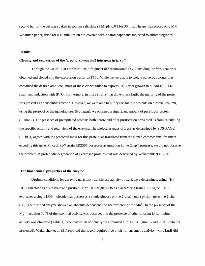
Cloning and expression of the N. gonorrhoeae F62 lgtE gene in E. coli
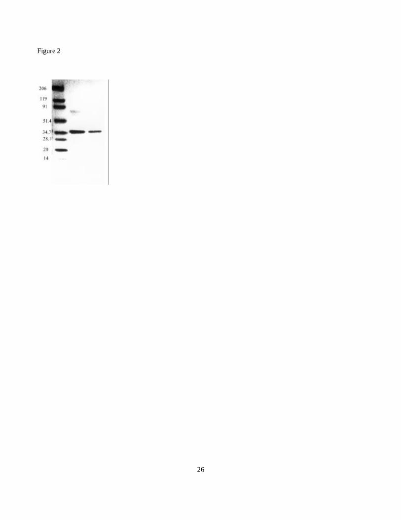
Through the use of PCR amplification, a fragment of chromosomal DNA encoding the lgtE gene was
obtained and cloned into the expression vector pET15b. While we were able to isolate numerous clones that
contained the desired amplicon, most of these clones failed to express LgtE after growth in E. coli ER2566
strain and induction with IPTG. Furthermore, in those strains that did express LgtE, the majority of the protein
was present as an insoluble fraction. However, we were able to purify the soluble proteins on a Nickel column,
using the protocol of the manufacturer (Novagen); we obtained a significant amount of pure LgtE protein
(Figure 2). The presence of precipitated proteins both before and after purification prevented us from calculating
the specific activity and total yield of the enzyme. The molecular mass of LgtE as determined by SDS-PAGE
(35 kDa) agreed with the predicted mass for this protein, as translated from the cloned chromosomal fragment
encoding this gene. Since E. coli strain ER2566 possesses as mutation in the OmpT protease, we did not observe
the problem of proteolytic degradation of expressed proteins that was described by Wakarchuk et al. (31).
The biochemical properties of the enzyme.
Optimal conditions for assaying galactosyl-transferase activity of LgtE were determined, using [3H]-
UDP-galactose as a substrate and purified F62? LgtA? LgtE LOS as a acceptor. Strain F62? LgtA? LgtE
expresses a single LOS molecule that possesses a single glucose on the ? -chain and a phosphate as the ? -chain
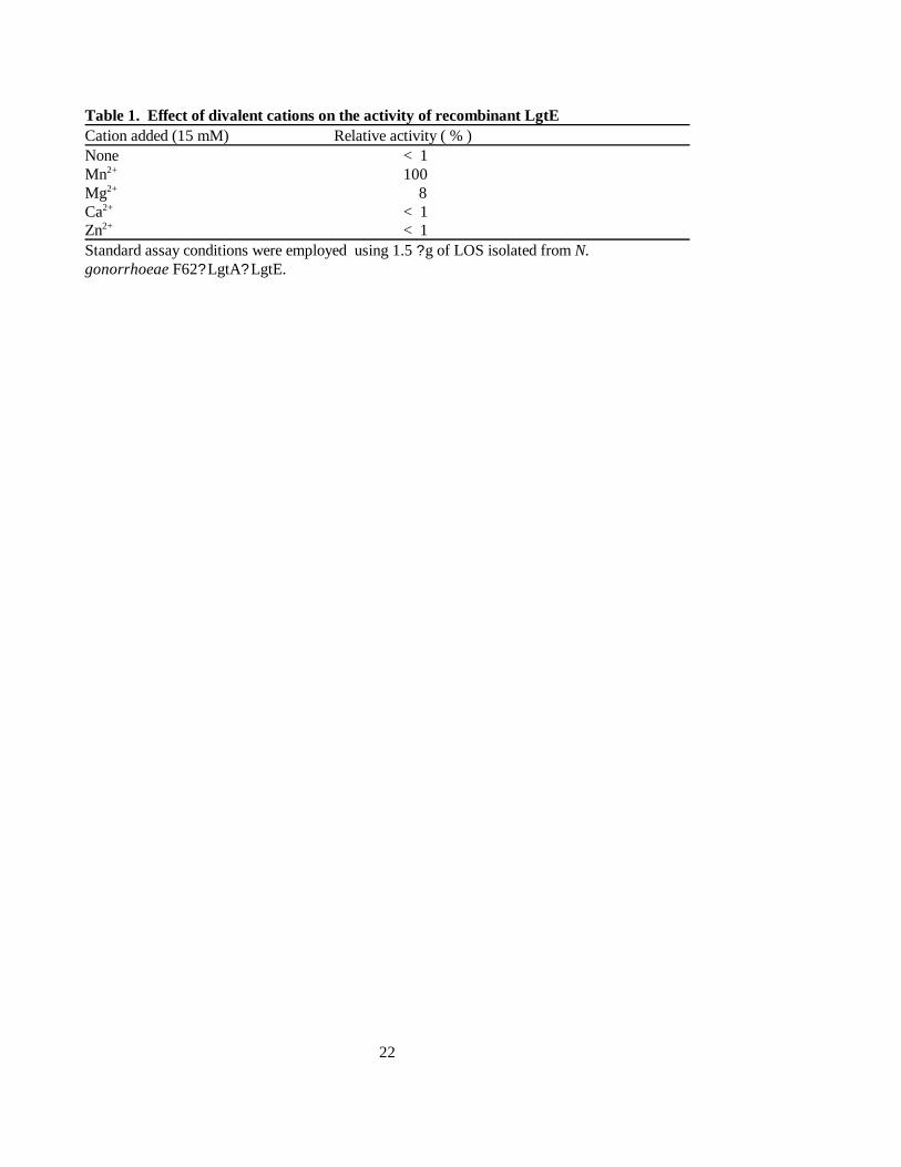
(28). The purified enzyme showed an absolute dependence on the presence of the Mn2+. In the presence of the
Mg2+ less then 10 % of the maximal activity was observed; in the presence of other divalent ions, minimal
activity was observed (Table 1). The maximum of activity was obtained at pH 7.5 (Figure 3) and 30 oC (data not
presented). Wakarchuk et al. (31) reported that LgtC required free thiols for enzymatic activity, while LgtB did
Page 9
9
not. We tested our preparation of LgtE to determine if it required free thiols for activity. The data presented in
figure 4 indicate that purified LgtE showed a linear rate of activity up to 90 min.; we concluded that this
enzyme did not possess a thiol requirement.
Substrate specificity.
Since some of the other gonococcal glycosyl transferases possessed broad substrate specificity, we used
various radiolabeled compounds to test for their ability to act as a substrate for LgtE. The data indicate that the
enzyme was only able to efficiently use UDP-Gal as a substrate (Table 2). Incorporation of radioactivity into
purified LOS using UDP-GalNAc and UDP-Glc occurred at an efficiency of less then 1 % of the level seen for
UDP-Gal. This is in contrast to LgtA from N. meningiditis that can use both UDP-GlcNAc and UDP-GalNAc as
a substrate at almost the same efficiency (3).
Acceptor specificity.
The genetic data indicate that the natural acceptor for LgtE should be the LOS structure present in strain
F62? lgtA? lgtE and the activity of this enzyme should be the addition of a galactose onto the ? -chain glucose.
The LOS structure present in the wild-type F62 strain lacks a free ? or ? -chain glucose; hence it should not be a
acceptor for LgtE. To test this prediction we used the LOS isolated from several derivatives of N. gonorrhoeae
F62 strain and from N. subflava 44. The data presented in Table 3 show that LgtE could mediate the addition of
radiolabeled Gal onto LOS isolated from a variety of strains in addition to the one that possessed an LOS
structure that was predicted to be the biosynthetic intermediate. The efficiency of transfer of Gal residues to
F62? lgtA? lgtE and F62 showed the same efficiency in the reactions containing decreased concentrations of
acceptor LOS structures (Fig. 4). To prove that the LOS from both strains possessed the same acceptor
specificity, an autoradiography experiment was performed. After radiolabeling, LOSs were analyzed by
electrophoresis on a SDS-Tris-Tricine gel and then subjected to autoradiography. While the data clearly
Page 10
10
indicated that radioactivity was incorporated into the LOS samples, the width of the signal on the autoradiogram
prevented us from determining which LOS structure was serving as an acceptor (Data not shown)
As an alternate approach to demonstrating the addition of galactose onto Neisserial LOS’s, we utilized
unlabeled UDP-galactose as a substrate, and various LOS acceptors as recipients. Strain F62? lgtA? lgtE
expresses an LOS structure that is the natural acceptor for LgtE. The data presented in figure 6A (Lanes 4 and 6)
indicate that LgtE is able to modify LOS expressed by F62? lgtA? lgtE. These data indicate that under the
experimental conditions we employed, LgtE appeared to mediate the addition of multiple galactose residues.
The data presented in table 3 indicate that strain LOS F62? lgtA? RfaK? LgtF LOS was able to serve as a
acceptor for galactose from UDP-galactose. Since this strain’s LOS is truncated and contain only two heptoses,
it should not possess the acceptor structure for LgtE. In an effort to visualize this addition, whole cells were
incubated with purified LgtE as described above. The data presented in Fig. 6, panel B, lane 2, clearly shows
that incubation of whole cells with LgtE results in the appearance of a new LOS band. Since the mobility of this
band is slower than that of LOS isolated from LOS F62? lgtA, it suggests that this LOS has had three or more
galactose moieties added to it. However, only a small percentage of the molecules were acted upon by LgtE.
While the addition of galactose onto this acceptor was unexpected, the addition of multiple galactose moieties is
consisted with the data seen in figure 6, panel A.
In order to determine if LgtE mediates the addition of galactose onto both the ? and ? chains of
gonococcal LOS, we tested whether purified LgtE was able to modify LOS isolated from N. subflava strain 44.
This strain makes two LOS structures, with the smaller LOS structure possessing a glucose on both the ? and ?
chains (28). LOS isolated from this strain fails to bind the Mab 3G9; reactivity with this Mab requires that the ?
and ? chains consist of lactose (35). The data presented in Fig. 6, panel C indicate that LgtE is able to modify
the LOS expressed by N. subflava 44.
Page 11
11
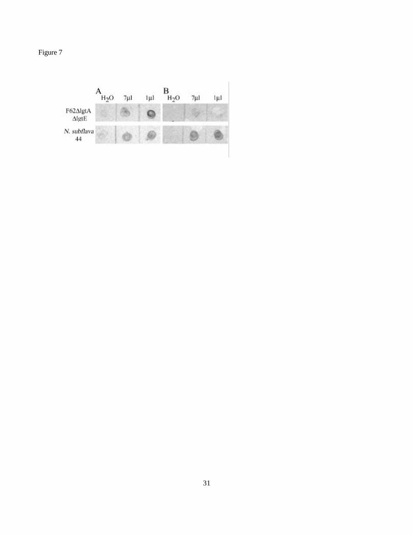
In order to determine if LgtE added galactose onto both chains of LOS expressed by N. subflava 44, we
used our in vitro assay to modify LOS isolated from this strain, and then tested the reaction product for its
acquisition of reactivity with various Mabs. The data presented in Figure 7, panel A indicates that while
F62? lgtA? lgtE LOS could be modified by purified LgtE, and that these reaction products now bind Mab 2-1-
L8, the resulting reactants failed to bind Mab 3G9 (Figure 7, Panel B). N. subflava 44 LOS, when modified in
vitro, clearly acquired the ability to bind both Mabs. From these data, we concluded that LgtE is able to mediate
the addition of galactose onto both the ? and ? chain glucose moieties.
Discussion
Many genes have been identified that are involved in LOS expression in the Neisseria. However, few
biochemical studies have been performed that demonstrate a clear structure/function relationship between the
gene product and its predicted biochemical function. A set of linked genes (lgtA-E) has been identified that seem
to encode the majority of glycosyl transferases needed to synthesize the carbohydrate portion of the ? -chain
(11). The biochemical functions of LgtA-D have been demonstrated, and correspond to the functions inferred
from analysis of mutations in these genes (3, 30, 31). While there is significant genetic evidence supporting the
function of LgtE as the glycosyl transferase responsible for the addition of galactose ? -1, 4 to glucose (6, 11, 14,
26), biochemical data supporting this assignment has been lacking. Furthermore, since the addition of galactose
? -1, 4 to glucose can occur on both the ? and ? chains, it is unclear if LgtE is responsible for both of these
biosynthetic processes.
Erwin et al. (6) demonstrated that in lgtE mutants, galactose was not added to either the ? or ? chains.
While their data clearly indicated that LgtE activity is required for galactose addition, the results could not rule
out the possibility that the ? -chain addition of galactose was mediated by an additional unlinked enzyme, whose
activity required before the addition of the ? -chain galactose could occur. As a first step in characterizing the
lgtE gene product, we used a gene cloning strategy to isolate a functional LgtE protein. While we were readily
Page 12
12
able to isolate seemingly intact DNA fragments into a lac-regulated expression vector, most of the recombinant
clones failed to express detectable levels of protein, even after induction with IPTG. Furthermore, expressing
clones seemed to lose the ability to express the protein after prolonged incubation, or storage. While we did not
investigate the reason for this instability, we believe that it is probably related to the fact that the LgtE protein is
able to modify LPS biosynthetic intermediates in E. coli, and the accumulation of these modified intermediates
are toxic.
Strain F62? lgtA? lgtE produces an LOS that contains a single glucose on the ? -chain and a single
phosphate as the ? -chain (28). As such, this strain produces an LOS with the predicted acceptor structure for
LgtE. The data shown in Table 3 and Fig. 6 demonstrate that this LOS can serve as an acceptor for galactose
from UDP-Galactose. Surprisingly, many other LOS structures were also able to serve as acceptor molecules for
LgtE. Furthermore, in our in vitro experiments, multiple LOS bands were obtained after incubation of
F62? lgtA? lgtE LOS with purified LgtE and UDP-galactose. The SDS-PAGE profile of the elongation product
suggests that two galactose residues are added onto F62? lgtA? lgtE LOS.
The ability of LgtE to add galactose onto a variety of LOS structures was unexpected. Most surprising
perhaps was its ability to add sugars onto LOS isolated from LOS F62? lgtA? RfaK? LgtF. The OS of this LOS
consists of two heptose molecules, and elongation of this LOS by LgtE in vivo has not been reported.
Furthermore, when galactose is added to the base OS, the resulting band has an SDS-PAGE mobility consistent
with the addition of three galactose residues. These data indicate that depending on the nature of the starting
LOS, it appears that 2 or 3 galactose residues were added. By varying the amount of LgtE added to the reaction
mixture, we could change the relative ratio of the elongation product; more enzyme increased the intensity of the
higher molecular weight components (data not shown). Since the aberrant additions only occurred during in
vitro reactions, and were influenced by the amount of exogenous LgtE added, this suggests that in vivo, the
Page 13
13
amount of enzyme expressed is quite low. Additional studies from our laboratory indicate that this hypothesis is
correct (DCS, unpublished observations).
Wakarchuk and coworkers demonstrated that purified LgtE was unable to add galactose onto synthetic
? -Glc acceptors (30). In light of our observation that LgtE is able to mediate the addition to a variety of
molecules that possess a lipid base, indicates that the failure observed by Wakarchuk et al. was most likely due
to the inability of the synthetic intermediates to form a stable interaction with LgtE.
Certain LOS immunotypes of N. meningitidis possess LOS with variations in the structure of the ? -
chain. The observation that excess amount of LgtE in a reaction results in homopolymer additions suggests a
mechanism that can explain how the L5 LOS immunotype might arise. If LgtF were overexpressed , it might
result in the addition of a second glucose residue at the base of the ? -chain. It is interesting to note that the SDS-
PAGE mobility of LOS isolated from L5 strains possess a significant amount of a truncated biosynthetic
product. This may reflect the limitation in the ability of LgtE to add galactose onto the diglucosyl structure.
The ability of purified LgtE to modify LOS isolated from N. Subflava 44 to reactivity with Mab 3G9
clearly indicates that LgtE is able to mediate the addition of galactose onto the ? -chain. Since LgtG expressing
strains that express LgtA (N. gonorrhoeae 15253 or F62LgtG+) do not add sugars onto the ? -chain beyond the
galactose, this indicates that LgtA possesses a structural requirement that biases its addition to the ? -chain. It is
possible that overexpression of LgtA could result in the elongation of both the ? and ? chains, similar to what
we have seen with LgtE. Since the level of expression of the various glycosyl-transferases in the gonococcus is
quite low (DCS, unpublished observations), it further suggests an additional mechanism of phenotypic
modulation, where subtle changes in the growth rate of the organism would modulate the absolute level of the
various proteins, and these results would be translated into small differences in LOS expression. It has been
Page 14
14
shown by several investigators that alteration in growth conditions effects LOS expression (8, 19, 20, 22), and
we believe that this modulation is due to small changes in the level of the various glycosyltransferases.
Page 15
15
Acknowledgments
The work described in this publication was supported by a grant from the National Institutes of Health to DCS,
AI24452. The authors wish to thank Anne Corriveau for her excellent technical assistance.
References
1. Arking, D., Y. Tong, and D. C. Stein. 2001. Analysis of lipooligosaccharide biosynthesis in the
Neisseriaceae. J. Bacteriol. 183:934-941.
2. Banerjee, A., R. Wang, Uljohn.S., P. A. Rice, E. C. Gotschlich, and D. C. Stein. 1998. Identification
of the gene (lgtG) encoding the lipooligosaccharide ? chain synthesizing glucosyl transferase from
Neisseria gonorrhoeae. Proc. Natl. Acad. Sci. 95:10872-10877.
3. Blixt, O., I. van Die, T. Norberg, and D. H. van den Eijnden. 1999. High-level expression of the
Neisseria meningitidis lgtA gene in Escherichia coli and characterization of the encoded N-
acetylglucosaminyltransferase as a useful catalyst in the synthesis of GlcNAc ? 1-->3Gal and GalNAc
? 1-->3Gal linkages. Glycobiol. 9:1061-1071.
4. Burch, C. L., R. J. Danaher, and D. C. Stein. 1997. Antigenic variation in Neisseria gonorrhoeae:
production of multiple lipooligosaccharides. J. Bacteriol. 179:982-986.
5. Danaher, R. J., J. C. Levin, D. Arking, C. L. Burch, R. Sandlin, and D. C. Stein. 1995. Genetic
basis of Neisseria gonorrhoeae lipooligosaccharide antigenic variation. J. Bacteriol. 177:7275-7279.
6. Erwin, A. L., P. A. Haynes, P. A. Rice, and E. C. Gotschlich. 1996. Conservation of the
lipooligosaccharide synthesis locus lgt among strains of Neisseria gonorrhoeae: requirement for lgtE in
synthesis of the 2C7 epitope and of the beta chain of strain 15253. J. Exp. Med. 184:1233-1241.
7. Fermer, C., B. E. Kristiansen, O. Skold, and G. Swedberg. 1995. Sulfonamide resistance in Neisseria
meningitidis as defined by site- directed mutagenesis could have its origin in other species. J Bacteriol
177:4669-75.
Page 16
16
8. Frangipane, J. V., and R. F. Rest. 1993. Anaerobic growth and cytidine 5'-monophospho-N-
acetylneuraminic acid act synergistically to induce high-level serum resistance in Neisseria
gonorrhoeae. Infect Immun 61:1657-66.
9. Gibson, B. W., W. Melaugh, N. J. Phillips, M. A. Apicella, A. A. Campagnari, and J. M. Griffiss.
1993. Investigation of the structural heterogeneity of lipooligosaccharides from pathogenic
Haemophilus and Neisseria species and of R-type lipopolysaccharides from Salmonella typhimurium by
electrospray mass spectrometry. J. Bacteriol. 175:2702-2712.
10. Gibson, B. W., J. W. Webb, R. Yamasaki, S. J. Fisher, A. L. Burlingame, R. E. Mandrell, H.
Schneider, and J. M. Griffiss. 1989. Structure and heterogeneity of the oligosaccharides from the
lipopolysaccharides of a pyocin-resistant Neisseria gonorrhoeae. Proc. Natl. Acad. Sci .U S A 86:17-
21.
11. Gotschlich, E. C. 1994. Genetic locus for the biosynthesis of the variable portion of Neisseria
gonorrhoeae lipooligosaccharide. J. Exp. Med. 180:2181-2190.
12. Griffiss, J. M., H. Schneider, R. E. Mandrell, R. Yamasaki, G. A. Jarvis, J. J. Kim, B. W. Gibson,
R. Hamadeh, and M. A. Apicella. 1988. Lipooligosaccharides: the principal glycolipids of the
neisserial outer membrane. Rev. Infect. Dis. 10:S287-295.
13. Jennings, M. P., D. W. Hood, I. R. Peak, M. Virji, and E. R. Moxon. 1995. Molecular analysis of a
locus for the biosynthesis and phase-variable expression of the lacto-N-neotetraose terminal
lipopolysaccharide structure in Neisseria meningitidis. Mol. Microbiol. 18:729-740.
14. Jennings, M. P., Y. N. Srikhanta, E. R. Moxon, M. Kramer, J. T. Poolman, B. Kuipers, and P. van
der Ley. 1999. The genetic basis of the phase variation repertoire of lipopolysaccharide immunotypes in
Neisseria meningitidis. Microbiol. 145:3013-3021.
15. John, C. M., J. M. Griffiss, M. A. Apicella, R. E. Mandrell, and B. W. Gibson. 1991. The structural
basis for pyocin resistance in Neisseria gonorrhoeae lipooligosaccharides. J. Biol. Chem. 266:19303-
19311.
Page 17
17
16. Jones, D. M., R. Borrow, A. J. Fox, S. Gray, K. A. Cartwright, and J. T. Poolman. 1992. The
lipooligosaccharide immunotype as a virulence determinant in Neisseria meningitidis. Microb. Pathog.
13:219-224.
17. Kahler, C. M., R. W. Carlson, M. M. Rahman, L. E. Martin, and D. S. Stephens. 1996. Two
glycosyltransferase genes, lgtF and rfaK, constitute the lipooligosaccharide ice (inner core extension)
biosynthesis operon of Neisseria meningitidis. J. Bacteriol. 178:6677-6684.
18. Kerwood, D. E., H. Schneider, and R. Yamasaki. 1992. Structural analysis of lipooligosaccharide
produced by Neisseria gonorrhoeae, strain MS11mk (variant A): a precursor for a gonococcal
lipooligosaccharide associated with virulence. Biochemistry 31:12760-8.
19. McGee, D. J., and R. F. Rest. 1996. Regulation of gonococcal sialyltransferase, lipooligosaccharide,
and serum resistance by glucose, pyruvate and lactate. Infect. Immun. 64:4630-4637.
20. Morse, S. A., C. S. Mintz, S. K. Sarafian, L. Bartenstein, M. Bertram, and M. A. Apicella. 1983.
Effect of dilution rate on lipopolysaccharide and serum resistance of Neisseria gonorrhoeae grown in
continuous culture. Infect. Immun 41:74-82.
21. Persson, K., H. D. Ly, M. Dieckelmann, W. W. Wakarchuk, S. G. Withers, and N. C. Strynadka.
2001. Crystal structure of the retaining galactosyltransferase LgtC from Neisseria meningitidis in
complex with donor and acceptor sugar analogs. Nat. Struct. Biol. 8:166-175.
22. Pettit, R. K., E. S. Martin, S. M. Wagner, and V. J. Bertolino. 1995. Phenotypic modulation of
gonococcal lipooligosaccharide in acidic and alkaline culture. Infect. Immun. 63:2773-2775.
23. Sambrook, J., E. F. Fritsch, and T. Maniatis. 1989. Molecular Cloning: a laboratory manual (2nd
ed.). Cold Spring Harbor Laboratory Press, Cold Spring Harbor, NY.
24. Sandlin, R., and D. C. Stein. 1991. Structural heterogeneity of lipopolysaccharides the Neisseriaceae.
FEMS Microbiol. Let. 90:69-72.
Page 18
18
25. Sandlin, R. C., M. A. Apicella, and D. C. Stein. 1993. Cloning of a gonococcal DNA sequence that
complements the lipooligosaccharide defects of Neisseria gonorrhoeae 1291d and 1291e. Infect. Immun.
61:3360-3368.
26. Song, W., L. Ma, W. Chen, and D. C. Stein. 2000. Role of lipooligosaccharide in opa-independent
invasion of Neisseria gonorrhoeae into human epithelial cells. J. Exp. Med. 191:949-959.
27. Tong, Y., D. Arking, S. Ye, B. Reinhold, V. Reinhold, and D. C. Stein. 2002. Neisseria gonorrhoeae
strain PID2 simultaneously expresses six chemically related lipooligosaccharide structures. Glycobiol.
In Press.
28. Tong, Y., B. Reinhold, V. Reinhold, B. Brandt, and D. C. Stein. 2001. Structural and
immunochemical characterization of the lipooligosaccharides expressed by Neisseria subflava 44. J.
Bact. 183:942-950.
29. Tsai, C. M., and C. E. Frasch. 1982. A sensitive silver stain for detecting lipooligosaccharide in
polyacrylamide gels. Anal. Biochem. 119:115-119.
30. Wakarchuk, W., A. Martin, M. P. Jennings, E. R. Moxon, and J. C. Richards. 1996. Functional
relationships of the genetic locus encoding the glycosyltransferase enzymes involved in expression of
the lacto-N-neotetraose terminal lipopolysaccharide structure in Neisseria meningitidis. J. Biol. Chem.
271:19166-19173.
31. Wakarchuk, W. W., A. Cunningham, D. C. Watson, and N. M. Young. 1998. Role of paired basic
residues in the expression of active recombinant galactosyltransferases from the bacterial pathogen
Neisseria meningitidis. Protein. Eng. 11:295-302.
32. Westphal, O., and K. Jann. 1972. Bacterial Lipopolysaccharides: extraction with phenol-water and
further applications of the procedure. Meth. Carbohydr. Chem. 5:83-91.
33. White, L. A., and D. S. Kellogg, Jr. 1965. Neisseria gonorrhoeae identification in direct smears by a
fluorescent antibody counterstain method. Appl. Microbiol. 13:171-174.
Page 19
19
34. Yamasaki, R., B. E. Bacon, W. Nasholds, H. Schneider, and J. M. Griffiss. 1991. Structural
determination of oligosaccharides derived from lipooligosaccharide of Neisseria gonorrhoeae F62 by
chemical, enzymatic, and two-dimensional NMR methods. Biochem. 30:10566-10575.
35. Yamasaki, R., D. E. Kerwood, H. Schneider, K. P. Quinn, J. M. Griffiss, and R. E. Mandrell.
1994. The structure of lipooligosaccharide produced by Neisseria gonorrhoeae, strain 15253, isolated
from a patient with disseminated infection. Evidence for a new glycosylation pathway of the gonococcal
lipooligosaccharide. J. Biol. Chem. 269:30345-30351.
36. Yamasaki, R., W. Nasholds, H. Schneider, and M. A. Apicella. 1991. Epitope expression and partial
structural characterization of F62 lipooligosaccharide (LOS) of Neisseria gonorrhoeae: IgM
monoclonal antibodies (3F11 and 1-1-M) recognize non-reducing termini of the LOS components. Mol.
Immunol. 28:1233-1242.
37. Yang, Q. L., and E. C. Gotschlich. 1996. Variation of gonococcal lipooligosaccharide structures is due
to alterations in poly-G tracts in lgtgenes encoding glycosyl transferases. J. Exp. Med. 183:323-327.
Page 20
20
Figures Legends
Fig. 1. Possible LOS structures seen in N. gonorrhoeae. The data in the figure are a compilation of published
LOS structures. The question marks in the figure indicate structures that have been identified, but the gene
responsible for the addition and the exact chemical composition of the sugar remain unknown. The labels above
the structures indicate various monoclonal antibodies that have been used as markers for the presence of various
components that terminate with te indicated sugars. The genes predicted to be responsible for the various sugar
additions are indicated in italic.
Fig. 2. SDS-PAGE of purified LgtE. E. coli strains were grown to mid log phase and LgtE expression was
induced by the addition of IPTG. Recombinant proteins were purified on a Nickel column. The lanes represent:
Lane 1, molecular weight markers; lane 2 and 3, purified enzyme (two consecutive fractions obtained after
elution from the column. The sizes of the molecular mass markers are given in kDa. A single protein of 35 kDa
is present in the preparation.
Fig. 3. Effect of the pH on the activity of recombinant LgtE. The activity was assayed using 1.5 ? g of LOS
purified from N. gonorrhoeae F62? LgtA? LgtE strain as an acceptor using standard incubation conditions
except for the variation in buffer pH.
Fig. 4. Linearity with time of the reaction catalyzed by recombinant LgtE. Standard assay conditions were
employed using 1.5 ? g of LOS purified from N. gonorrhoeae FA62? LgtA? LgtE strain.
Fig. 5. Dependence of the efficiency of transfer of donor UDP-Gal residue to different concentrations of
the LOS structures isolated from F62? LgtA? LgtE and F62 strains. The ability of the LgtE protein to
transfer galactose to various concentrations of LOS was determined by incubating in 30 ? l of reaction buffer (50
mM MES buffer, pH 7.3, 10 mM MnCl2, and 1 mM UDP-galactose) different concentrations of LOS. The
Page 21
21
reactions were incubated for 2 hr. at 30 oC and terminated by the addition of 10 ? l of 20 % SDS and heating at
70 oC for 3 min. (triangles denotes LOS from F62, squares from F62? LgtA? LgtE)
Fig. 6. Transfer of galactose into Neisserial LOS. The ability of the LgtE protein to transfer galactose to
various LOS structures was determined by incubating in 30 ? l of reaction buffer (50 mM MES buffer, pH 7.3,
10 mM MnCl2, 5 x 10⁶ whole cells, and 1 mM UDP-galactose). The reactions were incubated overnight at 30 oC
and terminated by the addition of 10 ? l of 20 % SDS and heating at 70 oC for 3 min. A 10 ? l of each reaction
was analyzed on a 16.5%Tris-tricine gel. Panel A. The lanes represent LOS isolated from: 1) F62? LgtA; 2)
F62? LgtA? LgtE; 3) F62? LgtA? LgtE incubated with reagents, minus LgtE; 4) F62? LgtA? LgtE, plus 1 ? l
LgtE preparation; 5) F62? LgtA? LgtE incubated with reagents, minus LgtE; 6) F62? LgtA? LgtE, plus 7 ? l
LgtE preparation. Panel B. The lanes represent LOS isolated from: 1) F62? rfaKA; 2) F62? rfaK incubated with
reagents, plus 7 ? l LgtE preparation; 3) F62? LgtA; and 4) F62? LgtA? LgtE. Panel C. The lanes represent: 1)
F62? LgtA; 2) N. subflava 44; 3) N. subflava 44 incubated with LgtE; 4) F62? LgtA? LgtE.
Fig. 7. Reactivity of in vitro modified LOS with various Mabs. Cells were incubated with purified LgtE (1
or 7 ? l) plus UDP-galactose in standard reaction buffer, and an aliquot was spotted onto a nitrocellulose filter.
Reactivity to various Mabs was determined using our colony blotting procedure. Panel A represents samples
that had been exposed to Mab 2-1-L8; Panel B represents samples that had been exposed to Mab 3G9.
Page 22
22
Table 1. Effect of divalent cations on the activity of recombinant LgtECation added (15 mM) Relative activity ( % )None < 1Mn2+ 100Mg2+ 8 Ca2+ < 1Zn2+ < 1Standard assay conditions were employed using 1.5 ? g of LOS isolated from N.gonorrhoeae F62? LgtA? LgtE.
Page 23
23
Table 2. Nucleotide-sugar substrate specificity of recombinant LgtE Substrate (0.18 mM ) Relative enzyme activity ( % )UDP-Gal 100UDP-GalNAc < 1UDP-Glc < 1Standard assay conditions were employed using 1.5 ? g of LOS isolated from N.gonorrhoeae F62? LgtA? LgtE.
Page 24
24
Table 3. LOS acceptor specificity of recombinant LgtE protein Acceptor (5 ? g) Relative activity ( % ) LOS F62? LgtA? LgtE 100 LOS F62 80 LOS F62? LgtA? RfaK? LgtF 70 LOS F62? LgtA? LgtG 50 LOS N. subflava 44 50Standard assay conditions were used. Reactions differed only in the LOS that was employed.