44
Biochemistry 3070 – Oxygen Transport Proteins 1 Oxygen Transport Proteins: Myoglobin & Hemoglobin Biochemistry 3070
| Date post: | 17-Dec-2015 |
| Category: |
Documents |
| Upload: | colleen-short |
| View: | 243 times |
| Download: | 4 times |
Biochemistry 3070 – Oxygen Transport Proteins 1
Oxygen Transport Proteins:
Myoglobin & Hemoglobin
Biochemistry 3070

Biochemistry 3070 – Oxygen Transport Proteins 2
Myoglobin
• Myoglobin functions as an oxygen transport protein in tissues. It also provides a local oxygen storage site by enhancing the solubility of oxygen.
• Hemoglobin is also an oxygen transport protein, but functions in erythrocytes, enhancing the solubility of oxygen in the blood.
• Both these proteins are excellent examples to study the relationship between protein structure and function.
Biochemistry 3070 – Oxygen Transport Proteins 3
Myoglobin Structure
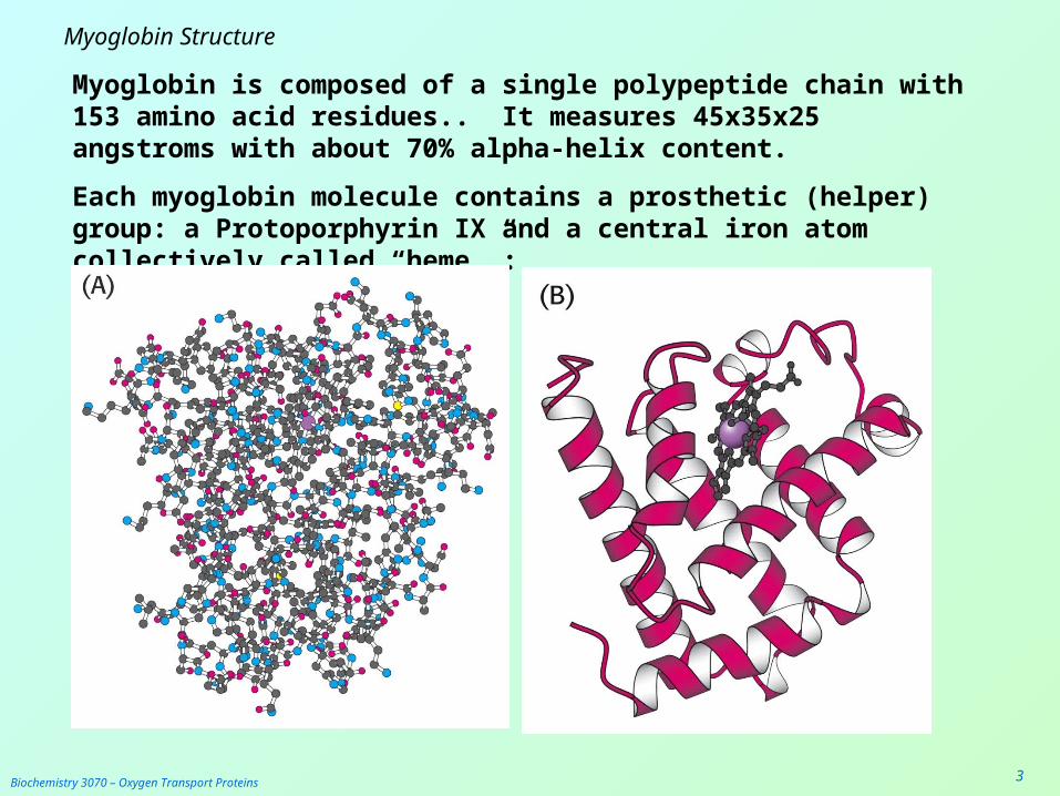
Myoglobin is composed of a single polypeptide chain with 153 amino acid residues.. It measures 45x35x25 angstroms with about 70% alpha-helix content.
Each myoglobin molecule contains a prosthetic (helper) group: a Protoporphyrin IX and a central iron atom collectively called “heme.”:

Biochemistry 3070 – Oxygen Transport Proteins 4
Myoglobin Structure
As with most water-soluble proteins, its polar amino acids are located on on the external surface of the protein, to maximize interactions with water. Non-polar amino acids are located almost entirely on the interior of the protein, leaving very little space inside. (Blue = charged amino acids; Yellow=hydrophobic amino acids.)
Biochemistry 3070 – Oxygen Transport Proteins 5
Myoglobin Structure
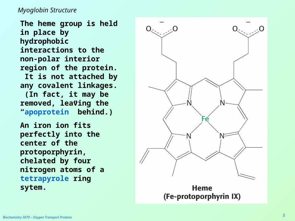
The heme group is held in place by hydrophobic interactions to the non-polar interior region of the protein. It is not attached by any covalent linkages. (In fact, it may be removed, leaving the “apoprotein” behind.)
An iron ion fits perfectly into the center of the protoporphyrin, chelated by four nitrogen atoms of a tetrapyrole ring sytem.
Biochemistry 3070 – Oxygen Transport Proteins 6
Myoglobin Structure
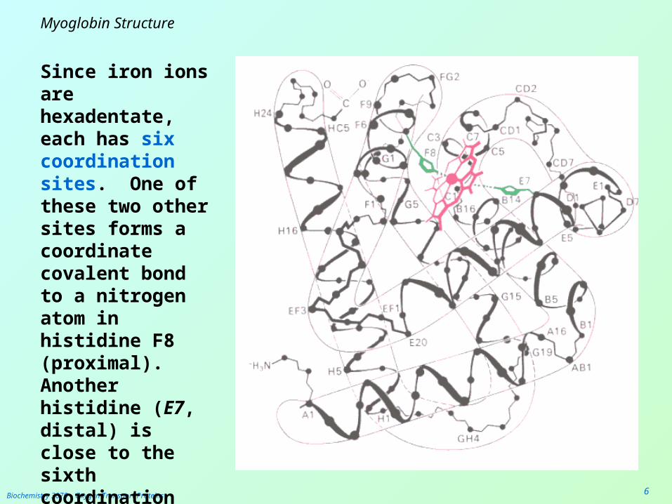
Since iron ions are hexadentate, each has six coordination sites. One of these two other sites forms a coordinate covalent bond to a nitrogen atom in histidine F8 (proximal). Another histidine (E7, distal) is close to the sixth coordination position.

Biochemistry 3070 – Oxygen Transport Proteins 7
Myoglobin Structure The iron ion is the binding site for oxygen molecules. The iron ion often converts between the free Fe2+ (ferrous ion) state and the bound Fe3+ (ferric ion) state.
In the unbound state, the iron atom is slightly proximal (above) the plane of the protoporphyrin. As oxygen binds to the distal side of the ring, it pulls the iron atom about 0.2 angstrom closer to the plane of the ring.
Although this distance is small, the movement is amplified, causing significant shifts throughout the teritary structure of the protein.

Biochemistry 3070 – Oxygen Transport Proteins 8
Myoglobin Structure
The position of the distal histidine (E7) prevents O2
from binding too strongly to the iron atom.
Maximal binding strength is achieved when the three atoms [Fe-O=O] form a linear sequence. However, the distal histidine prevents this from occurring, and the diatomic oxygen binds in a “bent” configuration.
Carbon monoxide also binds to the iron atom in myoglobin. In fact, it will displace oxygen and form a much tighter bond than oxygen, due to its more polar bond.
Even low concentrations of CO can displace O2. This explains how even low concentrations of CO can cause asphyxiation in the presence of O2!
Biochemistry 3070 – Oxygen Transport Proteins 9
Myoglobin Structure
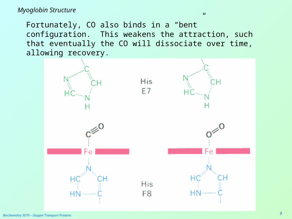
Fortunately, CO also binds in a “bent” configuration. This weakens the attraction, such that eventually the CO will dissociate over time, allowing recovery.
Biochemistry 3070 – Oxygen Transport Proteins 10
Hemoglobin
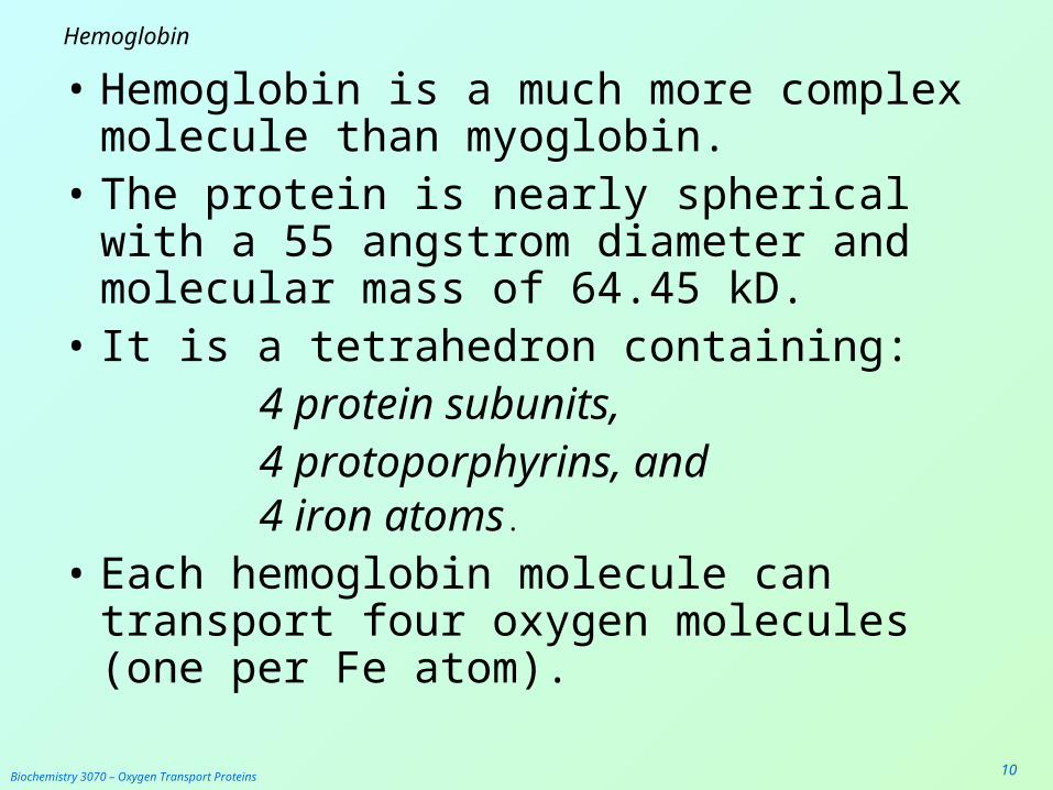
• Hemoglobin is a much more complex molecule than myoglobin.
• The protein is nearly spherical with a 55 angstrom diameter and molecular mass of 64.45 kD.
• It is a tetrahedron containing:4 protein subunits, 4 protoporphyrins, and4 iron atoms.
• Each hemoglobin molecule can transport four oxygen molecules (one per Fe atom).

Biochemistry 3070 – Oxygen Transport Proteins 11
Hemoglobin – Tetrameric Structure
The two alpha subunits have 141 amino acids, while the two beta subunits contain 146 residues.
Biochemistry 3070 – Oxygen Transport Proteins 12
Hemoglobin – Tetrameric Structure
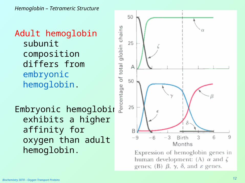
Adult hemoglobin subunit composition differs from embryonic hemoglobin.
Embryonic hemoglobin exhibits a higher affinity for oxygen than adult hemoglobin.
Biochemistry 3070 – Oxygen Transport Proteins 13
Hemoglobin – Tetrameric Structure
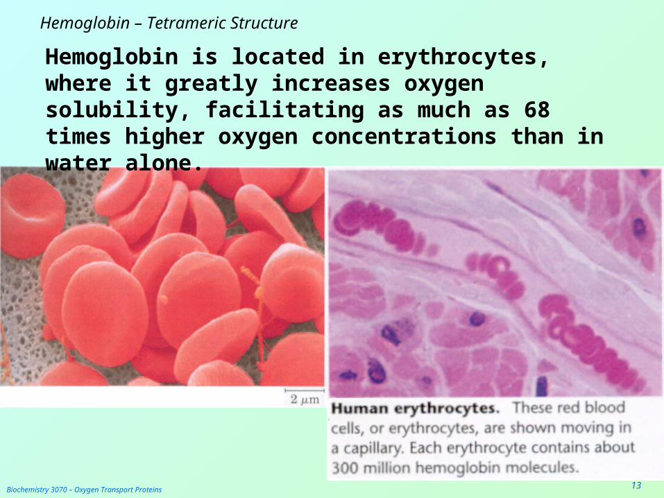
Hemoglobin is located in erythrocytes, where it greatly increases oxygen solubility, facilitating as much as 68 times higher oxygen concentrations than in water alone.
Biochemistry 3070 – Oxygen Transport Proteins 14
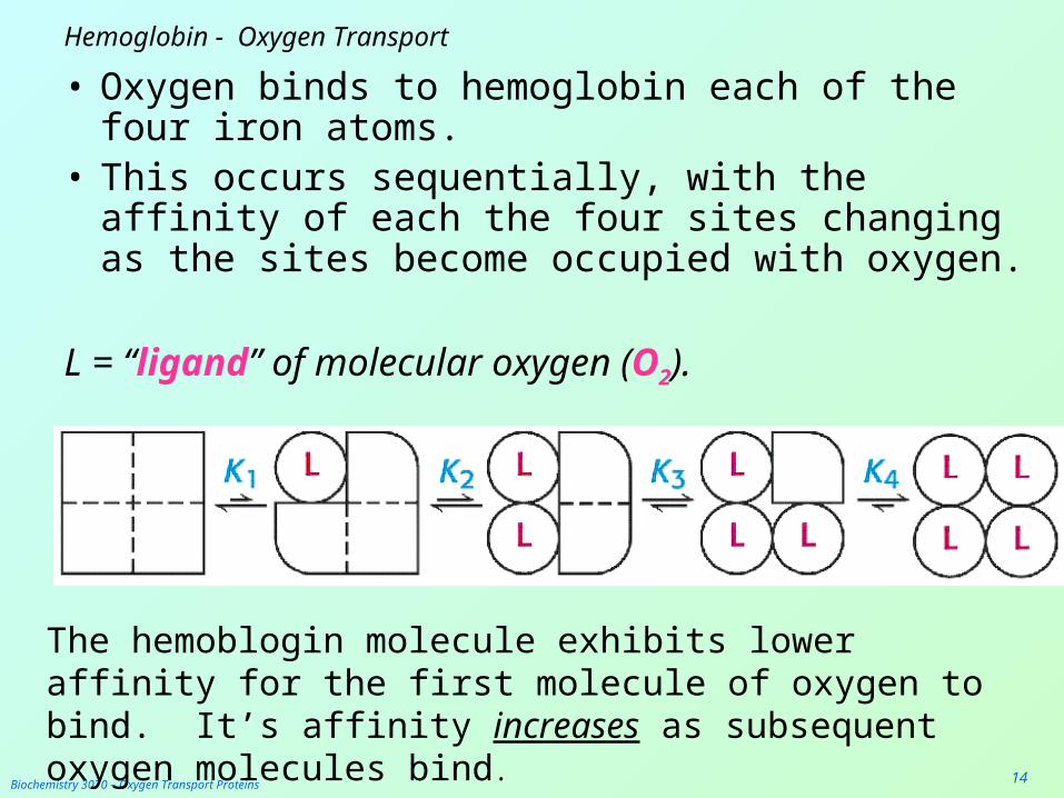
Hemoglobin - Oxygen Transport
• Oxygen binds to hemoglobin each of the four iron atoms.
• This occurs sequentially, with the affinity of each the four sites changing as the sites become occupied with oxygen.
L = “ligand” of molecular oxygen (O2).
The hemoblogin molecule exhibits lower affinity for the first molecule of oxygen to bind. It’s affinity increases as subsequent oxygen molecules bind.
Biochemistry 3070 – Oxygen Transport Proteins 15
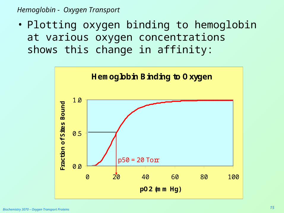
Hemoglobin - Oxygen Transport
• Plotting oxygen binding to hemoglobin at various oxygen concentrations shows this change in affinity:
Hemoglobin Binding to Oxygen
0.0
0.5
1.0
0 20 40 60 80 100
pO2 (mm Hg)
Frac
tio
n o
f S
ite
s B
ou
nd
p50 = 20 Torr
Hb

Biochemistry 3070 – Oxygen Transport Proteins 16
Hemoglobin - Oxygen Transport
• When the binding curve for myoglobin is compared to hemoglobin, a distinctly different binding profile is observed:
Mb & Hb Binding to Oxygen
0.0
0.5
1.0
0 20 40 60 80 100
pO2 (mm Hg)
Frac
tio
n o
f S
ite
s B
ou
nd
p50=20Torr
HbMb
p50= 2 Torr

Biochemistry 3070 – Oxygen Transport Proteins 17
Oxygen Binding: Myoglobin vs. Hemoglobin
Mb & Hb Binding to Oxygen
0.0
0.5
1.0
0 20 40 60 80 100pO2 (mm Hg)
Frac
tio
n o
f S
ite
s B
ou
nd HbMb
Capillary Lungs
At lower concentrations of oxygen (as in the capillary), myoglobin has higher affinity for oxygen than does hemoglobin:

Biochemistry 3070 – Oxygen Transport Proteins 18
Oxygen Binding: Myoglobin vs. Hemoglobin
Mb & Hb Binding to Oxygen
0.0
0.5
1.0
0 20 40 60 80 100pO2 (mm Hg)
Frac
tio
n o
f S
ite
s B
ou
nd
HbMb
Capillary Lungs
Questions:
1. When Mb and Hb are present in the same solution, which would compete more effectively for oxygen?
2. What if Mb and Hb solutions were separated by a semi-permeable membrane?
3. What if a solution of O2-saturated Hb were placed on one side of a membrane and a soolution of free (unbound) Mb were placed on the other side of the membrane. (pO2=10Torr)?
Biochemistry 3070 – Oxygen Transport Proteins 19
Hemoglobin – Oxygen Transport
• Discussion Questions:
1. Why is it beneficial for hemoglobin to exhibit such different affinities for oxygen?
2. What good is an oxygen “transport” protein that never delivers its oxygen to the tissues?

Biochemistry 3070 – Oxygen Transport Proteins 20
Hemoglobin – Oxygen Transport
• Transport of oxygen by both myoglobin and hemoglobin can be modeled mathematically.
• This relatively simple algebraic formula is: pO2
n
Y = pO2
n + p50n Where
Y = fraction of heme sites bound to oxygen,
pO2 = partial pressure of oxygen,
P50 = paritial pressure of oxygen at which 50% of sites are bound
N = cooperativity coefficient (Hill coefficient)

Biochemistry 3070 – Oxygen Transport Proteins 21
Hemoglobin – Oxygen Transport
• Oxygen Binding In the Pulmonary System:Assume that p50 = 35 Torr in the alveolar capillaries: pO2 = 100 Torr:
1003
Yalv = = 0.985 1003 + 353
• Oxygen Binding in the Perphieral Tissues:Assume that p50 = 35 Torr in the capillaries and pO2 = 20 Torr:
203
Yalv = = 0.157 203 + 353
Difference: (“Delta Y”) = 0.985 – 0.157 = 0.828
This means that 82.8% of the hemoglobin sites transported
oxygen to the tissues.

Biochemistry 3070 – Oxygen Transport Proteins 22
Myoglobin – Oxygen Transport
What if myoglobin were utilized to transport oxygen? (n=1)
• Oxygen Binding In the Pulmonary System:
Assume that p50 = 35 Torr in the alveolar capillaries: pO2 = 100 Torr:
1001
Yalv = = 0.741 1001 + 351
• Oxygen Binding in the Perphieral Tissues:Assume that p50 = 35 Torr in the capillaries and pO2 = 20 Torr:
201
Yalv = = 0.364 201 + 351
Difference: (“Delta Y”) = 0.741 – 0.364 = 0.377
In this case only 37.7% of the myoglobin sites transported
oxygen to the tissues!
Biochemistry 3070 – Oxygen Transport Proteins 23
Hemoglobin – Oxygen Binding “Cooperativity”
• The key to the dramatic difference in oxygen transport effeciency is hemoglobin’s “cooperativity.”
• The exponent in the prior equation is often called the “Hill Cooperativity Coefficient.”
• As “n” changes, so does the sigmoidal shape of the oxygen binding curve.
• Let’s examine the graphical effect of changing values of n:
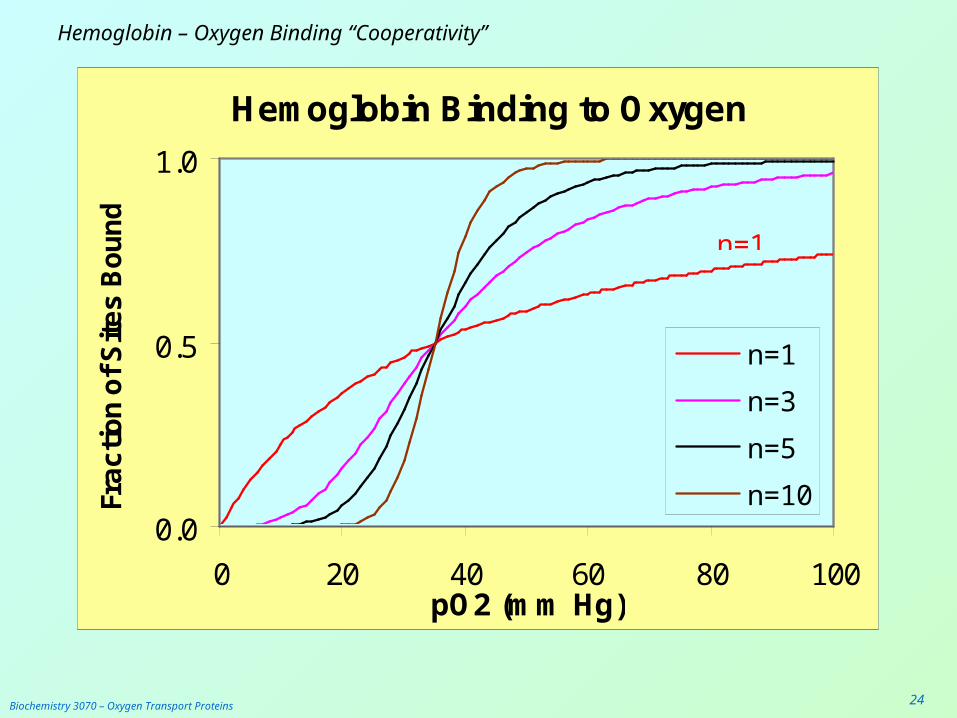
Biochemistry 3070 – Oxygen Transport Proteins 24
Hemoglobin – Oxygen Binding “Cooperativity”
Hemoglobin Binding to Oxygen
0.0
0.5
1.0
0 20 40 60 80 100pO2 (mm Hg)
Fra
cti
on
of
Sit
es
Bo
un
d
n=1
n=3
n=5
n=10
n=1
n=10

Biochemistry 3070 – Oxygen Transport Proteins 25
Hemoglobin – Oxygen Binding “Cooperativity”
• Assignment: Calculate four values for the oxygen binding to hemoglobin. Assume the following set of variables:
p50 = 20Torr
n=1 n=3
pO2 = 100 Torr Yalv=
(Alveolar Capillaries)
pO2 = 20 Torr Ycap=
(Peripheral Capillaries)
Difference Delta Y =(Fraction Transporting O2)
Biochemistry 3070 – Oxygen Transport Proteins 26
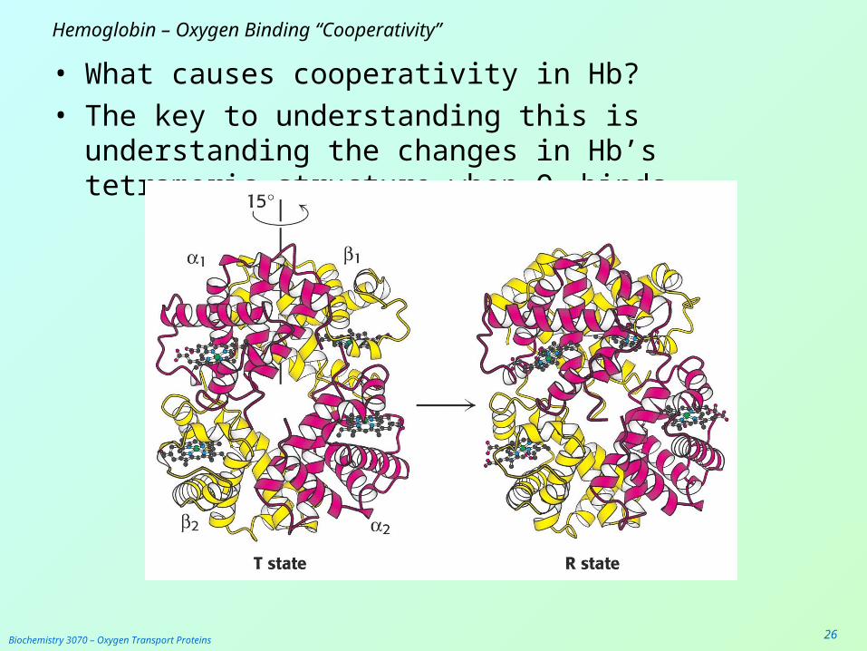
Hemoglobin – Oxygen Binding “Cooperativity”
• What causes cooperativity in Hb?• The key to understanding this is understanding the
changes in Hb’s tetrameric structure when O2 binds.

Biochemistry 3070 – Oxygen Transport Proteins 27
Hemoglobin – Oxygen Binding “Cooperativity”
• When the first O2 molecule binds to one of the four heme groups a number of structural changes occur:– The movement of the Fe atom into the heme plane
also draws in the F8 [promixal] histidine, leveraging a big change in its subunit.
– The alpha and beta groups rotate ~ 15° with respect to one another, disrupting non-covalent linkages between its neighboring subunits.
– The open “channel” in the center of the subunits becomes much smaller, bringing the beta chains much closer than before
• These structural changes increase affinity for oxygen in the remaining three subunits.

Biochemistry 3070 – Oxygen Transport Proteins 28
Hemoglobin – Oxygen Binding “Cooperativity”
• Simple dialysis revealed a key mechanism in Hb cooperativity:
• When erythrocyte contents are dialyzed, the Hill coeffecient drops to n=1! When the permeate is remixed with hemoglobin, the value of n returns to normal (~ 3.3).
• What conclusion can be drawn from this experiment?
Biochemistry 3070 – Oxygen Transport Proteins 29
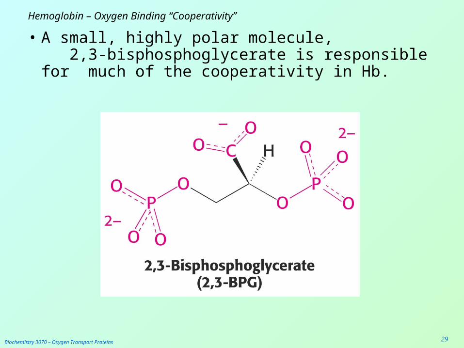
Hemoglobin – Oxygen Binding “Cooperativity”
• A small, highly polar molecule, 2,3-bisphosphoglycerate is responsible for much of the cooperativity in Hb.

Biochemistry 3070 – Oxygen Transport Proteins 30
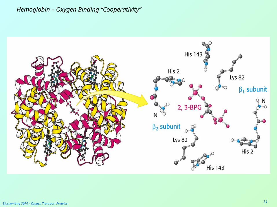
Hemoglobin – Oxygen Binding “Cooperativity”
• 2,3-BPG binds inside the cavity between the four subunits of Hb.
• 2,3-BPG binds only to deoxygenated Hb, when there is room in the cavity.
• When one or more O2 molecules are bound, 2,3-BPG can not fit into the smaller cavity.
• Therefore, the binding of O2 and 2,3-BPG is mutually exclusive (only one or the other). Both can not bind at the same time.
Biochemistry 3070 – Oxygen Transport Proteins 31
Hemoglobin – Oxygen Binding “Cooperativity”
Biochemistry 3070 – Oxygen Transport Proteins 32
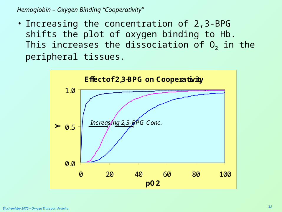
Hemoglobin – Oxygen Binding “Cooperativity”
• Increasing the concentration of 2,3-BPG shifts the plot of oxygen binding to Hb. This increases the dissociation of O2 in the peripheral tissues.
Effect of 2,3-BPG on Cooperativity
0.0
0.5
1.0
0 20 40 60 80 100pO2
Y
Increasing 2,3-BPG Conc.
Biochemistry 3070 – Oxygen Transport Proteins 33
Hemoglobin – Oxygen Binding “Cooperativity”
Assignment Question:When we change altitude, during the next
few days, the body responds by adjusting Hb cooperativity. This can be accomplished by changing the concentration of 2,3-BPG in the blood.
After moving from a low altitude to a higher altitude, does 2,3,-BPG increase or decrease?
Explain how this affects oxygen transport capacity.
Biochemistry 3070 – Oxygen Transport Proteins 34
Hemoglobin – Oxygen Binding “Cooperativity”
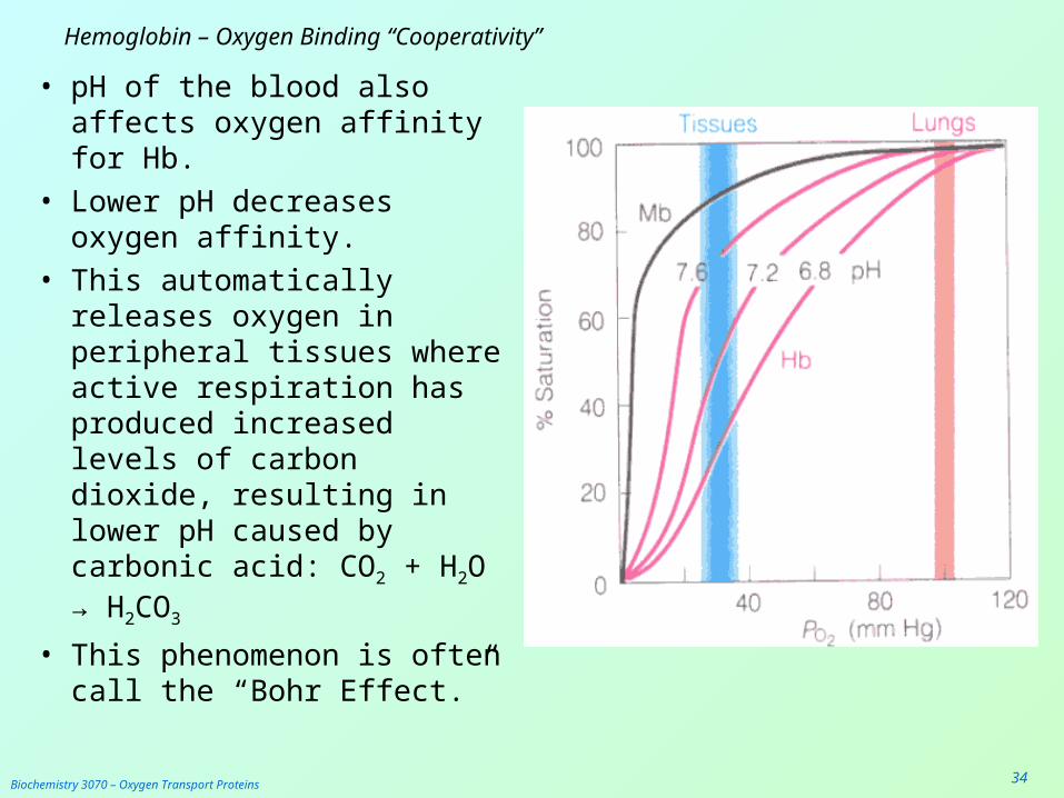
• pH of the blood also affects oxygen affinity for Hb.
• Lower pH decreases oxygen affinity.
• This automatically releases oxygen in peripheral tissues where active respiration has produced increased levels of carbon dioxide, resulting in lower pH caused by carbonic acid: CO2 + H2O → H2CO3
• This phenomenon is often call the “Bohr Effect.”

Biochemistry 3070 – Oxygen Transport Proteins 35
Hemoglobin – Oxygen Binding “Cooperativity”
• The result of the Bohr Effect is to deliver more total oxygen between the lungs and the peripheral tissues at lower pH:

Biochemistry 3070 – Oxygen Transport Proteins 36
Hemoglobin - Carbon Dioxide Transport
• In addition to its role in oxygen transport, hemoglobin also transports CO2 from the tissues back to the lungs for disposal.
• CO2 is not transported by the heme group. Rather, it binds to the terminal amino groups by forming “carbamates.”
• In the alveolae, the equlibrium shifts and the carbamates revert to free amines, releasing CO2.
Biochemistry 3070 – Oxygen Transport Proteins 37
Hemoglobin – Oxygen Binding “Cooperativity”
• Fetal hemoglobin’s “gamma” chains have serine in place of histidine 143 (adult beta chains). This serine is near the binding site for 2,3-BPG and reduces the affinity of fetal Hb for 2,3-BPG.
• The result of this change and reduced affinity for 2,3-BPG is an increased affinity for oxygen.
• Therefore, a fetus can effectively draw oxygen across the placental membrane.

Biochemistry 3070 – Oxygen Transport Proteins 38
Hemoglobin – Oxygen Binding “Cooperativity”

Biochemistry 3070 – Oxygen Transport Proteins 39
Hemoglobin – Sickle Cell Anemia
• A slight change in amino acid composition causes “Sickle Cell Anemia.”
• This genetic disease drastically impedes oxygen transport and is manifested as distorted “sickled ” erythrocyte shapes:
Biochemistry 3070 – Oxygen Transport Proteins 40
Hemoglobin – Sickle Cell Anemia
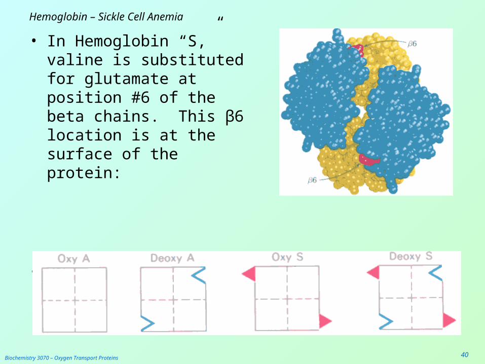
• In Hemoglobin “S,” valine is substituted for glutamate at position #6 of the beta chains. This β6 location is at the surface of the protein:
• This results in “sticky” sites on the deoxy beta chains:
Biochemistry 3070 – Oxygen Transport Proteins 41
Hemoglobin – Sickle Cell Anemia
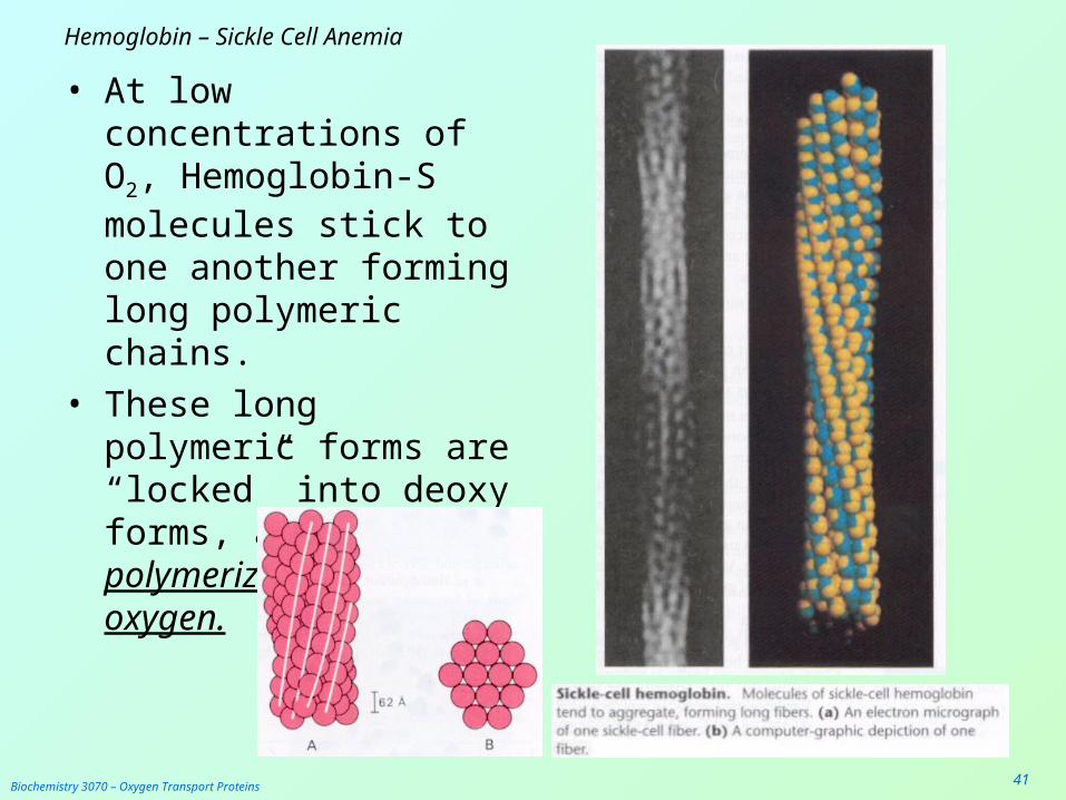
• At low concentrations of O2, Hemoglobin-S molecules stick to one another forming long polymeric chains.
• These long polymeric forms are “locked” into deoxy forms, and while polymerized can not bind oxygen.
Biochemistry 3070 – Oxygen Transport Proteins 42
Hemoglobin – Sickle Cell Anemia
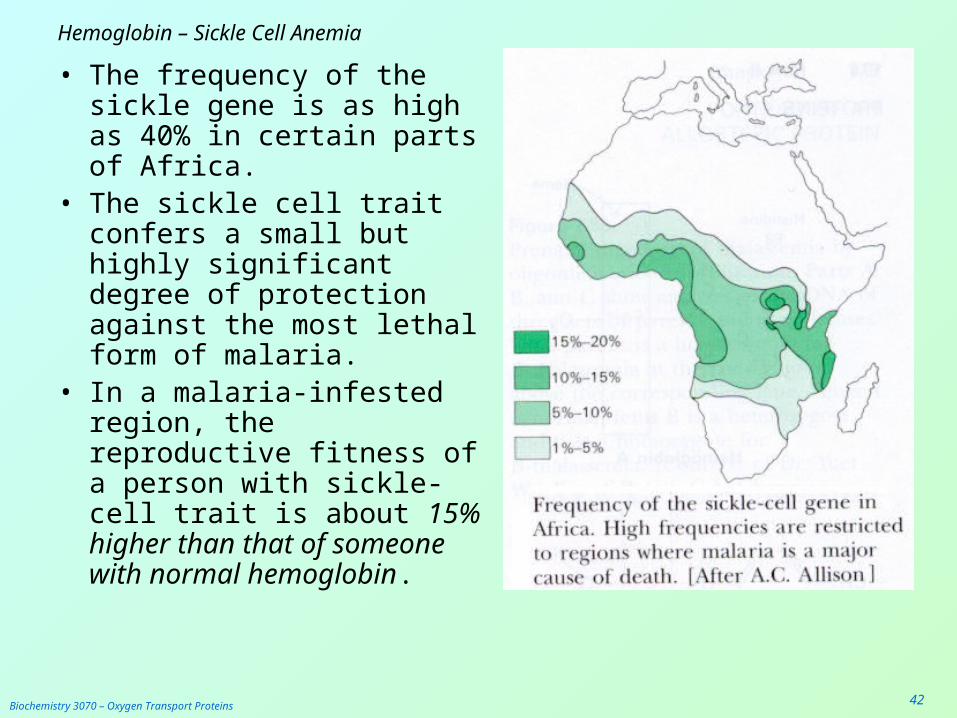
• The frequency of the sickle gene is as high as 40% in certain parts of Africa.
• The sickle cell trait confers a small but highly significant degree of protection against the most lethal form of malaria.
• In a malaria-infested region, the reproductive fitness of a person with sickle-cell trait is about 15% higher than that of someone with normal hemoglobin.

Biochemistry 3070 – Oxygen Transport Proteins 43
Hemoglobin – Sickle Cell Anemia• The Sickle Cell trait can be identified through DNA testing:
• Restriction enzyme digestion with MstII yields three fragments for a normal β gene, but only two for the sickle-cell gene:
Biochemistry 3070 – Oxygen Transport Proteins 44
End of Lecture Slides for
Oxygen Transport Proteins
Credits: Most of the diagrams used in these slides were taken from Stryer, et.al, Biochemistry, 5 th Ed., Freeman Press, Chapter 10 (in our course textbook) and from prior editions of this work.


