Biocompatibility and in vivo operation of implantable mesoporous PVDF-based nanogenerators Yanhao Yu a , Haiyan Sun b , Hakan Orbay c , Feng Chen b , Christopher G. England d , Weibo Cai b,d , Xudong Wang a,n a Department of Materials Science and Engineering, University of Wisconsin-Madison, Madison, WI 53706, USA b Department of Radiology, University of Wisconsin-Madison, WI 53705, USA c Department of Surgery, University of California-Davis, Sacramento, CA 95817, USA d Department of Medical Physics, University of Wisconsin-Madison, Madison, WI 53705, USA article info Article history: Received 19 May 2016 Received in revised form 24 June 2016 Accepted 13 July 2016 Available online 16 July 2016 Keywords: Biocompatibility In vivo biomechanical energy harvesting Mesoporous PVDF Piezoelectric nanogenerator abstract The rapid developments of implantable biomedical electronics give rise to the motivation of exploring efficient and durable self-powered charging system. In this paper, we report a mesoporous poly- vinylidene fluoride (PVDF)-based implantable piezoelectric nanogenerator (NG) for in vivo biomecha- nical energy harvesting. The NG was built with a sponge-like mesoporous PVDF film and encapsulated by polydimethylsiloxane (PDMS). After embedding this NG into rodents, a V oc of 200 mV was produced from the gentle movement of rodent muscle. Meanwhile, no toxicity or incompatibility sign was found in the host after carrying the packaged NG for 6 weeks. Moreover, the electric output of this NG was ex- tremely stable and exhibited no deterioration after 5 days of in vivo operation or 1.512 10 8 times mechanical deformation. This NG device could practically output a constant voltage of 52 mV via a 1 μF capacitor under living circumstance. The outstanding efficiency, magnificent durability and exceptional biocompatibility promise this mesoporous PVDF-based NG in accomplishing self-powered bioelectronics with potentially lifespan operation period. & 2016 Published by Elsevier Ltd. 1. Introduction Piezoelectric nanogenerator (NG) is a promising technology for harvesting mechanical energy from ambient environment relying on the electromechanical coupling effect of piezoelectric materials [1–4]. Due to the broad material selection and flexible structural design, NGs have shown superior capability in scavenging me- chanical energy in various forms including acoustic waves, physi- cal deflections, fluid or gas flows, and human activities [5–13]. Among them, in vivo biomechanical energy harvesting is parti- cularly attractive since this approach could lead to a sustainable energy source for implantable biomedical devices, which could significantly reduce the volume of powering component and avoid battery replacement [14–16]. Considerable endeavors have been devoted in employing NGs to harvesting mechanical energy from the motion of muscle or organs like heart, lung and diaphragm [16–21]. Most NGs tested inside living organisms used piezo- electric ceramics, such as zinc oxide, [18,19] barium titanate, [22] and lead zirconate titanate (PZT) [17,21,23]. In order to achieve high flexibility, these materials were typically constructed in na- nostructure and/or nanocomposite forms. On the other hand, piezoelectric polymers were directly used in NG design owing to their intrinsic superior flexibility and elasticity. β-phase poly- vinylidene fluoride (PVDF) is the most widely used piezoelectric polymers with merits of high piezoelectric coefficients (20–30 pC/ N for d 33 and 16 pC/N for d 31 ), [24] excellent mechanical property and good biocompatibility [25–31]. These advantages encouraged a number of NG developments based on the piezoelectric PVDF [32–35]. Nevertheless, disregarding the great number of demonstrations of implantable NG designs, study of the biocompatibility and op- eration in a closed living biological environment is still very lim- ited. The dynamic in vivo conditions could give rise to a number of critical issues that may not be a concern for in vitro tests. For example, the bio-fluid might respond to the polarized NG surfaces. Ions might infiltrate into the NG and deteriorate the piezoelectric property. The soft and irregular tissue surface might easily cause repelling or detachment of the NG component during normal body movement. It is also unclear how the biological surface would response to the high local piezoelectric potential generated in its vicinity. In this paper, we report a systematic study of the bio- compatibility and in vivo operation of a mesoporous PVDF-based Contents lists available at ScienceDirect journal homepage: www.elsevier.com/locate/nanoen Nano Energy http://dx.doi.org/10.1016/j.nanoen.2016.07.015 2211-2855/& 2016 Published by Elsevier Ltd. n Corresponding author. E-mail address: [email protected](X. Wang). Nano Energy 27 (2016) 275–281
Transcript
Nano Energy 27 (2016) 275–281
Contents lists available at ScienceDirect
Nano Energy
http://d2211-28
n CorrE-m
journal homepage: www.elsevier.com/locate/nanoen
Biocompatibility and in vivo operation of implantable mesoporousPVDF-based nanogenerators
Yanhao Yu a, Haiyan Sun b, Hakan Orbay c, Feng Chen b, Christopher G. England d,Weibo Cai b,d, Xudong Wang a,n
a Department of Materials Science and Engineering, University of Wisconsin-Madison, Madison, WI 53706, USAb Department of Radiology, University of Wisconsin-Madison, WI 53705, USAc Department of Surgery, University of California-Davis, Sacramento, CA 95817, USAd Department of Medical Physics, University of Wisconsin-Madison, Madison, WI 53705, USA
a r t i c l e i n f o
Article history:Received 19 May 2016Received in revised form24 June 2016Accepted 13 July 2016Available online 16 July 2016
Keywords:BiocompatibilityIn vivo biomechanical energy harvestingMesoporous PVDFPiezoelectric nanogenerator
x.doi.org/10.1016/j.nanoen.2016.07.01555/& 2016 Published by Elsevier Ltd.
The rapid developments of implantable biomedical electronics give rise to the motivation of exploringefficient and durable self-powered charging system. In this paper, we report a mesoporous poly-vinylidene fluoride (PVDF)-based implantable piezoelectric nanogenerator (NG) for in vivo biomecha-nical energy harvesting. The NG was built with a sponge-like mesoporous PVDF film and encapsulated bypolydimethylsiloxane (PDMS). After embedding this NG into rodents, a Voc of �200 mV was producedfrom the gentle movement of rodent muscle. Meanwhile, no toxicity or incompatibility sign was found inthe host after carrying the packaged NG for 6 weeks. Moreover, the electric output of this NG was ex-tremely stable and exhibited no deterioration after 5 days of in vivo operation or 1.512�108 timesmechanical deformation. This NG device could practically output a constant voltage of 52 mV via a 1 μFcapacitor under living circumstance. The outstanding efficiency, magnificent durability and exceptionalbiocompatibility promise this mesoporous PVDF-based NG in accomplishing self-powered bioelectronicswith potentially lifespan operation period.
& 2016 Published by Elsevier Ltd.
1. Introduction
Piezoelectric nanogenerator (NG) is a promising technology forharvesting mechanical energy from ambient environment relyingon the electromechanical coupling effect of piezoelectric materials[1–4]. Due to the broad material selection and flexible structuraldesign, NGs have shown superior capability in scavenging me-chanical energy in various forms including acoustic waves, physi-cal deflections, fluid or gas flows, and human activities [5–13].Among them, in vivo biomechanical energy harvesting is parti-cularly attractive since this approach could lead to a sustainableenergy source for implantable biomedical devices, which couldsignificantly reduce the volume of powering component and avoidbattery replacement [14–16]. Considerable endeavors have beendevoted in employing NGs to harvesting mechanical energy fromthe motion of muscle or organs like heart, lung and diaphragm[16–21]. Most NGs tested inside living organisms used piezo-electric ceramics, such as zinc oxide, [18,19] barium titanate, [22]and lead zirconate titanate (PZT) [17,21,23]. In order to achieve
).
high flexibility, these materials were typically constructed in na-nostructure and/or nanocomposite forms. On the other hand,piezoelectric polymers were directly used in NG design owing totheir intrinsic superior flexibility and elasticity. β-phase poly-vinylidene fluoride (PVDF) is the most widely used piezoelectricpolymers with merits of high piezoelectric coefficients (20–30 pC/N for d33 and 16 pC/N for d31), [24] excellent mechanical propertyand good biocompatibility [25–31]. These advantages encourageda number of NG developments based on the piezoelectric PVDF[32–35].
Nevertheless, disregarding the great number of demonstrationsof implantable NG designs, study of the biocompatibility and op-eration in a closed living biological environment is still very lim-ited. The dynamic in vivo conditions could give rise to a number ofcritical issues that may not be a concern for in vitro tests. Forexample, the bio-fluid might respond to the polarized NG surfaces.Ions might infiltrate into the NG and deteriorate the piezoelectricproperty. The soft and irregular tissue surface might easily causerepelling or detachment of the NG component during normal bodymovement. It is also unclear how the biological surface wouldresponse to the high local piezoelectric potential generated in itsvicinity. In this paper, we report a systematic study of the bio-compatibility and in vivo operation of a mesoporous PVDF-based
flexible NGs. The NG was built with a sponge-like mesoporousPVDF film and encapsulated by polydimethylsiloxane (PDMS). Thepackaged NG was implanted under the skin of living mice and rats.Histological examination revealed no signs of toxicity or in-compatibility during weeks of implantation. Appreciable piezo-electric output was obtained from the implanted NGs, which ex-hibited no deterioration after long-term in vivo operation. In vivoelectricity routing and capacitor charging were also demonstratedinside the rat's body. This work provides valuable insights towardthe biocompatibility and in vivo operation of flexible NGs forpractically applications as an implantable biomechanical energyharvesting system.
2. Experimental section
2.1. Fabrication of Sponge-Like PVDF Thin Films
PVDF powder (Sigma Aldrich) was first dissolved in N,N-di-methylformamide (DMF) solvent with a concentration of 10 wt% at70 °C. The PVDF solution was then mixed with ZnO nanoparticles(35–45 nm, US Research Nanomaterials, Inc.). The mass ratio be-tween ZnO nanoparticles and PVDF was 50%. A uniformly dis-tributed PVDF/ZnO suspension was obtained after treating themixture in ultrasonic bath for 30 min. The suspension was thencast into a petri dish and dried in oven at 75 °C for approximate10 min (depending on the film thickness). The films were subse-quently immersed in a 37 wt% HCl solution for 3 h to completelyremove the ZnO template. After acid etching and deionized waterwashing, the mesoporous PVDF thin films with a film thickness of�30 mm were obtained.
2.2. Assembly and Measurement of PVDF NG
The mesoporous PVDF film was first cut to 1 cm�2 cm and100 nm-thick interdigitated surface gold electrode was depositedby electron beam evaporation with a shadow mask. Then, thePVDF film was poled using the interdigitated electrodes in an oilbath with an electric field of 60 V/μm for 2 h. After that, the PVDFfilm was sandwiched between two PDMS films with distinct filmthicknesses (100 mm and 1 mm). The layered structure was thensealed by dipping and curing the liquid PDMS elastomer and cross-linker. During the performance measurement, a periodical de-flecting force was applied on the NG device using a computercontrolled shaker with a frequency of 20 Hz and a force of 6 N(quantified by a Force Gauge HF-500 N). The strain applied on theNG device was calculated to be �0.1% on the basis of the canti-lever model. The PDMS control sample was fabricated in a similarway by replacing the PVDF film with a PDMS film with identicalfilm thickness. The voltage outputs were recorded using an AgilentDSO1012A oscilloscope. The current outputs were measured usingan Autolab PGSTAT302N station. SEM image of mesoporous PVDFfilm was acquired with a LEO 1530 scanning electron microscope.FTIR characterizations of the PVDF films were performed by theBruker Tensor 27 spectrometer.
2.3. Implantation of the PVDF NG
All animal studies were conducted under a protocol approvedby the University of Wisconsin Institutional Animal Care and UseCommittee. Adult Sprague-Dawley rats and six-week-old im-printing control region (ICR) mice (Harlan, Indianapolis, IN) wereused to investigate the function of the NG in biological environ-ment. Briefly, anesthesia was induced by inhalation of 2–5% iso-flurane and maintained with 2% isoflurane. Following anesthesia,animals were fixed in the right lateral decubitus position. A 1–
3 cm surgical incision was made to place the device. The NG wasplaced between the epithelial and muscle layer in the thigh orback region. The incision was sutured and the animal was con-nected to oscilloscope for measuring the voltage output. The NGwas left under the skin and animals were allowed to recover. Afterthe conclusion of studies, animals were euthanized and the NG,along with muscle, skin, and other tissues was collected for ana-lysis. Computer tomography (CT) scans were performed using anInveon microPET/microCT rodent model scanner (Siemens MedicalSolutions USA, Inc.).
2.4. Biocompatibility safety assessment of NG in mice
In vivo toxicity was assessed in mice by surgically extractingthe NG from the surgical site at select time points post-im-plantation. Tissue containing the muscle and skin, with the devicebetween these two layers, was obtained for histological analysis.The NG was removed before tissues were paraffin embedded andsectioned at 5 mm thickness for analysis. For comparison, similartissues were extracted from the contralateral leg, which containedno devices. Tissue sections were hematoxylin and eosin (H&E)stained before analysis. Toxicity of the tissue was assessed insections using four criteria, including: (1) degree of inflammatoryinfiltrate; (2) degree of fibrosis; (3) presence of muscle degen-eration; and (4) presence and degree of cellular toxicities at im-plantation device (e.g., cellular shrinkage, condensation of chro-matin, cell membrane viability, and presence of apoptotic bodies).
3. Results and discussion
The mesoporous PVDF thin films were fabricated using ZnOnanoparticles (NPs) as the sacrificial template, following the ap-proach we reported previously [34]. Removing the ZnO NPs leftrandomly distributed pores with sizes of 30–500 nm in the PVDFfilms (Fig. S1). The phase of the mesoporous PVDF films wascharacterized by the Fourier transform infrared (FTIR) spectrum(Fig. S2), where representative peaks of the piezoelectric β-phaseat 840 and 1280 cm�1 could be clearly identified. The spontaneousformation of the β-phase was attributed to the interactions be-tween the PVDF dipoles and surface charges on ZnO surfaces[7,34]. A fully assembled PVDF NG for implantation is shown inFig. 1A. The mesoporous PVDF film was coated with a pair of in-terdigitated gold electrodes for piezoelectric charge collection, andthe entire structure was packaged inside PDMS. According to ourprevious research, compared with the solid PVDF, introducingconsiderable porosity to the PVDF film could significantly increaseits flexibility, [34] which is highly favorable for biomechanicalenergy harvesting. The interdigitated electrode was adopted toenable charge collection from the piezo vector of d33, which hasthe highest piezoelectric coefficient for PVDF [24]. Meanwhile, thethicknesses of PDMS films that covered the top and bottom sidesof the PVDF film were designed to be 100 mm and 1 mm, respec-tively. This variation of PDMS film thicknesses was able to increasethe tension and compression portion of internal dipoles and thusimprove device performance.
The energy harvesting capability of the completely-packagedNG was first characterized by periodically deflecting the NG in acantilever mode. The open-circuit voltage (Voc) and short-circuitcurrent (Isc) are presented in Fig. 1B and C, respectively. When theshaker was operating at 20 Hz with a force of 6 N and a strain of�0.1%, the average values of Voc and Isc reached 3.8 V and 3.5 μA,respectively, which outperformed a number of reported PVDF-based NGs [6,30,32,33,36]. This superior performance was mostlyattributed to the optimized device geometry and structural ad-vantage of the mesoporous PVDF thin film. In comparison, after
Fig. 1. In vitro electrical output of the PDMS-packaged PVDF NG. (A) A photograph of the PVDF NG before implantation. (B,C) The voltage (B) and current (C) output of thePVDF NG responding to a 20 Hz periodical deflection in a cantilever mode under in vitro condition.
Y. Yu et al. / Nano Energy 27 (2016) 275–281 277
replacing PVDF with PDMS (i.e., a representative triboelectricpolymer with no piezoelectric response), the peak Voc sharplyreduced to �0.1 V (Fig. S3), meaning the output of this NG designwas predominately from the piezoelectric effects rather than tri-boelectric effects of the PVDF film. The high piezoelectric outputalso suggested that the PDMS packaging had minimal influence tothe mechanical-to-electrical energy conversion.
To evaluate the biocompatibility, a PVDF NG with a size of6 mm�6 mm was inserted under the skin of mice's right leg(Fig. 2A and S4). The bottom layer of NG was attached to thequadriceps femoris muscle surface. Fig. 2B shows a CT scan of themouse that carried the NG for 5 days, where the metal electrodecould be clearly observed revealing the steady implantation
Fig. 2. Biocompatibility assessment of the PDMS-packaged PVDF NG. (A) A surgery pictlayer in the thigh region. Inset is an enlarged picture showing the implanted PVDF NG witrevealing the steady implantation position of the NG under mouse's normal activity. (C) S6 after implantation. No signs of toxicity or incompatibility induced by the NG were ob
position of the NG under the mouse's normal activity. The micewith implanted NG were sacrificed at week 1, 2, 3 and 6 for his-tological examination. Surgery and sham control groups wereextracted from the right and left legs of the animal, respectively.As shown in Fig. 2C, surgical pathology analysis revealed no overalldifferences between the NG-implanted and sham thigh in terms ofskin histology, as both sections appeared normal. There werelimited inflammatory infiltrates in both groups. In addition, therewere no signs of cellular toxicity or muscular atrophy/degenera-tion in tissue sections. As expected, the surgical thigh and shamthigh showed regional disruption in the epidermal layer, whichwas attributed to the formation of scar tissue and wound healing.Overall, there were no signs of toxicity or incompatibility induced
ure of the mouse with the PVDF NG embedded between the epithelial and muscleh integrated surface electrode. (B) CT scan of a mouse that carried the NG for 5 days,urgical pathology analysis of the NG-implanted and sham group at week 1, 2, 3 andserved in terms of muscle and skin histology.
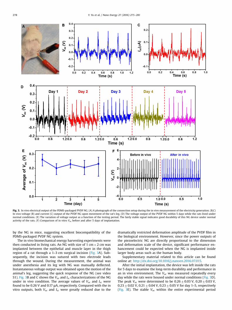
Fig. 3. In vivo electrical output of the PDMS-packaged PVDF NG. (A) A photograph of the connection setup during the in vivo measurement of the electricity generation. (B,C)In vivo voltage (B) and current (C) output of the PVDF NG upon movement of the rat's leg. (D) The voltage output of the PVDF NG within 5 days while the rats lived undernormal conditions. (E) The variation of voltage output as a function of the testing period. The fairly stable signal indicates good durability of this NG device under normalactivity of the rats. (F) Comparison of in vitro Voc before and after 5 days of implantation.
Y. Yu et al. / Nano Energy 27 (2016) 275–281278
by the NG in mice, suggesting excellent biocompatibility of thePDMS-packaged PVDF NG system.
The in vivo biomechanical energy harvesting experiments werethen conducted in living rats. An NG with size of 1 cm�2 cm wasimplanted between the epithelial and muscle layer in the thighregion of a rat through a 1–3 cm surgical incision (Fig. 3A). Sub-sequently, the incision was sutured with two electrode leadsthrough the wound. During the measurement, the animal wasunder anesthesia and its leg with NG was manually deflected.Instantaneous voltage output was obtained upon the motion of theanimal's leg, suggesting the quick response of the NG (see videoS1). Fig. 3B and C shows the Voc and Isc characterizations of the NGunder in vivo condition. The average values of Voc and Isc werefound to be 0.26 V and 0.17 μA, respectively. Compared with the invitro outputs, both Voc and Isc were greatly reduced due to the
dramatically restricted deformation amplitude of the PVDF film inthe biological environment. However, since the power outputs ofthe piezoelectric NG are directly proportional to the dimensionand deformation scale of the device, significant performance en-hancement could be expected when the NG is implanted insidelarger body areas such as the human body.
Supplementary material related to this article can be foundonline at: http://dx.doi.org/10.1016/j.nanoen.2016.07.015.
After the initial implantation, the device was left inside the ratsfor 5 days to examine the long-term durability and performance inan in vivo environment. The Voc was measured repeatedly everyday while the rats were housed under normal conditions (Fig. 3D).The peak Voc were determined to be 0.2670.03 V, 0.2070.03 V,0.2370.02 V, 0.2170.04 V, 0.2370.05 V for day 1–5, respectively(Fig. 3E). The stable Voc within the entire experimental period
Fig. 4. Long-term durability evaluation of the PDMS-packaged PVDF NG. (A) Schematic illustration of the mimic bio-environment with ultrasonic wave as the vibrationsource. (B,C) The voltage output of the PVDF NG at the beginning (B) and after 1 h of ultrasound (C). On and off represent with and without ultrasonic vibration. (D,E)Comparison of in vitro Voc before (D) and after (E) the ultrasound.
Y. Yu et al. / Nano Energy 27 (2016) 275–281 279
suggested that the NG device could function normally withoutnoticeable decay, despite the constant motion of the animal duringthe five days. After 5 days of embedment, the PVDF NG was takenout from the rat's body and its piezoelectric output was re-char-acterized under the same in vitro deflection conditions. No ob-vious discrepancy was observed from the Voc signals of NG before(black) and after (blue) 5 days of implantation in live rats (Fig. 3F).This comparison confirmed the well preserved functionality of theNG after experiencing long time exposure to the body fluid andtremendous/constant irregular mechanical deformation.
Nevertheless, the desired life time for an implantable NGshould be at least multiple years in order to effectively minimizethe chance of surgical replacement. Using heart beating as anexample, given the typically beating frequency being 60 perminute, 5 years of operation requires �1.577�108 straining ac-tions. In order to evaluate the possible life time of the packagedNG for converting heart motion to electricity within a reasonabletime frame, ultrasonic wave was introduced as the vibrationsource to mimic the straining from heart beating over life time. Asschematically shown in Fig. 4A, the entire NG device was im-mersed in the 0.9% NaCl solution and was subjected to continuousstraining in an ultrasonic bath. The ultrasonic source was operat-ing at a frequency of 42 kHz. By considering one strike by theacoustic wave as one straining action to the NG, one hour of ul-trasound bath ideally corresponded to 1.512�108 straining ac-tions, which was about the same as the number of heart beat in5 years. It should be noted that this estimation was establishedwithout considering the variable displacement scales of ultrasonicwave and human heart beat. The voltage output was measuredwhen the ultrasonic source was turned on and off. Clear voltageoutput was identified when turning on the ultrasonic equipment.
The average peak value of Voc was found to be �0.270.1 V, whichwas comparable to that of in vivo measurement (Fig. 4B). Thisrelatively small value was a consequence of the limited strainingamplitude from ultrasonic wave. After 1 h of continuous operation,the NG was able to produce a more consistent Voc of�0.1870.03 V (Fig. 4C). This comparison revealed that the PDMSpackaging was highly robust and was able to protect the PVDF NGfrom bio-fluids under long-term mechanical deformation. Thepiezoelectric voltage outputs of the NG were further comparedbefore and after the ultrasonic agitation outside of bio-fluids undera mechanical shaker. As shown in Fig. 4D and E, the Voc curveswere almost identical before and after the ultrasonic bath, con-firming the piezoelectric performance of the packaged PVDF NGwere not impaired by long-term straining actions in bio-fluids.This study demonstrated the excellent durability of the PDMS-packaged PVDF NG and endowed practical application potential ofthis NG system as an implantable power source of biomedicaldevices.
The efficient and stable electric output promised the PVDF NGin charging or powering small electronics in vivo. To demonstratethis point, an energy storage system comprised of a miniaturizedbridge circuit and a ceramic capacitor (1 mF) was packaged(Fig. 5A). As shown in Fig. 5B and C, the NG was implanted fol-lowing the same procedure as abovementioned implantation ex-periments. The output leads of the NG was wired through theepithelial and muscle layer in the thigh and back region, and ex-tended out from the back of the rat where the motion is mini-mized. The energy storage pack was then connected through theleads and fixed on the back of the rat. Upon the movement of therat's leg, the NG was straining and generating electricity as ex-pected. After the rectification, the alternating current (AC) was
Fig. 5. Demonstration of practical biomechanical energy conversion and storage in live rats. (A) A photograph of the energy storage package composed of a miniaturizedbridge circuit and a ceramic capacitor (1 mF). (B) A photograph of implanted PVDF NG in a rat with extended output leads. (C) Overall setup of the in vivo electricitygeneration and storage in rats. (D) DC voltage output from the PVDF NG after rectification. (E) Constant voltage output from NG charged energy storage package.
Y. Yu et al. / Nano Energy 27 (2016) 275–281280
converted to direct current (DC) signal without observable lossthrough the in vivo electricity transport (Fig. 5D). The peak valueof Voc after rectification reached about 200 mV, which was similaras the AC signal. This output was converted to a 52 mV DC voltagethrough a 1 μF capacitor (Fig. 5E), suggesting good potential ofusing NG to power biomedical electronics in vivo.
4. Conclusion
In summary, we developed an efficient, flexible and robustpiezoelectric polymer-based NG system with the capability ofharvesting biomechanical energy in living organism. The size,structure and packaging of the NG were designed delicately foradapting the harsh in vivo environment. Due to the optimizeddevice configuration and excellent flexibility of the sponge-likemesoporous PVDF film, the average Voc and Isc of this NG devicereached 3.8 V and 3.5 μA, respectively, outperforming a number ofreported PVDF-based NGs. By implanting the packaged NG to thequadriceps femoris muscle surface of a rat, a Voc of �200 mV wasgenerated from the gentle motions of the rat's leg; and this signalexhibited no reduction after 5 days of in vivo operation. Years-longoperation of the packaged NG in biological environment waspredicted by testing the NG in ultrasonic bio-fluid bath. Theelectricity produced from animal's leg movement was transportedin vivo by buried leads and connected to an energy storage packlocated remotely. The generated AC signals were rectified and a DCoutput of �52 mV was obtained through a capacitor. This studydemonstrated that packaged PVDF NG could be a feasible strategyto serve as implantable powering system with practically long lifetime.
Acknowledgments
Research reported in this publication was supported, in part, bythe National Institutes of Health (R01EB021336, R01CA169365,P30CA014520, and T32CA009206). The content is solely the re-sponsibility of the authors and does not necessarily represent theofficial views of the National Institutes of Health.
Appendix A. Supporting information
Supplementary data associated with this article can be found inthe online version at http://dx.doi.org/10.1016/j.nanoen.2016.07.015.
References
[1] Z.L. Wang, J. Song, Science 312 (2006) 242–246.[2] X. Wang, J. Song, J. Liu, Z.L. Wang, Science 316 (2007) 102–105.[3] X. Wang, Nano Energy 1 (2012) 13–24.[4] Y. Hu, Z.L. Wang, Nano Energy 14 (2015) 3–14.[5] S.N. Cha, J.S. Seo, S.M. Kim, H.J. Kim, Y.J. Park, S.W. Kim, J.M. Kim, Adv. Mater. 22
(2010) 4726–4730.[6] B.J. Hansen, Y. Liu, R. Yang, Z.L. Wang, ACS Nano 4 (2010) 3647–3652.[7] M. Lee, C.Y. Chen, S. Wang, S.N. Cha, Y.J. Park, J.M. Kim, L.J. Chou, Z.L. Wang,
Adv. Mater. 24 (2012) 1759–1764.[8] K.I. Park, M. Lee, Y. Liu, S. Moon, G.T. Hwang, G. Zhu, J.E. Kim, S.O. Kim, D.
K. Kim, Z.L. Wang, K.J. Lee, Adv. Mater. 24 (2012) 2999–3004.[9] J.-H. Lee, K.Y. Lee, B. Kumar, N.T. Tien, N.-E. Lee, S.-W. Kim, Energy Environ. Sci.
6 (2013) 169–175.[10] S. Siddiqui, D.-I. Kim, L.T. Duy, M.T. Nguyen, S. Muhammad, W.-S. Yoon, N.-
E. Lee, Nano Energy 15 (2015) 177–185.[11] Y. Yu, Z. Li, Y. Wang, S. Gong, X. Wang, Adv. Mater. 27 (2015) 4938–4944.[12] M. Zhang, T. Gao, J. Wang, J. Liao, Y. Qiu, Q. Yang, H. Xue, Z. Shi, Y. Zhao,
Z. Xiong, L. Chen, Nano Energy 13 (2015) 298–305.[13] Y. Yu, X. Wang, Extreme Mech. Lett. (2016), http://dx.doi.org/10.1016/j.
[14] Q. Zheng, B. Shi, F. Fan, X. Wang, L. Yan, W. Yuan, S. Wang, H. Liu, Z. Li, Z.L. Wang, Adv. Mater. 26 (2014) 5851–5856.
[15] G.T. Hwang, D. Im, S.E. Lee, J. Lee, M. Koo, S.Y. Park, S. Kim, K. Yang, S.J. Kim,K. Lee, K.J. Lee, ACS Nano 7 (2013) 4545–4553.
[16] G.T. Hwang, Y. Kim, J.-H. Lee, S. Oh, C.K. Jeong, D.Y. Park, J. Ryu, H.S. Kwon, S.-G. Lee, B. Joung, D. Kim, K.J. Lee, Energy Environ. Sci. 8 (2015) 2677–2684.
[17] S.R. Platt, S. Farritor, K. Garvin, H. Haider, IEEE/ASME Trans. Mechatron. 10(2005) 455–461.
[18] R. Yang, Y. Qin, C. Li, G. Zhu, Z.L. Wang, Nano Lett. 9 (2009) 1201–1205.[19] Z. Li, G. Zhu, R. Yang, A.C. Wang, Z.L. Wang, Adv. Mater. 22 (2010) 2534–2537.[20] A. Zurbuchen, A. Pfenniger, A. Stahel, C.T. Stoeck, S. Vandenberghe, V.M. Koch,
R. Vogel, Ann. Biomed. Eng. 41 (2013) 131–141.[21] C. Dagdeviren, B.D. Yang, Y. Su, P.L. Tran, P. Joe, E. Anderson, J. Xia,
V. Doraiswamy, B. Dehdashti, X. Feng, B. Lu, R. Poston, Z. Khalpey, R. Ghaffari,Y. Huang, M.J. Slepian, J.A. Rogers, Proc. Natl. Acad. Sci. 111 (2014) 1927–1932.
[22] K.I. Park, S. Xu, Y. Liu, G.T. Hwang, S.J. Kang, Z.L. Wang, K.J. Lee, Nano Lett. 10(2010) 4939–4943.
[23] Y. Qi, J. Kim, T.D. Nguyen, B. Lisko, P.K. Purohit, M.C. McAlpine, Nano Lett. 11(2011) 1331–1336.
[24] P. Ueberschlag, Sens. Rev. 21 (2001) 118–126.[25] R.A. Whiter, V. Narayan, S. Kar-Narayan, Adv. Energy Mater. 4 (2014) 1400519.
[26] B.S. Lee, B. Park, H.S. Yang, J.W. Han, C. Choong, J. Bae, K. Lee, W.R. Yu, U. Jeong,U.I. Chung, J.J. Park, O. Kim, ACS Appl. Mater. Interfaces 6 (2014) 3520–3527.
[27] S. Garain, T.K. Sinha, P. Adhikary, K. Henkel, S. Sen, S. Ram, C. Sinha,D. Schmeisser, D. Mandal, ACS Appl. Mater. Interfaces 7 (2015) 1298–1307.
[28] N.R. Alluri, B. Saravanakumar, S.J. Kim, ACS Appl. Mater. Interfaces 7 (2015)9831–9840.
[29] X. Chen, H. Tian, X. Li, J. Shao, Y. Ding, N. An, Y. Zhou, Nanoscale 7 (2015)11536–11544.
[30] J.-H. Lee, H.-J. Yoon, T.Y. Kim, M.K. Gupta, J.H. Lee, W. Seung, H. Ryu, S.-W. Kim,Adv. Funct. Mater. 25 (2015) 3203–3209.
[31] C. Chang, V.H. Tran, J. Wang, Y.K. Fuh, L. Lin, Nano Lett. 10 (2010) 726–731.[32] S.H. Bae, O. Kahya, B.K. Sharma, J. Kwon, H.J. Cho, B. Ozyilmaz, J.H. Ahn, ACS
Nano 7 (2013) 3130–3138.[33] M. Baniasadi, J. Huang, Z. Xu, S. Moreno, X. Yang, J. Chang, M.A. Quevedo-
Lopez, M. Naraghi, M. Minary-Jolandan, ACS Appl. Mater. Interfaces 7 (2015)5358–5366.
[34] Y. Mao, P. Zhao, G. McConohy, H. Yang, Y. Tong, X. Wang, Adv. Energy Mater. 4(2014) 130624.
[35] C. Sun, J. Shi, D.J. Bayerl, X. Wang, Energy Environ. Sci. 4 (2011) 4508.[36] J.H. Lee, K.Y. Lee, M.K. Gupta, T.Y. Kim, D.Y. Lee, J. Oh, C. Ryu, W.J. Yoo, C.Y. Kang,