93
BIOGENESIS OF PEROXISOMAL INTRINSIC MEMBRANE PROTEINS: UNVEILING THE ROLE OF PEX19P MANUEL ANTÓNIO BOTELHO PEREIRA PINTO Dissertação de doutoramento em Ciências Biomédicas 2008

BIOGENESIS OF PEROXISOMAL INTRINSIC MEMBRANE PROTEINS: UNVEILING THE ROLE OF PEX19P
MANUEL ANTÓNIO BOTELHO PEREIRA PINTO
Dissertação de doutoramento em Ciências Biomédicas
2008

MANUEL ANTÓNIO BOTELHO PEREIRA PINTO
BIOGENESIS OF PEROXISOMAL INTRINSIC MEMBRANE PROTEINS: UNVEILING THE ROLE OF PEX19P
Dissertação de Candidatura ao grau de Doutor em Ciências Biomédicas submetida ao Instituto de Ciências Biomédicas Abel Salazar da Universidade do Porto.
Orientador – Prof. Doutor Jorge Eduardo da Silva Azevedo Categoria – Professor associado com agregação Afiliação – Instituto de Ciências Biomédicas Abel Salazar da Universidade do Porto.
Co-orientadora – Doutora Maria Clara Pereira de Sá Miranda Categoria – Investigadora do Instituto de Biologia Molecular e Celular Afiliação – Instituto de Biologia Molecular e Celular.

TABLE OF CONTENTS
PRECEITOS LEGAIS..........................................................................................................1 AGRADECIMENTOS/ACKNOWLEDGEMENTS.................................................................2 ABSTRACT..........................................................................................................................3 RESUMO.............................................................................................................................4 RÉSUMÉ .............................................................................................................................6 ABBREVIATIONS................................................................................................................8 1. INTRODUCTION ...........................................................................................................10
1.1. Structure and function of peroxisomes ...................................................................11 1.2. Peroxisomal disorders ............................................................................................12 1.3. Peroxisome biogenesis...........................................................................................14
1.3.1. Matrix protein import ........................................................................................16 1.3.2. Peroxisome proliferation ..................................................................................17 1.3.3. Peroxisomal membrane biogenesis.................................................................17
1.3.3.1. Biogenesis of peroxisomal membrane proteins ........................................17 1.3.3.2. Origin of the peroxisomal membrane........................................................18 1.3.3.3. General properties of Pex3p .....................................................................21 1.3.3.4. Pex19p domain architecture and its interactions with PMPs ....................22 1.3.3.5. PMP sorting to peroxisomes and its dependence on Pex19p ..................23 1.3.3.6. Pex19p interactions with Pex3p................................................................24 1.3.3.7. Models of Pex19p role in biogenesis ........................................................25
2. OBJECTIVES ................................................................................................................28 3. EXPERIMENTAL PROCEDURES.................................................................................30
3.1. Construction of plasmids encoding the reporter PMP.............................................31 3.2. Cell culture, transfections and immunofluorescence microscopy ...........................32 3.3. Expression and purification of recombinant proteins ..............................................32 3.4. In vitro synthesis of radiolabeled proteins...............................................................33 3.5. Preparation of rat liver post-nuclear supernatants ..................................................33 3.6. In vitro import experiments .....................................................................................34 3.7. Extraction of membranes with alkali and high/low ionic strength solutions.............34 3.8. Density gradient centrifugation ...............................................................................35 3.9. Immunoprecipitation of the imported reporter protein.............................................35 3.10. Native polyacrylamide gel electrophoresis ...........................................................36 3.11. Antibodies .............................................................................................................36 3.12. Miscellaneous .......................................................................................................37

4. RESULTS ......................................................................................................................38 4.1. Construction and in vivo sorting of the reporter PMP, GFP-P24 ............................39 4.2. A domain of GFP-P24 becomes protease-resistant upon in vitro membrane
insertion .........................................................................................................................41 4.3. Characterization of GFP-P24 membrane interaction..............................................44 4.4. In vitro specific import of GFP-P24 into peroxisomes.............................................46 4.5. Involvement of Pex3p and Pex19p in the in vitro import of GFP-P24 into the
peroxisomal membrane .................................................................................................48 4.6. Pex19p interacts with GFP-P24 in the cytosol and mediates its insertion into the
peroxisome membrane ..................................................................................................51 4.7. Native gel analysis of Pex19p interactions with Pex3p...........................................52 4.8. GFP-P24 forms trimeric complexes involving Pex19p and Pex3p..........................54 4.9. Cargo PMP dependence of Pex19p binding affinity for Pex3p...............................56
5. DISCUSSION ................................................................................................................58 6. FUTURE PERSPECTIVES............................................................................................67 7. REFERENCES ..............................................................................................................70

1
PRECEITOS LEGAIS
De acordo com o disposto no Decreto-Lei nº 216/92 de 13 de Outubro, esclarece-
se serem da nossa responsabilidade a execução das experiências que estiveram na
origem dos resultados apresentados neste trabalho, assim como a sua interpretação,
discussão e redacção.
Nesta tese foram apresentados os resultados contidos no artigo já publicado e
seguidamente discriminado:
Pinto, M. P., Grou, C. P., Alencastre, I. S., Oliveira, M. E., Sá-Miranda, C., Fransen, M.
and Azevedo, J. E. (2006) The import competence of a peroxisomal membrane protein is
determined by Pex19p before the docking step. J. Biol. Chem. 281, 34492-34502.

2
AGRADECIMENTOS/ACKNOWLEDGEMENTS
Aproveito para agradecer a todas as pessoas que me apoiaram ao longo destes
anos e que de alguma forma contribuíram para este trabalho.
Agradeço ao Prof. Doutor Jorge Azevedo pela excelente orientação que prestou e
pela disponibilidade e incentivos constantes. As suas discussões científicas foram
sempre muito enriquecedoras e a sua motivação e entusiasmo contagiantes. O seu
acompanhamento permanente foi essencial para a realização deste trabalho.
Agradeço também à Doutora Clara Sá Miranda por todo o seu apoio e
preocupação em proporcionar as melhores condições de trabalho possíveis.
Aos elementos que fizeram ou fazem parte deste laboratório, um agradecimento
pelo bom ambiente e pelo apoio e ajuda que recebi. Em especial, um profundo obrigado à
Cláudia que trabalhou directamente comigo, pela sua preciosa ajuda e dedicação.
I am very grateful to Dr. Marc Fransen for giving me the opportunity to work in his
laboratory at the Katholieke Universiteit Leuven, and for receiving me so well in Belgium.
His teaching and support were extremely helpful. I am also thankful to Chantal for helping
me in the laboratory and to Dr. Paul Van Veldhoven. Many thanks to the people I met in
the lab’s PhD student room that made my stay in Leuven a wonderful experience.
Agradeço ainda à Fundação para a Ciência e Tecnologia pela bolsa de
doutoramento atribuída (SFRH/BD/18089/2004), e à Fundação Calouste Gulbenkian pelo
apoio financeiro para participação em congressos científicos.
Por fim, muito obrigado à minha família que sempre me apoiou e zelou pelo meu
bem-estar.

3
ABSTRACT
Biogenesis of the peroxisome membrane is still a poorly understood process with
many of the fundamental aspects being disputed. Paradoxically, such process seems to
depend on a quite simple machinery comprising only three proteins: Pex3p, Pex16p and
Pex19p. Pex3p and Pex16p are both integral proteins of the peroxisomal membrane
whereas Pex19p is a soluble protein apparently distributed between the cytosol and the
organelle membrane. Pex19p displays the capacity to interact with all the peroxisomal
membrane proteins investigated thus far. However, it remains unclear whether Pex19p
acts as a cycling import receptor for peroxisomal membrane proteins or as a membrane
chaperone assisting the assembly/disassembly of these proteins at the organelle surface.
In this work, an in vitro system was developed to characterize the import pathway
of peroxisomal membrane proteins. For this purpose, a PMP24-based reporter protein
was first produced. Using a post-nuclear supernatant from rat liver, this reporter protein
(GFP-P24) was shown to be specifically inserted into the peroxisomal membrane despite
the presence of all the other cellular organelles in the in vitro import reactions. Indeed,
GFP-P24 was detected in mature peroxisomes after only 7.5 min of import suggesting that
it is directly inserted from the cytosol into the peroxisomal membrane without any passage
through the endoplasmic reticulum. Furthermore, the import process shows a strong
dependence on both time and temperature but does not require ATP or GTP hydrolysis.
The insertion of GFP-P24 into the peroxisome membrane is blocked by IgGs
directed to Pex3p and by the presence of very low concentrations of a recombinant
version of its cytosolic domain, indicating that such mechanism is Pex3p-dependent.
Importantly, in experiments where different recombinant versions of Pex19p were added
either to the in vitro synthesis of the reporter protein or to the organelle suspensions, it
was demonstrated that import-competent Pex19p-cargo complexes are formed during or
shortly after the translation step in the cytosol and afterwards recognized by the
peroxisomal docking and insertion machinery.
In addition, the binding properties of Pex19p for Pex3p were analyzed by native
polyacrylamide gel electrophoresis. In these gels, dimeric complexes between Pex19p
and GFP-P24 and trimeric complexes composed of Pex19p, GFP-P24 and Pex3p were
detected. The trimeric complex is likely to reflect the docking of cargo-loaded Pex19p to
peroxisomal Pex3p. Moreover, the binding of a cargo to Pex19p was shown to markedly
increase its affinity towards Pex3p. The results presented here support a role for Pex19p
in the sorting of newly synthesized peroxisomal membrane proteins to the organelle
membrane and thus strongly favour the cycling receptor model for the biogenesis of this
class of proteins.

4
RESUMO
A biogénese da membrana peroxissomal é ainda pouco conhecida e muitos dos
seus aspectos fundamentais são controversos. Paradoxalmente, este processo parece
depender de uma maquinaria proteica bastante simples, envolvendo apenas três
proteínas: Pex3p, Pex16p e Pex19p. A Pex3p e a Pex16p são ambas proteínas
intrínsecas da membrana peroxissomal enquanto que a Pex19p é uma proteína solúvel
aparentemente distribuída entre o citosol e a membrana do organelo. A Pex19p possui a
capacidade de interactuar com todas as proteínas da membrana peroxissomal
investigadas até ao momento. Contudo, não é claro se a Pex19p actua como um receptor
cíclico na importação das proteínas da membrana peroxissomal, ou como um chaperone
membranar promovendo a formação de complexos destas proteínas na superfície do
organelo.
Neste trabalho, foi desenvolvido um sistema de importação in vitro para
caracterizar os mecanismos de endereçamento das proteínas da membrana
peroxissomal. Com esta finalidade, começou-se por produzir uma proteína da membrana
peroxissomal repórter a partir da PMP24. Usando um sobrenadante pós-nuclear de
fígado de rato, demonstrou-se que esta proteína repórter (GFP-P24) é inserida
especificamente na membrana do peroxissoma apesar da presença de todos os
organelos restantes nas reacções de importação in vitro. De facto, a GFP-P24 foi
detectada em peroxissomas maduros após 7,5 min de importação apenas, sugerindo que
é inserida directamente na membrana peroxissomal sem qualquer passagem pelo
retículo endoplasmático. O processo de importação apresenta ainda uma grande
dependência tanto do tempo como da temperatura, mas não requer a hidrólise de ATP ou
GTP.
A inserção da GFP-P24 na membrana peroxissomal é bloqueada por IgGs
dirigidas para a Pex3p e pela presença de concentrações muito baixas de uma versão
recombinante do seu domínio citosólico, indicando que o mecanismo é dependente de
Pex3p. Em experiências onde diferentes versões recombinantes da Pex19p foram
adicionadas às reacções de síntese in vitro da proteína repórter ou às suspensões
organelares, demonstrou-se que os complexos Pex19p-carga competentes para
importação se formam durante ou imediatamente após o passo de tradução no citosol e
são posteriormente reconhecidos pela maquinaria peroxissomal de recrutamento e
inserção.
As propriedades de ligação da Pex19p à Pex3p foram também analisadas
recorrendo a electroforese em gel nativo de poliacrilamida. Esta técnica permitiu a
detecção de complexos diméricos entre a Pex19p e a GFP-P24 e de complexos

5
triméricos compostos pela Pex19p, GFP-P24 e Pex3p. O complexo trimérico corresponde
provavelmente ao passo de reconhecimento da Pex19p carregada pela Pex3p
peroxissomal. Demonstrou-se ainda que a ligação de uma proteína da membrana
peroxissomal à Pex19p resulta num aumento significativo da sua afinidade para a Pex3p.
Os resultados apresentados neste trabalho indicam que a Pex19p actua no
endereçamento para o organelo das proteínas da membrana peroxissomal recém-
sintetizadas, deste modo favorecendo fortemente o modelo do receptor cíclico na
biogénese desta classe de proteínas.

6
RÉSUMÉ
La biogenèse de la membrane du peroxysome est un processus encore peu
connu, des nombreux aspects fondamentaux étant encore controversés. Paradoxalement,
ce processus semble être dépendant d’une machinerie protéique très simple, qui
comprend seulement trois protéines: Pex3p, Pex16p et Pex19p. Les protéines Pex3p et
Pex16p sont toutes les deux des protéines intrinsèques de la membrane peroxysomiale,
par contre Pex19p est une protéine soluble apparemment distribuée entre le cytosol et la
membrane de l’organelle. Pex19p a la capacité d’interagir avec toutes les protéines de la
membrane peroxysomiale étudiées jusqu’à présent. Cependant, il reste toujours à
déterminer si Pex19p agit comme récepteur de recyclage dans l’importation des protéines
de la membrane peroxysomiale, ou comme une chaperonne de la membrane assistant
l’assemblage/désassemblage de ces protéines à la surface de l’organelle.
Au cours de ce travail nous avons développé un système in vitro dans le but de
caractériser les mécanismes d’importation des protéines de la membrane du peroxysome.
Pour cela nous avons tout d’abord produit une protéine rapporteuse basée sur la PMP24.
En utilisant un surnageant post-nucléaire d’extrait de foie de rat, nous avons pu démontrer
que cette protéine rapporteuse (GFP-P24) s’insère spécifiquement dans la membrane du
peroxysome, en dépit de la présence de l’ensemble des autres organelles cellulaires
durant ces réactions d’importation in vitro. En effet, GFP-P24 a été détectée dans des
peroxysomes matures, seulement 7,5 minutes après l’importation, suggérant que GFP-
P24 soit insérée directement dans la membrane du peroxysome sans passage à travers
le réticulum endoplasmique. Le processus d’importation apparaît fortement dépendant de
la température et du temps, mais indépendant de l’hydrolyse de l’ATP ou du GTP.
L’insertion de GFP-P24 dans la membrane du peroxysome est bloquée par des
IGgs dirigées contre Pex3p et par une forme recombinante de son domaine cytosolique
en concentrations très basses, indiquant que ce mécanisme est dépendant de Pex3p.
Notamment, dans des expériences dans lesquelles différentes formes recombinantes de
Pex19p ont été rajoutées aux réactions de synthèse in vitro de la protéine rapporteuse ou
à des suspensions d’organelles, il a été démontré que des complexes Pex19p-cargo
compétents pour l’importation se forment pendant où immédiatement après l’étape de
traduction dans le cytosol et qu’ils sont reconnus postérieurement par la machinerie
peroxysomiale de recrutement et d’insertion.
De plus, les propriétés de liaison de Pex19p à Pex3p ont été analysées par
électrophorèse sur gel de polyacrylamide en conditions natives. Cette technique a permis
la détection de complexes dimériques composés de Pex19p et GPF-P24, ainsi que de
complexes trimériques composés de Pex19p, GFP-P24 et Pex3p. Le complexe trimérique

7
doit correspondre à l’étape de reconnaissance par la Pex3p du peroxysome de Pex19p
chargée. De plus, il a été démontré que la liaison d’une protéine de la membrane du
peroxysome à Pex19p augmente significativement son affinité pour Pex3p. Les résultats
présentés ici montrent que la Pex19p joue un rôle important dans le cheminement des
protéines membranaires du peroxysome synthétisées de novo vers la membrane de
l’organelle et supportent donc le modèle du récepteur de recyclage pour la biogenèse de
cette classe de protéines.

8
ABBREVIATIONS
AAA ATPases associated with diverse cellular activities
A. thaliana Arabidopsis thaliana
ATPγS Adenosine 5’-O-(thiotriphosphate)
CAAX Farnesylation motif where C is a cysteine, A is an aliphatic amino
acid and X is glutamine, cysteine, serine, alanine or methionine
CAT Catalase
CG Complementation group
CHO Chinese hamster ovary
CoA Coenzyme A
COP Coat protein complex
DHAP Dihydroxyacetone phosphate
DLP Dynamin-like protein
DTT Dithiothreitol
E2 Ubiquitin-conjugating enzyme
E-64 N-(trans-epoxysuccinyl)-L-leucine 4-guanidinobutylamide
E. coli Escherichia coli
EDTA Ethylenediaminetetraacetic acid
ER Endoplasmic reticulum
GAL1 Galactokinase
GFP Green fluorescent protein
GTPγS Guanosine 5'-O-(thiotriphosphate)
HcRed Far-red fluorescent protein from Heteractis crispa
His6 Hexahistidine
Hs Homo sapiens
IC50 Inhibitory concentration 50%
IgG Immunoglobulin G
IPTG Isopropyl 1-thio-β-D-galactopyranoside
IRD Infantile Refsum disease
KD Dissociation constant
MOPS 4-Morpholinepropanesulfonic acid
mPTS Peroxisomal membrane protein targeting signal
NALD Neonatal adrenoleukodystrophy
PAGE Polyacrylamide gel electrophoresis
PBD Peroxisome biogenesis disorders
PCR Polymerase chain reaction

9
Pex Peroxin
PMP Peroxisomal membrane protein
PMSF Phenylmethylsulfonyl fluoride
PNS Post-nuclear supernatant
PTS1 Peroxisomal targeting signal type 1
PTS2 Peroxisomal targeting signal type 2
RCDP1 Rhizomelic chondrodysplasia punctata type 1
RING Really interesting new gene
S. cerevisiae Saccharomyces cerevisiae
SDS Sodium dodecylsulfate
SH3 Src homology domain 3
TCP1 Tailless complex polypeptide 1
TEV Tobacco etch virus protease cleavage site
TPR Tetratricopeptide repeats
TRiC TCP1 ring complex
Tris Tris(hydroxymethyl)aminomethane
WD Domain containing the motif tryptophan-aspartate
YFP Yellow fluorescent protein
Y. lipolytica Yarrowia lipolytica
ZS Zellweger syndrome

10
1. INTRODUCTION

1. Introduction
11
1. INTRODUCTION
1.1. Structure and function of peroxisomes
Peroxisomes are a class of intracellular organelles nearly ubiquitous within
eukaryotic cells [1]. These structures were first described by Rhodin in 1954 as
microbodies [2] and later named peroxisomes by de Duve and Baudhuin in 1966 [3] after
its conserved hydrogen peroxide-based respiration. These organelles are surrounded by a
single membrane and display specialized functions and a diverse morphology among
different tissues and organisms [1, 4]. Peroxisome specializations include the
glyoxysomes in plants (responsible for the glyoxylate cycle) [5], the glycosomes in
trypanosomes (contain part of the glycolysis pathway) [6] and the Woronin body in
Neurospora crassa, which helps maintaining cellular integrity by preventing cytoplasmic
bleeding [7]. Although they can display tubular and branched shapes, peroxisomes are
typically spherical and range from 0.1-1 µm in diameter, presenting a very electron-dense
matrix with a paracrystalline core observed in rat liver cells [4] (Figure 1).
Figure 1. Purified rat liver peroxisomes observed under the electron microscope.
Bar, 0.5 µm x 38,000 (extracted from [8]).
Besides presenting a hydrogen peroxide-based respiration, another general
feature of peroxisomal biochemistry regards fatty acid β-oxidation [1]. Indeed, in plants
and yeasts peroxisomes are the sole site of fatty acid β-oxidation, whereas in mammals

1. Introduction
12
this function is accomplished both by peroxisomes and mitochondria [9]. In higher
eukaryotes, the organelle also catalyzes a set of additional metabolic functions which are
not present in lower eukaryotes. Mammalian peroxisomes comprise around 50 different
enzymes that participate in multiple metabolic pathways. Most of these enzymatic
activities are exclusively peroxisomal but some are shared with the mitochondria and the
cytosol (reviewed in [10]). Many of the peroxisomal enzymes are oxidases that generate
hydrogen peroxide which is afterwards disposed by other enzymes of the organelle, most
notably catalase [11]. Furthermore, other reactive oxygen and nitrogen species are also
produced and detoxified within peroxisomes [10, 11].
Mammalian peroxisomes are able to promote β-oxidation of several substrates.
While medium-chain fatty acids are only degraded in mitochondria and long-chain fatty
acids are β-oxidized in both organelles, very-long-chain fatty acids are solely metabolized
in peroxisomes [12]. Other substrates that also undergo β-oxidation only in peroxisomes
include pristanic acid, bile acid intermediates, long-chain dicarboxylic acids, some
polyunsaturated fatty acids, certain prostaglandins and leukotrienes, a few xenobiotics
and vitamins E and K [10, 13]. Fatty acids that hold a methyl group at position 3 cannot
undergo β-oxidation and need to be α-oxidized first. In contrast to β-oxidation, fatty acid α-
oxidation is restricted to peroxisomes [14]. In mammals, peroxisomes are also involved in
the oxidation of D-amino acids, L-lysine and its metabolite L-pipecolate, uric acid
(nonprimates) and polyamines (spermine and spermidine) [10]. The toxic metabolite
glyoxylate is converted into glycine by the alanine:glyoxylate aminotransferase which
localizes exclusively to peroxisomes in humans [15]. The biosynthesis of ether-
phospholipids is initiated in peroxisomes, where the ether linkage is introduced, while the
final steps are performed in the endoplasmic reticulum [16, 17]. Although the participation
of peroxisomes in isoprenoid and cholesterol biosynthesis was generally accepted for
several years, recent data suggest no direct involvement of the organelle in these
pathways [18-20].
1.2. Peroxisomal disorders
The importance of peroxisomes in human development and its essential role in
metabolism is well illustrated by a group of inherited diseases in which one or more
peroxisomal functions are impaired. The peroxisomal disorders are classified in two
groups, comprising the peroxisomal biogenesis disorders and the single enzyme
deficiencies [21-23]. The latter group of disorders currently includes ten different diseases
and can be divided into subgroups on the basis of the metabolic pathway affected by the
1. Introduction
13
mutated enzyme (Table 1). The archetype and most common single enzyme disease is X-
linked adrenoleukodystrophy (X-ALD) with an incidence of about 1:20,000 newborn males
[24, 25]. This disorder affects peroxisomal β-oxidation and is generally characterized by a
progressive degeneration of the central nervous system and the adrenal gland [26].
Table 1. Peroxisomal single enzyme deficiencies
Metabolic pathway affected Peroxisomal disease Enzyme defect
Ether phospholipid synthesis Rhizomelic chondrodysplasia
punctata Type 2
DHAP acyltransferase (DHAPAT)
Rhizomelic chondrodysplasia
punctata Type 3
Alkyl-DHAP synthase (ADHAPS)
Peroxisomal β-oxidation X-linked adrenoleukodystrophy Adrenoleukodystrophy protein (ALDP)
Acyl-CoA oxidase deficiency Acyl-CoA oxidase-1 (ACOX1)
D-bifunctional protein deficiency D-bifunctional protein (DBP)
2-Methylacyl-CoA racemase
deficiency
2-Methylacyl-CoA racemase (AMACR)
Sterol carrier protein X deficiency Sterol carrier protein X (SCPx)
Peroxisomal α-oxidation Refsum disease Phytanoyl-CoA hydroxylase (PAHX)
Glyoxylate detoxification Hyperoxaluria Type 1 Alanine:glyoxylate aminotransferase
(AGT)
H2O2-metabolism Acatalasaemia Catalase (CAT)
DHAP, dihydroxyacetone phosphate (adapted from [22]).
The peroxisomal biogenesis disorders (PBD) are a genetically diverse group of
diseases wherein peroxisome assembly and multiple metabolic functions are impaired
[23]. This group of diseases occurs approximately in 1:50,000 live births and consists of
four clinical phenotypes: the Zellweger syndrome (cerebro-hepato-renal syndrome) (ZS),
neonatal adrenoleukodystrophy (NALD), infantile Refsum disease (IRD) and rhizomelic
chondrodysplasia punctata type 1 (RCDP1) [21, 27]. The first three syndromes were
included in the so-called Zellweger spectrum after it became clear that mutations in the
same gene can lead to any of the disorders. Indeed, these overlapping phenotypes
represent three degrees of severity, with the ZS as the most severe form, followed by
NALD, and IRD at the milder end of the spectrum [21, 23]. Typical phenotypic features
within Zellweger spectrum patients include facial dysmorphism, ocular abnormalities, liver
and neurological diseases, hearing loss and premature death [28]. The fourth clinical
phenotype, RCDP1, shows more specific characteristics [29]. In fact, this disorder
involves mutations in one single gene, PEX7, which only affects the limited subset of
PTS2 enzymes [30] (see section 1.3.1.). In contrast to RCDP1, the Zellweger spectrum is
characterized by the impairment of most, if not all, peroxisomal functions and has been

1. Introduction
14
associated with defects in 12 distinct genes [21]. Complementation studies with PBD
patient fibroblasts together with transfection experiments using peroxin-encoding plasmids
(see section below) have been used to identify the affected genes (PEX genes). The
presently known complementation groups (CG) are shown in Table 2 [21, 27].
Table 2. PEX gene defects and complementation groups in PBD
Gene CG-Dutch CG-Japan CG-KKI Clinical phenotypes Proportion of ZSS*
PEX1 2 E 1 ZS NALD IRD 70%
PEX2 5 F 10 ZS IRD 3%
PEX3 G 12 ZS <1%
PEX5 4 2 ZS NALD <2%
PEX6 3 C 4,6 ZS NALD IRD 10%
PEX7 1 R 11 RCDP1 -
PEX10 B 7 ZS NALD 3%
PEX12 3 ZS NALD IRD 5%
PEX13 H 13 ZS NALD <1%
PEX14 K ZS <1%
PEX16 D 9 ZS <1%
PEX19 J 14 ZS <1%
PEX26 A 8 ZS NALD IRD 5%
*Estimates derived from KKI data; CG, complementation group; Dutch, group at University
of Amsterdam; Japan, group at Gifu University School of Medicine; KKI, Kennedy Krieger
Institute; ZSS, Zellweger syndrome spectrum; ZS, Zellweger syndrome; NALD, neonatal
adrenoleukodystrophy; IRD, infantile Refsum disease; RCDP1, rhizomelic
chondrodysplasia punctata type 1 (adapted from [21]).
1.3. Peroxisome biogenesis
The proteins encoded by PEX genes are required for peroxisome biogenesis and
are collectively designated peroxins. These proteins are represented by the acronym Pex
followed by a number reflecting its order of discovery [31]. At present, over 30 different
peroxins are known, many of which are conserved from lower to higher eukaryotes,
whereas for some others no mammal homologs were found [32, 33] (Table 3). Among the
lower eukaryotes, most of the additional peroxins are specific to one species and seem to
be functional redundant. This is specially the case for peroxins involved in peroxisome
proliferation in fungi [32]. According to their roles in peroxisomal biogenesis, peroxins are
usually divided into three classes: the ones required for the peroxisomal membrane
assembly, those responsible for the import of matrix proteins into the organelle and the
ones that regulate peroxisomal proliferation.

1. Introduction
15
Table 3. Proteins implicated in the biogenesis of peroxisomes (Peroxins)
Peroxin Organisms Localization Domains Proposed function
Pex1p m, p, f, y cytosol/membrane AAA ATPase Matrix protein import, export of Pex5p
Pex2p m, p, f, y membrane Zinc RING finger Matrix protein import, translocation
Pex3p m, p, f, y membrane Membrane biogenesis, PMP import
Pex4pa p, f, y cytosol/membrane E2 Ubiquitin-
conjugating enzyme
Matrix protein import , Pex5p
ubiquitination
Pex5pb m, p, f, y cytosol/membrane TPRs Matrix protein import, PTS1 (and PTS2
in m, p) receptor
Pex6p m, p, f, y cytosol/membrane AAA ATPase Matrix protein import, export of Pex5p
Pex7p m, p, f, y cytosol/membrane WD repeats Matrix protein import, PTS2 receptor
Pex8p f, y matrix/membrane Matrix protein import
Pex9p Yl (ORF wrongly identified, antisense sequence of Pex26p)
Pex10p m, p, f, y membrane Zinc RING finger Matrix protein import, translocation
Pex11pc m, p, f, y membrane Division and proliferation
Pex12p m, p, f, y membrane Zinc RING finger Matrix protein import, translocation
Pex13p m, p, f, y membrane SH3 Matrix protein import, docking
Pex14p m, p, f, y membrane Coiled-coil Matrix protein import, docking
Pex15p Sc membrane Matrix protein import, Pex1p/Pex6p
anchor
Pex16p m, p, f, Yl membrane Membrane biogenesis
Pex17p y membrane Coiled-coil Matrix protein import, docking
Pex18p Sc cytosol/membrane Matrix protein import, PTS2 import
Pex19p m, p, f, y cytosol/membrane Farnesylation motif Membrane biogenesis, PMP import
Pex20p f, y cytosol/membrane Matrix protein import, PTS2 import
Pex21p Sc cytosol/membrane Matrix protein import, PTS2 import
Pex22p p, f, y membrane Matrix protein import, Pex4p anchor
Pex23p f, y membrane Dysferlin Proliferation
Pex24p f, y membrane Proliferation
Pex25p y membrane Proliferation
Pex26p m, f, yd membrane Matrix protein import, Pex1p/Pex6p
anchor
Pex27p Sc membrane Proliferation
Pex28p Sc membrane Proliferation (Pex24p ortholog)
Pex29p y membrane Proliferation
Pex30p Sc membrane Dysferlin Proliferation (Pex23p ortholog)
Pex31p Sc membrane Dysferlin Proliferation
Pex32p y membrane Dysferlin Proliferation
m, mammals; p, plants; f, filamentous fungi; y, yeasts; Yl, Yarrowia lipolytica only; Sc, Saccharomyces
cerevisiae only; aIn mammals the E2 acting on Pex5p is the multipurpose UbcH5a/b/c [34]; bMammals
contain two isoforms, Pex5pS and Pex5pL, the latter harbouring a Pex7p-binding site; cMammalian cells
contain three PEX11 genes encoding Pex11pα, Pex11pβ and Pex11pγ; dPex26p is absent in Sc and related
yeasts [1, 23, 32, 33, 35].

1. Introduction
16
1.3.1. Matrix protein import
Peroxisomal matrix proteins are synthesized on cytosolic ribosomes and post-
translationally imported into the lumen of the organelle [1, 36, 37]. Interestingly,
peroxisomes are able to import proteins that are already folded and even oligomerized
[38]. Newly synthesized matrix proteins are directed to the organelle through their
peroxisomal targeting signal (PTS1 or PTS2). Most of these proteins contain a PTS1
signal which consists of the C-terminal tripeptide S-K-L, or a conserved variant, and that is
recognized by the cycling receptor Pex5p in the cytosol [39-41]. A small fraction of matrix
proteins contains an N-terminal PTS2 targeting signal comprising the consensus
sequence (R/K)-(L/I/V)-X5-(H/Q)-(L/A/F) which interacts with Pex7p [42-44]. In mammals,
this peroxin binds to the long isoform of Pex5p (Pex5pL), an interaction that is essential
for targeting PTS2-containing proteins into the organelle [45, 46]. Thus, in these
organisms Pex5p is in charge of sorting both PTS1- and PTS2-containing proteins.
The binding of a cargo to Pex5p is believed to induce conformational alterations at
its N terminus, triggering the interaction of the receptor-cargo complex with the
docking/translocation machinery at the peroxisomal membrane [36, 47]. The core
components of this machinery are Pex13p, Pex14p and the three Zinc RING finger
proteins Pex2p, Pex10p and Pex12p [48-51]. The interaction of the Pex5p-cargo complex
with this apparatus ultimately results in Pex5p insertion into the peroxisomal membrane
and release of the cargo protein into the organelle matrix [36, 52, 53]. Strikingly, the
docking/translocation steps do not require ATP hydrolysis, suggesting that its driving force
is provided solely by the strong protein-protein interactions involving Pex5p and some
components of the docking/translocation machinery, such as Pex14p [36, 54].
In order to be exported from the membrane, Pex5p has to undergo
monoubiquitination at cysteine 11, an unusual type of ubiquitination [55]. Both plants and
yeasts/fungi harbour Pex4p, a specialized ubiquitin-conjugating enzyme (E2) acting on
Pex5p [56-58]. In contrast, mammals do not contain such a peroxisomal-specialized E2
and Pex5p is instead ubiquitinated by the promiscuous UbcH5 family, specifically
UbcH5a/b/c (E2D1/2/3) [34]. The identity of the ubiquitin-ligase(s) (E3) involved in the
ubiquitin transfer remains unknown, although the RING finger peroxins are the most
obvious candidates. Finally, ubiquitinated Pex5p is dislocated back to the cytosol in an
energy-dependent process [54] by the action of the AAA ATPases Pex1p and Pex6p [59,
60], which are anchored to the peroxisomal membrane by Pex26p [61]. After the export
step, Pex5p is once again available for promoting further cycles of protein transportation.

1. Introduction
17
1.3.2. Peroxisome proliferation
Peroxisomes are highly dynamic organelles that increase in size and number in
response to a wide variety of both internal and external stimuli [62, 63]. In mammals, three
peroxins, the isoforms Pex11pα, Pex11pβ and Pex11pγ, have been implicated in
peroxisomal proliferation [64-66]. Pex11pα expression is induced by peroxisome
proliferating agents [67] whereas both Pex11pβ and Pex11pγ are constitutively
expressed, though the latter seems to be tissue-specific [65, 66]. Therefore, Pex11pα is
apparently involved in peroxisome proliferation following external stimuli while Pex11pβ is
responsible for constitutive peroxisome biogenesis. In contrast, yeasts only have one
Pex11p but contain several Pex11p-like proteins and other peroxins that regulate the size
and abundance of the organelle [33, 62] (see Table 3). Pex11pβ promotes peroxisome
growth and elongation but the division itself requires the dynamin-like protein DLP1 [68,
69]. Fis1p is a DLP1-interacting protein also involved in the fission process that recruits
DLP1 to the peroxisomal membrane [70]. Interestingly, DLP1 and Fis1p participate in
mitochondria division as well, representing shared components of the fission machineries
of both organelles [71].
1.3.3. Peroxisomal membrane biogenesis
1.3.3.1. Biogenesis of peroxisomal membrane proteins
The biogenesis of peroxisomal membrane proteins involves a machinery which is
distinct from the one required for matrix protein import. This partition is well illustrated by
the existence of two cellular phenotypes within the CGs of peroxisome biogenesis
disorders [27]. Most of these cell lines contain peroxisomal remnant structures lacking
most or all of their matrix protein content but apparently retaining the normal peroxisomal
membrane protein (PMP) repertoire [23, 72]. In merely three CGs comprising defects in
Pex3p, Pex16p or Pex19p, no identifiable peroxisomal structures were found, suggesting
that these peroxins are responsible for the organelle membrane biogenesis [73-77].
Strikingly, Pex16p is absent in yeasts with the exception of Yarrowia lipolytica,
reducing the proteins required for peroxisome membrane biogenesis in those organisms
to only Pex3p and Pex19p [32, 78]. Furthermore, Y. lipolytica Pex16p is thought to
regulate the organelle proliferation rather than the assembly of the peroxisomal
membrane [79]. In mammals, Pex16p is reported to have a role in the early steps of the
peroxisomal membrane biogenesis (see section 1.3.3.2.), upstream of Pex3p [80, 81].

1. Introduction
18
Nevertheless, the mechanistic role of Pex16p in this process remains very poorly
understood. Taken together, it seems that biogenesis of PMPs depends on a very simple
machinery, comprising as few as three proteins or even only two in the case of yeast
species.
1.3.3.2. Origin of the peroxisomal membrane
The early stages of peroxisome biogenesis have been a subject of much
controversy [82, 83]. A longstanding concept proposed that peroxisomes are autonomous
organelles deriving from pre-existing ones by growth and division (see section 1.3.2.).
This model was supported by a large amount of data indicating that both peroxisomal
matrix proteins (see section 1.3.1.) and at least some PMPs [37, 84, 85] are synthesized
on free cytosolic ribosomes and post-translationally imported into the organelle, through
peroxisome-specific protein machineries. In addition, the source of phospholipids for the
peroxisomal membrane was proposed to be the endoplasmic reticulum (ER), like in the
case of mitochondria [37, 82]. The phospholipid transfer could occur either via small
vesicles or in sites of close apposition between peroxisomes and the ER [82], previously
observed in morphological studies [86].
However, cell lines defective in Pex3p, Pex16p or Pex19p lacking any detectable
peroxisomal remnants are still able to restore peroxisome formation when the wild-type
version of the gene is reintroduced [75, 77]. Some authors explained this reappearance by
postulating the existence of undetected membrane remnants termed protoperoxisomes as
membrane donors [82]. Conversely, this regeneration capacity led to the proposal of the
ER involvement in de novo formation and maintenance of peroxisomes [83, 87]. In this
regard, recent data on the intracellular trafficking of Pex3p in yeast cells have been
collected and used to sustain this alternative model [88-90] (see Figure 2). Nonetheless,
the evidence remains inconclusive and should be interpreted with great care. Most of
these studies use engineered overexpressed membrane proteins, truncated or tagged
versions, all of which are frequently associated with mistargeting events, generally to the
ER [1, 91]. Moreover, PMPs are also known to mislocalize to other membrane systems in
peroxisome-deficient cells [92, 93]. Since most of these studies use such mutant cell lines
the attained conclusions may not apply to a normal cell. Another important aspect is
related to the long time windows of the experiments. In fact, the fate of the protein is often
followed several hours upon expression when steady-state levels are reached and
therefore miss the early kinetics of the protein. For instance, PMP70 becomes associated
with peroxisomes in vivo with a half-time of 3 minutes [85].

1. Introduction
19
Figure 2. Models of peroxisome biogenesis and multiplication. Conflicting data have given rise to different concepts regarding peroxisome formation and maintenance.
According to some authors, peroxisomes derive de novo from the ER and mature into functional organelles
through the import of PMPs followed by the import of the matrix content. Within this perspective, the division
process has been suggested to occur either at an ER subdomain before the maturation pathways [83], or only
after the ER-derived membrane structures mature into functional peroxisomes [63]. On the other hand, the
longstanding view considers that peroxisomes arise from growth of pre-existing ones via PMP and matrix
protein import and subsequent fission of the enlarged organelles. In such model, membrane components
could be provided by the ER [82] (adapted from [62, 94]).
One of these studies tried to prevent artefacts from high expression by using the
GAL1 promoter to limit the induction of Pex3p to the steady-state levels of the
endogenous peroxin, in Saccharomyces cerevisiae [88]. However, the authors did not
consider the much higher rate of expression with this promoter. YFP-tagged Pex3p was
shown to route from the ER to newly formed peroxisomes over a period of 120 minutes in
a PEX3-deficient strain. However, when the authors repeated these experiments using
wild-type cells no clear result could be obtained. Furthermore, the Pex3p version used in
this study contains a bulky YFP tag at the N terminus next to its putative transmembrane
domain (see section 1.3.3.3.), rendering it unlikely to cross a lipid bilayer and more prone
ER subdomain Mature peroxisome
PMP import
Matrix proteinimport
Division?
Division?
PMP import Matrix protein import
Division
ER-derived de novo formation
Growth and division of mature peroxisomes

1. Introduction
20
to mistargeting. In another study, an engineered Pex3p variant harbouring an ER signal
peptide coupled to a FLAG epitope was used [89]. This signal peptide was shown to be
cleaved in the ER and only the processed peroxin was found to rescue peroxisome
formation in a PEX3-deficent strain. Despite trafficking through the ER, this tagged protein
is less efficient and may be processed without actually being inserted, becoming later
available for peroxisomal membranes.
In contrast to yeasts, mammalian Pex3p mistargets to mitochondria when
overexpressed in both normal and PEX19-deficient cells [92, 95-97] and so far has not
been found in the ER compartment. Indeed, data suggesting ER participation in
mammalian peroxisome biogenesis are scarce. Early morphological observations showed
peroxisomes juxtaposed to the ER with possible membrane continuities pointing to an ER
origin [98]. More recently, these observations were extended in mouse dendritic cells
which display unique elaborate peroxisomal structures in the vicinity of the ER [99]. The
authors detected two PMPs (Pex13p and PMP70) apparently in specialized ER regions
but the results and specificity of the antibodies used were unclear. Instead of tracking
Pex3p, one study followed Pex16p travelling from the ER to peroxisomes, in both wild-
type and PEX16-deficient mammalian cells, by using a photo-activatable GFP version
[81]. However, the assays included highly expressed tagged versions and long incubation
periods of many hours. In previous studies Pex16p was observed readily and exclusively
in peroxisomes [77, 100]. Furthermore, the authors report that in wild-type cells most new
peroxisomes are formed de novo [81]. Unfortunately, these experiments comprised again
overexpression of tagged Pex16p which may cause peroxisome proliferation. Under these
unbalanced conditions, it is possible that the de novo pathway is induced and contributes
considerably to the peroxisomal figures, but remains uncertain whether this is also the
case in normal cells. Both in this study and in the yeast ones, it was assumed that Pex3p
or Pex16p generate new peroxisomes through vesicles budding out of the ER, although
such process was never shown. In fact, peroxisome biogenesis does not depend on
COPI- and COPII-mediated vesicle budding within the early secretory pathway [77, 93,
100] nor on the translocation factors Sec61p and Ssh1p which are required for protein
import into the ER compartment [101]. Hence, these data support an ER independent
biogenesis though its involvement through a thus far unknown pathway remains possible.
In a recent study, Motley and Hettema analyzed the contribution of de novo
formation from the ER against growth and fission of pre-existing mature peroxisomes, in
S. cerevisiae [94]. By using matting assays and pre-labelled peroxisomes with GFP-PTS1
and HcRed-PTS1, the authors demonstrated that in wild-type cells these organelles arise
solely by division of pre-existing ones without any de novo formation. Another experiment
showed that Pex3p-GFP mislocalized to the ER in a peroxisome-deficient strain redirects

1. Introduction
21
to existing peroxisomes after cell fusion and does not generate de novo organelles.
Therefore, these results indicate that if there is an ER to peroxisome pathway in wild-type
cells, it provides existing organelles with membrane components rather than produce new
ones. Nevertheless, in cells that lost their peroxisomes due to a segregation deficiency,
the organelles did reappear [94]. Altogether, it seems more likely that de novo formation of
peroxisomes represents a backup system of the cell to cope with a situation where the
organelle is lost and may reflect its evolutionary origin.
1.3.3.3. General properties of Pex3p
As stated above, the biogenesis of PMPs is still quite elusive despite relying on a
rather simple protein machinery with Pex3p and Pex19p as the core components. Over
the past few years, several properties of these two proteins have been described [78].
Pex3p behaves as an intrinsic protein of the peroxisomal membrane displaying a number
of predicted transmembrane segments and a topology that seems to vary among species
[97, 102-104]. However, these differences should be regarded with care because epitope-
tagged proteins were used in these experiments, which may affect the protein topology. In
the case of human Pex3p, a 373-amino acid protein, it has been suggested that a single
transmembrane domain at the N terminus anchors this cytosolic-exposed protein to the
peroxisomal membrane [97, 105]. In addition, the targeting information of Pex3p is
confined to the first 46 residues or less in all the studied organisms [90, 97, 102, 104, 106,
107]. Interestingly, this targeting domain does not interact with Pex19p suggesting that
Pex3p is sorted to the peroxisomal membrane in a Pex19p-independent way [97, 108-
110], differently from all the other PMPs (see section 1.3.3.5.). As discussed in section
1.3.3.2., some authors proposed that Pex3p transits through the ER mediating the
budding of new immature peroxisomes, before the Pex19p-dependent assembly of the
other PMPs. Nevertheless, Pex3p does bind to Pex19p being this peroxin its only known
interacting partner. The Pex19p-binding site resides within the cytosolic domain and
comprises amino acid stretches 120-136 and 148-307 [95, 108]. In addition, a domain
containing tryptophan 104 of Pex3p was recently shown to be essential for Pex19p
binding [111].

1. Introduction
22
1.3.3.4. Pex19p domain architecture and its interactions with PMPs
Pex19p, the other central peroxin of peroxisome membrane biogenesis, exhibits
many distinct features. It is a hydrophilic and acidic protein with a partially disordered
structure that comprises 299 amino acid residues in humans [74, 112]. This peroxin is
mostly cytosolic though it also localizes to a smaller extent to the peroxisomal membrane
[92, 113, 114]. Additionally, human Pex19p disperses as a monomer in solution upon
analytical ultracentrifugation [112]. On the other hand, in the plant Arabidopsis thaliana the
protein was reported to form a dimer [115]. However, this dimeric species was detected
only under nonreducing conditions and was susceptible to DTT. The true nature of this
species remains to be determined.
Figure 3. Domain organization of human Pex19p.
The domain structure of human Pex19p was basically predicted from protein-protein interaction studies such
as two-hybrid and pull-down assays. The interaction domains depicted in boxes are in agreement with the
domain sizes mapped in [116]. The box “PMPs” refers to the domain of Pex19p displaying the broad
specificity of PMP binding. This cargo-binding region is indicated as well as the putative peroxisomal
membrane docking domain. The second Pex3p-interacting site which seems to overlap with the N-terminal
portion of the PMP-binding region is also shown. “CAAX” refers to the CAAX box farnesylation motif consisting
of the C-terminal residues CLIM in the human peroxin. The disordered N-terminal half and the more structured
region as determined by limited proteolysis [112] are indicated as well (adapted from [78, 116]).
Remarkably, Pex19p displays a multi-domain disposition with the ability to interact
with all the PMPs investigated [92, 108, 110, 112, 116-120]. The structure of human
Pex19p can be divided into a flexible and disordered N-terminal half (1-155) and a more
compact C-terminal region (156-296), by limited proteolysis [112] (Figure 3). Furthermore,
circular dichroism studies showed a low amount of secondary structure in the N-terminal
half whereas a 55% content of α-helix was estimated for the C-terminal domain. In
agreement, this 35-kDa protein elutes in gel filtration chromatography experiments with a
retention time consistent with a molecular weight of 111 kDa, which is indicative of a non-
globular conformation [112]. The N-terminal region further includes a Pex3p-binding site
within the first 51 amino acids and a Pex14p-interacting region between residues 60 and
N C
CA
AX
Pex3p Pex3p Pex14p PMPs
disordered region structured region
membrane docking domain
cargo-binding domain

1. Introduction
23
93 [116] (Figure 3). With respect to the C-terminal two-thirds, it harbours the broad binding
specificity of Pex19p for all PMPs, except Pex14p, and a second binding site for Pex3p
[116, 119]. Interestingly, this second Pex3p-interacting site appears to be weaker and is
distinct from the one for the other PMPs, although these regions largely overlap and could
not be physically separated. The PMP-binding domain encompasses amino acid residues
124-299 while the interaction with Pex3p seems to require only the initial portion of this
domain [116] (Figure 3).
Finally, Pex19p also presents a CAAX box at the C terminus which has been
shown to be a site of farnesylation [74, 113, 121]. This farnesylation consensus motif is
found in most Pex19p sequences but is absent in the trypanosomatid homologs [122,
123]. In fact, the requisite of this post-translation modification for Pex19p function is
controversial [74, 92, 108, 113, 114, 119]. Over the years, several studies have diverged
on whether or not Pex19p farnesylation affects its affinity for PMPs [108, 113, 118, 119,
124]. Similarly, complementation assays of PEX19-deficient cell lines with variants holding
a disrupted CAAX box have reached different conclusions regarding peroxisome-restoring
activity [74, 92, 113, 118, 119]. Yet, it becomes apparent from those studies that such
Pex19p versions are able to rescue peroxisome biogenesis whenever overexpressed.
Recently, Vastiau and colleagues aimed at clarifying this subject and addressed Pex19p
farnesylation in both yeasts and mammals [125]. The authors reported no significant
differences between nonfarnesylated and farnesylated Pex19p proteins in the affinity for
PMPs, restoration of biogenesis and peroxisome protein import. Therefore, the authors
suggested that farnesylation of this peroxin is dispensable for its function and may play an
ancillary role. Moreover, it was shown that this motif is not required for the peroxisomal
localization of Pex19p [95, 118, 125].
1.3.3.5. PMP sorting to peroxisomes and its dependence on Pex19p
The trafficking of newly synthesized PMPs to the peroxisomal membrane is
secured by the existence of cis-acting targeting signals, designated mPTSs [126]. In
contrast to the targeting signals of peroxisomal matrix proteins (see section 1.3.1.), the
mPTSs present a wide variety both in length and primary sequence [117, 126]. Indeed,
the gathered evidence indicates that the peroxisomal sorting information of PMPs resides
in physicochemical properties and structural elements which presently cannot be defined
by an amino acid sequence. In general, the mPTS is composed of a targeting segment
containing a cluster of basic amino acids and at least one transmembrane domain that
anchors the protein to the peroxisomal membrane (reviewed in [126]). Notably, many

1. Introduction
24
multispanning PMPs display several independent mPTSs that may function cooperatively
[108, 127, 128]. In addition, the sorting of PMPs to the peroxisomal membrane is
evolutionary conserved among distantly related species [126]. This feature is illustrated by
the finding that some PMPs from yeasts and mammals are correctly targeted to
peroxisomes when expressed in the other organism [129, 130].
Interestingly, most of the mPTSs are also involved in the interaction with Pex19p,
directly implicating this peroxin in PMP targeting [117, 126, 129, 131]. Some authors
suggested that these Pex19p-binding sites can be shortened to a helical motif of 11 amino
acids comprising hydrophobic and positively charged residues [131]. Although the pursuit
of a consensus sequence for the mPTS of PMPs has proven unsuccessful [126], those
authors developed a yeast-based prediction matrix to detect Pex19p-binding sites in these
proteins [131]. This algorithm was used to predict some Pex19p-interacting regions that
were experimentally confirmed [117, 123, 129, 131, 132]. Nonetheless, this in silico
approach should be regarded with care since it renders high numbers of false positive
results.
The involvement of Pex19p in PMP sorting was further analyzed by transient
inhibition of Pex19p expression using RNA interference [109]. The authors proposed the
existence of two distinct PMP import mechanisms and two classes of mPTSs. The class 1
mPTSs, which are present in most PMPs, bind Pex19p and depend on this peroxin for
targeting whereas class 2 mPTSs function independently of Pex19p [109]. Currently,
Pex3p is the only identified class 2 PMP [109] which is supported by data showing that its
mPTS does not interact with Pex19p (see section 1.3.3.3.). This distinct sorting pathway
of Pex3p is consistent with an early role in peroxisome membrane biogenesis.
1.3.3.6. Pex19p interactions with Pex3p
The observations depicted above reflect a complex interaction mode of Pex3p
towards Pex19p, different from all the other PMPs. In agreement, ternary complexes
involving Pex3p, Pex19p and a PMP were detected in vitro, wherein Pex19p interacts with
both Pex3p and the PMP [112, 133]. Furthermore, in vivo studies have shown that Pex3p
recruits Pex19p to the peroxisomal membrane or even to other organelles when the
former is artificially mistargeted [95, 96]. In experiments with transient inhibition of Pex3p
expression, Pex19p is no longer detected on peroxisomes. Conversely, the targeting of
the Pex3p mPTS is unaffected by this inhibition, further corroborating its distinct import
mechanism [95]. Together with data from other studies, these results indicate that Pex3p
is responsible for the docking of Pex19p at the peroxisome membrane [95, 96, 113, 116,

1. Introduction
25
118]. As described in section 1.3.3.4., two separate Pex3p-binding sites have been
mapped on Pex19p (see Figure 3). The N-terminal Pex3p-interacting region was reported
to be essential and sufficient to dock Pex19p at the peroxisome surface [95, 116]. In
contrast, the other Pex3p-binding site at the C-terminal domain does not appear to be
involved in docking and probably serves a different purpose such as facilitating the
release of the cargo PMP [116]. Altogether, the available data indicate that Pex19p and
Pex3p compose, at least transiently, a structural and functional unit.
1.3.3.7. Models of Pex19p role in biogenesis
Taken as a whole, Pex19p emerges as a key peroxin participating in the PMP
import pathway. However, despite all the data collected over the recent years, the role of
this protein is still disputed [78]. At present, it is clear that Pex19p has the capacity to bind
a broad range of newly synthesized PMPs. For instance, an overexpressed Pex19p
version holding a nuclear localization signal led to the mislocalization of newly synthesized
PMPs to the nucleus [92]. By using a cell-free translation system, it was suggested that
this peroxin not only interacts with newly synthesized PMPs but also prevents them from
aggregating [112]. In fact, in vivo studies have shown that PMPs are mistargeted or
degraded in the absence of Pex19p [91, 92, 109, 118]. Thus, it is widely accepted that
Pex19p behaves as a chaperone-like protein which binds and stabilizes PMPs by masking
their hydrophobic transmembrane domains and preventing them from misfolding and
aggregation [78, 108, 109, 112]. Nevertheless, divergence regarding the true physiological
site of the Pex19p-PMP interaction (cytosol or peroxisomal membrane) raised two distinct
models on the role of Pex19p in peroxisome membrane biogenesis (Figure 4).
Some authors have proposed that Pex19p binds newly synthesized PMPs in the
cytosol in a chaperone-like function and transports them to the peroxisomal membrane
acting as cycling receptor. At the organelle surface, Pex19p docks to Pex3p and mediates
the PMP insertion into the lipid bilayer [92, 95, 109, 133]. This model is supported by
several lines of evidence described above (sections 1.3.3.5. and 1.3.3.6). However, the
cycling receptor hypothesis has still not met general acceptance and faces a few
reservations. Indeed, this model is primarily based on the bimodal distribution of Pex19p
which is not fully established [92, 134]. Moreover, it is assumed that the Pex19p-PMP
complexes observed in the cytosol are in fact import-competent and recognized by the
docking/insertion machinery. In addition, those studies cannot exclude the possibility that
these observations represent an artefact from Pex19p overexpression. On the other hand,
it was reported that for a few PMPs other than Pex3p, the mPTSs and the Pex19p-binding

1. Introduction
26
sites are separate and nonoverlapping entities [108, 110, 120, 135]. Therefore, these
authors suggested that Pex19p cannot function as a targeting receptor for PMPs and
postulated that Pex19p interacts with pre-existing PMPs at the peroxisomal membrane,
acting as membrane chaperone in the assembly or disassembly of PMP complexes [110,
120, 134, 136]. This second model is largely based on cross-linking experiments showing
no difference between the amounts of Pex19p-PMP complexes in the presence or
absence of new protein synthesis, together with the inability to find such complexes in the
cytosol [110]. However, the assays were performed under steady-state conditions where
the cytosolic pool of PMPs in transit is certainly much smaller than the steady-state PMP
population residing at the peroxisomal membrane. In addition, the presented conclusions
bear the problem of deriving solely from negative results. This concern also applies to the
data suggesting that the mPTSs and the Pex19p-binding sites fall into distinct regions of
the PMP. Indeed, the finding that Pex19p does not bind to some functional mPTSs arise
from two-hybrid assays wherein negative results do not necessarily denote lack of
interaction and should be interpreted with great care. Similarly, the Pex19p-binding sites
that fail to target to peroxisomes in vivo may simply reflect the absence or disruption of the
domain(s) within the mPTS required for membrane insertion. As mentioned in section
1.3.3.5., some PMPs appear to harbour multiple mPTSs, underlining the complexity of the
receptor-cargo interaction and, ultimately, PMP insertion into the peroxisome membrane.
Clearly, further work is necessary to clarify the role of Pex19p in peroxisome biogenesis.

1. Introduction
27
peroxisome
cytosol
nascent PMP
N Pex19p C
Import receptor?
Membrane chaperone?
PMP complexes
Figure 4. The role of Pex19p in the biogenesis of PMPs.
Two distinct models have been proposed regarding the role of Pex19p in PMP biogenesis. At the basis of
such models is the dispute on whether the Pex19p-PMP interaction occurs primarily in the cytosol or at the
peroxisomal membrane, under physiological conditions. According to the first model, Pex19p binds newly
synthesized PMPs in the cytosol in a chaperone-like manner and transports them to the peroxisomal
membrane acting as an import receptor. At the organelle surface Pex19p docks to Pex3p, delivers the PMP
and recycles back to the cytosol. The second model states that Pex19p binds pre-existing PMPs at the
peroxisome membrane, where it functions as a membrane chaperone assisting the assembly/disassembly of
PMP complexes.
28
2. OBJECTIVES

2. Objectives
29
2. OBJECTIVES
Biogenesis of the peroxisomal membrane remains a barely understood process
and a matter of much debate. In this context, Pex19p appears as the key component
involved in the PMP import pathway. Despite all the gathered data, it is still unclear
whether this peroxin plays a role as a cytosolic chaperone and cycling receptor for PMPs,
or as a membrane chaperone assisting the assembly or disassembly of PMP complexes
at the peroxisomal surface.
Most of the available data on the role of Pex19p is derived from mutated cell lines
and steady-state studies using overexpressed proteins. Alternatively, other studies have
inferred Pex19p function from its protein-protein interactions determined essentially by
two-hybrid, pull-down or immunoprecipitation assays. Therefore, a kinetic strategy should
shed new light into these issues. The main objective of this study was the development of
an in vitro import system to characterize the sorting pathway of a PMP into the
peroxisomal membrane. In vitro import systems present two major advantages in relation
to other assays. First and foremost, it provides a kinetic perspective because experimental
time windows of minutes can be used. Secondly, these systems are inherently open which
permits an easy addition or neutralization of components at any given time point.
In the past, this kind of strategy was employed and specific insertion of two PMPs,
PMP22 and PMP70, into the peroxisomal membrane was shown [84, 85, 137]. However,
when these studies were performed very little was known regarding peroxisome
membrane biogenesis and the identity of the involved components. Currently,
considerably more aspects about this subject are known and probably all the implicated
peroxins have been identified. Thus, the aim of this work was to extend previous
observations and further investigate the mechanisms of PMP import using other tools
such as recombinant proteins and antibodies.
For this purpose, an appropriate reporter PMP was first constructed and validated
in vivo. The in vitro import system was then developed using a post-nuclear supernatant
(PNS) from rat liver. This in vitro system was next used to address the involvement of the
central peroxins Pex3p and Pex19p in the PMP import pathway. In addition, the binding
properties of Pex19p towards Pex3p and the reporter PMP were also analyzed.

30
3. EXPERIMENTAL PROCEDURES

3. Experimental Procedures
31
3. EXPERIMENTAL PROCEDURES
3.1. Construction of plasmids encoding the reporter PMP
From the N to the C terminus, the reporter PMP used in this work comprises the
green fluorescent protein (GFP), a linker of 12 amino acids (SGLRSRAQASNS), the first
175 amino acid residues of human PMP24 [138] and three c-Myc epitopes. In order to
construct an expression plasmid encoding this fusion protein, the plasmid yf48b10.r1
(IMAGE Consortium ID 25369) was used as a template in a PCR reaction using the
forward primer HsPMP24Fw and the reverse primer HsPMP24.175Rv. The amplified
fragment, containing the PMP24 moiety, was digested with EcoRI and BglII and cloned
into the EcoRI/BamHI sites of pEGFP-C1 (Clontech), originating pMP13. The latter
plasmid was digested with BamHI and SalI and ligated to a DNA fragment obtained by
annealing the primers 3xc-MycFw and 3xc-MycRv, producing pMP15. This plasmid
encodes the desired PMP24-fusion protein and was used in transfection experiments. To
generate a plasmid for expression of the reporter PMP in the in vitro
transcription/translation reactions, pMP15 was subjected to a PCR reaction using the
primer pair GFP-P24Fw and GFP-P24Rv. The resulting PCR product was digested with
SalI and XmaI and inserted into the SalI/XmaI-digested pGEM-4 vector (Promega),
yielding pMP17. In order to produce a DNA fragment encoding GFP along with the linker
(see above) and preceded by the T7 RNA polymerase promoter, pMP17 was also used as
a template in a PCR using the forward primer pGEM-4Fw and the reverse primer
GFPlinkerRv.
Table 4. Oligonucleotides used in PCR reactions of the reporter PMP
Primer Sequence
HsPMP24Fw 5'-GGGGGAATTCTATGGCAGCCCCGCCGCAG-3'
HsPMP24.175Rv 5'-CGGAGATCTCAGGTCGACGCGGCCGCGGATCCCCTTACGACCTCGGTGATACTC
AAAGAGCCAC-3'
3xc-MycFw 5'-GATCATGGGACAGAAGCTGATCTCAGAGGAGGACCTGGAGCAGAAACTCATCTCT
GAAGAAGATCTGGAACAAAAGTTGATTTCAGAAGAAGATCTG-3'
3xc-MycRv 5'-TCGACAGATCTTCTTCTGAAATCAACTTTTGTTCCAGATCTTCTTCAGAGATGAGTT
TCTGCTCCAGGTCCTCCTCTGAGATCAGCTTCTGTCCCAT-3'
GFP-P24Fw 5'-GCGCGCGTCGACCACCATGGTGAGCAAG-3'
GFP-P24Rv 5'-TCCCCCCGGGCTACAGATCTTCTTCTGAAATC-3'
pGEM-4Fw 5'-AGTCAGTGAGCGAGGAAGCGGAAGAGC-3'
GFPlinkerRv 5'-CGCCACGCCTAAGAATTCGAAGCTTGAG-3'

3. Experimental Procedures
32
3.2. Cell culture, transfections and immunofluorescence microscopy
Chinese hamster ovary (CHO) cells were cultured in alpha minimal essential
medium supplemented with 10% (v/v) fetal calf serum, 100 µg/ml penicillin G, 100 µg/ml
streptomycin sulfate and 0.25 µg/ml amphotericin B in a humidified incubator at 37°C in
the presence of 5% CO2. The cells were transferred to coverslips and afterwards
transfected by employing the Magnetofection transfection technology (OZ Biosciences) or
Lipofectamine Plus (Invitrogen). Indirect immunofluorescence analyses were performed
as described [139], using the anti-Pex14p antibody. The subcellular localization of the
GFP-fusion proteins was determined by co-localization with Pex14p. Fluorescence was
observed under a Leica DMR microscope equipped with fluorescein
isothiocyanate/RSGP/Bodipy/Fluo3/DIO and Texas Red filters. The membrane topologies
of the GFP-fusion proteins were assessed by indirect immunofluorescence with antibodies
directed to GFP or the c-Myc epitope, in the presence of streptolysin O to selectively
permeabilize the plasma membrane or Triton X-100 to permeabilize all membranes, as
described [140].
3.3. Expression and purification of recombinant proteins
The plasmids encoding the recombinant versions of human Pex19p,
His6HsPex19p (pMF119) [108], His6HsPex19p(31-299) (pTW151) [108], His6HsPex19p(1-
124) (pMF956) [141] and His6-TEV-HsPex3p(34-373) (pMF1259) [141], were provided by
Dr. Marc Fransen (Katholieke Universiteit Leuven, Leuven, Belgium). His6HsPex19p,
His6HsPex19p(31-299) and His6HsPex19p(1-124) were expressed in the Escherichia coli
strains M15, TOP10 and BL21 (DE3), respectively. Induction of 100-ml cultures with 1 mM
isopropyl 1-thio-β-D-galactopyranoside (IPTG) (His6HsPex19p and His6HsPex19p(1-124))
or 2% (w/v) L-arabinose (His6-HsPex19p(31-299)) was carried out for 3 h at 37 ºC.
Bacterial pellets were cooled on ice and lysed by sonication in 1.5 ml of 50 mM Tris-HCl,
pH 8.0, 150 mM NaCl, 1 mM DTT, 0.1 mg/ml phenylmethylsulfonyl fluoride (PMSF) and
1:500 (v/v) mammalian protease inhibitor mixture (Sigma). Insoluble material and debris
were removed by centrifugation at 10,000 g for 15 min, and the clarified supernatant was
applied to 100 µl (bed volume) of HIS-SelectTM nickel affinity gel (Sigma) and incubated
for 2 h at 4 ºC. The nickel beads were washed three times with 1.5 ml of 50 mM sodium
phosphate, pH 8.0, 150 mM NaCl and the His-tagged proteins eluted by three washes
with 200 µl of 50 mM sodium phosphate, pH 8.0, 150 mM NaCl and 100 mM imidazole.
The eluted proteins were concentrated to ~100 µl using 10,000-molecular weight cut-off

3. Experimental Procedures
33
polyethersulfone Vivaspin concentrators (Vivascience), and the buffer was exchanged to
SEM (0.25 M sucrose, 20 mM MOPS-KOH, pH 7.4, 1 mM EDTA-NaOH, pH 7.4), 1 mM
DTT by three cycles of concentration/dilution. These Pex19p recombinant proteins were
further purified by anion-exchange chromatography using a linear gradient of 25-500 mM
NaCl in 50 mM Tris-HCl, pH 8.0 and 1-ml Econo-Pac® high capacity ion exchange
cartridges (Bio-Rad). The fractions containing the purified proteins were pooled and
subjected to five rounds of concentration/dilution with SEM, 1mM DTT, using the Vivaspin
columns. Protein aliquots at 5-10 mg/ml were frozen in liquid N2 and stored at -70 ºC.
His6-TEV-HsPex3p(34-373) was expressed in the BL21 (DE3) strain of E. coli, as
described [112]. Bacterial cells of 100-ml culture were induced with 1 mM IPTG for 24 h at
18 ºC. The recombinant protein was purified using the HIS-SelectTM nickel affinity gel as
described above except that the NaCl concentration was reduced to 75 mM in all buffers.
His6-TEV-HsPex3p(34-373) was concentrated and diluted with SEM, 1 mM DTT as
described above.
3.4. In vitro synthesis of radiolabeled proteins
The 35S-labeled proteins were synthesized using the TNT® Quick Coupled
Transcription/Translation System (Promega) in the presence of RedivueTM L-
[35S]methionine (specific activity >1000 Ci/mmol; Amersham) according to the
manufacturer’s protocol. When specified, purified recombinant versions of Pex19p in SEM
buffer were included in the transcription/translation reactions at 2.4 µM final
concentrations. In experiments involving different reticulocyte lysates, the concentrations
of the 35S-labeled reporter protein were previously quantified by SDS-PAGE and
autoradiography. Reticulocyte lysates showing a variation in the yield of the radioactive
protein higher than 20% were not used in the experiments.
3.5. Preparation of rat liver post-nuclear supernatants
PNS fractions were prepared from the liver of male Wistar rats with 1-2 months of
age, fasted overnight, essentially as described [52]. Briefly, the livers were quickly
removed and homogenized in SEM buffer supplemented with 2 µg/ml N-(trans-
epoxysuccinyl)-L-leucine 4-guanidinobutylamide (E-64). The PNS fractions were obtained
after centrifuging the homogenates twice at 600 g for 10 min at 4 ºC, using the SS-34
rotor in a Sorvall® (DuPont Instruments) centrifuge. Aliquots at 40-60 mg/ml of PNS

3. Experimental Procedures
34
protein were frozen in liquid N2 and stored at -70 ºC. These PNS aliquots preserve their
import competence for at least one month.
3.6. In vitro import experiments
In vitro import reactions were performed in a 100-µl final volume of import buffer
(0.25 M sucrose, 50 mM KCl, 20 mM MOPS-KOH, pH 7.4, 3 mM MgCl2, 0.2% (w/v) lipid-
free bovine serum albumin, 80 µM methionine, 2 µg/ml E-64) using 450 µg of rat liver PNS
protein and 0.15-0.25 µl of a rabbit reticulocyte lysate containing the 35S-labeled reporter
protein. Unless indicated otherwise, the reactions were carried out at 37 ºC for 30 min.
Where specified, nucleotides were included at 5 mM (ATP or ATPγS) or 0.5 mM (GTP or
GTPγS) final concentrations. Treatment of reticulocyte lysates and PNS fractions with
apyrase (20 U/ml; grade VII; Sigma) or heat-inactivated apyrase (10 min at 95 ºC) were
performed in import buffer for 5 min at 37 ºC. In antibody inhibition experiments, PNS
fractions in import buffer were preincubated with 8 µg of purified IgGs in SEM buffer for 20
min on ice, before starting the import reaction by adding the 35S-labeled reporter protein.
At the end of the import reactions, the samples were processed as described [52, 53].
Accordingly, import reactions were treated with proteinase K at 400 µg/ml final
concentration for 30 min on ice. Where indicated, the samples received detergents (1%
(w/v) Triton X-100, 0.5% (w/v) sodium deoxycholate) before adding the protease.
Proteinase K was inhibited with 0.5 mg/ml PMSF for 2 min on ice. The import reactions
were diluted to 1 ml with SEM buffer and the organelles were isolated by centrifugation at
15,000 g for 15 min at 4 ºC. Protein in the samples was precipitated with 10% (w/v)
trichloroacetic acid for 30 min on ice. The precipitated protein was pelleted at 15,000 g for
15 min at 4 ºC, washed with acetone and subjected to SDS-PAGE analysis. Typically, the
gels were blotted onto a nitrocellulose membrane and the radioactive proteins detected by
autoradiography. Upon exposure, the membrane was probed with the relevant antibodies.
All in vitro import experiments were performed at least five times.
3.7. Extraction of membranes with alkali and high/low ionic strength solutions
After proteinase K treatment, organelle pellets from standard import reactions were
extracted with alkaline carbonate [142] or with solutions of high or low ionic strengths [53],
as described. In brief, the organelle pellets were either resuspended in 0.1 M Na2CO3, pH
11.5 and incubated for 30 min on ice, or resuspended in SEM buffer or 0.5 M NaCl in

3. Experimental Procedures
35
SEM buffer followed by sonication (three times for 10 s). The samples were halved and
one half was kept on ice as a control, whereas the other was separated into membrane
and soluble fractions by centrifugation at 135,000 g for 1 h. The proteins in the samples
were precipitated with 10% (w/v) trichloroacetic acid and analyzed by SDS-PAGE followed
by autoradiography and immunoblotting, as described above (section 3.6.).
3.8. Density gradient centrifugation
Import reactions analyzed by density gradient centrifugation were prepared in a
four-fold scale-up of the standard reactions. Proteinase K-treated import suspensions
were diluted to 1.5 ml with SEM buffer and applied onto the top of step Nycodenz
gradients (1.5 ml of 45% (w/v), 6 ml of 30% (w/v), 2 ml of 25% (w/v) and 2 ml of 20% (w/v)
Nycodenz in 5 mM MOPS-KOH, pH 7.2 and 1 mM EDTA-NaOH, pH 7.2). The gradients
were centrifuged at 48,000 g for 2 h at 4 ºC, using the vertical rotor STEPSAVERTM 65V13
(Sorvall®). Fourteen equal fractions of ~920 µl were collected from the bottom to the top
of the gradients and aliquots of 250 µl were withdrawn for trichloroacetic acid precipitation
and SDS-PAGE analysis. The gradient distribution of the organelles was assessed by
immunoblotting with marker antibodies and the radioactive proteins were detected by
autoradiography.
3.9. Immunoprecipitation of the imported reporter protein
In the immunoprecipitation experiments of import reactions, a five-fold scale-up of
the standard reaction was used. Protease-treated import reactions and reticulocyte
lysates containing the 35S-labeled reporter protein were diluted in immunoprecipitation
buffer (50 mM Tris-HCl, pH 8.0, 1 mM EDTA-NaOH, pH 8.0, 150 mM NaCl, 1% (w/v)
Nonidet P-40, 0.5% (w/v) sodium deoxycholate, 500 µg/ml PMSF and 1:100 (v/v)
mammalian protease inhibitor mixture (Sigma)) and incubated for 30 min at 4 ºC. After
centrifuging the samples at 15,000 g for 15 min, the clarified supernatants were subjected
to immunoprecipitation using Protein G-Sepharose 4 Fast Flow (Amersham) beads
preincubated with the respective antibodies, as described [50]. After incubation for 3 h at 4
ºC, the beads were washed four times with immunoprecipitation buffer, and the
immunoprecipitated proteins were eluted with Laemmli sample buffer [143] and subjected
to SDS-PAGE analysis.

3. Experimental Procedures
36
3.10. Native polyacrylamide gel electrophoresis
For native gel analysis, the recombinant proteins were incubated for 30 min at
room temperature in 20 µl of 50 mM Tris-HCl, pH 8.0, 2 mM DTT. At the end of the
incubation, the samples received 2 µl of 0.17% (w/v) bromphenol blue, 50% (w/v) sucrose
and were centrifuged for 10 min at 15,000 g. The protein samples were subsequently
loaded onto Tris nondenaturing discontinuous 9 or 10% polyacrylamide gels [144]. These
gels were run at 250 V at 4 ºC and stained with Coomassie Brilliant Blue R-250. In
experiments containing 35S-labeled proteins, the gels were transferred to nitrocellulose
membranes and exposed to an X-ray film or fixed, incubated with AmplifyTM Fluorographic
Reagent (Amersham) and dried following the manufacturer’s instructions.
3.11. Antibodies
The rabbit antibodies directed to human Pex3p and Pex16p [141] were produced
by Dr. Márcia Oliveira (Universidade do Porto, Porto, Portugal), the anti-Pex13p antibody
[145] was produced by Dr. Alexandra Gouveia (Universidade do Porto, Porto, Portugal)
and the antibody directed to rat PMP24 [138] was produced by Dr. Carlos Reguenga
(Universidade do Porto, Porto, Portugal). The rabbit antibodies directed to GFP [108] and
rat Pex14p [146] were provided by Dr. Marc Fransen (Katholieke Universiteit Leuven,
Leuven, Belgium). The anti-PMP70 antibody [147] was a kind gift of Dr. Wilhelm W. Just
(Universität Heidelberg, Heidelberg, Germany). The antibodies directed to catalase
(catalog no. RDI-CATALASEabr; Research Diagnostics, Inc.), KDEL (catalog no.
ab12223; Abcam), cytochrome c (catalog no. 556433; BD Pharmingen) and c-Myc
(catalog no. 11667149001; Roche Applied Science) were purchased. On Western blotting
analysis, rabbit and mouse antibodies were detected with alkaline phosphatase-
conjugated anti-rabbit and anti-mouse antibodies (Sigma), respectively. Anti-Pex3p and
anti-Pex16p IgGs were purified by affinity chromatography by Dr. Márcia Oliveira
(Universidade do Porto, Porto, Portugal). Total IgGs were purified from rabbit anti-PMP70
and nonimmune sera using Protein A-Sepharose beads according to the manufacturer’s
instructions (Sigma). IgG preparations were diluted and concentrated for five times with
SEM, using the Vivaspin columns as described above (section 3.3.).

3. Experimental Procedures
37
3.12. Miscellaneous
Concentrations of recombinant proteins were determined from the absorbance at
280 nm in a buffer containing 6 M guanidine, 20 mM sodium phosphate, pH 6.5, by
employing the respective extinction coefficients calculated by the ProtParam tool available
at the ExPASy website (http://www.expasy.org).
Protein samples subjected to SDS-PAGE were heated in Laemmli sample buffer
[143], centrifuged for 2 min at 15,000 g to remove insoluble material and loaded onto the
gel. SDS-PAGE separation was performed in 0.75- or 1-mm-thick 12-16% polyacrylamide
gels using the Laemmli discontinuous buffer system [143]. At the end of the
electrophoresis, the gels were stained with Coomassie Brilliant Blue R-250 or blotted onto
nitrocellulose membranes (Schleicher & Schuell) using a semi-dry system (The W.E.P.
Company) according to the manufacturer’s instructions.
Densitometric analysis of X-ray films was performed using the UN-SCAN-IT
automated system. Apparent KD and IC50 values were determined from the results of,
respectively, three and five independent experiments, using the GraphPad Prism 4
software.

38
4. RESULTS

4. Results
39
4. RESULTS
4.1. Construction and in vivo sorting of the reporter PMP, GFP-P24
The first step in the development of an in vitro import system involves obtaining a
suitable reporter protein. Such reporter protein has to be efficiently targeted and inserted
into the peroxisomal membrane. Importantly, it is essential that the correctly imported
protein can be distinguished from the nonimported fraction. However, none of the
mammalian PMPs are known to be processed or undergo any covalent modification (e.g.
glycosylation or signal sequence processing) throughout their assembly pathway into the
organelle membrane. Protease protection assays are often used to assess the insertion of
a protein into a given membrane system. Ideally, a defined portion of the correctly inserted
protein should be accessible and sensitive to the protease to produce an insertion
“signature”. Such pattern is related to the membrane topology of the protein. Without this
criterion, the observed protected protein fraction may rather reflect the use of a low
protease concentration or nonspecific adsorption to organelle membranes. Thus, the
required reporter protein should be a PMP that provides a protease “signature” upon
import into the peroxisome membrane.
In previous works with mouse and rat liver peroxisomes, several PMPs were
subjected to protease protection assays (C. P. Guimarães, A. M. Gouveia and J. E.
Azevedo, unpublished results). However, no protected fragments detected by
immunoblotting were found for most of these PMPs. In contrast, rat liver PMP24 fully
resisted to both trypsin and proteinase K digestion (C. Reguenga and J. E. Azevedo,
unpublished results). This 24-kDa PMP is a highly basic protein that comprises 212 amino
acid residues [138]. Moreover, PMP24 appears to have several transmembrane segments
(Figure 5), probably with small cytosolic loops inaccessible to the protease. Thus, the
fusion of a soluble and protease-sensitive domain to a PMP24 moiety might generate a
reporter PMP with the required proteolytic “signature”. Several PMP24-derived fusion
proteins holding soluble tag domains were constructed. As discussed previously (see
“Introduction”, section 1.3.3.2.), truncations or addition of tags to a PMP can interfere with
its targeting. Therefore, the in vivo subcellular localization of these modified PMP24
versions was verified upon transfection of CHO cells. This intracellular sorting was
assessed by co-localization with the peroxisomal membrane marker Pex14p in
immunofluorescence studies. One of the constructs displaying an exclusive peroxisomal
localization (Figure 6) was selected and used in the subsequent experiments. This fusion
protein was designated GFP-P24 and harbours the first 175 amino acid residues of

4. Results
40
human PMP24, which includes all the predicted membrane-spanning domains (Figure 5).
The reporter protein further comprises an N-terminal GFP domain, followed by a 12-amino
acid linker, the PMP24 region and three C-terminal c-Myc epitopes (Figure 6B).
The membrane topology of GFP-P24 was analyzed by differential permeabilization
of CHO cells. Under conditions of selective permeabilization of the plasma membrane,
GFP was recognized by the anti-GFP antibody indicating that this N-terminal domain is
exposed to the cytosol. Conversely, the c-Myc epitopes were detected only after
permeabilization of all membranes, which suggests that these C-terminal tags are facing
the peroxisomal matrix. On the whole, GFP-P24 fulfils the requisites needed for a valid
reporter PMP to be tested in in vitro import experiments.
Figure 5. Sequence alignment of PMP24 proteins and their putative transmembrane domains.
Human (Hs) PMP24 is aligned with protein homologs from Gallus gallus (Gg), Tetraodon nigroviridis (Tn),
Caenorhabditis elegans (Ce), Ustilago maydis (Um), Cryptococcus neoformans (Cn), Gibberella zeae (Gz),
Aspergillus niger (An), Yarrowia lipolytica (Yl), Candida albicans (Ca), Debaryomyces hansenii (Dh),
Dictyostelium discoideum (Dd). Transmembrane domains predicted using HMMTOP
(http://www.enzim.hu/hmmtop) are shaded in yellow. The asterisk indicates arginine 175 of human PMP24.
*

4. Results
41
4.2. A domain of GFP-P24 becomes protease-resistant upon in vitro membrane insertion
The in vivo results confirm that GFP-P24 is correctly targeted to peroxisomes,
rendering it a suitable reporter PMP candidate for the in vitro import experiments. In the
next step, GFP-P24 was synthesized in vitro in the presence of [35S]methionine using a
rabbit reticulocyte lysate. In this cell-free system, the fusion protein was properly
C
1 239 252 426 432 462
L2 L1 PMP24(1-175) N 3xc-Myc GFP
GFP-P24 Pex14p
merged
A
B
Figure 6. The reporter fusion protein GFP-P24 localizes to peroxisomes in vivo. A. CHO cells were transiently transfected with a plasmid expressing the fusion protein GFP-P24 and
examined by direct and indirect fluorescence microscopy to determine the intracellular localization. The
punctuate structures observed with GFP-P24 are peroxisomes, as shown by their co-localization with
endogenous Pex14p, a peroxisome membrane marker. B. Domain organization of the GFP-P24 fusion
protein. From the N to the C terminus, GFP-P24 comprises GFP, a linker of 12 amino acids (L1,
SGLRSRAQASNS), the first 175 amino acids of human PMP24, a short second linker (L2, GRKGI) and
three copies of the c-Myc epitope.

4. Results
42
expressed and thus could be used in the in vitro import assays. To assemble the import
reaction, the radiolabeled reporter protein was incubated with a PNS from rat liver as a
source of organelles, in import buffer containing ATP (see “Experimental Procedures”,
section 3.6.), for 20 min at 37 ºC (see the scheme in Figure 7). As a negative control, a
protein consisting of amino acid residues 1-251 of GFP-P24 (thus comprising the soluble
GFP and the linker of 12 amino acids but not the PMP24 moiety, see Figure 6B) was also
used in these experiments. At the end of the incubation, the samples were centrifuged
and the organelle pellets were gently resuspended in SEM buffer. The organelle
suspensions were then divided into four identical aliquots. The first one was kept
untreated and the second, the third and the fourth aliquot received detergents, proteinase
K, and detergents plus proteinase K, respectively, as indicated in Figure 7. The protease
treatment was performed on ice for 30 min, as described in “Experimental Procedures".
When present, proteinase K was inhibited by the addition of PMSF to all aliquots. The
samples that received no detergents were further separated by centrifugation into
organelle pellet and supernatant. The proteins from all aliquots were precipitated with
trichloroacetic acid and analyzed by SDS-PAGE followed by autoradiography.
As shown in Figure 7, a portion of the control GFP protein still sedimented with the
organelles after the first and the second centrifugations (lanes 3 and 1, respectively),
despite being soluble and not a membrane protein. This result illustrates the need of using
a protease “signature” and shows that sedimentation with organelles alone is an
insufficient criterion to assess specificity in this type of assays. Proteinase K digestion of
samples containing this protein generated a smaller fragment of 28 kDa. The presence of
this fragment is consistent with a tight GFP structure largely resistant to proteolysis [148].
This 28-kDa protease-resistant domain is present in the detergent-treated aliquot (lane 6)
and in the supernatant fraction (lane 5) but is absent from the organelle pellet (lane 4).
Therefore, the protease treatment eliminates the nonspecific adsorption to membranes of
the control protein. Regarding GFP-P24, a large amount of this protein sedimented with
the organelles after both the first and second centrifugations (lanes 9 and 7, respectively).
Most important, treatment of import reactions containing GFP-P24 with proteinase K yields
two protease-resistant fragments. One of the fragments displays the same gel migration
as the one of the control GFP protein, and is also observed in the supernatant fraction
(lane 11) and in the detergent-solubilized sample (lane 12) but not in the organelle pellet
(lane 10). Indeed, this fragment of 28 kDa corresponds to GFP as it could be
immunoprecipitated with the anti-GFP antibody (Figure 8, lane 3). The other fragment has
a smaller mass (14 kDa) and presents distinct features. This fragment sediments with the
organelles (Figure 7, lane 10) and can be immunoprecipitated using the anti-PMP24
antibody (Figure 8, lane 2). Thus, it contains the PMP24 moiety.

4. Results
43
Figure 7. A fragment of 35S-labeled GFP-P24 becomes protease-resistant and is found in the organelle fraction upon incubation with a rat liver PNS.
An in vitro synthesized 35S-labeled GFP control protein (GFP’, lanes 1-6) or 35S-labeled GFP-P24 (lanes 7-
12) was incubated with PNS fractions in import buffer supplemented with ATP for 20 min at 37ºC. After the
import reaction, the organelles were sedimented, resuspended in SEM buffer and equally divided into four
aliquots. The first aliquot was kept untreated and afterwards separated into organelle pellet (P, lanes 1 and
7) and supernatant (S, lanes 2 and 8). The second aliquot was treated with detergents as described in
“Experimental Procedures” (T, lanes 3 and 9). The third aliquot was subjected to proteinase K digestion and
later separated into an organelle pellet (P, lanes 4 and 10) and a soluble fraction (S, lanes 5 and 11). The
fourth aliquot was treated both with detergents and proteinase K (T, lanes 6 and 12). At the end of the
experiment, the total proteins in all samples were recovered by trichloroacetic acid precipitation. After SDS-
PAGE separation, the gels were blotted onto nitrocellulose membranes followed by autoradiography. Lanes
I: 2.5% of the reticulocyte lysates containing the 35S-labeled proteins added to the initial import reactions.
Note that lanes T and lanes P plus S represent one-fourth of the initial import reactions. Numbers to the left
indicate the molecular masses of protein standards in kDa. 14F: 14-kDa protease-resistant fragment of GFP-
P24.

4. Results
44
Furthermore, the 14-kDa domain is completely degraded by proteinase K in the presence
of detergents (Figure 7, lane 12). This result shows that the acquired protease-protected
status derives from membrane insertion rather than from intrinsic resistance of the domain.
Finally, the yield of radiolabeled GFP-P24 conversion into the 14-kDa fragment during the
import assays was estimated to be 15-20% from the densitometric analysis of dried gels.
Taken together, these data indicate that 35S-labeled GFP-P24 was successfully inserted
into organelle membranes through the PMP24 region with the GFP domain facing the
cytosol and accessible to proteases, as expected from the in vivo observations. Moreover,
the membrane insertion shields a 14-kDa domain from proteinase K degradation providing
the proteolytic “signature” to monitor the import process.
4.3. Characterization of GFP-P24 membrane interaction
The properties of the 14-kDa protease-resistant fragment were further
characterized. Despite several attempts, this 14-kDa domain could not be
immunoprecipitated by the anti-c-Myc antibody. Strikingly, full-length GFP-P24 from
reticulocyte lysates also did not immunoprecipitate with this antibody. The reasons behind
this behaviour are presently unknown. Thus, no additional information regarding the
membrane topology of this domain of the reporter PMP could be obtained.
Figure 8. Identification of the protease-resistant fragments of 35S-labeled GFP-P24. A PNS fraction was incubated with 35S-labeled GFP-P24 in import buffer for 30 min at 37 ºC, using a five-
fold increase of the standard import reaction. After proteinase K treatment, the organelle suspensions were
diluted 10 times with immunoprecipitation buffer containing protease inhibitors (see “Experimental
Procedures”). The solubilized samples were centrifuged and the clarified supernatant was divided into three
aliquots. The first aliquot (1/5 of total) was kept on ice throughout the experiment and then subjected to
trichloroacetic acid precipitation (lane 1). The second and the third aliquots (2/5 of total each) were
immunoprecipitated by using the anti-PMP24 (lane 2) or the anti-GFP (lane 3) antibody, respectively. An
autoradiograph of an SDS-PAGE gel blotted onto a nitrocellulose membrane is shown. Numbers to the right
indicate the molecular masses of protein standards in kDa. 14F: 14-kDa protease-resistant fragment of GFP-
P24.
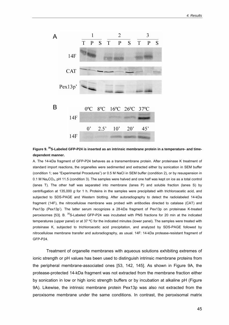
4. Results
45
Figure 9. 35S-Labeled GFP-P24 is inserted as an intrinsic membrane protein in a temperature- and time-dependent manner.
A. The 14-kDa fragment of GFP-P24 behaves as a transmembrane protein. After proteinase K treatment of
standard import reactions, the organelles were sedimented and extracted either by sonication in SEM buffer
(condition 1; see “Experimental Procedures”) or 0.5 M NaCl in SEM buffer (condition 2), or by resuspension in
0.1 M Na2CO3, pH 11.5 (condition 3). The samples were halved and one half was kept on ice as a total control
(lanes T). The other half was separated into membrane (lanes P) and soluble fraction (lanes S) by
centrifugation at 135,000 g for 1 h. Proteins in the samples were precipitated with trichloroacetic acid, and
subjected to SDS-PAGE and Western blotting. After autoradiography to detect the radiolabeled 14-kDa
fragment (14F), the nitrocellulose membrane was probed with antibodies directed to catalase (CAT) and
Pex13p (Pex13p’). The latter serum recognizes a 28-kDa fragment of Pex13p on proteinase K-treated
peroxisomes [53]. B. 35S-Labeled GFP-P24 was incubated with PNS fractions for 20 min at the indicated
temperatures (upper panel) or at 37 ºC for the indicated minutes (lower panel). The samples were treated with
proteinase K, subjected to trichloroacetic acid precipitation, and analyzed by SDS-PAGE followed by
nitrocellulose membrane transfer and autoradiography, as usual. 14F: 14-kDa protease-resistant fragment of
GFP-P24.
Treatment of organelle membranes with aqueous solutions exhibiting extremes of
ionic strength or pH values has been used to distinguish intrinsic membrane proteins from
the peripheral membrane-associated ones [53, 142, 145]. As shown in Figure 9A, the
protease-protected 14-kDa fragment was not extracted from the membrane fraction either
by sonication in low or high ionic strength buffers or by incubation at alkaline pH (Figure
9A). Likewise, the intrinsic membrane protein Pex13p was also not extracted from the
peroxisome membrane under the same conditions. In contrast, the peroxisomal matrix
A
B

4. Results
46
protein catalase was found in the soluble phase under all conditions employed. Hence,
the 14-kDa domain behaves as an intrinsic membrane protein, additionally confirming that
GFP-P24 is in fact inserted into organelle membranes.
The acquisition of resistance to proteinase K by the 14-kDa domain reflects the
membrane insertion of GFP-P24. A kinetic analysis of this process was performed (Figure
9B). These experiments show that membrane insertion of GFP-P24 is both temperature-
and time-dependent. After 20 min of incubation at 37 ºC, high amounts of the reporter
PMP acquire a protease-resistant status whereas no detectable import occurs at 0 or 8
ºC. At 37 ºC, the import of GFP-P24 converges rapidly to steady-state values, with a large
fraction inserted after only 10 min of incubation.
In any biological process, an important aspect that must be studied to understand
its mechanism regards the energetics of that process. To characterize the energetics of
the membrane insertion of GFP-P24, import reactions were carried out in the presence of
ATP, GTP or nonhydrolyzable analogs (ATPγS or GTPγS). In other import reactions, both
the PNS fractions and the GFP-P24-containing reticulocyte lysates were treated with
apyrase, an NTP-degrading enzyme. Strikingly, no significant differences were observed
among these distinct conditions (Figure 10). Therefore, energy in the form of ATP or GTP
is not required to drive the reporter PMP import process.
4.4. In vitro specific import of GFP-P24 into peroxisomes
The above described experiments strongly suggest that the reporter protein, GFP-
P24, is inserted into some membrane system in a process that is time- and temperature-
Figure 10. In vitro import of 35S-labeled GFP-P24 is independent of ATP and GTP hydrolysis. In vitro import reactions were performed at 37 ºC without exogenously added nucleotides (lane 1), with both
PNS fractions and reticulocyte lysates (containing 35S-labeled GFP-P24) treated with apyrase (lane 2) or
with heat-inactivated apyrase (lane 3), or in the presence of 5 mM ATP (lane 4), 5 mM ATPγS (lane 5), 0.5
mM GTP (lane 6) or 0.5 mM GTPγS (lane 7). After proteinase K treatment, the samples were precipitated
with trichloroacetic acid and processed for SDS-PAGE separation followed by blotting onto a nitrocellulose
membrane. Upon autoradiography, the membrane was probed with an antibody directed to the peroxisomal
enzyme catalase (CAT) to control loadings on gel. 14F: 14-kDa protease-resistant fragment of GFP-P24.

4. Results
47
dependent but does not require ATP or GTP hydrolysis. However, those experiments do
not reveal the identity of the membrane system(s) in which insertion of GFP-P24 is
occurring. As discussed in “Introduction” (section 1.3.3.2.), much controversy subsists on
whether PMPs are imported directly to peroxisomes or sorted first to the ER. Thus,
considering the small time windows used in the developed assay and the fast GFP-P24
membrane insertion (see Figure 9B), the identification of the membrane system targeted
by the reporter PMP gains further interest. In order to clarify this issue, PNS fractions were
subjected to standard import reactions with radiolabeled GFP-P24 for 7.5 min at 37 ºC.
After this short incubation, the organelle suspension was treated with proteinase K and
loaded onto the top of a discontinuous Nycodenz gradient (see “Experimental
Procedures”, section 3.8.). At the end of the centrifugation, the gradient was fractionated
and analyzed by SDS-PAGE and immunoblotting. The gradient distribution of
peroxisomes, ER and mitochondria was assessed with organelle-specific antibodies
(Figure 11).
In these gradients, peroxisomes present a double distribution appearing in two
different regions, as reported before in another study [52]. One population is found in
fractions 8-10, which corresponds to the 25% (w/v) Nycodenz step, wherein large portions
of both ER and mitochondria are also present. In contrast, the other peroxisome
population is highly pure and is detected in fractions 2-3, corresponding to the 30%-45%
(w/v) Nycodenz interface, where none of the two other organelles are found. As shown in
Figure 11, the dual distribution of peroxisomes in these gradients is clearly illustrated by
Pex13p, an intrinsic protein of the peroxisomal membrane [149-151]. Although catalase is
also a peroxisomal protein, a slightly different distribution is observed, with an additional
population remaining at the top of the gradient (fractions 11-14). Since catalase is a
soluble matrix enzyme, this behaviour is explained by leakage of peroxisomes during the
preparation of PNS fractions. The gradient distribution of the protease-resistant fragments
of 35S-labeled GFP-P24 yields a very clear result. The 28-kDa GFP fragment stays at the
top of the gradient (fractions 11-14) along with other soluble proteins like free catalase. In
sharp contrast, the 14-kDa domain distribution perfectly matches the one of Pex13p,
showing that GFP-P24 is specifically inserted into the peroxisome membrane despite the
presence of all the other organelles in these import reactions. In addition, the parallelism
between GFP-P24 and Pex13p profiles, after only 7.5 min of import, strongly suggests
that the reporter PMP is inserted into the membrane of mature peroxisomes from the very
beginning with no passage through the ER.
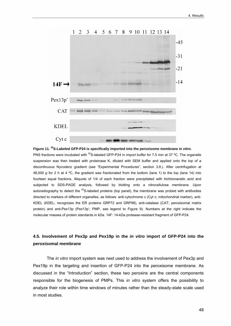
4. Results
48
4.5. Involvement of Pex3p and Pex19p in the in vitro import of GFP-P24 into the peroxisomal membrane
The in vitro import system was next used to address the involvement of Pex3p and
Pex19p in the targeting and insertion of GFP-P24 into the peroxisome membrane. As
discussed in the “Introduction” section, these two peroxins are the central components
responsible for the biogenesis of PMPs. This in vitro system offers the possibility to
analyze their role within time windows of minutes rather than the steady-state scale used
in most studies.
Figure 11. 35S-Labeled GFP-P24 is specifically imported into the peroxisome membrane in vitro.
PNS fractions were incubated with 35S-labeled GFP-P24 in import buffer for 7.5 min at 37 ºC. The organelle
suspension was then treated with proteinase K, diluted with SEM buffer and applied onto the top of a
discontinuous Nycodenz gradient (see “Experimental Procedures”, section 3.8.). After centrifugation at
48,000 g for 2 h at 4 ºC, the gradient was fractionated from the bottom (lane 1) to the top (lane 14) into
fourteen equal fractions. Aliquots of 1/4 of each fraction were precipitated with trichloroacetic acid and
subjected to SDS-PAGE analysis, followed by blotting onto a nitrocellulose membrane. Upon
autoradiography to detect the 35S-labeled proteins (top panel), the membrane was probed with antibodies
directed to markers of different organelles, as follows: anti-cytochrome c (Cyt c; mitochondrial marker), anti-
KDEL (KDEL; recognizes the ER proteins GRP72 and GRP98), anti-catalase (CAT; peroxisomal matrix
protein) and anti-Pex13p (Pex13p’; PMP, see legend to Figure 9). Numbers at the right indicate the
molecular masses of protein standards in kDa. 14F: 14-kDa protease-resistant fragment of GFP-P24.

4. Results
49
Pex3p has been proposed to act as a docking factor for Pex19p at the peroxisomal
surface (see “Introduction”, section 1.3.3.6.). The involvement of this peroxin was first
assessed by preincubating PNS fractions with IgGs directed to Pex3p and then subjecting
the organelle suspensions to standard import reactions with radiolabeled GFP-P24.
Similarly, import reactions using PNS fractions pretreated with anti-Pex16p, anti-PMP70
or preimmune IgGs were performed as well. While none of the latter IgGs affected the
import of GFP-P24, the anti-Pex3p IgGs caused a strong inhibition in the insertion of the
reporter PMP (Figure 12A). This result directly implies Pex3p on the mechanism of GFP-
P24 import into the peroxisomal membrane. Furthermore, a recombinant version of Pex3p
consisting of its cytosolic domain (thus lacking the N-terminal transmembrane domain,
see “Introduction”, section 1.3.3.3.), Pex3p(34-373), was also included in these assays.
Interestingly, the addition of this Pex3p fragment to the assays also resulted in the
inhibition of the import of GFP-P24, displaying a 10 nM IC50 (Figure 12B, upper panel).
This observation suggests that the recombinant Pex3p version interacts with some
component of the PMP import pathway hampering its function, and further corroborates
the participation of this peroxin in the GFP-P24 import process. A Pex19p complex is the
most obvious candidate for a target of this recombinant Pex3p protein.
In order to verify the involvement of Pex19p in the GFP-P24 sorting pathway,
standard import reactions were carried out in the presence of increasing amounts of
recombinant Pex19p. Remarkably, rather than any stimulation in the import of GFP-P24,
the opposite effect is observed at higher concentrations of the recombinant protein (Figure
12B, lower panel). Indeed, this recombinant Pex19p protein exhibits a 260 nM IC50 for the
insertion of GFP-P24 into the peroxisomal membrane. Most likely, high amounts of
recombinant Pex19p start interfering with the import mechanism of GFP-P24, possibly by
hindering Pex3p at the peroxisomal membrane (see “Discussion”). Interestingly, when
recombinant Pex19p is previously subjected to heat treatment, the inhibitory activity of the
protein in the import reactions is preserved (Figure 12B, lower panel, lane ∆+). Such
treatment is expected to irreversibly denature a globular protein and disrupt its function, as
observed for Pex3p (Figure 12B, upper panel, lane ∆+). However, a major portion of
Pex19p polypeptide chain is natively unfolded ([112], see section 1.3.3.4. of “Introduction”)
and thus insensitive to this treatment. The observation that heat-treated Pex19p retains its
properties on native PAGE analysis and its interaction with Pex3p (I. S. Alencastre, C. P.
Grou and J. E. Azevedo, unpublished observations) corroborates this interpretation.

4. Results
50
A
B
Figure 12. Pex3p and Pex19p participate in the in vitro import of 35S-labeled GFP-P24.
A. PNS fractions were treated for 20 min on ice with 8 µg of immunopurified IgGs directed to Pex3p (lane 1)
or Pex16p (lane 2), or total IgGs purified from an anti-PMP70 (lane 3) or nonimmune (lane 4) serum. As a
control, a PNS fraction was preincubated under the same conditions with SEM buffer (lane 5). After the
preincubation, the PNS fractions were subjected to standard import reactions with 35S-labeled GFP-P24. The
samples were processed as described in legend to Figure 10. B. 35S-Labeled GFP-P24 was incubated with
PNS fractions in import buffer containing increasing amounts of recombinant Pex3p(34-373) (upper panel) or
recombinant Pex19p (lower panel) for 20 min at 37 ºC. The numbers refer to the concentration in nM of the
recombinant protein used in each import reaction. In parallel, other import reactions were performed in the
presence of the highest amount of the respective recombinant protein pretreated by heat at 95 ºC for 5 min
(lane ∆+). After proteinase K treatment, the samples were processed as described in legend to Figure 10.
CAT: catalase. 14F: 14-kDa protease-resistant fragment of GFP-P24.

4. Results
51
4.6. Pex19p interacts with GFP-P24 in the cytosol and mediates its insertion into the peroxisome membrane
The observation that addition of recombinant Pex19p to the import reactions does
not stimulate GFP-P24 insertion into the peroxisomal membrane may have several
explanations (see previous section). For instance, this recombinant version of Pex19p
could lack the capacity to bind GFP-P24. Alternatively, it is possible that the interaction of
Pex19p with PMPs has to occur earlier, during or shortly after their translation. In order to
address this possibility, different recombinant versions of Pex19p (full-length Pex19p,
Pex19p(31-299) or Pex19p(1-124)) were added at different points in the life of the reporter
PMP. Thus, in the experiment presented in Figure 13, the recombinant proteins were
added either to the reticulocyte lysates during the in vitro synthesis of GFP-P24 (lanes 2,
4 and 6), or to the PNS fractions at the in vitro import assays (lanes 1, 3 and 5). In each
sample, the final concentration of the recombinant protein in the import reaction was the
same, 6 nM. As shown in lanes 1, 3 and 5, inclusion of any of these recombinant Pex19p
proteins in import mixtures comprising standard GFP-P24-containing lysates (i.e. GFP-
P24 synthesized in the absence of recombinant proteins) has no effect in the efficiency of
the import process. Strikingly, dramatic changes in the import efficiencies are observed
depending on whether GFP-P24 was synthesized in the presence or absence of the
recombinant proteins. In fact, inclusion of full-length Pex19p in the in vitro translation of
GFP-P24 leads to a 2.5-fold increase in the peroxisomal membrane insertion of the
reporter PMP (compare lanes 1 and 2). In sharp contrast, addition of recombinant
Pex19p(31-299) to the translation reaction results in a strong decrease in the amount of
imported GFP-P24 (lane 4). This truncated version of Pex19p lacks the strongest Pex3p-
binding site presumably necessary for docking at peroxisomal membrane, but preserves
the integrity of the cargo-binding region (see “Introduction”, sections 1.3.3.4.-1.3.3.6.).
Furthermore, when recombinant Pex19p(1-124) is added to the in vitro translation of GFP-
P24 (lane 6), no significant differences in the amount of protease-protected 14-kDa
fragment are obtained. In the case of Pex19p(1-124), the Pex3p-docking domain is
present whereas the cargo-binding region is absent (see “Introduction”, sections 1.3.3.4.-
1.3.3.6.). Altogether, these results indicate that the import competence of GFP-P24 is
ensured by Pex19p molecules present at the translation level. Therefore, the presented
data suggest that Pex19p forms a complex with newly synthesized GFP-P24 in the
cytosol, and that this complex is the substrate for the import machinery at the peroxisomal
membrane.

4. Results
52
4.7. Native gel analysis of Pex19p interactions with Pex3p
The experiments involving radiolabeled GFP-P24 translated in the presence of
recombinant versions of Pex19p strongly suggest that protein complexes comprising the
reporter PMP and these recombinant proteins are formed within a cytosolic fraction, the
reticulocyte lysate. Moreover, the nature of the recombinant proteins used had major
effects in the import competence of GFP-P24. Indeed, full-length Pex19p yields a strong
stimulation in the import process while the truncated version Pex19p(31-299), lacking the
putative Pex3p-docking domain, has a large inhibitory outcome. Thus, these protein-
protein interactions were further analyzed by employing a different method, the native
PAGE. The behaviours of recombinant Pex19p, Pex19p(31-299), Pex19p(1-124) and
Pex3p(34-373) under this electrophoretic technique were first characterized. Each of the
recombinant Pex19p versions was preincubated with increasing amounts of Pex3p(34-
373) and subjected to native PAGE analysis (Figure 14). As expected, both full-length
Pex19p and Pex19p(1-124) interacted with Pex3p(34-373) (Figure 14, panels A and B,
respectively) since these versions include the strongest N-terminal Pex3p-binding domain.
Figure 13. Inclusion of recombinant versions of Pex19p in the in vitro translation of 35S-labeled GFP-P24 strongly influences its import efficiency. 35S-Labeled GFP-P24 was synthesized in vitro using a reticulocyte lysate supplemented with recombinant
Pex19p (lane 2), Pex19p(31-299) (lane 4) or Pex19p(1-124) (lane 6), and afterwards subjected to standard
in vitro import reactions with PNS fractions. Alternatively, 35S-labeled GFP-P24 synthesized in the absence
of recombinant proteins was incubated with PNS fractions containing recombinant Pex19p (lane 1),
Pex19p(31-299) (lane 3) or Pex19p(1-124) (lane 5) and subjected to standard import reactions as well. In all
samples, the final concentration of the recombinant proteins in the import assay was 6 nM. The yield of the
different reticulocyte lysates in 35S-labeled GFP-P24 was previously quantified by SDS-PAGE and
autoradiography, so that the amounts of the radiolabeled reporter used in each import reaction did not vary
more than 20%. Proteinase K-treated samples were processed as described in legend to Figure 10. CAT:
catalase. 14F: 14-kDa protease-resistant fragment of GFP-P24.

4. Results
53
A
B
C
Figure 14. Interactions of Pex19p, Pex19p(1-124) and Pex19p(31-299) with Pex3p(34-373) by native PAGE analysis.
A constant amount (1 µg) of recombinant Pex19p (P19; panel A), Pex19p(1-124) (P19(1-124); panel B) or
Pex19p(31-299) (P19(31-299); panel C) was incubated with increasing amounts of recombinant Pex3p(34-
373) (P3; 0, 1, 2, 4, 6, 8 and 10 µg in lanes 1-7, respectively) for 30 min at room temperature. After this
incubation, the samples were subjected to native PAGE as described in “Experimental Procedures” (section
3.10.). Coomassie-blue stained gels are shown. In panel C, 10 µg of recombinant Pex3p(34-373) alone were
applied to lane 8. The arrows refer to the migration position of the recombinant proteins and formed
complexes.

4. Results
54
Interestingly, Pex19p seems to display a higher affinity towards Pex3p(34-373) when
compared to its N-terminal fragment Pex19p(1-124), as suggested by their estimated KD
values of ~3.4 µM and ~26 µM, respectively. This affinity difference probably derives from
the second, though weaker, Pex3p-binding site which is absent in Pex19p(1-124) (see
“Introduction”, section 1.3.3.4.). Although Pex19p(31-299) harbours this second domain,
no interaction with Pex3p(34-373) was detected under these electrophoretic conditions
(Figure 14C). Therefore, this Pex3p-binding site alone is insufficient to promote complex
formation, at least under the used conditions. Taken together, these results corroborate
the Pex3p-binding properties previously reported for these recombinant versions of
Pex19p [116] and render native PAGE a useful approach to study such protein-protein
interactions.
4.8. GFP-P24 forms trimeric complexes involving Pex19p and Pex3p
As mentioned in the previous section, the experiments which included GFP-P24
synthesized in the presence of recombinant Pex19p versions suggest that protein
complexes between the reporter PMP and the recombinant proteins were formed in vitro.
Therefore, protein complexes involving radiolabeled GFP-P24 were also analyzed using
the native PAGE technique. When a standard GFP-P24-containing reticulocyte lysate (i.e.
no recombinant proteins were added during the translation) is subjected to this
electrophoretic analysis, a slow-migrating cluster of radioactive sharp bands is observed
(Figure 15, arrow a). In addition, a faint and diffuse smear with a faster migration is also
detected (lane 1). These two radioactive populations were confirmed as GFP-P24 after
subjecting a full lane from a native gel to a second dimension by SDS-PAGE separation.
The sharp bands observed at the top of the gel may result from precipitation of the
partially hydrophobic GFP-P24 protein. Alternatively, they can represent a high molecular
mass GFP-P24-containing complex with rabbit proteins from the lysate, possibly the
chaperonin TCP1 ring complex (TRiC). In fact, such complex was previously identified for
PMP22 using a rabbit reticulocyte lysate as well [137]. In the same study, PMP22 was
also detected in another complex comprising an unidentified 40-kDa protein. Of these two
PMP22-containing complexes, the latter showed much higher import efficiency [137].
Therefore, this 40-kDa protein seems to correspond to rabbit Pex19p. It should be noted
that the rabbit peroxin is present during the in vitro translation of GFP-P24 despite being
apparently in limiting amounts, as judged from the effect of the recombinant Pex19p
proteins.

4. Results
55
In these native gels, GFP-P24 synthesized in the presence of recombinant Pex19p
displays a distinct pattern (lane 2). The group of slow-migrating sharp bands is now
considerably weaker, whereas most of the reporter protein concentrates in a more intense
smear migrating behind a sharp band (arrow c). Remarkably, the intensity of this sharp
band is greatly increased when a GFP-P24-containing reticulocyte lysate is supplemented
with recombinant Pex19p prior to electrophoresis (lanes 3 and 4). These results show that
Pex19p abundance both during the in vitro synthesis and the gel run strongly increases
the fraction of GFP-P24 that can migrate in this sharp band, probably by maintaining the
reporter PMP in a soluble state and preventing its aggregation. Moreover, these
observations also suggest that such complexes are somewhat labile under the
electrophoretic conditions used. Therefore, high amounts of recombinant Pex19p in the
gels prevent complex dissociation to a larger extent.
Interestingly, inclusion of recombinant Pex3p(34-373) in these samples leads to a
shift of the sharp band to an upper region of gel (lanes 5 and 6, arrow b). This result
corroborates the former band as the dimeric complex GFP-P24-Pex19p (arrow c), and
indicates that the new band (arrow b) corresponds to a trimeric complex comprising GFP-
P24, Pex19p and Pex3p(34-373). The truncated Pex19p(31-299) version was also
Figure 15. 35S-Labeled GFP-P24 forms dimeric complexes with Pex19p and trimeric complexes involving Pex19p and Pex3p(34-373) in native PAGE analysis. 35S-Labeled GFP-P24 was synthesized in vitro using a rabbit reticulocyte lysate under standard conditions
(lanes 1, 3, 5, 7 and 9) or supplemented with recombinant Pex19p (lanes 2, 4 and 6) or Pex19p(31-299)
(lanes 8 and 10). Before electrophoresis, most samples received 1 µg of recombinant Pex19p (lanes 3-6) or
Pex19p(31-299) (lanes 7-10). Some samples also received 1.5 µg of recombinant Pex3p(34-373) (lanes 5,
6, 9 and 10). After the addition of recombinant proteins, the samples were subjected to native PAGE as
described in “Experimental Procedures” (section 3.10.). A fluorograph of a dried gel is shown. The arrows
indicate a cluster of sharp bands at the top of the gel containing GFP-P24 (arrow a), a complex comprising
GFP-P24, Pex19p proteins and Pex3p(34-373) (arrow b) and a complex involving GFP-P24 and Pex19p
proteins (arrow c).

4. Results
56
included in these experiments both during the translation of the reporter PMP and prior to
electrophoresis (lanes 7 and 8). In these samples, the observed pattern is similar to the
one obtained with full-length Pex19p, although the sharp band displays a slightly lower
mobility. However, when the same samples receive recombinant Pex3p(34-373), only a
small amount of the GFP-P24 complex is shifted to an upper region of the gel (lane 10).
Most likely, this different behaviour reflects the absence or disruption of the strongest
Pex3p-binding domain in Pex19p(31-299). Nevertheless, a small portion of trimeric
complex is indeed observed, which suggests that Pex19p(31-299) is still able to bind
Pex3p(34-373) although with lower affinity. As shown above, however, these two
recombinant proteins alone did not form a complex under the same electrophoretic
conditions (Figure 14C). Therefore, the interaction between Pex19p(31-299) and
Pex3p(34-373) in these native gels seems to be promoted by GFP-P24.
4.9. Cargo PMP dependence of Pex19p binding affinity for Pex3p
Native PAGE analysis allowed the detection of dimeric complexes between GFP-
P24 and Pex19p, and trimeric complexes comprising the reporter PMP, Pex19p and
Pex3p(34-373). Thus, this technique can be used to analyze how the presence of the
cargo PMP, GFP-P24, affects the binding properties of Pex19p for Pex3p. To address this
issue, steady amounts of recombinant Pex19p and of a reticulocyte lysate containing
radiolabeled GFP-P24 were mixed and incubated with increasing quantities of
recombinant Pex3p(34-373). The samples were then subjected to native PAGE analysis
followed by blotting onto a nitrocellulose membrane and exposure to an X-ray film. In
Figure 16, a Ponceau S-stained membrane (lower panel) and an autoradiograph (upper
panel) of the same experiment are shown. At lower substoichiometric concentrations of
Pex3p(34-373), unloaded recombinant Pex19p is essentially observed in a free state
(lower panel). Conversely, Pex19p molecules loaded with GFP-P24 readily bind
Pex3p(34-373) under exactly the same conditions (upper panel). Therefore, these
observations indicate that Pex19p interactions with Pex3p are modulated by the cargo
PMP whose binding greatly increases the affinity of Pex19p for Pex3p.

4. Results
57
Figure 16. Binding of GFP-P24 increases the affinity of Pex19p for Pex3p.
Increasing amounts of recombinant Pex3p(34-373) (0, 93.5, 187, 325, 750, 1500 and 3000 ng in lanes 1-7,
respectively) were incubated with recombinant Pex19p (2 µg) and 35S-labeled GFP-P24 synthesized in the
presence of recombinant Pex19p. After incubation for 30 min at room temperature, the samples were
subjected to native PAGE followed by blotting onto a nitrocellulose membrane and autoradiography (see
“Experimental Procedures”, section 3.10.). An autoradiograph (upper panel) and a Ponceau S-stained
membrane (lower panel) of the same experiment are shown. Lane 8 contains 2 µg of recombinant Pex19p
whereas lane 9 holds 3 µg of Pex3p(34-373). The arrows refer to the gel migration of recombinant Pex19p
(P19), recombinant Pex3p(34-373) (P3) and the complexes formed with 35S-labeled GFP-P24 (PMP). The
asterisk in the lower panel indicates the hemoglobin band derived from the rabbit reticulocyte lysate.

58
5. DISCUSSION

5. Discussion
59
5. DISCUSSION
In this work, an in vitro import system was developed to characterize the specific
targeting and insertion of a PMP into the peroxisome membrane. As previously discussed,
this kind of systems has the advantage of being experimentally open and providing a
kinetic perspective. Conversely, most of the other studies have used steady-state
strategies to address this subject. The first step in the development of the in vitro import
system was the construction of a reporter PMP that could provide a protease “signature”
to assess the correctly inserted protein fraction. For this purpose, an engineered version
of PMP24 was constructed and validated in vivo to be used as reporter in the in vitro
assay. This fusion PMP, GFP-P24, fulfils the protease “signature” criterion and was shown
to be specifically imported into the peroxisomal membrane. In addition to protease
resistance, the membrane insertion of the reporter PMP was also corroborated in
experiments in which organelle membranes were extracted by sonication in low or high
ionic strength solutions or incubated with alkali. A kinetic characterization of the in vitro
GFP-P24 import was performed showing a time- and temperature-dependent membrane
insertion. These experiments revealed a strong dependence on temperature with high
levels of insertion at 37 ºC, but largely decreasing at 26 ºC or 16 ºC and essentially
undetected at lower temperatures. Furthermore, this import occurs very rapidly at 37 ºC,
with a substantial amount of inserted GFP-P24 being detected after only 10 min of
incubation. Hence, the PMP import pathway appears to be completed within a very short
time window.
Several years ago, a similar strategy was used to follow the insertion of in vitro
translated PMP22 and PMP70 into the membrane of peroxisomes [84, 85]. To assess
membrane integration of PMP22, the authors digested the samples with subtilisin. Indeed,
imported PMP22 becomes entirely resistant to this protease [84]. In the case of PMP70,
the authors relied solely on carbonate extraction [85]. A major difference between the
previous systems and the one developed in this work concerns the fact that purified
peroxisomes were used in those studies, whereas the present one used PNS fractions
from rat liver. The reason behind this choice derives from the fact that PMP import into
purified peroxisomes does not rule out the hypothesis that such pathway represents a
minor contribution to mature peroxisomes, whereas in the presence of all of the other
organelles the primary sorting pathway may be distinct, for instance, via the ER (see
“Introduction”, section 1.3.3.2.). Thus, the finding that a reporter PMP is imported into
peroxisomes, even though all the other abundant subcellular organelles are present in the
PNS fraction used, demonstrates the specificity of the system.

5. Discussion
60
This observation gains special relevance considering the current dispute regarding
the ER involvement in peroxisome membrane biogenesis. As discussed in “Introduction"
(section 1.3.3.2.), the longstanding model states that PMPs are synthesized on free
cytosolic ribosomes and post-translationally inserted into the organelle membrane [37,
82], whereas some other authors have proposed that peroxisomes bud from the ER with
at least some PMPs travelling first through this compartment [83, 87]. This last concept is
supported by some recent data suggesting that two PMPs, particularly yeast Pex3p and
mammalian Pex16p, reach the peroxisomal membrane via the ER [81, 88-90]. Although
these studies raise many reservations (see "Introduction”, section 1.3.3.2.), it should be
emphasized that Pex3p and Pex16p are not ordinary PMPs since they participate in the
biogenesis of the remaining PMPs. Therefore, it is possible that these two peroxins are
sorted through a pathway distinct from all the other PMPs. Indeed, it was suggested that
Pex3p is imported independently of Pex19p [109]. Nevertheless, such traffic of Pex3p or
Pex16p through the ER could also imply that newly synthesized PMPs are primarily
targeted and inserted in this compartment, reaching the peroxisomes at a later stage.
However, the observation that, after only 7.5 min of import reaction, GFP-P24 localizes to
mature peroxisomes and not to the ER strongly suggests that it is inserted directly from
the cytosol into the peroxisomal membrane. In fact, this very short time period renders a
putative passage through the ER highly unlikely.
At the time the studies of PMP22 and PMP70 import were performed, the data
regarding peroxisome membrane biogenesis were still very limited and the protein
components involved were unknown. Presently, our understanding of this process has
improved and the peroxins mediating the PMP sorting pathway are identified. Thus, these
mechanisms could be more thoroughly analyzed. As shown in Figure 12B (lower panel),
addition of increasing amounts of recombinant Pex19p to the in vitro import reactions has
no stimulatory effect but rather inhibits GFP-P24 insertion into the peroxisomal membrane
at higher concentrations. Although Pex19p is directly involved in the PMP sorting
mechanism, this result is not surprising. The absence of stimulation of the import process
is consistent with the properties of the Pex19p-PMP interaction. Pex19p was reported to
have a chaperone-like activity in binding newly synthesized PMPs and preventing their
aggregation by shielding hydrophobic domains [112]. Moreover, the results from Figure 13
indicate that these complexes must be formed during or immediately after PMP translation
(see below). Therefore, additional Pex19p molecules available at the import level do not
contribute to the membrane insertion of the reporter PMP. The inhibition observed when
using large concentrations of recombinant Pex19p is probably related to the fact that
recombinant Pex19p still interacts with Pex3p(34-373) in its unloaded state. This
recombinant version of Pex3p contains the cytosolic exposed domain involved in Pex19p

5. Discussion
61
binding (see “Introduction”, section 1.3.3.3.). Thus, recombinant Pex19p is also likely to
interact with peroxisomal Pex3p in the in vitro import assays, hampering its function when
present at high concentrations and preventing the docking of Pex19p-cargo complexes.
This ability of Pex19p to bind Pex3p even when no PMP is being carried does not
appear to be restricted to this recombinant version. Indeed, overexpressed Pex19p
versions show a Pex3p-dependent peroxisomal localization although no peroxisomal
proliferation is observed in these cells [74, 95, 96]. Furthermore, a truncated version
comprising the Pex3p docking domain but lacking the PMP-binding region displayed
exactly the same behaviour [95]. Altogether, these observations suggest that free Pex19p
is in fact capable of interacting with Pex3p when the intracellular levels of the former are
artificially raised. However, under normal physiological concentrations, the affinity of
unloaded Pex19p towards peroxisomal Pex3p is probably insignificant. In the in vitro
import assays, recombinant Pex19p inhibited GFP-P24 import with an IC50 value of 260
nM, whereas the native PAGE KD value estimated for the Pex19p-Pex3p(34-373)
interaction was much higher, ~3.4 µM. Considering that in this electrophoretic technique
the samples are subjected to nonequilibrium and nonphysiological conditions, this
apparent KD value is likely to be overestimated. Indeed, in a recent study this interaction
was reported to have a KD value in the nanomolar range (3.4 nM), determined by surface
plasmon resonance [111]. Nevertheless, the apparent KD figures estimated by native
PAGE analysis are definitely valuable when comparing the binding affinities of the
different recombinant versions of Pex19p towards Pex3p(34-373).
The inclusion of recombinant Pex19p versions in the in vitro translation of GFP-
P24 subsequently subjected to in vitro import reactions yielded results of most important
consequence. The addition of full-length Pex19p to the in vitro synthesis of the reporter
PMP leads to a marked stimulation in its peroxisomal import, whereas the presence of
Pex19p(31-299) during translation produces a strong inhibition of the import process. In
stark contrast, no differences in the normal import levels are obtained with exactly the
same final concentrations of the recombinant proteins in import reactions using GFP-P24
translated in the absence of exogenous Pex19p. Therefore, these data indicate that
Pex19p proteins form a complex with GFP-P24 at the translation level that determines the
PMP import competence. Additional free Pex19p molecules (recombinant or the rat liver
ones) available at later stages do not contribute to this import competence. These
observations imply that those complexes formed during GFP-P24 synthesis correspond to
the substrates recognized by the import machinery at the peroxisome membrane. The
properties of the Pex19p proteins included in these complexes then establish the final
outcome. When full-length Pex19p is present, the complexes interact with Pex3p, and
GFP-P24 is eventually inserted into the peroxisomal membrane. In contrast, Pex19p(31-

5. Discussion
62
299) lacks the strongest Pex3p-binding domain apparently required for docking at the
peroxisomal surface, and thus is incapable of delivering the reporter PMP and mediating
its insertion. Taken together, these results are the first mechanistic evidence that Pex19p
functions primarily in binding newly synthesized PMPs in the cytosol and transporting
them to the peroxisome membrane. In line with these conclusions, both IgGs directed to
Pex3p and recombinant Pex3p(34-373) resulted in a decrease of imported GFP-P24. The
anti-Pex3p IgGs hinder the peroxin at the peroxisome membrane and therefore the
docking sites for the Pex19p-cargo complexes. On the other hand, native PAGE analyses
confirmed that recombinant Pex3p(34-373) is capable of interacting both with free and
cargo-loaded Pex19p. Hence, this Pex3p version, comprising the cytosolic-exposed
domain, probably sequesters the Pex19p-cargo complexes preventing their docking at the
peroxisomal import machinery.
A relevant aspect regarding the experiments involving recombinant Pex19p lies in
the concentrations used when compared to the endogenous values. At present, the
physiological concentration of Pex19p remains unknown for all organisms. Nevertheless,
a few considerations should be made on this subject. In this regard, morphometric data
available for the rat hepatocyte may provide some helpful hints [152]. From this data, one
Pex19p protein per peroxisome corresponds to a cytosolic concentration of 0.23 nM.
Therefore, 6 nM (the amount used in the import assays from Figure 13) of cytosolic
Pex19p would represent 26 molecules per peroxisome in these cells. Moreover, rat liver
Pex3p seems to have the same abundance of Pex14p [153] which was estimated in a
previous study to represent 0.25% of the total rat liver peroxisomal protein [50]. In
agreement, a band of 38 kDa from highly purified rat liver peroxisome membranes,
consisting of Pex3p and PMP52 [154], represented 0.37% of total peroxisomal protein
[145]. The in vitro import assays described in this work are performed in 100 µl final
volume containing 450 µg of PNS protein, wherein peroxisomes correspond to 2.5% of
total rat liver protein. According to these data, the endogenous 42-kDa Pex3p from rat
peroxisomes is estimated to be at 6.6 nM in the import reaction. This concentration value
is roughly the same of recombinant Pex19p in the experiments from Figure 13. Thus, the
concentrations of recombinant Pex19p used in those assays are apparently not in a molar
excess towards peroxisomal Pex3p.
The native PAGE method employed in this study emerged as a useful technique
when investigating the interactions involving Pex19p, Pex3p and a cargo PMP (GFP-P24).
As a first approach, the binding properties of Pex19p for Pex3p were analyzed using
different recombinant versions of Pex19p and Pex3p(34-373). In these experiments, full-
length Pex19p binds Pex3p(34-373) with a higher affinity than does Pex19p(1-124). On
the other hand, Pex19p(31-299) interacts quite weakly with Pex3p(34-373) since, under

5. Discussion
63
the electrophoretic conditions, a complex was only detected in the presence of the cargo
GFP-P24. These results are compatible with the proposed existence of two Pex3p-binding
domains on Pex19p, a strong one at the N terminus and a weaker one partially
overlapping with the cargo-interacting region (see “Introduction”, section 1.3.3.4.). Thus,
the observed binding properties for these recombinant proteins are in agreement with the
ones previously obtained in the two-hybrid system [116]. Furthermore, dimeric complexes
between Pex19p and GFP-P24 and trimeric complexes comprising Pex3p, Pex19p and
GFP-P24 were also detected in these native gels. Hence, the dimeric complexes
correspond to cargo-loaded Pex19p whereas the trimeric complexes may represent the
docking of cargo-loaded Pex19p to peroxisomal Pex3p. Similar trimeric complexes
involving other cargo PMPs were also described in other studies [112, 133]. Considering
that all these protein complexes could be observed under native PAGE, the effect of the
cargo PMP on Pex19p affinity for Pex3p was analyzed as well. As shown in Figure 16, the
binding of the cargo GFP-P24 to Pex19p increases its affinity for Pex3p. This property is
expected so that only cargo-loaded Pex19p is targeted to the docking and insertion
machinery, and free Pex19p is not occupying Pex3p docking sites. Thus, the binding of
the cargo may induce some conformational changes in Pex19p enhancing the affinity of
its N-terminal domain for Pex3p. Alternatively, it is possible that Pex3p directly interacts
with the cargo protein within the Pex19p-PMP complex, mediating the subsequent
insertion steps. Elucidation of this issue will surely help understanding the mechanisms of
docking and insertion into the peroxisomal membrane. Importantly, this difference in
affinity may have major implications on the energetics of Pex19p export back to the
cytosol. Indeed, the strong decrease in the affinity of Pex19p for Pex3p after the cargo
PMP is delivered probably drives this export step. Therefore, it is likely that no ATP or
GTP hydrolysis is required for recycling Pex19p.
The in vitro import system developed in this work was also used to assess the
energetics of the docking and insertion mechanisms of the reporter PMP into the
peroxisomal membrane. Remarkably, the import process was insensitive to high amounts
of ATP, GTP or their nonhydrolyzable analogs, and also to apyrase which catalyzes the
hydrolysis of ATP and other nucleotides. Hence, these results indicate that none of the
steps from the peroxisomal docking of Pex19p-cargo complexes until the ultimate PMP
membrane insertion involve energy in the form of ATP or GTP. Furthermore, once the
Pex19p-cargo complex is formed during translation or shortly after it, the whole PMP
import pathway may be energy independent. Indeed, all the subsequent steps are likely to
be energetically favourable. Thus, considering that PMPs are highly hydrophobic, their
membrane integration may drive the docking and insertion steps. The export of Pex19p
back into the cytosol is probably ensured by the decrease in its affinity for Pex3p upon

5. Discussion
64
cargo release (see above). These findings are consistent with the energetics reported for
the in vitro import of rat PMP22 and PMP70 [84, 85] and A. thaliana PMP22 [155],
although the latter authors suggest that ATP may stimulate the assembly of the PMP at
the peroxisomal membrane. In sharp contrast, Matsuzono and co-workers recently
suggested that cargo-loaded Pex19p is targeted to peroxisomes in an ATP-dependent
manner [133]. This apparent discrepancy may be related with the features of the in vitro
system used in that study. In fact, the authors did not use a reporter PMP but rather
followed the traffic of radiolabeled Pex19p between the organelle pellet and the
supernatant of import reactions. Therefore, the cargo PMPs transported by radioactive
Pex19p are presumably supplied by the PNS fraction used in those assays which, in fact,
was never demonstrated. Here, it was shown that import-competent Pex19p-cargo
complexes have to be formed during or immediately after the translation step of the PMP.
Thus, an ATP-requiring step may be related to the PMP synthesis itself or to a putative
transfer from a chaperone to Pex19p. Nevertheless, the experimental criteria used by
Matsuzono and colleagues raise many reservations regarding the relevance of those
observations. As discussed before, the in vitro import system developed here relies on a
protease protection assay in which a protease-resistant fragment of the reporter PMP
represents the protein fraction that is correctly inserted into the peroxisome membrane.
However, Matsuzono and co-workers defined the import event solely by sedimentation of
the radiolabeled protein with the organelle fraction, which is a clearly insufficient criterion.
Moreover, such organelle pellets were obtained upon ultracentrifuging 250-µl samples at
100,000 g for 30 min [133]. In contrast, the organelles pellets from the in vitro import
assays performed in the present work were obtained by centrifuging the 1-ml samples at
15,000 g for 15 min in a typical tabletop microcentrifuge. The problem of relying on such
criterion is well illustrated by the results with a control GFP protein shown in Figure 7.
Although this protein is highly soluble, it still sediments with organelles even after two
centrifugation steps in standard conditions (i.e. at 15,000 g for 15 min).
The results described in the present work provide a new insight into PMP
biogenesis and clarify some essential aspects of the Pex19p-mediated import pathway.
Indeed, it was shown that import-competent Pex19p-cargo complexes are formed in the
cytosol during or shortly after PMP translation and represent the substrates recognized by
the peroxisome docking and insertion machinery. Altogether, the presented data
constitute compelling evidence supporting a Pex19p role as PMP cycling receptor. In this
model, Pex19p interacts with the nascent PMP either during or immediately after its
translation on free cytosolic ribosomes (see Figure 17). The second possibility may
require a chaperone such as TRiC to assist the protein folding and transfer to Pex19p.
This peroxin loaded with the PMP then acts as a cycling receptor and transports it to

5. Discussion
65
peroxisomes. At the organelle membrane, cargo-loaded Pex19p (but not the free peroxin)
docks on Pex3p and delivers the PMP which ultimately becomes inserted into the lipid
bilayer, via ATP- and GTP-independent mechanisms. It remains to be determined whether
Pex19p is also involved in subsequent steps such as membrane integration or
assembly/disassembly of protein complexes. After releasing the PMP, free Pex19p is
exported back to the cytosol because its affinity for Pex3p is now much smaller.
Therefore, this step is probably energetically favourable as well. In the cytosol, exported
Pex19p binds once more to newly synthesized PMPs and catalyzes further cycles of
transportation. Since Pex19p displayed all the properties of a mobile receptor for this
particular PMP, it is likely that the same biogenesis pathway also applies to the other
PMPs, perhaps with the exception of Pex3p and Pex16p as these peroxins appear to be
directly involved in this process. However, it is still possible that a different protein
mediates the sorting of other PMPs to the peroxisomal membrane, specially the ones
which appear to have separate mPTSs and Pex19p-binding sites. Bearing in mind the
concerns regarding these observations (see “Introduction”, section 1.3.3.7.), such
hypothesis seems unlikely. Taken as a whole, the developed in vitro import system and
the native PAGE analysis presented in this work have proven to be extremely helpful
strategies to study the PMP import pathway and their protein components.

5. Discussion
66
Figure 17. Model for the Pex19p-mediated import of PMPs.Step 1: Pex19p (19) interacts with a nascent PMP through its C-terminal domain (light blue). This cargo
binding occurs either during or immediately after PMP synthesis on cytosolic ribosomes and may be
assisted by chaperones. Step 2: Pex19p then functions as an import receptor and transports the PMP to the
peroxisomal membrane. Cargo-loaded Pex19p displays a higher affinity for peroxisomal Pex3p (3), where it
docks via its N terminus (dark blue). Step 3: After the docking, Pex19p delivers the PMP which eventually
becomes inserted into the lipid bilayer. Both the docking and insertion steps are ATP and GTP independent.
Pex16p (16) may also participate in these steps. Free Pex19p has much lower affinity for Pex3p and is
released back to the cytosol, apparently without the need of an energy input. Step 4: In the cytosol, free
Pex19p is now again available to interact with newly synthesized PMPs and mediate further cycles of PMP
import.
peroxisome
19
16
19
PMP3
19 PMP
16
3
peroxisome
16
PMP
19
3
peroxisome
cytosol cytosol
cytosol
cytosol
nascent PMP
step 1
step 4
step 2 step 3

67
6. FUTURE PERSPECTIVES

6. Future Perspectives
68
6. FUTURE PERSPECTIVES
Despite the progress in recent years together with the significant contribution from
this work, many important questions concerning peroxisome membrane biogenesis
remain to be answered. The Pex19p-mediated PMP import pathway was characterized
here using a modified version of PMP24 as a reporter cargo protein. Considering that
PMP24 is an ordinary PMP, the attained conclusions probably also apply to most or even
all of the other PMPs, except Pex3p and Pex16p which are directly implicated in the
biogenesis of this class of proteins. Nevertheless, a yet unknown different sorting pathway
may exist for some other PMPs. Therefore, other reporter PMPs should be tested in the in
vitro import system developed in this work. In this regard, Pex14p, which also yields a
protease-resistant fragment upon proteinase K digestion of the organelles [156], was
subjected to import reactions. This PMP exhibited the same behaviour of GFP-P24 in
these in vitro import assays (C. P. Grou, M. P. Pinto and J. E. Azevedo, unpublished
results). Additional PMPs will also be tested with this in vitro system. Another important
aspect regards whether a chaperone and/or other factors mediate the folding and transfer
of the nascent PMP from the ribosome to Pex19p. Furthermore, the production of a
soluble Pex19p-binding peptide, capable of maintaining the peroxin in a loaded state,
would surely be a useful tool for various purposes such as competition studies and
analysis of binding properties. These issues are currently being addressed.
As previously discussed, the biogenesis of PMPs apparently constitutes a
remarkably simplified system comprising only three protein components, Pex3p, Pex16p
and Pex19p. In yeast species, Pex16p is either absent or serves a different purpose (Y.
lipolytica), rendering Pex3p and Pex19p as the only proteins of such machinery [32, 79].
Moreover, mammalian Pex16p is thought to regulate early events in peroxisomal
membrane biogenesis, especially in de novo formation [80, 81], and has not been linked
to import of PMPs into mature peroxisomes. In addition, considering that the docking and
insertion steps are independent of ATP and GTP hydrolysis, no other protein factors seem
to be required. Therefore, a minimum in vitro import system was envisaged consisting of
Pex3p reconstituted into liposomes, Pex19p and a reporter PMP. Thus, reconstitution of
full-length Pex3p into liposomes resembling the phospholipid content of the peroxisomal
membrane [157, 158] was attempted. For this purpose, full-length Pex3p was first
expressed in bacterial cells. However, in contrast to its cytosolic domain, Pex3p(34-373),
the expression levels of the full-length peroxin were extremely low. Subsequently, several
strategies were followed in an attempt to produce recombinant Pex3p. Recently, these
expression problems were overcome and reasonable amounts of the protein were
obtained in a mostly insoluble state. Following solubilization with detergents, Pex3p was

6. Future Perspectives
69
then subjected to reconstitution in the presence of different ratios of phospholipids.
Unfortunately, so far Pex3p failed to reconstitute in these experiments. Presently,
reconstitution assays are being performed using different solubilization conditions. A
successful reconstitution of Pex3p into liposomes would allow carrying out in vitro import
reactions using Pex19p loaded with a reporter PMP. Then, it could be assessed whether
such minimum in vitro reconstituted system is sufficient to promote PMP membrane
insertion. Specific import of a reporter PMP in this in vitro reconstituted system would
have major implications. In fact, such result would imply that Pex16p is not essential in
this process and rather plays a distinct role in peroxisomal membrane biogenesis.
Additionally, this observation would indicate that no further proteins are required. In the
case of a negative outcome, coupled reconstitution of Pex3p and Pex16p should be
attempted. In summary, reconstitution of a minimum PMP in vitro import system would
certainly be enlightening. Indeed, reconstitution of a biological system is the ultimate goal
in biochemistry.

70
7. REFERENCES

7. References
71
7. REFERENCES [1] Purdue, P. E. and Lazarow, P. B. (2001) Peroxisome biogenesis. Annu. Rev. Cell
Dev. Biol. 17, 701-752.
[2] Rhodin, J. (1954) Correlation of ultrastructural organization and function in normal
and experimentally changed proximal convoluted tubule cells of the mouse kidney.
Ph.D. thesis. Karolinska Institutet, Aktiebolaget Godvil, Stockholm.
[3] De Duve, C. and Baudhuin, P. (1966) Peroxisomes (microbodies and related
particles) Physiol. Rev. 46, 323-357.
[4] Eckert, J. H. and Erdmann, R. (2003) Peroxisome biogenesis. Rev. Physiol.
Biochem. Pharmacol. 147, 75-121.
[5] Kunze, M., Pracharoenwattana, I., Smith, S. M. and Hartig, A. (2006) A central role
for the peroxisomal membrane in glyoxylate cycle function. Biochim. Biophys. Acta
1763, 1441-1452.
[6] Michels, P. A., Bringaud, F., Herman, M. and Hannaert, V. (2006) Metabolic
functions of glycosomes in trypanosomatids. Biochim. Biophys. Acta 1763, 1463-
1477.
[7] Jedd, G. and Chua, N. H. (2000) A new self-assembled peroxisomal vesicle
required for efficient resealing of the plasma membrane. Nat. Cell Biol. 2, 226-231.
[8] Fujiki, Y., Fowler, S., Shio, H., Hubbard, A. L. and Lazarow, P. B. (1982)
Polypeptide and phospholipid composition of the membrane of rat liver
peroxisomes: comparison with endoplasmic reticulum and mitochondrial
membranes. J. Cell Biol. 93, 103-110.
[9] Kunau, W. H., Dommes, V. and Schulz, H. (1995) beta-oxidation of fatty acids in
mitochondria, peroxisomes, and bacteria: a century of continued progress. Prog.
Lipid Res. 34, 267-342.

7. References
72
[10] Wanders, R. J. and Waterham, H. R. (2006) Biochemistry of mammalian
peroxisomes revisited. Annu. Rev. Biochem. 75, 295-332.
[11] Schrader, M. and Fahimi, H. D. (2006) Peroxisomes and oxidative stress. Biochim.
Biophys. Acta 1763, 1755-1766.
[12] Reddy, J. K. and Hashimoto, T. (2001) Peroxisomal beta-oxidation and
peroxisome proliferator-activated receptor alpha: an adaptive metabolic system.
Annu. Rev. Nutr. 21, 193-230.
[13] Van Veldhoven, P. P., Casteels, M., Mannaerts, G. P. and Baes, M. (2001) Further
insights into peroxisomal lipid breakdown via alpha- and beta-oxidation. Biochem.
Soc. Trans. 29, 292-298.
[14] Casteels, M., Foulon, V., Mannaerts, G. P. and Van Veldhoven, P. P. (2003)
Alpha-oxidation of 3-methyl-substituted fatty acids and its thiamine dependence.
Eur. J. Biochem. 270, 1619-1627.
[15] Danpure, C. J. (2006) Primary hyperoxaluria type 1: AGT mistargeting highlights
the fundamental differences between the peroxisomal and mitochondrial protein
import pathways. Biochim. Biophys. Acta 1763, 1776-1784.
[16] Brites, P., Waterham, H. R. and Wanders, R. J. (2004) Functions and biosynthesis
of plasmalogens in health and disease. Biochim. Biophys. Acta 1636, 219-231.
[17] Gorgas, K., Teigler, A., Komljenovic, D. and Just, W. W. (2006) The ether lipid-
deficient mouse: tracking down plasmalogen functions. Biochim. Biophys. Acta
1763, 1511-1526.
[18] Hogenboom, S., Tuyp, J. J., Espeel, M., Koster, J., Wanders, R. J. and Waterham,
H. R. (2004) Human mevalonate pyrophosphate decarboxylase is localized in the
cytosol. Mol. Genet. Metab. 81, 216-224.
[19] Hogenboom, S., Tuyp, J. J., Espeel, M., Koster, J., Wanders, R. J. and Waterham,
H. R. (2004) Mevalonate kinase is a cytosolic enzyme in humans. J. Cell Sci. 117,
631-639.

7. References
73
[20] Hogenboom, S., Tuyp, J. J., Espeel, M., Koster, J., Wanders, R. J. and Waterham,
H. R. (2004) Phosphomevalonate kinase is a cytosolic protein in humans. J. Lipid
Res. 45, 697-705.
[21] Steinberg, S. J., Dodt, G., Raymond, G. V., Braverman, N. E., Moser, A. B. and
Moser, H. W. (2006) Peroxisome biogenesis disorders. Biochim. Biophys. Acta
1763, 1733-1748.
[22] Wanders, R. J. and Waterham, H. R. (2006) Peroxisomal disorders: the single
peroxisomal enzyme deficiencies. Biochim. Biophys. Acta 1763, 1707-1720.
[23] Weller, S., Gould, S. J. and Valle, D. (2003) Peroxisome biogenesis disorders.
Annu. Rev. Genomics Hum. Genet. 4, 165-211.
[24] Bezman, L., Moser, A. B., Raymond, G. V., Rinaldo, P., Watkins, P. A., Smith, K.
D., Kass, N. E. and Moser, H. W. (2001) Adrenoleukodystrophy: incidence, new
mutation rate, and results of extended family screening. Ann. Neuro. 49, 512-517.
[25] Kemp, S., Pujol, A., Waterham, H. R., van Geel, B. M., Boehm, C. D., Raymond,
G. V., Cutting, G. R., Wanders, R. J. and Moser, H. W. (2001) ABCD1 mutations
and the X-linked adrenoleukodystrophy mutation database: role in diagnosis and
clinical correlations. Hum. Mutat. 18, 499-515.
[26] Berger, J. and Gartner, J. (2006) X-linked adrenoleukodystrophy: clinical,
biochemical and pathogenetic aspects. Biochim. Biophys. Acta 1763, 1721-1732.
[27] Sacksteder, K. A. and Gould, S. J. (2000) The genetics of peroxisome biogenesis.
Annu. Rev. Genet. 34, 623-652.
[28] Raymond, G. V. (1999) Peroxisomal disorders. Curr. Opin. Pediatr. 11, 572-576.
[29] Bams-Mengerink, A. M., Majoie, C. B., Duran, M., Wanders, R. J., Van Hove, J.,
Scheurer, C. D., Barth, P. G. and Poll-The, B. T. (2006) MRI of the brain and
cervical spinal cord in rhizomelic chondrodysplasia punctata. Neurology 66, 798-
803; discussion 789.
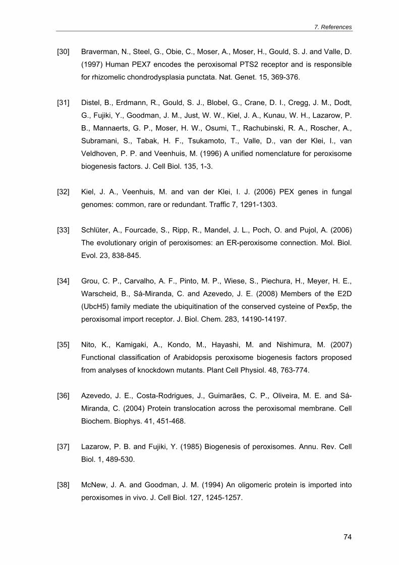
7. References
74
[30] Braverman, N., Steel, G., Obie, C., Moser, A., Moser, H., Gould, S. J. and Valle, D.
(1997) Human PEX7 encodes the peroxisomal PTS2 receptor and is responsible
for rhizomelic chondrodysplasia punctata. Nat. Genet. 15, 369-376.
[31] Distel, B., Erdmann, R., Gould, S. J., Blobel, G., Crane, D. I., Cregg, J. M., Dodt,
G., Fujiki, Y., Goodman, J. M., Just, W. W., Kiel, J. A., Kunau, W. H., Lazarow, P.
B., Mannaerts, G. P., Moser, H. W., Osumi, T., Rachubinski, R. A., Roscher, A.,
Subramani, S., Tabak, H. F., Tsukamoto, T., Valle, D., van der Klei, I., van
Veldhoven, P. P. and Veenhuis, M. (1996) A unified nomenclature for peroxisome
biogenesis factors. J. Cell Biol. 135, 1-3.
[32] Kiel, J. A., Veenhuis, M. and van der Klei, I. J. (2006) PEX genes in fungal
genomes: common, rare or redundant. Traffic 7, 1291-1303.
[33] Schlüter, A., Fourcade, S., Ripp, R., Mandel, J. L., Poch, O. and Pujol, A. (2006)
The evolutionary origin of peroxisomes: an ER-peroxisome connection. Mol. Biol.
Evol. 23, 838-845.
[34] Grou, C. P., Carvalho, A. F., Pinto, M. P., Wiese, S., Piechura, H., Meyer, H. E.,
Warscheid, B., Sá-Miranda, C. and Azevedo, J. E. (2008) Members of the E2D
(UbcH5) family mediate the ubiquitination of the conserved cysteine of Pex5p, the
peroxisomal import receptor. J. Biol. Chem. 283, 14190-14197.
[35] Nito, K., Kamigaki, A., Kondo, M., Hayashi, M. and Nishimura, M. (2007)
Functional classification of Arabidopsis peroxisome biogenesis factors proposed
from analyses of knockdown mutants. Plant Cell Physiol. 48, 763-774.
[36] Azevedo, J. E., Costa-Rodrigues, J., Guimarães, C. P., Oliveira, M. E. and Sá-
Miranda, C. (2004) Protein translocation across the peroxisomal membrane. Cell
Biochem. Biophys. 41, 451-468.
[37] Lazarow, P. B. and Fujiki, Y. (1985) Biogenesis of peroxisomes. Annu. Rev. Cell
Biol. 1, 489-530.
[38] McNew, J. A. and Goodman, J. M. (1994) An oligomeric protein is imported into
peroxisomes in vivo. J. Cell Biol. 127, 1245-1257.

7. References
75
[39] Brocard, C. and Hartig, A. (2006) Peroxisome targeting signal 1: is it really a
simple tripeptide? Biochim. Biophys. Acta 1763, 1565-1573.
[40] Gould, S. J., Keller, G. A., Hosken, N., Wilkinson, J. and Subramani, S. (1989) A
conserved tripeptide sorts proteins to peroxisomes. J. Cell Biol. 108, 1657-1664.
[41] McCollum, D., Monosov, E. and Subramani, S. (1993) The pas8 mutant of Pichia
pastoris exhibits the peroxisomal protein import deficiencies of Zellweger
syndrome cells--the PAS8 protein binds to the COOH-terminal tripeptide
peroxisomal targeting signal, and is a member of the TPR protein family. J. Cell
Biol. 121, 761-774.
[42] Lazarow, P. B. (2006) The import receptor Pex7p and the PTS2 targeting
sequence. Biochim. Biophys. Acta 1763, 1599-1604.
[43] Osumi, T., Tsukamoto, T., Hata, S., Yokota, S., Miura, S., Fujiki, Y., Hijikata, M.,
Miyazawa, S. and Hashimoto, T. (1991) Amino-terminal presequence of the
precursor of peroxisomal 3-ketoacyl-CoA thiolase is a cleavable signal peptide for
peroxisomal targeting. Biochem. Biophys. Res. Commun. 181, 947-954.
[44] Rehling, P., Marzioch, M., Niesen, F., Wittke, E., Veenhuis, M. and Kunau, W. H.
(1996) The import receptor for the peroxisomal targeting signal 2 (PTS2) in
Saccharomyces cerevisiae is encoded by the PAS7 gene. EMBO J. 15, 2901-
2913.
[45] Braverman, N., Dodt, G., Gould, S. J. and Valle, D. (1998) An isoform of pex5p,
the human PTS1 receptor, is required for the import of PTS2 proteins into
peroxisomes. Hum. Mol. Genet. 7, 1195-1205.
[46] Otera, H., Harano, T., Honsho, M., Ghaedi, K., Mukai, S., Tanaka, A., Kawai, A.,
Shimizu, N. and Fujiki, Y. (2000) The mammalian peroxin Pex5pL, the longer
isoform of the mobile peroxisome targeting signal (PTS) type 1 transporter,
translocates the Pex7p.PTS2 protein complex into peroxisomes via its initial
docking site, Pex14p. J. Biol. Chem. 275, 21703-21714.

7. References
76
[47] Gouveia, A. M., Guimarães, C. P., Oliveira, M. E., Sá-Miranda, C. and Azevedo, J.
E. (2003) Insertion of Pex5p into the peroxisomal membrane is cargo protein-
dependent. J. Biol. Chem. 278, 4389-4392.
[48] Agne, B., Meindl, N. M., Niederhoff, K., Einwächter, H., Rehling, P., Sickmann, A.,
Meyer, H. E., Girzalsky, W. and Kunau, W. H. (2003) Pex8p: an intraperoxisomal
organizer of the peroxisomal import machinery. Mol. Cell 11, 635-646.
[49] Azevedo, J. E. and Schliebs, W. (2006) Pex14p, more than just a docking protein.
Biochim. Biophys. Acta 1763, 1574-1584.
[50] Reguenga, C., Oliveira, M. E., Gouveia, A. M., Sá-Miranda, C. and Azevedo, J. E.
(2001) Characterization of the mammalian peroxisomal import machinery: Pex2p,
Pex5p, Pex12p, and Pex14p are subunits of the same protein assembly. J. Biol.
Chem. 276, 29935-29942.
[51] Williams, C. and Distel, B. (2006) Pex13p: docking or cargo handling protein?
Biochim. Biophys. Acta 1763, 1585-1591.
[52] Gouveia, A. M., Guimarães, C. P., Oliveira, M. E., Reguenga, C., Sá-Miranda, C.
and Azevedo, J. E. (2003) Characterization of the peroxisomal cycling receptor,
Pex5p, using a cell-free in vitro import system. J. Biol. Chem. 278, 226-232.
[53] Gouveia, A. M., Reguenga, C., Oliveira, M. E., Sá-Miranda, C. and Azevedo, J. E.
(2000) Characterization of peroxisomal Pex5p from rat liver. Pex5p in the Pex5p-
Pex14p membrane complex is a transmembrane protein. J. Biol. Chem. 275,
32444-32451.
[54] Oliveira, M. E., Gouveia, A. M., Pinto, R. A., Sá-Miranda, C. and Azevedo, J. E.
(2003) The energetics of Pex5p-mediated peroxisomal protein import. J. Biol.
Chem. 278, 39483-39488.
[55] Carvalho, A. F., Pinto, M. P., Grou, C. P., Alencastre, I. S., Fransen, M., Sá-
Miranda, C. and Azevedo, J. E. (2007) Ubiquitination of mammalian Pex5p, the
peroxisomal import receptor. J. Biol. Chem. 282, 31267-31272.

7. References
77
[56] Platta, H. W., El Magraoui, F., Schlee, D., Grunau, S., Girzalsky, W. and Erdmann,
R. (2007) Ubiquitination of the peroxisomal import receptor Pex5p is required for
its recycling. J. Cell Biol. 177, 197-204.
[57] Williams, C., van den Berg, M., Sprenger, R. R. and Distel, B. (2007) A conserved
cysteine is essential for Pex4p-dependent ubiquitination of the peroxisomal import
receptor Pex5p. J. Biol. Chem. 282, 22534-22543.
[58] Zolman, B. K., Monroe-Augustus, M., Silva, I. D. and Bartel, B. (2005)
Identification and functional characterization of Arabidopsis PEROXIN4 and the
interacting protein PEROXIN22. Plant Cell 17, 3422-3435.
[59] Miyata, N. and Fujiki, Y. (2005) Shuttling mechanism of peroxisome targeting
signal type 1 receptor Pex5: ATP-independent import and ATP-dependent export.
Mol. Cell. Biol. 25, 10822-10832.
[60] Platta, H. W., Grunau, S., Rosenkranz, K., Girzalsky, W. and Erdmann, R. (2005)
Functional role of the AAA peroxins in dislocation of the cycling PTS1 receptor
back to the cytosol. Nat. Cell Biol. 7, 817-822.
[61] Matsumoto, N., Tamura, S. and Fujiki, Y. (2003) The pathogenic peroxin Pex26p
recruits the Pex1p-Pex6p AAA ATPase complexes to peroxisomes. Nat. Cell Biol.
5, 454-460.
[62] Schrader, M. and Fahimi, H. D. (2006) Growth and division of peroxisomes. Int.
Rev. Cytol. 255, 237-290.
[63] Thoms, S. and Erdmann, R. (2005) Dynamin-related proteins and Pex11 proteins
in peroxisome division and proliferation. FEBS J. 272, 5169-5181.
[64] Abe, I. and Fujiki, Y. (1998) cDNA cloning and characterization of a constitutively
expressed isoform of the human peroxin Pex11p. Biochem. Biophys. Res.
Commun. 252, 529-533.

7. References
78
[65] Schrader, M., Reuber, B. E., Morrell, J. C., Jimenez-Sanchez, G., Obie, C., Stroh,
T. A., Valle, D., Schroer, T. A. and Gould, S. J. (1998) Expression of PEX11beta
mediates peroxisome proliferation in the absence of extracellular stimuli. J. Biol.
Chem. 273, 29607-29614.
[66] Tanaka, A., Okumoto, K. and Fujiki, Y. (2003) cDNA cloning and characterization
of the third isoform of human peroxin Pex11p. Biochem. Biophys. Res. Commun.
300, 819-823.
[67] Li, X., Baumgart, E., Dong, G. X., Morrell, J. C., Jimenez-Sanchez, G., Valle, D.,
Smith, K. D. and Gould, S. J. (2002) PEX11alpha is required for peroxisome
proliferation in response to 4-phenylbutyrate but is dispensable for peroxisome
proliferator-activated receptor alpha-mediated peroxisome proliferation. Mol. Cell.
Biol. 22, 8226-8240.
[68] Koch, A., Schneider, G., Luers, G. H. and Schrader, M. (2004) Peroxisome
elongation and constriction but not fission can occur independently of dynamin-like
protein 1. J. Cell Sci. 117, 3995-4006.
[69] Koch, A., Thiemann, M., Grabenbauer, M., Yoon, Y., McNiven, M. A. and
Schrader, M. (2003) Dynamin-like protein 1 is involved in peroxisomal fission. J.
Biol. Chem. 278, 8597-8605.
[70] Koch, A., Yoon, Y., Bonekamp, N. A., McNiven, M. A. and Schrader, M. (2005) A
role for Fis1 in both mitochondrial and peroxisomal fission in mammalian cells.
Mol. Biol. Cell 16, 5077-5086.
[71] Schrader, M. (2006) Shared components of mitochondrial and peroxisomal
division. Biochim. Biophys. Acta 1763, 531-541.
[72] Santos, M. J., Imanaka, T., Shio, H., Small, G. M. and Lazarow, P. B. (1988)
Peroxisomal membrane ghosts in Zellweger syndrome--aberrant organelle
assembly. Science 239, 1536-1538.
[73] Honsho, M., Tamura, S., Shimozawa, N., Suzuki, Y., Kondo, N. and Fujiki, Y.
(1998) Mutation in PEX16 is causal in the peroxisome-deficient Zellweger
syndrome of complementation group D. Am. J. Hum. Genet. 63, 1622-1630.
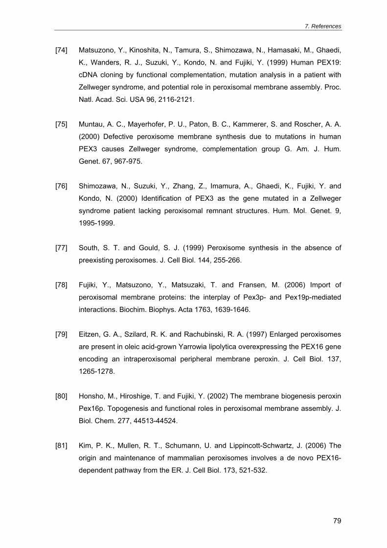
7. References
79
[74] Matsuzono, Y., Kinoshita, N., Tamura, S., Shimozawa, N., Hamasaki, M., Ghaedi,
K., Wanders, R. J., Suzuki, Y., Kondo, N. and Fujiki, Y. (1999) Human PEX19:
cDNA cloning by functional complementation, mutation analysis in a patient with
Zellweger syndrome, and potential role in peroxisomal membrane assembly. Proc.
Natl. Acad. Sci. USA 96, 2116-2121.
[75] Muntau, A. C., Mayerhofer, P. U., Paton, B. C., Kammerer, S. and Roscher, A. A.
(2000) Defective peroxisome membrane synthesis due to mutations in human
PEX3 causes Zellweger syndrome, complementation group G. Am. J. Hum.
Genet. 67, 967-975.
[76] Shimozawa, N., Suzuki, Y., Zhang, Z., Imamura, A., Ghaedi, K., Fujiki, Y. and
Kondo, N. (2000) Identification of PEX3 as the gene mutated in a Zellweger
syndrome patient lacking peroxisomal remnant structures. Hum. Mol. Genet. 9,
1995-1999.
[77] South, S. T. and Gould, S. J. (1999) Peroxisome synthesis in the absence of
preexisting peroxisomes. J. Cell Biol. 144, 255-266.
[78] Fujiki, Y., Matsuzono, Y., Matsuzaki, T. and Fransen, M. (2006) Import of
peroxisomal membrane proteins: the interplay of Pex3p- and Pex19p-mediated
interactions. Biochim. Biophys. Acta 1763, 1639-1646.
[79] Eitzen, G. A., Szilard, R. K. and Rachubinski, R. A. (1997) Enlarged peroxisomes
are present in oleic acid-grown Yarrowia lipolytica overexpressing the PEX16 gene
encoding an intraperoxisomal peripheral membrane peroxin. J. Cell Biol. 137,
1265-1278.
[80] Honsho, M., Hiroshige, T. and Fujiki, Y. (2002) The membrane biogenesis peroxin
Pex16p. Topogenesis and functional roles in peroxisomal membrane assembly. J.
Biol. Chem. 277, 44513-44524.
[81] Kim, P. K., Mullen, R. T., Schumann, U. and Lippincott-Schwartz, J. (2006) The
origin and maintenance of mammalian peroxisomes involves a de novo PEX16-
dependent pathway from the ER. J. Cell Biol. 173, 521-532.

7. References
80
[82] Lazarow, P. B. (2003) Peroxisome biogenesis: advances and conundrums. Curr.
Opin. Cell Biol. 15, 489-497.
[83] Tabak, H. F., Hoepfner, D., Zand, A., Geuze, H. J., Braakman, I. and Huynen, M.
A. (2006) Formation of peroxisomes: present and past. Biochim. Biophys. Acta
1763, 1647-1654.
[84] Diestelkotter, P. and Just, W. W. (1993) In vitro insertion of the 22-kD peroxisomal
membrane protein into isolated rat liver peroxisomes. J. Cell Biol. 123, 1717-1725.
[85] Imanaka, T., Shiina, Y., Takano, T., Hashimoto, T. and Osumi, T. (1996) Insertion
of the 70-kDa peroxisomal membrane protein into peroxisomal membranes in vivo
and in vitro. J. Biol. Chem. 271, 3706-3713.
[86] Novikoff, A. and Shin, W. Y. (1964) The endoplasmic reticulum in the Golgi zone
and its relation to microbodies, Golgi apparatus and autophagic vacuoles in rat
liver cells. J. Microsc. 3, 187-206.
[87] Kunau, W. H. (2005) Peroxisome biogenesis: end of the debate. Curr. Biol. 15,
R774-R776.
[88] Hoepfner, D., Schildknegt, D., Braakman, I., Philippsen, P. and Tabak, H. F.
(2005) Contribution of the endoplasmic reticulum to peroxisome formation. Cell
122, 85-95.
[89] Kragt, A., Voorn-Brouwer, T., van den Berg, M. and Distel, B. (2005) Endoplasmic
reticulum-directed Pex3p routes to peroxisomes and restores peroxisome
formation in a Saccharomyces cerevisiae pex3Delta strain. J. Biol. Chem. 280,
34350-34357.
[90] Tam, Y. Y., Fagarasanu, A., Fagarasanu, M. and Rachubinski, R. A. (2005) Pex3p
initiates the formation of a preperoxisomal compartment from a subdomain of the
endoplasmic reticulum in Saccharomyces cerevisiae. J. Biol. Chem. 280, 34933-
34939.
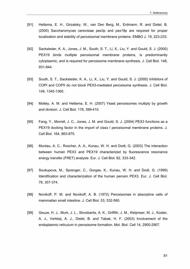
7. References
81
[91] Hettema, E. H., Girzalsky, W., van Den Berg, M., Erdmann, R. and Distel, B.
(2000) Saccharomyces cerevisiae pex3p and pex19p are required for proper
localization and stability of peroxisomal membrane proteins. EMBO J. 19, 223-233.
[92] Sacksteder, K. A., Jones, J. M., South, S. T., Li, X., Liu, Y. and Gould, S. J. (2000)
PEX19 binds multiple peroxisomal membrane proteins, is predominantly
cytoplasmic, and is required for peroxisome membrane synthesis. J. Cell Biol. 148,
931-944.
[93] South, S. T., Sacksteder, K. A., Li, X., Liu, Y. and Gould, S. J. (2000) Inhibitors of
COPI and COPII do not block PEX3-mediated peroxisome synthesis. J. Cell Biol.
149, 1345-1360.
[94] Motley, A. M. and Hettema, E. H. (2007) Yeast peroxisomes multiply by growth
and division. J. Cell Biol. 178, 399-410.
[95] Fang, Y., Morrell, J. C., Jones, J. M. and Gould, S. J. (2004) PEX3 functions as a
PEX19 docking factor in the import of class I peroxisomal membrane proteins. J.
Cell Biol. 164, 863-875.
[96] Muntau, A. C., Roscher, A. A., Kunau, W. H. and Dodt, G. (2003) The interaction
between human PEX3 and PEX19 characterized by fluorescence resonance
energy transfer (FRET) analysis. Eur. J. Cell Biol. 82, 333-342.
[97] Soukupova, M., Sprenger, C., Gorgas, K., Kunau, W. H. and Dodt, G. (1999)
Identification and characterization of the human peroxin PEX3. Eur. J. Cell Biol.
78, 357-374.
[98] Novikoff, P. M. and Novikoff, A. B. (1972) Peroxisomes in absorptive cells of
mammalian small intestine. J. Cell Biol. 53, 532-560.
[99] Geuze, H. J., Murk, J. L., Stroobants, A. K., Griffith, J. M., Kleijmeer, M. J., Koster,
A. J., Verkleij, A. J., Distel, B. and Tabak, H. F. (2003) Involvement of the
endoplasmic reticulum in peroxisome formation. Mol. Biol. Cell 14, 2900-2907.

7. References
82
[100] Voorn-Brouwer, T., Kragt, A., Tabak, H. F. and Distel, B. (2001) Peroxisomal
membrane proteins are properly targeted to peroxisomes in the absence of COPI-
and COPII-mediated vesicular transport. J. Cell Sci. 114, 2199-2204.
[101] South, S. T., Baumgart, E. and Gould, S. J. (2001) Inactivation of the endoplasmic
reticulum protein translocation factor, Sec61p, or its homolog, Ssh1p, does not
affect peroxisome biogenesis. Proc. Natl. Acad. Sci. USA 98, 12027-12031.
[102] Ghaedi, K., Tamura, S., Okumoto, K., Matsuzono, Y. and Fujiki, Y. (2000) The
peroxin pex3p initiates membrane assembly in peroxisome biogenesis. Mol. Biol.
Cell 11, 2085-2102.
[103] Haan, G. J., Faber, K. N., Baerends, R. J., Koek, A., Krikken, A., Kiel, J. A., van
der Klei, I. J. and Veenhuis, M. (2002) Hansenula polymorpha Pex3p is a
peripheral component of the peroxisomal membrane. J. Biol. Chem. 277, 26609-
26617.
[104] Hunt, J. E. and Trelease, R. N. (2004) Sorting pathway and molecular targeting
signals for the Arabidopsis peroxin 3. Biochem. Biophys. Res. Commun. 314, 586-
596.
[105] Kammerer, S., Holzinger, A., Welsch, U. and Roscher, A. A. (1998) Cloning and
characterization of the gene encoding the human peroxisomal assembly protein
Pex3p. FEBS Lett. 429, 53-60.
[106] Baerends, R. J., Faber, K. N., Kram, A. M., Kiel, J. A., van der Klei, I. J. and
Veenhuis, M. (2000) A stretch of positively charged amino acids at the N terminus
of Hansenula polymorpha Pex3p is involved in incorporation of the protein into the
peroxisomal membrane. J. Biol. Chem. 275, 9986-9995.
[107] Wiemer, E. A., Luers, G. H., Faber, K. N., Wenzel, T., Veenhuis, M. and
Subramani, S. (1996) Isolation and characterization of Pas2p, a peroxisomal
membrane protein essential for peroxisome biogenesis in the methylotrophic yeast
Pichia pastoris. J. Biol. Chem. 271, 18973-18980.

7. References
83
[108] Fransen, M., Wylin, T., Brees, C., Mannaerts, G. P. and Van Veldhoven, P. P.
(2001) Human pex19p binds peroxisomal integral membrane proteins at regions
distinct from their sorting sequences. Mol. Cell. Biol. 21, 4413-4424.
[109] Jones, J. M., Morrell, J. C. and Gould, S. J. (2004) PEX19 is a predominantly
cytosolic chaperone and import receptor for class 1 peroxisomal membrane
proteins. J. Cell Biol. 164, 57-67.
[110] Snyder, W. B., Koller, A., Choy, A. J. and Subramani, S. (2000) The peroxin
Pex19p interacts with multiple, integral membrane proteins at the peroxisomal
membrane. J. Cell Biol. 149, 1171-1178.
[111] Sato, Y., Shibata, H., Nakano, H., Matsuzono, Y., Kashiwayama, Y., Kobayashi,
Y., Fujiki, Y., Imanaka, T. and Kato, H. (2008) Characterization of the interaction
between recombinant human peroxin Pex3p and Pex19p: identification of TRP-
104 IN Pex3p as a critical residue for the interaction. J. Biol. Chem. 283, 6136-
6144.
[112] Shibata, H., Kashiwayama, Y., Imanaka, T. and Kato, H. (2004) Domain
architecture and activity of human Pex19p, a chaperone-like protein for
intracellular trafficking of peroxisomal membrane proteins. J. Biol. Chem. 279,
38486-38494.
[113] Gotte, K., Girzalsky, W., Linkert, M., Baumgart, E., Kammerer, S., Kunau, W. H.
and Erdmann, R. (1998) Pex19p, a farnesylated protein essential for peroxisome
biogenesis. Mol. Cell. Biol. 18, 616-628.
[114] Snyder, W. B., Faber, K. N., Wenzel, T. J., Koller, A., Luers, G. H., Rangell, L.,
Keller, G. A. and Subramani, S. (1999) Pex19p interacts with Pex3p and Pex10p
and is essential for peroxisome biogenesis in Pichia pastoris. Mol. Biol. Cell 10,
1745-1761.
[115] Hadden, D. A., Phillipson, B. A., Johnston, K. A., Brown, L. A., Manfield, I. W., El-
Shami, M., Sparkes, I. A. and Baker, A. (2006) Arabidopsis PEX19 is a dimeric
protein that binds the peroxin PEX10. Mol. Membr. Biol. 23, 325-336.

7. References
84
[116] Fransen, M., Vastiau, I., Brees, C., Brys, V., Mannaerts, G. P. and Van Veldhoven,
P. P. (2005) Analysis of human Pex19p's domain structure by pentapeptide
scanning mutagenesis. J. Mol. Biol. 346, 1275-1286.
[117] Halbach, A., Landgraf, C., Lorenzen, S., Rosenkranz, K., Volkmer-Engert, R.,
Erdmann, R. and Rottensteiner, H. (2006) Targeting of the tail-anchored
peroxisomal membrane proteins PEX26 and PEX15 occurs through C-terminal
PEX19-binding sites. J. Cell Sci. 119, 2508-2517.
[118] Matsuzono, Y., Matsuzaki, T. and Fujiki, Y. (2006) Functional domain mapping of
peroxin Pex19p: interaction with Pex3p is essential for function and translocation.
J. Cell Sci. 119, 3539-3550.
[119] Mayerhofer, P. U., Kattenfeld, T., Roscher, A. A. and Muntau, A. C. (2002) Two
splice variants of human PEX19 exhibit distinct functions in peroxisomal assembly.
Biochem. Biophys. Res. Commun. 291, 1180-1186.
[120] Vizeacoumar, F. J., Vreden, W. N., Aitchison, J. D. and Rachubinski, R. A. (2006)
Pex19p binds Pex30p and Pex32p at regions required for their peroxisomal
localization but separate from their peroxisomal targeting signals. J. Biol. Chem.
281, 14805-14812.
[121] James, G. L., Goldstein, J. L., Pathak, R. K., Anderson, R. G. and Brown, M. S.
(1994) PxF, a prenylated protein of peroxisomes. J. Biol. Chem. 269, 14182-
14190.
[122] Banerjee, S. K., Kessler, P. S., Saveria, T. and Parsons, M. (2005) Identification of
trypanosomatid PEX19: functional characterization reveals impact on cell growth
and glycosome size and number. Mol. Biochem. Parasitol. 142, 47-55.
[123] Yernaux, C., Fransen, M., Brees, C., Lorenzen, S. and Michels, P. A. (2006)
Trypanosoma brucei glycosomal ABC transporters: identification and membrane
targeting. Mol. Membr. Biol. 23, 157-172.

7. References
85
[124] Gloeckner, C. J., Mayerhofer, P. U., Landgraf, P., Muntau, A. C., Holzinger, A.,
Gerber, J. K., Kammerer, S., Adamski, J. and Roscher, A. A. (2000) Human
adrenoleukodystrophy protein and related peroxisomal ABC transporters interact
with the peroxisomal assembly protein PEX19p. Biochem. Biophys. Res.
Commun. 271, 144-150.
[125] Vastiau, I. M., Anthonio, E. A., Brams, M., Brees, C., Young, S. G., Van de Velde,
S., Wanders, R. J., Mannaerts, G. P., Baes, M., Van Veldhoven, P. P. and
Fransen, M. (2006) Farnesylation of Pex19p is not essential for peroxisome
biogenesis in yeast and mammalian cells. Cell. Mol. Life Sci. 63, 1686-1699.
[126] Van Ael, E. and Fransen, M. (2006) Targeting signals in peroxisomal membrane
proteins. Biochim. Biophys. Acta 1763, 1629-1638.
[127] Brosius, U., Dehmel, T. and Gartner, J. (2002) Two different targeting signals
direct human peroxisomal membrane protein 22 to peroxisomes. J. Biol. Chem.
277, 774-784.
[128] Jones, J. M., Morrell, J. C. and Gould, S. J. (2001) Multiple distinct targeting
signals in integral peroxisomal membrane proteins. J. Cell Biol. 153, 1141-1150.
[129] Halbach, A., Lorenzen, S., Landgraf, C., Volkmer-Engert, R., Erdmann, R. and
Rottensteiner, H. (2005) Function of the PEX19-binding site of human
adrenoleukodystrophy protein as targeting motif in man and yeast. PMP targeting
is evolutionarily conserved. J. Biol. Chem. 280, 21176-21182.
[130] Landgraf, P., Mayerhofer, P. U., Polanetz, R., Roscher, A. A. and Holzinger, A.
(2003) Targeting of the human adrenoleukodystrophy protein to the peroxisomal
membrane by an internal region containing a highly conserved motif. Eur. J. Cell
Biol. 82, 401-410.
[131] Rottensteiner, H., Kramer, A., Lorenzen, S., Stein, K., Landgraf, C., Volkmer-
Engert, R. and Erdmann, R. (2004) Peroxisomal membrane proteins contain
common Pex19p-binding sites that are an integral part of their targeting signals.
Mol. Biol. Cell 15, 3406-3417.
7. References
86
[132] Saveria, T., Halbach, A., Erdmann, R., Volkmer-Engert, R., Landgraf, C.,
Rottensteiner, H. and Parsons, M. (2007) Conservation of PEX19-binding motifs
required for protein targeting to mammalian peroxisomal and trypanosome
glycosomal membranes. Eukaryot. Cell 6, 1439-1449.
[133] Matsuzono, Y. and Fujiki, Y. (2006) In vitro transport of membrane proteins to
peroxisomes by shuttling receptor Pex19p. J. Biol. Chem. 281, 36-42.
[134] Lambkin, G. R. and Rachubinski, R. A. (2001) Yarrowia lipolytica cells mutant for
the peroxisomal peroxin Pex19p contain structures resembling wild-type
peroxisomes. Mol. Biol. Cell 12, 3353-3364.
[135] Biermanns, M. and Gartner, J. (2001) Targeting elements in the amino-terminal
part direct the human 70-kDa peroxisomal integral membrane protein (PMP70) to
peroxisomes. Biochem. Biophys. Res. Commun. 285, 649-655.
[136] Fransen, M., Vastiau, I., Brees, C., Brys, V., Mannaerts, G. P. and Van Veldhoven,
P. P. (2004) Potential role for Pex19p in assembly of PTS-receptor docking
complexes. J. Biol. Chem. 279, 12615-12624.
[137] Pause, B., Diestelkotter, P., Heid, H. and Just, W. W. (1997) Cytosolic factors
mediate protein insertion into the peroxisomal membrane. FEBS Lett. 414, 95-98.
[138] Reguenga, C., Oliveira, M. E., Gouveia, A. M., Eckerskorn, C., Sá-Miranda, C. and
Azevedo, J. E. (1999) Identification of a 24 kDa intrinsic membrane protein from
mammalian peroxisomes. Biochim. Biophys. Acta 1445, 337-341.
[139] Fransen, M., Van Veldhoven, P. P. and Subramani, S. (1999) Identification of
peroxisomal proteins by using M13 phage protein VI phage display: molecular
evidence that mammalian peroxisomes contain a 2,4-dienoyl-CoA reductase.
Biochem. J. 340 (Pt 2), 561-568.
[140] Wendland, M. and Subramani, S. (1993) Cytosol-dependent peroxisomal protein
import in a permeabilized cell system. J. Cell Biol. 120, 675-685.

7. References
87
[141] Pinto, M. P., Grou, C. P., Alencastre, I. S., Oliveira, M. E., Sá-Miranda, C.,
Fransen, M. and Azevedo, J. E. (2006) The import competence of a peroxisomal
membrane protein is determined by Pex19p before the docking step. J. Biol.
Chem. 281, 34492-34502.
[142] Fujiki, Y., Hubbard, A. L., Fowler, S. and Lazarow, P. B. (1982) Isolation of
intracellular membranes by means of sodium carbonate treatment: application to
endoplasmic reticulum. J. Cell Biol. 93, 97-102.
[143] Laemmli, U. K. (1970) Cleavage of structural proteins during the assembly of the
head of bacteriophage T4. Nature 227, 680-685.
[144] Gallagher, S. R. (1999) One-dimensional electrophoresis using nondenaturing
conditions. In Current Protocols in Molecular Biology (Ausubel, F. M., Brent, R.,
Kingston, R. E., Moore, D. D., Seidman, J. G., Smith, J. A., and Struhl, K., eds),
pp. 10.2B.1-10.2B.11. John Wiley & Sons, Inc, New York.
[145] Gouveia, A. M., Reguenga, C., Oliveira, M. E., Eckerskorn, C., Sá-Miranda, C. and
Azevedo, J. E. (1999) Alkaline density gradient floatation of membranes:
polypeptide composition of the mammalian peroxisomal membrane. Anal.
Biochem. 274, 270-277.
[146] Fransen, M., Terlecky, S. R. and Subramani, S. (1998) Identification of a human
PTS1 receptor docking protein directly required for peroxisomal protein import.
Proc. Natl. Acad. Sci. USA 95, 8087-8092.
[147] Koster, A., Heisig, M., Heinrich, P. C. and Just, W. W. (1986) In vitro synthesis of
peroxisomal membrane polypeptides. Biochem. Biophys. Res. Commun. 137, 626-
632.
[148] Cubitt, A. B., Heim, R., Adams, S. R., Boyd, A. E., Gross, L. A. and Tsien, R. Y.
(1995) Understanding, improving and using green fluorescent proteins. Trends
Biochem. Sci. 20, 448-455.

7. References
88
[149] Elgersma, Y., Kwast, L., Klein, A., Voorn-Brouwer, T., van den Berg, M., Metzig,
B., America, T., Tabak, H. F. and Distel, B. (1996) The SH3 domain of the
Saccharomyces cerevisiae peroxisomal membrane protein Pex13p functions as a
docking site for Pex5p, a mobile receptor for the import PTS1-containing proteins.
J. Cell Biol. 135, 97-109.
[150] Erdmann, R. and Blobel, G. (1996) Identification of Pex13p a peroxisomal
membrane receptor for the PTS1 recognition factor. J. Cell Biol. 135, 111-121.
[151] Gould, S. J., Kalish, J. E., Morrell, J. C., Bjorkman, J., Urquhart, A. J. and Crane,
D. I. (1996) Pex13p is an SH3 protein of the peroxisome membrane and a docking
factor for the predominantly cytoplasmic PTs1 receptor. J. Cell Biol. 135, 85-95.
[152] Weibel, E. R., Staubli, W., Gnagi, H. R. and Hess, F. A. (1969) Correlated
morphometric and biochemical studies on the liver cell. I. Morphometric model,
stereologic methods, and normal morphometric data for rat liver. J. Cell Biol. 42,
68-91.
[153] Kikuchi, M., Hatano, N., Yokota, S., Shimozawa, N., Imanaka, T. and Taniguchi, H.
(2004) Proteomic analysis of rat liver peroxisome: presence of peroxisome-specific
isozyme of Lon protease. J. Biol. Chem. 279, 421-428.
[154] Wiese, S., Gronemeyer, T., Ofman, R., Kunze, M., Grou, C. P., Almeida, J. A.,
Eisenacher, M., Stephan, C., Hayen, H., Schollenberger, L., Korosec, T.,
Waterham, H. R., Schliebs, W., Erdmann, R., Berger, J., Meyer, H. E., Just, W.,
Azevedo, J. E., Wanders, R. J. and Warscheid, B. (2007) Proteomics
characterization of mouse kidney peroxisomes by tandem mass spectrometry and
protein correlation profiling. Mol. Cell. Proteomics 6, 2045-2057.
[155] Tugal, H. B., Pool, M. and Baker, A. (1999) Arabidopsis 22-kilodalton peroxisomal
membrane protein. Nucleotide sequence analysis and biochemical
characterization. Plant Physiol. 120, 309-320.
[156] Oliveira, M. E., Reguenga, C., Gouveia, A. M., Guimarães, C. P., Schliebs, W.,
Kunau, W. H., Silva, M. T., Sá-Miranda, C. and Azevedo, J. E. (2002) Mammalian
Pex14p: membrane topology and characterisation of the Pex14p-Pex14p
interaction. Biochim. Biophys. Acta 1567, 13-22.

7. References
89
[157] Hardeman, D., Versantvoort, C., van den Brink, J. M. and van den Bosch, H.
(1990) Studies on peroxisomal membranes. Biochim. Biophys. Acta 1027, 149-
154.
[158] Yang, J., Han, X. and Gross, R. W. (2003) Identification of hepatic peroxisomal
phospholipase A(2) and characterization of arachidonic acid-containing choline
glycerophospholipids in hepatic peroxisomes. FEBS Lett. 546, 247-250.


