Page 1
RESEARCH ARTICLE
Biogeographical venom variation in the Indian
spectacled cobra (Naja naja) underscores the
pressing need for pan-India efficacious
snakebite therapy
R. R. Senji LaxmeID1☯, Saurabh AttardeID
1☯, Suyog Khochare1☯, Vivek Suranse1,
Gerard MartinID2, Nicholas R. CasewellID
3, Romulus WhitakerID4, Kartik SunagarID
1*
1 Evolutionary Venomics Lab. Centre for Ecological Sciences, Indian Institute of Science, Bangalore,
Karnataka, India, 2 The Liana Trust, Survey #1418/1419 Rathnapuri, Hunsur, Karnataka, India, 3 Centre for
Snakebite Research & Interventions, Liverpool School of Tropical Medicine, Pembroke Place, Liverpool,
United Kingdom, 4 Madras Crocodile Bank Trust/Centre for Herpetology, Mamallapuram, Tamil Nadu, India
☯ These authors contributed equally to this work.
* [email protected]
Abstract
Background
Snake venom composition is dictated by various ecological and environmental factors, and
can exhibit dramatic variation across geographically disparate populations of the same spe-
cies. This molecular diversity can undermine the efficacy of snakebite treatments, as anti-
venoms produced against venom from one population may fail to neutralise others. India is
the world’s snakebite hotspot, with 58,000 fatalities and 140,000 morbidities occurring annu-
ally. Spectacled cobra (Naja naja) and Russell’s viper (Daboia russelii) are known to cause
the majority of these envenomations, in part due to their near country-wide distributions.
However, the impact of differing ecologies and environment on their venom compositions
has not been comprehensively studied.
Methods
Here, we used a multi-disciplinary approach consisting of venom proteomics, biochemical
and pharmacological analyses, and in vivo research to comparatively analyse N. naja ven-
oms across a broad region (>6000 km; seven populations) covering India’s six distinct bio-
geographical zones.
Findings
By generating the most comprehensive pan-Indian proteomic and toxicity profiles to date,
we unveil considerable differences in the composition, pharmacological effects and poten-
cies of geographically-distinct venoms from this species and, through the use of immunolog-
ical assays and preclinical experiments, demonstrate alarming repercussions on antivenom
therapy. We find that commercially-available antivenom fails to effectively neutralise
PLOS Neglected Tropical Diseases | https://doi.org/10.1371/journal.pntd.0009150 February 18, 2021 1 / 28
a1111111111
a1111111111
a1111111111
a1111111111
a1111111111
OPEN ACCESS
Citation: Senji Laxme RR, Attarde S, Khochare S,
Suranse V, Martin G, Casewell NR, et al. (2021)
Biogeographical venom variation in the Indian
spectacled cobra (Naja naja) underscores the
pressing need for pan-India efficacious snakebite
therapy. PLoS Negl Trop Dis 15(2): e0009150.
https://doi.org/10.1371/journal.pntd.0009150
Editor: Ulrich Kuch, Goethe University, GERMANY
Received: May 16, 2020
Accepted: January 18, 2021
Published: February 18, 2021
Copyright: © 2021 Senji Laxme et al. This is an
open access article distributed under the terms of
the Creative Commons Attribution License, which
permits unrestricted use, distribution, and
reproduction in any medium, provided the original
author and source are credited.
Data Availability Statement: All relevant data are
within the manuscript and its Supporting
Information files. The mass spectrometry data
generated in this study has been deposited to the
ProteomeXchange Consortium via the PRIDE
partner repository with data identifier: PXD020497
(https://www.ebi.ac.uk/pride/archive?keyword=
PXD020497). Additionally, an archive containing
the results of proteomics analyses in HTML format
has been added to S1 Data.
Page 2
envenomations by the pan-Indian populations of N. naja, including a complete lack of neu-
tralisation against the desert Naja population.
Conclusion
Our findings highlight the significant influence of ecology and environment on snake venom
composition and potency, and stress the pressing need to innovate pan-India effective anti-
venoms to safeguard the lives, limbs and livelihoods of the country’s 200,000 annual snake-
bite victims.
Author summary
Annually, India is burdened by the highest number of snake envenomations across the
globe, with over 58,000 fatalities and three times the number of morbidities, predomi-
nantly affecting the rural agrarian communities. The spectacled cobra (Naja naja) and
Russell’s viper (Daboia russelii) are responsible for the vast majority of envenomations in
the country, in part, due to their near country-wide distributions. In this study, we unveil
the astounding differences in venom composition of N. naja from six different bio-
geographical zones across the country (>6000 km). We provide a comprehensive account
of their disparate venom proteomic profiles, biochemical and pharmacological effects,
and the associated potencies. Our study uncovers alarming differences in the efficacy of
the marketed polyvalent antivenoms in neutralising these venoms, thereby, emphasising
the pressing need to develop dose-efficacious and pan-India effective antivenoms for the
treatment of snakebites in the country. This study also highlights the significant influence
of ecology and diverse environments on the venom variability, insinuating the necessity
for innovating cost-effective and pan-India efficacious solutions to safeguard the lives,
limbs and livelihoods of India’s two hundred thousand annual snakebite victims.
Introduction
Venom is an adaptive trait that has evolved multiple times across the animal kingdom to facili-
tate various ecological functions, including defence, predation, competition, or a combination
thereof [1–4]. Given their medical relevance to humans in the form of snakebite, and the tre-
mendous biodiscovery potential of their toxic molecules, snake venoms have received unparal-
leled research attention. In India, there are over 60 described snake species capable of
inflicting clinically significant envenomations in humans, among which 14 species have been
documented to cause human fatalities [5]. Nevertheless, existing antivenoms—only available
specific treatment for snakebite—are produced exclusively against the so-called ‘big four’
snakes: the spectacled cobra (Naja naja), common krait (Bungarus caeruleus), Russell’s viper
(Daboia russelii) and saw-scaled viper (Echis carinatus). Despite the availability of polyvalent
antivenom, snakebite continues to be a severe burden on the rural agrarian communities in
India, resulting in an annual toll greater than that of any other country [6,7].
The composition of venom, which is theorised to be influenced by various ecological and
environmental factors, including diet, predator pressure, climatic zones, and ontogenetic
shifts, can vary across the geographical distribution of snake species [8–12], even at very short
distances [13,14]. This variation not only underpins the ecological adaptations of the animal
but also severely impacts the efficacy of snakebite therapy. Commercial Indian antivenoms are
PLOS NEGLECTED TROPICAL DISEASES Hiss of death
PLOS Neglected Tropical Diseases | https://doi.org/10.1371/journal.pntd.0009150 February 18, 2021 2 / 28
Funding: K.S. was supported by the following
grants: Department for International Development
(https://www.gov.uk/world/organisations/dfid-
india) ([DFID: grant IAVI/BES/KASU/0002]. The
views expressed do not necessarily reflect the UK
Government’s official policies); the Department of
Biotechnology-IISc Partnership Program (http://
dbtindia.gov.in/); DST-INSPIRE Faculty Award
(DST/INSPIRE/04/2017/000071, http://online-
inspire.gov.in/), and the DST-FIST (SR/FST/LS-II/
2018/233, http://www.fist-dst.org/). N.R.C.
acknowledges support from a Sir Henry Dale
Fellowship (200517/Z/16/Z) jointly funded by the
Wellcome Trust and Royal Society. Venom
sampling equipment and expeditions were
supported by USV Private Limited (http://www.
usvindia.com). The funders had no role in study
design, data collection and analysis, decision to
publish, or preparation of the manuscript.
Competing interests: The authors have declared
that no competing interests exist.
Page 3
produced by hyperimmunising equines with the ‘big four’ snake venoms and purifying the
resultant anti-snake venom toxin antibodies. However, venoms are sourced from only a couple
of districts in the southern part of the country, which may therefore render them incapable of
neutralising the toxic effects of other more distant populations where venom composition may
vary [15]. While such variation has been noted in the venoms of selected populations of N.
naja [16–25], the true extent of biogeographic venom variation and its impact on the efficacy
of marketed antivenoms is yet to be comprehensively elucidated.
To address these shortcomings, we investigated the venoms of one of the most medically
important Indian snakes, N. naja, which has been reported to be responsible for the majority
of snakebite fatalities and disabilities in the Indian subcontinent [7]. We characterised the
composition and function of venom from this snake species from six distinct biogeographical
zones across the country (>6000 km), thereby, generating the most comprehensive proteomic
and toxicity profiles of this species to date. The results of our in vitro and in vivo experiments
revealed dramatic differences in toxin compositions, synergistic pharmacological effects, and
in vivo potency of the venoms. We also reveal the disturbing impact this variation has on the
effectiveness of commercial Indian antivenoms to neutralise venoms sourced from different
parts of the country. Our results highlight the significant impact that ecology and environment
can have in shaping these complex biochemical cocktails, and emphasise the urgent need to
develop pan-India effective snakebite therapies.
Methods
Ethics statement
The median lethal dose (LD50) of venoms and the median effective dose (ED50) of commer-
cially available antivenoms were determined as per World Health Organization (WHO)-rec-
ommended protocols at the Central Animal Facility, Indian Institute of Science (IISc),
Bangalore (Registration number 48/GO/ReBi/SL/1999 /CPCSEA; 11-03-1999). For these
assays, male CD-1 mice (18–22 g) were used with due approval from (i) the Committee for the
Purpose of Control and Supervision of Experiments on Animals (CPCSEA), Government of
India; and (ii) the Institutional Animal Ethics Committee (IAEC), IISc, Bangalore (CAF/Eth-
ics/642/2018; CAF/Ethics/643/2018). Based on the results of in vitro venom recognition exper-
iments (enzyme-linked immunosorbent assay and immunoblotting), a single commercial
antivenom was selected for the ED50 experiments to limit the numbers of experimental ani-
mals subjected to these severe-rated experiments. Animals were handled according to the insti-
tutional guidelines during and after the completion of the experiment. To evaluate snake
venom-induced coagulopathies on human blood, ethical permission was obtained from the
Institute Human Ethical Committee (IHEC No: 5–24072019), IISc, Bangalore, and blood was
collected from healthy volunteers after explaining the details of the study.
Sampling permits, snake venoms and antivenoms
Snake venoms were collected from 80+ individuals across a range of 6000 km from the follow-
ing regions with appropriate permissions from the respective State Forest Departments: North
(Punjab: #3615;11/10/12), South (Tamil Nadu), Southeast (Andhra Pradesh:#13526/2017/WL-
3), East (West Bengal: 386/WL/4R-6/2017), West (Rajasthan: P.3(3)Forest/2004), Southwest
(Maharashtra: Desk-22(8)/Research/CR-80(16–17) /943/2017-18), and Central (Madhya Pra-
desh: #/TK-1/48-II/606) India. The venom samples were collected from individuals with or
without pooling, flash-frozen, and stored at -80˚ C following lyophilisation, until use (S1A
Table). Details of the investigated Indian antivenoms produced by four major commercial
antivenom manufacturers are provided in the S1B Table.
PLOS NEGLECTED TROPICAL DISEASES Hiss of death
PLOS Neglected Tropical Diseases | https://doi.org/10.1371/journal.pntd.0009150 February 18, 2021 3 / 28
Page 4
Protein concentration
Following reconstitution in molecular grade water, protein concentrations of the venoms were
estimated using the Bradford method, with bovine serum albumin (BSA) as standard [[26];
S1A Table]. The antivenom vials were reconstituted as per the manufacturer’s guidelines, and
the total IgG content was estimated using the bovine gamma globulin (BGG) standard curve
(S1B Table).
Gel electrophoresis
Venom samples were normalised for protein content (12 μg), and the components were sepa-
rated using sodium dodecyl sulfate-polyacrylamide gel electrophoresis (SDS-PAGE) under
reducing conditions [27]. Coomassie Brilliant Blue R-250 (Sisco Research Laboratories Pvt.
Ltd, India) stained gels were visualised in an iBright CL1000 (Thermo Fisher Scientific, USA)
gel documentation system.
Reversed-phase high-performance liquid chromatography (RP-HPLC)
The reconstituted venoms were fractionated using a Shimadzu LC-20AD series HPLC system
(Kyoto, Japan) using a previously described protocol with modifications [28]. 200 μg of each
venom was loaded onto a 4.6 × 250 mm, C18 (5 μm, 300 Å) reversed-phase column (Shi-
madzu, Japan), and equilibrated with solution A [0.1% trifluoroacetic acid (TFA) in water (v/
v)]. The fractions were eluted at a flow rate of 1 ml/min using the following concentration gra-
dients of solution B [0.1% TFA in 100% acetonitrile (v/v)]: 5–15%, 15–45% and 45–70% for 10,
60 and 10 min, respectively, and the absorbance was monitored at 215 nm.
Liquid chromatography-tandem mass spectrometry (LC-MS/MS)
The proteomic profiles of the collected HPLC fractions (40 μg) were characterised via electro-
spray ionisation tandem mass spectrometry (ESI-MS/MS). Following reduction with dithio-
threitol (DTT; 10 mM), alkylation using iodoacetamide (IAA; 30 mM), and an overnight
trypsin (0.2 μg/μl) digestion, each HPLC fraction was desalted. Liquid chromatography of
these processed samples was performed using a Thermo EASY nLC 1200 series system
(Thermo Fisher Scientific, MA, USA) with a 50 cm × 75 μm, C18 (3 μm, 100 Å) nano-LC col-
umn. The sample (injection volume of 2 μl) was run at a flow rate of 300 nL/min in buffer A
(0.1% formic acid in HPLC grade water) and buffer B (0.1% formic acid in 80% acetonitrile)
solutions. The gradient of buffer B used for the elution was 10–45% over the first 98 min, 45–
95% over the next 4 min, followed by 95% over the last 18 min. Mass spectrometric analyses of
the samples were performed using the Thermo Orbitrap Fusion Mass Spectrometer (Thermo
Fisher Scientific, MA, USA). For the MS scan, the following parameters were used: scan range
(m/z) of 375–1700 with a resolution of 120000 and maximum injection time of 50 ms. For the
fragment scans, an ion trap detector was used with high collision energy fragmentation (30%),
scan range (m/z) of 100–2000, and maximum injection time of 35 ms. The raw MS/MS spectra
were searched against the SwissProt database (www.uniprot.org) using PEAKS Studio X (Bio-
informatics Solutions Inc., ON, Canada) with the following parameters: parent and fragment
mass error tolerance limits of 10 ppm and 0.6 Da, respectively; ‘monoisotopic’ precursor ion
search type; and ‘semispecific’ trypsin digestion. Carbamidomethylation and oxidation were
specified as fixed and variable modifications, respectively. Error in the identification of pep-
tides was minimised by fixing the False Discovery Rate (FDR) for peptide-spectrum matching
at 0.1% and the corresponding -10lgP cutoff value was automatically determined by PEAKS
Studio. Only hits with one or more unique peptides were considered for downstream analyses.
PLOS NEGLECTED TROPICAL DISEASES Hiss of death
PLOS Neglected Tropical Diseases | https://doi.org/10.1371/journal.pntd.0009150 February 18, 2021 4 / 28
Page 5
The mass spectrometry data generated in this study have been deposited to the ProteomeX-
change Consortium via the PRIDE partner repository [29], with data identifier: PXD020497.
The relative abundance of each toxin hit in a fraction was determined by estimating its area
under the spectral intensity curve (i.e., AUC) relative to the total AUC for all toxins in
that fraction. The AUC values obtained from PEAKS Studio analyses represented the mean
spectral intensities [30] and were normalised across fractions using the percentage of peak
areas for the respective RP-HPLC fractions [31]. Thus, the relative abundance of a toxin hit
(X) was calculated as follows (here, N indicates the number of fractions obtained from
RP-HPLC):
Relative abundance of X %ð Þ
¼XN
n¼1
AUC of X in Fraction Fn � AUC of the chromatographic fraction Fn ð%ÞTotal AUC of all toxin hits in Fraction Fn
Venom biochemistry
The biochemical activities of the various venom samples were evaluated in the following assays
using previously described methods [14].
Phospholipase A2 (PLA2) assay
Slightly modified turbidimetric assays were conducted to assess the PLA2 activities of venoms
as described previously [14,32]. A fresh chicken egg was used to prepare the egg-yolk substrate
solution in 0.9% NaCl solution, such that its absorbance at 740 nm corresponded to 1. A fixed
concentration of crude venom samples (1 μg) prepared in 20 mM Tris-HCl buffer, time-
dependent kinetic assays were performed in triplicate. Following the addition of 250 μl of the
egg yolk solution, absorbance was measured for 60 min at 740 nm in an EPOCH 2 microplate
spectrophotometer (BioTek Instruments, Inc., USA). Unit activity was calculated as the
amount of crude venom required to reduce the absorbance of the substrate by 0.01 OD unit
per min at the given wavelength [33].
Snake venom protease assay
Protease activity was estimated using azocasein as a substrate using the protocol described pre-
viously [34]. A known volume of crude venom was incubated with 80 μl of the substrate at 37˚
C for 90 min in triplicate. The reaction was stopped using 200 μl of trichloroacetic acid, and
the supernatant was obtained by centrifuging at 1000 × g for 5 min. To this, an equal volume
of 0.5 M NaOH was added, and the absorbance was measured at 440 nm. Purified protease
from bovine pancreas (Sigma-Aldrich, USA) was used as a positive control to calculate the rel-
ative protease activity of the crude venoms.
L-amino acid oxidase (LAAO) assay
LAAO activity was assessed using a previously described endpoint assay with slight modifica-
tions [14,35]. Briefly, the L-leucine substrate solution, containing Tris-HCl buffer (50 mM), L-
leucine (5 mM), horseradish peroxidase (5 IU/ml), and o-phenylenediamine dihydrochloride
(2 mM), was mixed with crude venom (10 μg) in a 9:1 ratio and incubated at 37˚ C for 60 min
in triplicates. The reaction was stopped by adding 2 M H2SO4, and the absorbance was mea-
sured at 492 nm with an EPOCH 2 microplate spectrophotometer.
PLOS NEGLECTED TROPICAL DISEASES Hiss of death
PLOS Neglected Tropical Diseases | https://doi.org/10.1371/journal.pntd.0009150 February 18, 2021 5 / 28
Page 6
DNase assay
To assay the DNase activities of venoms, a modified protocol was employed wherein purified
DNA from calf thymus (Sigma-Aldrich, USA) dissolved in phosphate buffer saline (PBS; pH
7.4) was incubated with a known concentration of crude venom at 37˚ C for 60 min. Post-
incubation, samples were subjected to agarose gel electrophoresis on 0.8% agarose gel, fol-
lowed by visualisation on an iBright CL1000 [14,36].
Fibrinogenolytic assay
Fibrinogenolytic activities of snake venoms against human fibrinogen were determined using
a method previously described by Ouyang and Teng [14,37]. The reaction mixture contained
15 μg of human fibrinogen (Sigma-Aldrich, USA) dissolved in PBS (pH 7.4), and a known
concentration of venom, ranging between 1 and 10 μg and was incubated at 37˚ C for 60 min.
After incubation, an equal volume of loading dye (1 M Tris-HCl pH 6.8, 50% glycerol, 0.5%
bromophenol blue, 10% SDS, 20% β-mercaptoethanol) was added and the samples heated at
70˚ C for 10 min. Subsequently, samples were separated by 15% SDS-PAGE, staining the gel
with Coomassie Brilliant Blue R-250, prior to visualisation in an iBright CL1000 (Thermo
Fisher Scientific, USA) gel documentation system. Results are interpreted with respect to a
negative control that only consists of human fibrinogen without venom, where all three bands
are seen intact.
Blood coagulation assays
The effect of snake venom on the two major coagulation cascades, namely, the extrinsic and
intrinsic pathways, were evaluated by measuring prothrombin time (PT) and activated partial
thromboplastin time (aPTT), respectively. In brief, platelet-poor plasma (PPP), obtained by
centrifuging human blood at 3000 × g for 10 min at 4˚ C, was mixed with different venom con-
centrations. A Hemostar XF 2.0 coagulometer and commercially available UNIPLASTIN and
LIQUICELIN-E diagnostic kits (Tulip Diagnostics, Mumbai) were used for conducting PT
and aPTT tests, respectively.
Haemolytic assay
Haemolytic activities of venoms were assessed as described previously [14,38]. For assaying
haemolytic activities of venoms, human red blood cells (RBC), obtained after the separation of
PPP, were washed five times with 1× PBS buffer (pH 7.4) and centrifuged at 3000 × g for 10
min at 4˚ C. Following the resuspension of the RBC pellet in PBS, samples were incubated
with different concentrations of venoms (5, 10, 20 and 40 μg) at 37˚ C for 24 hours in triplicate.
Thereafter, reaction mixtures were centrifuged at 3000 × g for 10 min at 4˚ C, and the absor-
bance of the supernatant was measured at 540 nm using an Epoch 2 microplate spectropho-
tometer. Triton X (0.5%) and PBS were used as positive and negative controls, respectively.
Enzyme-linked immunosorbent assay (ELISA)
Indirect ELISA experiments were used to quantify the in vitro binding titres between the ven-
oms and commercial antivenoms. ELISAs were performed using minor modifications of pre-
viously described protocols [14,39]. Venom samples (100 ng) were diluted in a carbonate
buffer (pH 9.6) and coated onto 96-well plates. After overnight incubation at 4˚ C, the
unbound venom was washed off using Tris-buffered saline (0.01 M Tris pH 8.5, 0.15 M NaCl)
containing 1% Tween 20 (TBST), and incubated with blocking buffer (5% skimmed milk in
TBST) for 3 hours at room temperature. Following another round of TBST washing, the
PLOS NEGLECTED TROPICAL DISEASES Hiss of death
PLOS Neglected Tropical Diseases | https://doi.org/10.1371/journal.pntd.0009150 February 18, 2021 6 / 28
Page 7
venom-bound plates were incubated overnight with different dilutions of commercial antiven-
oms at 4˚ C. All four antivenoms (Premium Serums, VINS, Bharat, and Haffkine), with
sequential fivefold dilutions (starting from 1:4 dilution) in blocking buffer (1 mg/ml), were
added to the plates in triplicates. Thereafter, unbound antibodies were removed by TBST
washing and the plates were incubated at room temperature for 2 hours following the addition
of horseradish peroxidase (HRP)-conjugated, rabbit anti-horse secondary antibody (Sigma-
Aldrich, USA), diluted at a ratio of 1:1000 in PBS. Finally, 100 μl of 2,2/-azino-bis (2-ethyl-
benzthiazoline-6-sulphonic acid) substrate solution (Sigma-Aldrich, USA) was added, the
resulting optical density measured at a wavelength of 405 nm for 40 min, and plotted against
the respective dilution. The 40th min was chosen as the endpoint based on the results of the
standardisation experiments that showed the highest binding at this time interval. The cut off
for non-specific binding was determined as described earlier, using IgG from unimmunised
(naïve) horses as a negative control [14].
Immunoblotting
Immunoblotting experiments were performed following the protocol described with modifica-
tions [14,39]. Venoms were first electrophoretically separated by SDS-PAGE (12.5% gel) and
then transferred to a nitrocellulose membrane at 25 V and 2.5 A for 7 min, following the man-
ufacturer’s protocol (BioRad, USA). Ponceau S reversible stain was used for assessing the
transfer efficiency, following which the non-specific regions on the membrane were blocked
overnight with 5% skimmed milk in TBST at 4˚ C. This was followed by six TBST washes over
a period of an hour, before an overnight incubation at 4˚ C following the addition of the
respective polyvalent antivenom at a 1:200 dilution in the blocking buffer. HRP-conjugated,
rabbit anti-horse secondary antibody was added at a dilution of 1:2000 following six TBST
washes to remove unbound antivenom. The binding of antivenom to venom was captured by
the addition of enhanced chemiluminescence substrate as per the manufacturer’s instructions
(Thermo Fisher Scientific, USA) and imaged in an iBright CL1000 (Thermo Fisher Scientific,
USA).
In vivo venom toxicity and antivenom efficacy assays
To evaluate the pan-India toxicity profiles of N. naja venoms, and the preclinical efficacy of
currently marketed Indian antivenoms against the lethal venom effects, we conducted in vivoneutralisation assays in murine models.
The intravenous median lethal dose (LD50)
The potency of the venom sample corresponding to a biogeographic zone was determined by
calculating the LD50 or the amount of venom required to kill 50% of the test population of
mice [40]. In brief, five different venom concentrations were prepared in physiological saline
(0.9% NaCl), followed by the intravenous injection into the tail vein of the mice (500 μl/
mouse). Five CD-1 mice in the weight range of 18–22 g were used per group, with one control
group receiving normal saline alone. Following injection, mice were kept under observation
for 24 hours, and the number of dead and surviving animals recorded for the calculation of
LD50 values using Probit statistics [41].
The median effective dose (ED50)
The preclinical efficacy of an antivenom, in effect, its capability to neutralise the lethal systemic
effects of snake venom, can be evaluated by calculating the ED50 value, which is defined as the
PLOS NEGLECTED TROPICAL DISEASES Hiss of death
PLOS Neglected Tropical Diseases | https://doi.org/10.1371/journal.pntd.0009150 February 18, 2021 7 / 28
Page 8
minimum amount of antivenom required to protect 50% of mice injected with lethal doses of
venom [40]. For these experiments, we used the Premium Serums antivenom, as this product
was found to recognise the Naja venoms to a greater extent than all of the other Indian mar-
keted antivenoms, as determined by our in vitro assays. We chose to test only the most promis-
ing of the marketed Indian antivenoms in these experiments to reduce the burden of suffering
on experimental animals. Venom doses equivalent to five LD50 determined in the experiments
above were used as the ‘challenge dose’. Different volumes of antivenom were mixed with the
challenge dose of venom, followed by an incubation period of 30 min at 37˚ C. Immediately
after incubation, each venom-antivenom mixture (n = 4 per venom) was intravenously
injected into a group of five male CD-1 mice (18–22 g). A group of five male mice injected
with 1× LD50 of venom, served as the positive control. The ED50 values of the antivenom
against each venom were calculated using Probit statistics [41]. Antivenom neutralisation
potency was calculated as described before [14,42].
Antivenom neutralisation potency mg=mlð Þ ¼ðn � 1Þ�LD50 of venom ðmg=mouseÞ
ED50 ðmlÞ
Here, n is equal to the number of LD50 used as the challenge dose.
Statistical analysis
One-way ANOVA and Two-way ANOVA with Tukey’s and Dunnett’s multiple comparison
tests were used for the statistical comparisons of biochemical assays and ELISA results, respec-
tively, and were performed in GraphPad Prism (GraphPad Software 8.0, San Diego, California
USA, www.graphpad.com).
Results
Venom proteomics
The proteomic profiles of N. naja venoms collected from seven populations in six distinct bio-
geographical zones across India (Fig 1A and S1A Table) were elucidated using SDS-PAGE and
RP-HPLC. In addition, three populations [i.e., the semi-arid (Punjab: PB), Gangetic Plains (West
Bengal: WB) and desert (Rajasthan: RJ) populations] were selected based on their unique HPLC
and toxicity profiles and were subjected to tandem mass spectrometry. While SDS-PAGE profiles
revealed molecular weights of toxins and the primary differences in the composition of venom
proteins between populations (Fig 1B), finer differences in venom composition were unravelled
by RP-HPLC (Fig 2). To identify venom components in each fraction, we further subjected indi-
vidual fractions to LC-MS/MS. Differences were not only noted in the number of fractions shared
between populations but also in their intensities, which corresponds to protein abundances.
Mass spectrometry of venom fractions identified between 48 to 59 non-redundant toxin
proteins from 11 toxin families in the pan-Indian populations of N. naja (S2A–S2C Table and
S1 Data). Tandem mass spectrometry identified a plethora of toxin protein families including
three-finger toxin (3FTx), cobra venom factor (CVF), phospholipase A2 (PLA2), Kunitz-type
serine protease inhibitor (Kunitz), cysteine-rich secretory proteins (CRISP), snake venom
metalloproteinase (SVMP), nerve growth factor (NGF), L-amino-acid oxidase (LAAO), 5’-
nucleotidase, vespryn and cystatin in the venoms of N. naja (S2A–S2C Table and S1 Data).
3FTx are a major family of functionally diverse low molecular weight toxins (6–9 kDa) that
target a wide range of receptors and ion channels [44–46]. 3FTxs were identified as the most
abundant venom protein family in all populations of N. naja across the Indian subcontinent.
They are abundantly secreted in the venoms of most Elapidae snakes and are known to inflict
PLOS NEGLECTED TROPICAL DISEASES Hiss of death
PLOS Neglected Tropical Diseases | https://doi.org/10.1371/journal.pntd.0009150 February 18, 2021 8 / 28
Page 9
a plethora of toxic effects in bite victims, including neurotoxicity, cytotoxicity, anti-platelet
activity and cardiotoxicity [44,47–50]. Here, we detected major differences in the amounts of
neurotoxic 3FTx (N-3FTx) between the pan-Indian populations of N. naja (Figs 2 and 3).
Mass spectrometric analyses revealed that, while this toxin type constituted 80% and 73.3% of
the venom profiles of semi-arid (PB) and Gangetic plain (WB) populations, respectively, only
~30% of the venom was comprised of N-3FTx in the desert (RJ) population of N. naja (Fig 3).
In contrast, the desert population (RJ) secreted 2 to 4 times more cytotoxic/cardiotoxic 3FTxs
(C-3FTx; 41.7%) in comparison to the Gangetic Plain (WB) and semi-arid (PB) populations
(23.6% and 10%, respectively; Fig 3). Interestingly, we observed that the abundance of PLA2
also varied significantly between populations (0.04 to 20%). While the abundance of PLA2 in
the desert population (RJ) was in line with the literature [18], the relatively lower abundances
in the semi-arid (PB) and Gangetic Plain (WB) populations highlight the remarkable biogeo-
graphic variations in the venoms of N. naja. Furthermore, minor differences were observed in
the abundance of CRISP (1.6 to 3.2%), vespryn (0.94 to 1.9%), SVMP (1.3 to 2.1%), Kunitz
(0.05 to 3.2%) and NGF (0.13 to 1.9%) across populations (Fig 3 and S2A–S2C Table). In con-
trast to previous reports [14,23,24], we detected limited amounts of CVF (<0.001 to 0.11) in
these populations. Another noteworthy discovery was the identification of the PLA2 inhibitor
(PLI) (Uniprot ID: Q7LZI1) from the Gangetic Plain (WB) population of N. naja. PLIs have
been previously identified in the blood of several snake species, and are implicated in prevent-
ing self-envenomation [51].
Fig 1. Sampling locations and SDS-PAGE profiles of N. naja venoms from distinct biogeographic zones of India. This figure depicts (A) the venom sampling
locations across distinct biogeographic zones of India and (B) SDS-PAGE profiles of N. naja venoms under reducing conditions. M: Protein marker (units in kDa); PB:
Punjab; TN: Tamil Nadu; AP: Andhra Pradesh; WB: West Bengal; RJ: Rajasthan; MH: Maharashtra; and MP: Madhya Pradesh. The map of India shown here was
prepared with QGIS 3.8 [43].
https://doi.org/10.1371/journal.pntd.0009150.g001
PLOS NEGLECTED TROPICAL DISEASES Hiss of death
PLOS Neglected Tropical Diseases | https://doi.org/10.1371/journal.pntd.0009150 February 18, 2021 9 / 28
Page 10
Venom biochemistry
Snakebite victims often present with a wide range of symptoms post-envenomation owing to
the compositional and functional diversity of toxins. In order to understand the biochemical
roles of toxins and the pharmacological implications associated with snakebite, we conducted
several enzymatic (PLA2, protease, LAAO, DNase, fibrinogenolytic and haemolytic) and phar-
macological (PT and aPTT) assays.
Fig 2. Biogeographic venom variability in N. naja. HPLC profiles of N. naja venoms from various biogeographic zones of India are depicted here. A plot of
absorbance values (mAU) at 215 nm against retention time (min) highlights the dramatic variation in the pan-Indian populations of this species. The doughnut charts
are based on the area under the curve of the respective fractions (uniquely encoded with colours and numbers).
https://doi.org/10.1371/journal.pntd.0009150.g002
PLOS NEGLECTED TROPICAL DISEASES Hiss of death
PLOS Neglected Tropical Diseases | https://doi.org/10.1371/journal.pntd.0009150 February 18, 2021 10 / 28
Page 11
Phospholipase A2 (PLA2) assay
Venoms of both elapid and viperid snakes are enriched with PLA2s, which are amongst the
most important snake venom toxin superfamilies [52]. The clinical manifestations resulting
from these may vary, depending on the relative abundance and types of PLA2 toxins present in
the venom [53]. Therefore, to evaluate the catalytic activities of venom PLA2s, we conducted
enzymatic assays on the venoms of geographically disparate N. naja populations. These experi-
ments revealed low to negligible differences (p> 0.05) in PLA2 activities between these popu-
lations (S1 Fig), despite the observed proteomic variation.
Snake venom protease and L-amino acid oxidase (LAAO) assays
Despite being secreted in limited amounts, elapid venom protease (SVMP and Snake Venom
Serine Protease) and LAAO venom proteins may contribute to toxicity by exerting a variety of
pharmacological effects. SVMPs in Elapidae snakes have been shown to affect haemostasis by
inhibiting the aggregation of platelets [54]. Similarly, SVSPs too could interfere with the clot-
ting cascade by exhibiting thrombin- and plasminogen-like activities [55–57]. In addition to
fibrinogenolysis, kallikrein-like SVSPs are also known to affect the blood pressure by inducing
the release of hypotensive bradykinin [58]. On the other hand, LAAO is responsible for cyto-
toxicity, cell death, haemorrhage and inhibition of platelet aggregation [59–61]. When crude
venoms of N. naja were assayed for their ability to cleave azocasein, none of the populations
showed significant activity (p> 0.05), consistent with the low abundance of venom proteases
(SVSP and SVMP) in these venoms (S1 Fig). In contrast, all populations of N. naja oxidised
the L-leucine substrate and exhibited significant intrapopulation differences (p< 0.05; S1 Fig).
Fig 3. Proteomic compositions of N. naja venoms from various biogeographic regions. Doughnut charts depicting the relative abundances of various toxins
comprising the venoms of N. naja are presented here. Individual toxins are colour coded, and their relative abundances are indicated in percentages.
https://doi.org/10.1371/journal.pntd.0009150.g003
PLOS NEGLECTED TROPICAL DISEASES Hiss of death
PLOS Neglected Tropical Diseases | https://doi.org/10.1371/journal.pntd.0009150 February 18, 2021 11 / 28
Page 12
However, further investigations are required to understand the precise biological and pharma-
cological consequence of such difference in activities.
DNase assay
Post-envenomation, the nuclear material released by the lysis of various cells of the bite victim
(e.g., neutrophils) can form an extracellular mesh to restrict toxins from entering circulation
[62]. DNases present in venoms of certain snakes have been shown to actively destruct these
extracellular traps by enzymatic cleavage of the nucleotides [63,64]. Considering this, we per-
formed DNase assays on the venoms of N. naja from various biogeographic zones. Not sur-
prisingly, all populations of N. naja were found to exhibit very high DNase activities (64–
100%), higher than even the purified DNase I from bovine pancreas, which served as the posi-
tive control (~78%) (S2 Fig).
Fibrinogenolytic assay
Fibrinogen is a precursor that undergoes catalytic activation into fibrin, which, in turn, initi-
ates clot formation upon injury. Many snake venoms are known to affect haemostasis by cleav-
ing fibrinogen, which can, in turn, help to prolong haemorrhage caused by other toxins [65].
Therefore, we evaluated the ability of Naja venoms to induce fibrinogenolysis. Human fibrino-
gen, which was used as a substrate in this assay, consists of three subunits—Aα, Bβ and γ—and
all are crucial for fibrin clot formation. Venoms from all populations of N. naja exhibited com-
plete degradation of the Aα subunit following incubation for an hour (S3 Fig), while the Bβ-
and γ-chains of human fibrinogen were unaffected. This contrasts with previous findings
showing that the eastern N. naja population exhibits negligible effects on human fibrinogen
[17].
Coagulation assay
Snake venom proteins can disrupt homeostasis by affecting various components of the blood
coagulation cascade [58,65,66], including proteolytic snake venom toxins that act on factors
that activate or inactivate either the intrinsic or extrinsic pathways [67]. As such toxins have
the potential to alter the clinical outcome of envenomation significantly, we evaluated the abili-
ties of pan-Indian Naja venoms to disrupt the coagulation cascade. We used measures of the
PT to test for perturbations in the extrinsic pathway, and the aPTT for the intrinsic pathway of
blood coagulation (Fig 4A and 4B). In line with previous findings described for many Najaspecies [17,22,68], venoms of all N. naja populations were found to exhibit potent anticoagu-
lant properties as they mostly affected the intrinsic coagulation cascade (aPTT; Fig 4B). Inter-
estingly, only one of the coastal populations (Tamil Nadu: TN) was found to affect the
extrinsic cascade (PT), as it delayed blood coagulation by 81 sec at a very low venom concen-
tration (40 μg) but had relatively lower effects on the intrinsic coagulation cascade (Fig 4A and
4B). Most other populations of N. naja, including the semi-arid (PB), desert (RJ), Western
Ghats (Maharashtra: MH), Deccan plateau (Madhya Pradesh: MP) and the other coastal popu-
lation (Andhra Pradesh: AP), significantly affected the intrinsic coagulation pathway and
delayed blood coagulation by ~568 sec at the 40 μg venom concentration (Fig 4B). Among
these, the Deccan plateau (MP) population was found to be the most potent anticoagulant as it
achieved strong anticoagulatory effects at the 5 μg venom concentration, closely followed by
the desert population (RJ; 10 μg). Interestingly, the Gangetic Plain (WB) population neither
significantly affected the intrinsic, nor the extrinsic blood coagulation cascades (Fig 4A and
4B). Considering the limitation of the coagulometer in recording clotting time beyond 600 sec,
and the ability of N. naja to inject very large amounts of venom in a single bite [on average
PLOS NEGLECTED TROPICAL DISEASES Hiss of death
PLOS Neglected Tropical Diseases | https://doi.org/10.1371/journal.pntd.0009150 February 18, 2021 12 / 28
Page 13
~300 mg of venom was obtained from 18 N. naja individuals across biogeographic zones, with
as much as 413 mg from a single individual in the Deccan plateau (MP) region], it is very likely
that these outcomes grossly underestimate the true anticoagulatory potential of Naja venoms.
Haemolytic assay
Snake venom toxins inflict various pharmacological effects that disrupt homeostasis. Secretory
PLA2s, for example, are known to cause cytotoxicity, myotoxicity, neurotoxicity, hypoxia and
platelet aggregation [53,69–72]. In addition, PLA2s are also known to cause haemolysis by
hydrolysing phospholipid molecules of the cellular membrane [73]. The breakdown of RBC by
venom PLA2s results in oxidative stress and inflammation, further accelerating tissue damage
and necrosis [74,75]. While this effect can also be induced by C-3FTx [76], PLA2s are known
to enhance the haemolytic potential of snake venoms [77–79]. When assayed for the ability to
break down erythrocytes, various populations of Naja venoms showed differing degrees of
haemotoxicity that were concentration-dependent (Fig 4C). At the highest concentration
tested (40 μg), N. naja venom from the Gangetic Plain (WB) showed the least activity (17%),
Fig 4. Pan-Indian N. naja venom-induced coagulopathies. The abilities of venoms of various populations of N. naja to cause perturbations to the blood coagulation
cascade via extrinsic (A) and intrinsic (B) pathways are depicted here as heatmaps. Numbers inside cells indicate the time (sec) required for the formation of the first fibrin
clot. A colour key representing time in sec is also provided for each heatmap. Haemolytic activities of N. naja (C) venoms, defined as the percentage relative activity of the
positive control (0.5% Triton X), are also shown.
https://doi.org/10.1371/journal.pntd.0009150.g004
PLOS NEGLECTED TROPICAL DISEASES Hiss of death
PLOS Neglected Tropical Diseases | https://doi.org/10.1371/journal.pntd.0009150 February 18, 2021 13 / 28
Page 14
while the highest activity was observed in the Western Ghats (MH) and the desert (RJ) popula-
tions (44% of the positive control for both; Fig 4C).
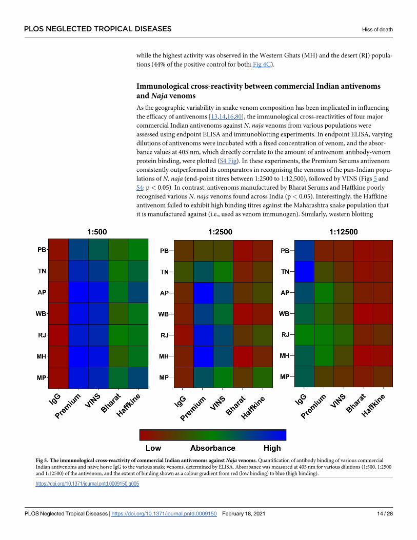
Immunological cross-reactivity between commercial Indian antivenoms
and Naja venoms
As the geographic variability in snake venom composition has been implicated in influencing
the efficacy of antivenoms [13,14,16,80], the immunological cross-reactivities of four major
commercial Indian antivenoms against N. naja venoms from various populations were
assessed using endpoint ELISA and immunoblotting experiments. In endpoint ELISA, varying
dilutions of antivenoms were incubated with a fixed concentration of venom, and the absor-
bance values at 405 nm, which directly correlate to the amount of antivenom antibody-venom
protein binding, were plotted (S4 Fig). In these experiments, the Premium Serums antivenom
consistently outperformed its comparators in recognising the venoms of the pan-Indian popu-
lations of N. naja (end-point titres between 1:2500 to 1:12,500), followed by VINS (Figs 5 and
S4; p< 0.05). In contrast, antivenoms manufactured by Bharat Serums and Haffkine poorly
recognised various N. naja venoms found across India (p< 0.05). Interestingly, the Haffkine
antivenom failed to exhibit high binding titres against the Maharashtra snake population that
it is manufactured against (i.e., used as venom immunogen). Similarly, western blotting
Fig 5. The immunological cross-reactivity of commercial Indian antivenoms against Naja venoms. Quantification of antibody binding of various commercial
Indian antivenoms and naive horse IgG to the various snake venoms, determined by ELISA. Absorbance was measured at 405 nm for various dilutions (1:500, 1:2500
and 1:12500) of the antivenom, and the extent of binding shown as a colour gradient from red (low binding) to blue (high binding).
https://doi.org/10.1371/journal.pntd.0009150.g005
PLOS NEGLECTED TROPICAL DISEASES Hiss of death
PLOS Neglected Tropical Diseases | https://doi.org/10.1371/journal.pntd.0009150 February 18, 2021 14 / 28
Page 15
experiments revealed that several venom components were unrecognised or exhibited low lev-
els of antibody binding when probed with the Bharat Serums and Haffkine antivenoms (S5A
and S5B Fig). Overall, both VINS and Premium Serums antivenoms exhibited increased recog-
nition of venom proteins, with the latter being relatively better than all other tested antivenoms
in terms of both end-point titres and absorbance values (p< 0.05). Incidentally, the naïve
horse IgG exhibited a degree of non-specific cross-reactivity against the largely abundant tox-
ins found in the high (25–50 kDa) and low (<10 kDa) molecular weight ranges (S5A and S5B
Fig). These findings suggest that a degree of non-specific binding occurs between the equine
antibodies and the venom proteins, in line with previous work [14]. It should be noted that the
low molecular weight toxins (e.g., 3FTx) are known to exhibit poor immunogenicity [81–83].
Hence, it is very likely that this further contributes to the lack of low molecular weight toxin
specific antibodies.
Venom potency by median lethal dose (LD50)
Snake venom compositions are predominantly shaped by the ecology and environment.
Resulting compositional differences in venoms, as a result of local adaptations, can signifi-
cantly alter the clinical pathogenesis observed in human snakebite victims. While investigating
the lethal effects of the N. naja venoms sourced from various Indian biogeographic popula-
tions, fascinating observations were made (S3A Table). While the Deccan plateau (MP: 0.22
mg/kg), Gangetic plain (WB: 0.27 mg/kg) and the semi-arid (PB: 0.33 mg/kg) populations of
N. naja venoms were determined to be extremely toxic to mice, the desert population (RJ: 2.53
mg/kg) proved to be dramatically less toxic (Fig 6A). In addition, the venom of one of the
coastal populations (AP: 0.55 mg/kg) was found to exhibit relatively lower venom potencies
(Fig 6A).
Fig 6. Toxicity profiles of N. naja from various biogeographic zones across India, and the neutralisation potencies of commercial Indian antivenom against these
venoms. Murine intravenous median lethal doses (expressed in mg/kg) of various populations of N. naja venoms (A) and the neutralising potencies (expressed in mg/ml)
of the Premium Serums commercial antivenom against these venoms (B). The vertical dotted lines in panel B indicate the marketed neutralising potency (0.60 mg/ml) of
commercial antivenoms against the N. naja venom.
https://doi.org/10.1371/journal.pntd.0009150.g006
PLOS NEGLECTED TROPICAL DISEASES Hiss of death
PLOS Neglected Tropical Diseases | https://doi.org/10.1371/journal.pntd.0009150 February 18, 2021 15 / 28
Page 16
Antivenom efficacy via median effective dose (ED50)
Considering that the Premium Serums antivenom exhibited the highest in vitro venom recog-
nition of the various marketed antivenom products tested in this study (S1B Table), we
selected this antivenom for in vivo venom neutralisation experiments. Despite this best-case
scenario, the results of our preclinical ED50 experiments highlighted poor pan-India venom
neutralisation efficacies of this product, with the estimated neutralising potencies observed
well below that of the marketed claims of neutralisation (0.6 mg/ml for N. naja; S3B Table).
With the exception of N. naja venom from the coastal population in Andhra Pradesh (0.80
mg/ml), the Premium Serums antivenom exhibited extremely low neutralising potencies
against the lethal effects of the venoms of all other biogeographical populations of this species
(0.28 to 0.38 mg/ml; Fig 6B). Alarmingly, this antivenom was found to be completely ineffec-
tive at protecting mice envenomed with 5× LD50 of venom from the desert population (RJ) of
N. naja, as even the highest antivenom doses tested (166.66 μl) failed to protect the experimen-
tal animals from the lethal effects of the venom. However, when the venom challenge dose was
reduced to 3× LD50, a neutralising potency slightly greater than that marketed (0.74 mg/ml),
was observed. It should be noted that the amount of venom injected by individual N. najasnakes can be very large (300 mg on average).
Discussions
Geographic variability in venom complexity and potency is dictated by
differing ecologies and environments
From a biogeographical perspective, India can be divided into ten zones: 1. Himalayas; 2.
Trans-Himalayas; 3. Semi-arid regions; 4. Desert; 5. Western Ghats; 6. Deccan plateau; 7. Gan-
getic plains; 8. Coasts; 9. Northeast India; and 10. Islands [84]. The remarkable adaptability of
N. naja is illustrated by its broad distribution across complex climatic conditions, including
hot and dry semi-arid and arid regions, tropical monsoon forests, hot and humid coastline,
and the fertile Gangetic plains. There are scarce reports of N. naja in northeastern India, albeit
from only the northern parts of West Bengal and southern Assam [85]. However, none of the
Indian ‘big four’ snake species are found in the Trans-Himalayas and the Andaman and Nico-
bar Islands. Despite such biogeographical variation, the influence of distinct ecologies and
environment on the venom composition of N. naja has not been previously investigated. To
address this shortcoming, venom samples from the pan-Indian populations of N. naja were
collected from six of the seven biogeographic zones of India inhabited by this species.
Proteomic characterisation unveiled dramatic differences in the venom compositions of
snakes from distinct biogeographical zones. For example, the venoms of N. naja showed
remarkable differences in relative amounts of 3FTx subtypes. Among the pan-Indian popula-
tions of N. naja, the semiarid (PB) and Gangetic Plain (WB) populations secreted the highest
amounts of N-3FTx in their venom. In contrast, the venoms of the desert (RJ) population
secreted relatively limited amounts of the N-3FTx, while largely being composed of cytotoxic/
cardiotoxic 3FTXs (Fig 3 and S2A–S2C Table). Large amounts of C-3FTxs have also been pre-
viously reported from the venoms of captive snakes sourced from Western India (Rajasthan
and Gujarat) [18]. Similarly, the venom of the Western Ghats (MH) population was previously
reported to be comprised of ~42% N-3FTx [14]. Such considerable differences in the amounts
of neurotoxins were found to significantly influence venom potencies towards mice (Fig 6A
and S3A Table). Populations with large amounts of neurotoxins, such as the semi-arid (PB)
and Gangetic Plain (WB), were characterised by increased venom toxicities (LD50: 0.33 and
0.27 mg/kg, respectively), whereas the Western Ghats (MH) and desert (RJ) populations were
PLOS NEGLECTED TROPICAL DISEASES Hiss of death
PLOS Neglected Tropical Diseases | https://doi.org/10.1371/journal.pntd.0009150 February 18, 2021 16 / 28
Page 17
characterised by relatively lower lethal potencies [LD50: 0.73 and 2.53 mg/kg, respectively;
[14]]. A correlation between the amounts of N-3FTx and venom potency has also been
reported before in the Southeast Asian Naja spp. [86]. Although the prey spectrum of various
Indian populations of N. naja is poorly understood, the extremely low potency of the desert
population towards mice (2.53 mg/kg) could be perceived as indicative of non-mammalian
prey animals chiefly featuring in the diet of this population. However, since the desert popula-
tion (RJ), despite having the least potent venom, caused murine lethality much more rapidly
(15 min) than all other populations (45–60 min), it may suggest the reliance of this population
on cytotoxic/cardiotoxic 3FTxs that constituted a large portion of the venom. Unlike this arid
population, N. naja from semi-arid (PB) regions secreted neurotoxic 3FTxs in abundance and,
hence, required minuscule amounts of the venom to inflict respiratory failure in mice. Thus,
albeit requiring different amounts of venom, both strategies seem to be equally effective in cap-
turing prey in harsh, arid environments. In contrast to the desert population (RJ), the extreme
potency of N. naja in the Deccan plateau region (MP: 0.22 mg/kg), and their ability to inject
large amounts of venom (as high as 413 mg), makes them one of the most medically important
‘big four’ snake populations in the country. While neurotoxins affect the nervous system of
prey animals, C-3FTxs induce cell necrosis and apoptosis by inflicting pores on the phospho-
lipid membrane [87,88]. Not surprisingly, in cell viability assays, the C-3FTx-rich western
Indian populations (desert and the Western Ghats) exhibited the highest haemolytic activity,
while the neurotoxin-rich eastern Indian population (Gangetic Plains) was the least haemo-
toxic (Figs 3 and 4C). Thus, the compositional and biochemical venom variation observed
here has the potential to result in pathological variation in cobra snakebite victims found
across different regions of India.
Venom pathology of N. naja is driven by complex synergistic actions
Snake venom is a concoction of diverse biochemical components that often work synergisti-
cally to facilitate effective prey capture [79,89–91]. Various enzymatic toxins, such as hyaluron-
idase and DNase, are known to function as ‘spreading factors’ [63,92]. Upon envenomation,
the cells of the host are lysed by cytolytic toxins (e.g., C-3FTx and PLA2), resulting in cell death
and the extrusion of nuclear DNA. The released genetic material, in turn, ensnares venom
components into extracellular traps that function as barriers, thereby restricting the venom
from accessing the blood circulation [63,64]. In order to overcome this barrier, N. naja seem-
ingly employs DNase enzymes that catalyse the breakdown of the traps and facilitate the rapid
spread of venom to the other parts of the body. Elapidae and Viperidae snakes employ distinct
strategies for killing their prey, with many elapid snakes secreting venoms enriched with neu-
rotoxins, while most viperid venoms predominantly contain components that cause haemody-
namic alteration, local tissue necrosis, and myotoxicity [93,94]. Therefore, an increase in
DNase activity could confer an evolutionary advantage to elapid snake venoms, as it may
enhance the diffusion of neurotoxic components. In support of this hypothesis, we observed
that the populations with increased amounts of N-3FTxs also exhibited the highest DNase
activities (S1 and S2 Figs).
Biogeographic venom variability negatively impacts upon snakebite
therapy
The polyvalent antivenoms available for the treatment of snakebites in India have been histori-
cally manufactured from the venoms of the south Indian (Tamil Nadu) population of the ‘big
four’ snakes. When the Premium Serums commercial antivenom, which exhibited relatively
increased in vitro venom cross-reactivity in comparison with the other antivenoms under
PLOS NEGLECTED TROPICAL DISEASES Hiss of death
PLOS Neglected Tropical Diseases | https://doi.org/10.1371/journal.pntd.0009150 February 18, 2021 17 / 28
Page 18
investigation, was tested for its in vivo efficacy against venoms from the pan-Indian popula-
tions of N. naja, alarming results were observed (Fig 6B and S3B Table). Among the five inves-
tigated populations of N. naja, only the venom from the coastal region (Andhra Pradesh, the
neighbouring state to Tamil Nadu) was neutralised at a dose comparable to the marketed ther-
apeutic potency (0.80 mg/ml). While the antivenom was able to neutralise the toxic effects of
the highly neurotoxic venoms from the Gangetic Plain (WB), semi-arid region (PB) and the
Deccan plateau (MP), very high doses were required, and thus the neutralising potency was
well below the marketed efficacy (0.28 to 0.38 mg/ml). Even more concerningly, the antivenom
completely failed to neutralise the lethal effects of the less toxic venom sourced from the desert
population (RJ), despite exhibiting a binding efficiency that was comparable to the efficiency
exhibited towards the coastal (Andhra Pradesh) population (Figs 5 and 6B and S3B Table).
Interestingly, similar observations were recently described for the cytotoxin/cardiotoxin-rich
venoms of monocled cobra (N. kaouthia) in northeast India (Arunachal Pradesh), where the
overall venom potency was low, but the tested Premium Serums antivenom completely failed
to neutralise the lethal effects in a murine model of envenomation [14]. These results are indic-
ative of the presence of novel toxin isoforms that are currently unrecognised by the commer-
cial Indian antivenom, which is exclusively produced against the southern population of ‘big
four’ snakes. Ultimately, the in vivo venom neutralisation experiments performed here reveal
disturbing deficiencies of the tested Indian antivenoms against most populations of N. naja.
Despite exhibiting better in vitro binding compared to other commercial antivenoms, Pre-
mium Serums antivenom performed poorly under in vivo conditions. Given the relatively
decreased venom recognition capabilities of the other commercial antivenoms tested in this
study, and the identical strategies of antivenom production that involves sourcing of venom
from a single population, it is highly unlikely that the other antivenoms will effectively neutral-
ise the lethal effects of the distant N. naja populations. Furthermore, this interpretation is sup-
ported by preclinical antivenom efficacy testing (VINS and Bharat antivenoms) on N. najavenoms (population undisclosed) [95]. Thus, it is essential to alter existing antivenom
manufacturing strategies to generate efficacious pan-Indian snakebite treatment.
The road map to pan-India effective antivenoms
In contrast to the rapid acquisition of knowledge relating to the composition and diversifica-
tion of snake venoms, antivenom manufacturing strategies have remained virtually unchanged
over the past century. To improve the plight of India’s million snakebite victims, significant
strategic changes are warranted in both the manufacturing and marketing of commercial
Indian antivenoms.
Immediate solutions: The development of region-specific antivenoms and
the implementation of vital health policy decisions
Commercial Indian antivenoms manufactured against the coastal Tamil Nadu population
(TN) of snakes, which secrete a very distinct venom cocktail in comparison to conspecifics in
other biogeographical regions, lack pan-India efficacy. In addition to providing evidence for
the ineffectiveness against various populations of N. naja in this study, we have previously
reported the inefficacy of the marketed antivenoms against the common krait (B. caeruleus)from Punjab [14]. This highlights the inability of the marketed antivenoms in neutralising ven-
oms of two of the ‘big four’ snake species from the northern Indian region (Fig 7). This unfor-
tunate outcome is a result of discounting the remarkable inter- and intra-specific venom
diversity in snakes and producing a single antivenom for use across the large Indian subconti-
nent. Given the considerable biotic and abiotic diversity in India, and the remarkable
PLOS NEGLECTED TROPICAL DISEASES Hiss of death
PLOS Neglected Tropical Diseases | https://doi.org/10.1371/journal.pntd.0009150 February 18, 2021 18 / 28
Page 19
geographic venom variability among snakes, the conventional antivenom is doomed to failure
in regions with disparate populations of ‘big four’ and/or other distinct venomous snake spe-
cies. An immediate solution to this problem could be the identification of medically important
snakes by regions, i.e., consideration of both the ‘big four’ and the ‘neglected many’ (medically
important yet neglected lineages of snakes), and the inclusion of their venoms in the immuni-
sation mixture for formulating regionally-effective antivenoms. Based on the outcomes of this
study, research on medically important yet neglected snakes [14,96], and the geographical
Fig 7. The negative impact of biogeographic venom variability on Indian snakebite therapy. This figure depicts the repercussions of biogeographic venom variability
on snakebite treatment in India. Antivenom vials indicate the relative differences in neutralisation potencies against the geographically distinct populations of N. naja(yellow), and B. caeruleus (purple) in comparison to the source population in southern India, where the red dotted line on the vials represents the marketed neutralising
potency of commercial Indian antivenoms. The intensity of purple clouds on the map is indicative of the estimated standardised snakebite death rates per million reported
by Suraweera et al. 2020 [6], where the brighter regions represent the major hotspots. Geographical locales are defined by the box in the top right. The map of India shown
here was prepared with QGIS 3.8 [43].
https://doi.org/10.1371/journal.pntd.0009150.g007
PLOS NEGLECTED TROPICAL DISEASES Hiss of death
PLOS Neglected Tropical Diseases | https://doi.org/10.1371/journal.pntd.0009150 February 18, 2021 19 / 28
Page 20
distribution of ‘big four’ snakes, several Indian regions can be identified that would benefit
from regional antivenoms: 1. North(west) India; 2. East India; 3. Northeast India; 4. Andaman
and Nicobar islands; 5. Central India; and 6. South India.
Since Indian antivenoms have never undergone clinical validation through formal clinical
trials, robust data on their efficacy and safety is currently unavailable. Given the potential for
significant batch-to-batch variation and treatment failure due to venom variation, stringent
evaluation of the preclinical efficacy of antivenoms, ideally by an independent external labora-
tory or at the very least the publication of manufacturer-generated data for independent assess-
ment, should be mandatory prior to marketing [14,97,98]. Moreover, the license to sell
commercial antivenoms in various Indian states is currently based on a tender system. Instead,
licensure should be strictly based on the outcomes of such rigorous preclinical evaluation. In
addition, the procurement and qualification guidelines for venoms used for immunisation
during the manufacturing process should take into account the influence of various ecological
and environmental factors on venom variability. Unfortunately, these factors are currently
being ignored during the commercial manufacture of Indian antivenoms. For example, ven-
oms that exhibit either very high or low potencies are generally not used in the immunisation
process by many Indian antivenom manufacturers (KS, personal communication with manu-
facturers). This could explain the complete lack of neutralisation against the desert population
of N. naja that exhibited very low potency in the murine model. Overall, in the absence of
broadly neutralising next-generation antivenoms, these measures can help improve the effi-
cacy of snakebite therapies in the country.
The long-term solution: Innovation of broadly neutralising recombinant
antivenoms
Immunisation of animals with crude ‘whole’ venoms that could potentially contain snakebite-
irrelevant antigens, e.g., bacteria, viruses and/or other impurities, along with the environmen-
tal antigens that the immunised animals get exposed to over their lifetime, increases the pro-
portion of non-toxin-specific redundant antibodies in the finished product. In addition to
toxins that result in severe pathophysiology in humans, snake venom cocktails also contain
venom components that target non-mammalian prey/predatory animals. Therefore, using
crude venoms for immunisation results in the inclusion of antibodies against such medically
unimportant toxins, and significantly lowers the proportion of therapeutically important IgGs
in the marketed product. This, in turn, significantly increases the number of antivenom vials
required to effect cure (typically >20 in India). Therefore, in addition to their inability to
counter toxic effects of pan-Indian populations of snakes, conventional serum therapy is
marred by other inadequacies, including dose inefficacy, inconsistent batch effectiveness, and
the risk of inducing fatal anaphylaxis via the intravenous delivery of animal IgG. Several
immunochromatographic techniques, such as immunoaffinity purification, which involves the
down-selection of antibodies using antigenic baits [99], could also help in improving the con-
centrations of therapeutically relevant antibodies in the marketed product.
Although regionally-effective antivenoms could serve as an interim solution to address
local variations in snake venom and species diversity, they would still suffer from the afore-
mentioned limitations. Hence, the discovery of broadly neutralising recombinant antivenom
offers a long-term solution for treating snakebites in India. Recombinant antibodies could be
developed by various approaches and in different formats (e.g., monoclonal, oligoclonal, intact
IgG, nanobodies, etc.), and could be human-derived or humanised, and engineered to specifi-
cally target clinically important toxins detected across distinct snake populations and species
[97,98]. Thus, recombinant therapy has the potential to deliver many advantages over
PLOS NEGLECTED TROPICAL DISEASES Hiss of death
PLOS Neglected Tropical Diseases | https://doi.org/10.1371/journal.pntd.0009150 February 18, 2021 20 / 28
Page 21
conventional antivenom therapy, including high dose efficacy, pan-Indian efficacy, and
improved safety profiles. The cost of production is the only current limitation of recombinant
therapy as this entirely depends on the number of neutralising antibodies in the commercial
antivenom concoction. However, this could be overcome by discovering and engineering
broadly effective/paraspecific antibodies. The recombinant expression of such broadly neutral-
ising antibodies should therefore be strongly pursued as long-term replacements of conven-
tional antivenoms to enable rural Indian communities to access safe and efficacious life-saving
snakebite therapies.
Limitations of the study
While a considerable amount of PLA2 (20%) was detected by tandem mass spectrometry of the
N. naja venom from the desert population (RJ), very limited differences were noted in phos-
pholipase activities of populations from distinct biogeographic regions. This could, indeed,
result from an overestimation of PLA2 in the venom of the desert (RJ) population or an under-
estimation of this toxin superfamily in other populations. It should be noted, however, that
these estimates are in line with the literature, where a similar abundance of PLA2 was reported
for Naja venoms sourced from Rajasthan and Gujarat [18]. Moreover, as the prominent role
of neurotoxic and cytotoxic 3FTxs in Naja envenomation has been very well-established, the
differences in the lowly abundant PLA2 toxins are unlikely to affect the major interpretations
and conclusions of this study. Consistently, SDS-PAGE analysis clearly shows the abundance
of 3FTxs in the molecular weight range of 6–9 kDa in all Naja venoms [46]. Further, while our
analyses recovered LAAO only from the desert population (RJ), acetylcholinesterase was not
detected in any of the populations subjected to mass spectrometry. The inability to detect such
minor components in Naja venoms is mostly due to the lack of well-characterised toxin
sequences from the medically important Indian snakes in the public repositories, highlighting
the importance of conducting venom gland transcriptomics studies of Indian snakes.
It should also be noted that given the limited approvals from the authorities and the logistic
and financial constraints associated with sampling, venom samples could not be collected
from multiple individuals of certain populations. For others, while venoms were pooled from
multiple individuals and were subjected to preliminary quality screening, we selected individ-
ual venoms for assessing the influence of biogeography on snake venom composition and
potency. Nonetheless, the results of our proteomic analyses (SDS-PAGE, HPLC and mass
spectrometry) are consistent with the literature and agree with the reported overall venom
compositions. Considering the possibility of individual variability, we do not claim that these
results necessarily represent an entire population/region. Future investigations incorporating
much larger sampling efforts, not just by collecting the venoms of four to five individuals from
the same location as reported in the literature, but by sampling many snakes across multiple
regions in a given biogeographic zone. Such studies may reveal further intrapopulation differ-
ences in venom compositions and activities, and the implications of such variation on the
effectiveness of antivenoms.
Conclusion
In conclusion, an array of in vitro and in vivo experiments performed in this study reveals sig-
nificant intraspecific differences in the venom proteomic composition and toxicities of N. najavenoms across six distinct biogeographical regions in India. Although in vitro antivenom
screening experiments revealed increased venom binding by the Premium Serums antivenom,
in comparison to those manufactured by VINS, Bharat Serums and Haffkine, in vivo anti-
venom neutralising experiments revealed alarming efficacy shortcomings of India’s snakebite
PLOS NEGLECTED TROPICAL DISEASES Hiss of death
PLOS Neglected Tropical Diseases | https://doi.org/10.1371/journal.pntd.0009150 February 18, 2021 21 / 28
Page 22
therapies. Antivenom was found to be incapable of effectively neutralising the venoms of most
N. naja populations (four out of five populations), including failing completely to prevent
against venom-induced lethality caused by the desert (RJ) population. These data highlight the
complexity and importance of understanding intra-specific venom variation and the impact
that it can have on snakebite treatment. Our findings emphasise the pressing need to develop
highly specific and dose-efficacious antivenoms for the treatment of snakebites in the Indian
subcontinent. While in the long term this can likely be achieved via the application of innova-
tive recombinant antibody technologies, in the interim, we strongly advocate for the produc-
tion of regionally effective antivenoms that can circumvent medically important inter- and
intra-specific differences in snake venoms found across the different biogeographical regions
of India.
Supporting information
S1 Fig. Biochemical variation in the pan-Indian populations of N. naja venoms.(PDF)
S2 Fig. Agarose gel electrophoresis showing DNase activities of N. naja venoms.
(PDF)
S3 Fig. Fibrinogenolytic activities of N. naja venoms from distinct locations across India.
(PDF)
S4 Fig. Immunological cross-reactivity between commercial Indian antivenoms and N.
naja venoms.
(PDF)
S5 Fig. A Western blotting of commercial Indian antivenoms against the venoms of pan-
Indian populations of N. naja. B Heatmap of venom recognition potential of commercial
Indian antivenoms against the venoms of pan-Indian populations of N. naja.
(PDF)
S1 Table. A Details of the N. naja venom samples tested. B Details of the investigated com-
mercial Indian antivenoms.
(PDF)
S2 Table. A-C Toxin compositions of N. naja venoms from various populations across India.
(PDF)
S3 Table. A The median lethal dose of the pan-Indian populations of N. naja. B Neutralising
potencies of Premium Serums antivenom against the pan-Indian populations of N. naja.
(PDF)
S1 Data. Results of mass spectrometry analyses for semi-arid (Punjab), Gangetic plain
(West Bengal) and desert population (Rajasthan) of Naja naja venoms (S1 Data.zip).
(ZIP)
Acknowledgments
The authors are thankful to Ashwin Iyer, Navneet Kaur, Aratrika Ray, Aditi Singh, and Bharat
Ahuja for assistance with biochemical experiments. Authors are also thankful to the following
State Forest Departments for the kind support and permits for venom collection: Punjab,
Tamil Nadu, Andhra Pradesh, West Bengal, Rajasthan, Maharashtra, and Madhya Pradesh.
For the invaluable assistance in the collection of samples, authors are thankful to Ajay Kartik
PLOS NEGLECTED TROPICAL DISEASES Hiss of death
PLOS Neglected Tropical Diseases | https://doi.org/10.1371/journal.pntd.0009150 February 18, 2021 22 / 28
Page 23
(MCBT), Sumanth Madhav (Humane Society International), Vivek Sharma, Joy Gardner, All-
win Jesudasan (MCBT), and P. Gowri Shankar (North Orissa University). For contributing
snake photographs in Fig 1 of the manuscript, the authors are thankful to Ajay Kartik (TN),
Chaitanya Shukla (MH), Gunjan Pancholi (RJ), and Vivek Sharma (all other populations).
Author Contributions
Conceptualization: Kartik Sunagar.
Data curation: R. R. Senji Laxme, Saurabh Attarde, Suyog Khochare.
Formal analysis: R. R. Senji Laxme, Saurabh Attarde, Suyog Khochare, Vivek Suranse, Kartik
Sunagar.
Funding acquisition: Kartik Sunagar.
Investigation: R. R. Senji Laxme, Saurabh Attarde, Suyog Khochare, Vivek Suranse, Kartik
Sunagar.
Methodology: R. R. Senji Laxme, Saurabh Attarde, Suyog Khochare, Kartik Sunagar.
Project administration: Kartik Sunagar.
Resources: Gerard Martin, Romulus Whitaker, Kartik Sunagar.
Supervision: Kartik Sunagar.
Visualization: Kartik Sunagar.
Writing – original draft: R. R. Senji Laxme, Kartik Sunagar.
Writing – review & editing: R. R. Senji Laxme, Saurabh Attarde, Suyog Khochare, Vivek Sur-
anse, Nicholas R. Casewell, Kartik Sunagar.
References1. Fry BG, Roelants K, Champagne DE, Scheib H, Tyndall JD, King GF, et al. The toxicogenomic multi-
verse: convergent recruitment of proteins into animal venoms. Annu Rev Genomics Hum Genet. 2009;
10:483–511. https://doi.org/10.1146/annurev.genom.9.081307.164356 PMID: 19640225
2. Casewell NR, Wuster W, Vonk FJ, Harrison RA, Fry BG. Complex cocktails: the evolutionary novelty of
venoms. Trends Ecol Evol. 2013; 28(4):219–29. https://doi.org/10.1016/j.tree.2012.10.020 PMID:
23219381
3. Sunagar K, Casewell N, Varma S, Kolla R, Antunes A, Moran Y. Deadly innovations: unraveling the
molecular evolution of animal venoms. Venom Genomics and Proteomics; Springer: Dordrecht, The
Netherlands. 2014:1–23.
4. Casewell NR, Jackson TNW, Laustsen AH, Sunagar K. Causes and Consequences of Snake Venom
Variation. Trends in Pharmacological Sciences. 2020; 41(8):570–81 https://doi.org/10.1016/j.tips.2020.
05.006 PMID: 32564899
5. Whitaker R, Martin G. Diversity and Distribution of Medically Important Snakes of India. In: Gopalakrish-
nakone P, Faiz A, Fernando R, Gnanathasan CA, Habib AG, Yang CC, editor. Clinical Toxinology in
Asia Pacific and Africa. Dordrecht: Springer Netherlands; 2015. p. 115–36.
6. Suraweera W, Warrell D, Whitaker R, Menon G, Rodrigues R, Fu SH, et al. Trends in snakebite deaths
in India from 2000 to 2019 in a nationally representative mortality study. Elife. 2020; 9.
7. Mohapatra B, Warrell DA, Suraweera W, Bhatia P, Dhingra N, Jotkar RM, et al. Snakebite mortality in
India: a nationally representative mortality survey. PLoS Negl Trop Dis. 2011; 5(4):e1018. https://doi.
org/10.1371/journal.pntd.0001018 PMID: 21532748
8. Daltry JC, Wuster W, Thorpe RS. Diet and snake venom evolution. Nature. 1996; 379(6565):537–40.
https://doi.org/10.1038/379537a0 PMID: 8596631
9. Holding ML, Biardi JE, Gibbs HL. Coevolution of venom function and venom resistance in a rattlesnake
predator and its squirrel prey. Proc Biol Sci. 2016; 283(1829).
PLOS NEGLECTED TROPICAL DISEASES Hiss of death
PLOS Neglected Tropical Diseases | https://doi.org/10.1371/journal.pntd.0009150 February 18, 2021 23 / 28
Page 24
10. Currier RB, Harrison RA, Rowley PD, Laing GD, Wagstaff SC. Intra-specific variation in venom of the
African Puff Adder (Bitis arietans): Differential expression and activity of snake venom metalloprotei-
nases (SVMPs). Toxicon. 2010; 55(4):864–73. https://doi.org/10.1016/j.toxicon.2009.12.009 PMID:
20026155
11. Rokyta DR, Margres MJ, Ward MJ, Sanchez EE. The genetics of venom ontogeny in the eastern dia-
mondback rattlesnake (Crotalus adamanteus). PeerJ. 2017; 5:e3249. https://doi.org/10.7717/peerj.
3249 PMID: 28462047
12. Zancolli G, Calvete JJ, Cardwell MD, Greene HW, Hayes WK, Hegarty MJ, et al. When one phenotype
is not enough: divergent evolutionary trajectories govern venom variation in a widespread rattlesnake
species. Proc Biol Sci. 2019; 286(1898):20182735. https://doi.org/10.1098/rspb.2018.2735 PMID:
30862287
13. Sunagar K, Undheim EA, Scheib H, Gren EC, Cochran C, Person CE, et al. Intraspecific venom varia-
tion in the medically significant Southern Pacific Rattlesnake (Crotalus oreganus helleri): biodiscovery,
clinical and evolutionary implications. J Proteomics. 2014; 99:68–83. https://doi.org/10.1016/j.jprot.
2014.01.013 PMID: 24463169
14. Senji Laxme RR, Khochare S, de Souza HF, Ahuja B, Suranse V, Martin G, et al. Beyond the ’big four’:
Venom profiling of the medically important yet neglected Indian snakes reveals disturbing antivenom
deficiencies. PLoS Negl Trop Dis. 2019; 13(12):e0007899. https://doi.org/10.1371/journal.pntd.
0007899 PMID: 31805055
15. Warrell DA, Gutierrez JM, Calvete JJ, Williams D. New approaches & technologies of venomics to meet
the challenge of human envenoming by snakebites in India. Indian J Med Res. 2013; 138:38–59. PMID:
24056555
16. Shashidharamurthy R, Kemparaju K. Region-specific neutralization of Indian cobra (Naja naja) venom
by polyclonal antibody raised against the eastern regional venom: A comparative study of the venoms
from three different geographical distributions. Int Immunopharmacol. 2007; 7(1):61–9. https://doi.org/
10.1016/j.intimp.2006.08.014 PMID: 17161818
17. Dutta S, Chanda A, Kalita B, Islam T, Patra A, Mukherjee AK. Proteomic analysis to unravel the com-
plex venom proteome of eastern India Naja naja: Correlation of venom composition with its biochemical
and pharmacological properties. J Proteomics. 2017; 156:29–39. https://doi.org/10.1016/j.jprot.2016.
12.018 PMID: 28062377
18. Sintiprungrat K, Watcharatanyatip K, Senevirathne WD, Chaisuriya P, Chokchaichamnankit D, Srisom-
sap C, et al. A comparative study of venomics of Naja naja from India and Sri Lanka, clinical manifesta-
tions and antivenomics of an Indian polyspecific antivenom. J Proteomics. 2016; 132:131–43. https://
doi.org/10.1016/j.jprot.2015.10.007 PMID: 26506536
19. Maduwage K, Silva A, O’Leary MA, Hodgson WC, Isbister GK. Efficacy of Indian polyvalent snake anti-
venoms against Sri Lankan snake venoms: lethality studies or clinically focussed in vitro studies. Sci
Rep. 2016; 6:26778. https://doi.org/10.1038/srep26778 PMID: 27231196
20. Wong KY, Tan CH, Tan NH. Venom and Purified Toxins of the Spectacled Cobra (Naja naja) from Paki-
stan: Insights into Toxicity and Antivenom Neutralization. Am J Trop Med Hyg. 2016; 94(6):1392–9.
https://doi.org/10.4269/ajtmh.15-0871 PMID: 27022154
21. Mukherjee AK, Kalita B, Mackessy SP. A proteomic analysis of Pakistan Daboia russelii russelii venom
and assessment of potency of Indian polyvalent and monovalent antivenom. J Proteomics. 2016;
144:73–86. https://doi.org/10.1016/j.jprot.2016.06.001 PMID: 27265321
22. Chanda A, Kalita B, Patra A, Senevirathne W, Mukherjee AK. Proteomic analysis and antivenomics
study of Western India Naja naja venom: correlation between venom composition and clinical manifes-
tations of cobra bite in this region. Expert Rev Proteomics. 2019; 16(2):171–84. https://doi.org/10.1080/
14789450.2019.1559735 PMID: 30556786
23. Chanda A, Mukherjee AK. Quantitative proteomics to reveal the composition of Southern India specta-
cled cobra (Naja naja) venom and its immunological cross-reactivity towards commercial antivenom. Int
J Biol Macromol. 2020; 160:224–32. https://doi.org/10.1016/j.ijbiomac.2020.05.106 PMID: 32439440
24. Chanda A, Patra A, Kalita B, Mukherjee AK. Proteomics analysis to compare the venom composition
between Naja naja and Naja kaouthia from the same geographical location of eastern India: Correlation
with pathophysiology of envenomation and immunological cross-reactivity towards commercial polyan-
tivenom. Expert Rev Proteomics. 2018; 15(11):949–61. https://doi.org/10.1080/14789450.2018.
1538799 PMID: 30345852
25. Choudhury M, McCleary RJR, Kesherwani M, Kini RM, Velmurugan D. Comparison of proteomic pro-
files of the venoms of two of the ’Big Four’ snakes of India, the Indian cobra (Naja naja) and the common
krait (Bungarus caeruleus), and analyses of their toxins. Toxicon. 2017; 135:33–42. https://doi.org/10.
1016/j.toxicon.2017.06.005 PMID: 28602829
PLOS NEGLECTED TROPICAL DISEASES Hiss of death
PLOS Neglected Tropical Diseases | https://doi.org/10.1371/journal.pntd.0009150 February 18, 2021 24 / 28
Page 25
26. Bradford MM. A rapid and sensitive method for the quantitation of microgram quantities of protein utiliz-
ing the principle of protein-dye binding. Anal Biochem. 1976; 72:248–54. https://doi.org/10.1006/abio.
1976.9999 PMID: 942051
27. Smith BJ. SDS Polyacrylamide Gel Electrophoresis of Proteins. Methods Mol Biol. 1984; 1:41–55.
https://doi.org/10.1385/0-89603-062-8:41 PMID: 20512673
28. Lomonte B, Calvete JJ. Strategies in ’snake venomics’ aiming at an integrative view of compositional,
functional, and immunological characteristics of venoms. J Venom Anim Toxins Incl Trop Dis. 2017;
23:26. https://doi.org/10.1186/s40409-017-0117-8 PMID: 28465677
29. Vizcaino JA, Deutsch EW, Wang R, Csordas A, Reisinger F, Rios D, et al. ProteomeXchange provides
globally coordinated proteomics data submission and dissemination. Nat Biotechnol. 2014; 32(3):223–
6. https://doi.org/10.1038/nbt.2839 PMID: 24727771
30. PEAKS Studio Technical Notes. Peptide Feature Area: Bioinformatics Solution Inc.; [Available from:
https://www.bioinfor.com/tutorial-peptide-feature-area/].
31. Tan NH, Wong KY, Tan CH. Venomics of Naja sputatrix, the Javan spitting cobra: A short neurotoxin-
driven venom needing improved antivenom neutralization. J Proteomics. 2017; 157:18–32. https://doi.
org/10.1016/j.jprot.2017.01.018 PMID: 28159706
32. Marinetti GV. The action of phospholipase A on lipoproteins. Biochim Biophys Acta. 1965; 98(3):554–
65. https://doi.org/10.1016/0005-2760(65)90152-9 PMID: 5891200
33. Joubert FJ, Taljaard N. Purification, some properties and amino-acid sequences of two phospholipases
A (CM-II and CM-III) from Naja naja kaouthia venom. Eur J Biochem. 1980; 112(3):493–9. https://doi.
org/10.1111/j.1432-1033.1980.tb06112.x PMID: 7460933
34. Chowdhury MA, Miyoshi S, Shinoda S. Purification and characterization of a protease produced by Vib-
rio mimicus. Infect Immun. 1990; 58(12):4159–62. https://doi.org/10.1128/IAI.58.12.4159-4162.1990
PMID: 2254038
35. Kishimoto M, Takahashi T. A spectrophotometric microplate assay for L-amino acid oxidase. Anal Bio-
chem. 2001; 298(1):136–9. https://doi.org/10.1006/abio.2001.5381 PMID: 11673909
36. Gerceker D, Karasartova D, Elyurek E, Barkar S, Kiyan M, Ozsan TM, et al. A new, simple, rapid test for
detection of DNase activity of microorganisms: DNase Tube test. J Gen Appl Microbiol. 2009; 55
(4):291–4. https://doi.org/10.2323/jgam.55.291 PMID: 19700923
37. Teng CM, Ouyang C, Lin SC. Species difference in the fibrinogenolytic effects of alpha- and beta-fibri-
nogenases from Trimeresurus mucrosquamatus snake venom. Toxicon. 1985; 23(5):777–82. https://
doi.org/10.1016/0041-0101(85)90008-x PMID: 4089873
38. Maisano M, Trapani MR, Parrino V, Parisi MG, Cappello T, D’Agata A, et al. Haemolytic activity and
characterization of nematocyst venom from Pelagia noctiluca (Cnidaria: Scyphozoa). Italian Journal of
Zoology. 2013; 80(2):168–76.
39. Casewell NR, Cook DA, Wagstaff SC, Nasidi A, Durfa N, Wuster W, et al. Pre-clinical assays predict
pan-African Echis viper efficacy for a species-specific antivenom. PLoS Negl Trop Dis. 2010; 4(10):
e851. https://doi.org/10.1371/journal.pntd.0000851 PMID: 21049058
40. World Health Organisation (WHO). World Health Organisation guidelines for the production, control and
regulation of snake antivenom immunoglobulins. World Health Organisation; 2018.
41. Finey D. Probit Analysis 3rd ed. London: Cambridge University Press; 1971.
42. Araujo HP, Bourguignon SC, Boller MA, Dias AA, Lucas EP, Santos IC, et al. Potency evaluation of anti-
venoms in Brazil: the national control laboratory experience between 2000 and 2006. Toxicon. 2008; 51
(4):502–14. https://doi.org/10.1016/j.toxicon.2007.11.002 PMID: 18155119
43. QGIS Development Team (2019). QGIS Geographic Information System. Open Source Geospatial
Foundation Project. http://qgis.osgeo.org.
44. Kini RM, Doley R. Structure, function and evolution of three-finger toxins: mini proteins with multiple tar-
gets. Toxicon. 2010; 56(6):855–67. https://doi.org/10.1016/j.toxicon.2010.07.010 PMID: 20670641
45. Sunagar K, Jackson TN, Undheim EA, Ali SA, Antunes A, Fry BG. Three-fingered RAVERs: Rapid
Accumulation of Variations in Exposed Residues of snake venom toxins. Toxins (Basel). 2013; 5
(11):2172–208. https://doi.org/10.3390/toxins5112172 PMID: 24253238
46. Utkin YN. Three-finger toxins, a deadly weapon of elapid venom—milestones of discovery. Toxicon.
2013; 62:50–5. https://doi.org/10.1016/j.toxicon.2012.09.007 PMID: 23000250
47. de Weille JR, Schweitz H, Maes P, Tartar A, Lazdunski M. Calciseptine, a peptide isolated from black
mamba venom, is a specific blocker of the L-type calcium channel. Proc Natl Acad Sci U S A. 1991; 88
(6):2437–40. https://doi.org/10.1073/pnas.88.6.2437 PMID: 1848702
48. Konshina AG, Dubovskii PV, Efremov RG. Structure and dynamics of cardiotoxins. Curr Protein Pept
Sci. 2012; 13(6):570–84. https://doi.org/10.2174/138920312803582960 PMID: 23004359
PLOS NEGLECTED TROPICAL DISEASES Hiss of death
PLOS Neglected Tropical Diseases | https://doi.org/10.1371/journal.pntd.0009150 February 18, 2021 25 / 28
Page 26
49. Pawlak J, Mackessy SP, Sixberry NM, Stura EA, Le Du MH, Menez R, et al. Irditoxin, a novel covalently
linked heterodimeric three-finger toxin with high taxon-specific neurotoxicity. FASEB J. 2009; 23
(2):534–45. https://doi.org/10.1096/fj.08-113555 PMID: 18952712
50. Chanda C, Sarkar A, Sistla S, Chakrabarty D. Anti-platelet activity of a three-finger toxin (3FTx) from
Indian monocled cobra (Naja kaouthia) venom. Biochem Biophys Res Commun. 2013; 441(3):550–4.
https://doi.org/10.1016/j.bbrc.2013.10.125 PMID: 24183721
51. Kochva E. Evolution and secretion of venom and its antidotes in snakes. Period Biol. 1978; 80 Suppl
1:11–23.
52. Tasoulis T, Isbister GK. A Review and Database of Snake Venom Proteomes. Toxins (Basel). 2017; 9
(9):290. https://doi.org/10.3390/toxins9090290 PMID: 28927001
53. Kini RM. Excitement ahead: structure, function and mechanism of snake venom phospholipase A2
enzymes. Toxicon. 2003; 42(8):827–40. https://doi.org/10.1016/j.toxicon.2003.11.002 PMID:
15019485
54. Kumar MS, Girish KS, Vishwanath BS, Kemparaju K. The metalloprotease, NN-PF3 from Naja naja
venom inhibits platelet aggregation primarily by affecting α2β1 integrin. Ann Hematol. 2011; 90(5):569–
77. https://doi.org/10.1007/s00277-010-1103-1 PMID: 20957364
55. Kalogeropoulos K, Treschow AF, Escalante T, Rucavado A, Gutierrez JM, Laustsen AH and Workman
CT. Protease Activity Profiling of Snake Venoms Using High-Throughput Peptide Screening. Toxins
(Basel). 2019; 11(3). https://doi.org/10.3390/toxins11030170 PMID: 30893860
56. Pirkle H, Stocker K. Thrombin-like enzymes from snake venoms: an inventory. For the Subcommittee
on Nomenclature of Exogenous Hemostatic Factors of the Scientific and Standardization Committee of
the International Society on Thrombosis and Haemostasis. Thromb Haemost. 1991; 65(4):444–50.
PMID: 2057929
57. Serrano SM, Maroun RC. Snake venom serine proteinases: sequence homology vs. substrate specific-
ity, a paradox to be solved. Toxicon. 2005; 45(8):1115–32. https://doi.org/10.1016/j.toxicon.2005.02.
020 PMID: 15922778
58. Matsui T, Fujimura Y, Titani K. Snake venom proteases affecting hemostasis and thrombosis. Biochim
Biophys Acta. 2000; 1477(1–2):146–56. https://doi.org/10.1016/s0167-4838(99)00268-x PMID:
10708855
59. Ande SR, Kommoju PR, Draxl S, Murkovic M, Macheroux P, Ghisla S, et al. Mechanisms of cell death
induction by L-amino acid oxidase, a major component of ophidian venom. Apoptosis. 2006; 11
(8):1439–51. https://doi.org/10.1007/s10495-006-7959-9 PMID: 16770529
60. Costa TR, Burin SM, Menaldo DL, de Castro FA, Sampaio SV. Snake venom L-amino acid oxidases:
an overview on their antitumor effects. J Venom Anim Toxins Incl Trop Dis. 2014; 20:23. https://doi.org/
10.1186/1678-9199-20-23 PMID: 24940304
61. Paloschi MV, Pontes AS, Soares AM, Zuliani JP. An Update on Potential Molecular Mechanisms
Underlying the Actions of Snake Venom L-amino Acid Oxidases (LAAOs). Curr Med Chem. 2018; 25
(21):2520–30. https://doi.org/10.2174/0929867324666171109114125 PMID: 29119915
62. Swethakumar B, NaveenKumar SK, Girish KS, Kemparaju K. The action of Echis carinatus and Naja
naja venoms on human neutrophils; an emphasis on NETosis. Biochim Biophys Acta Gen Subj.
2020;1864(6):129561. https://doi.org/10.1016/j.bbagen.2020.129561 PMID: 32068016
63. Katkar GD, Sundaram MS, NaveenKumar SK, Swethakumar B, Sharma RD, Paul M, et al. NETosis
and lack of DNase activity are key factors in Echis carinatus venom-induced tissue destruction. Nat
Commun. 2016; 7:11361. https://doi.org/10.1038/ncomms11361 PMID: 27093631
64. Stackowicz J, Balbino B, Todorova B, Godon O, Iannascoli B, Jonsson F, et al. Evidence that neutro-
phils do not promote Echis carinatus venom-induced tissue destruction. Nature Communications. 2018;
9(1):1–3. https://doi.org/10.1038/s41467-017-02088-w PMID: 29317637
65. Yamazaki Y, Morita T. Snake venom components affecting blood coagulation and the vascular system:
structural similarities and marked diversity. Curr Pharm Des. 2007; 13(28):2872–86. https://doi.org/10.
2174/138161207782023775 PMID: 17979732
66. Sanchez EF, Flores-Ortiz RJ, Alvarenga VG, Eble JA. Direct Fibrinolytic Snake Venom Metalloprotei-
nases Affecting Hemostasis: Structural, Biochemical Features and Therapeutic Potential. Toxins
(Basel). 2017; 9(12):392. https://doi.org/10.3390/toxins9120392 PMID: 29206190
67. Du XY, Sim DS, Lee WH, Zhang Y. Blood cells as targets of snake toxins. Blood Cells Mol Dis. 2006; 36
(3):414–21. https://doi.org/10.1016/j.bcmd.2006.03.001 PMID: 16631395
68. Bittenbinder MA, Zdenek CN, Op den Brouw B, Youngman NJ, Dobson JS, Naude A, Vonk FJ, Fry BG.
Coagulotoxic Cobras: Clinical Implications of Strong Anticoagulant Actions of African Spitting Naja Ven-
oms That Are Not Neutralised by Antivenom but Are by LY315920 (Varespladib). Toxins (Basel). 2018;
10(12):516.
PLOS NEGLECTED TROPICAL DISEASES Hiss of death
PLOS Neglected Tropical Diseases | https://doi.org/10.1371/journal.pntd.0009150 February 18, 2021 26 / 28
Page 27
69. Corin RE, Viskatis LJ, Vidal JC, Etcheverry MA. Cytotoxicity of crotoxin on murine erythroleukemia cells
in vitro. Invest New Drugs. 1993; 11(1):11–5. https://doi.org/10.1007/BF00873905 PMID: 8349431
70. Mora-Obando D, Diaz C, Angulo Y, Gutierrez JM, Lomonte B. Role of enzymatic activity in muscle dam-
age and cytotoxicity induced by Bothrops asper Asp49 phospholipase A2 myotoxins: are there addi-
tional effector mechanisms involved? PeerJ. 2014; 2:e569. https://doi.org/10.7717/peerj.569 PMID:
25276503
71. Osipov AV, Filkin SY, Makarova YV, Tsetlin VI, Utkin YN. A new type of thrombin inhibitor, noncytotoxic
phospholipase A2, from the Naja haje cobra venom. Toxicon. 2010; 55(2–3):186–94. https://doi.org/10.
1016/j.toxicon.2009.07.011 PMID: 19622365
72. Tsai IH, Lu PJ, Wang YM, Ho CL, Liaw LL. Molecular cloning and characterization of a neurotoxic phos-
pholipase A2 from the venom of Taiwan habu (Trimeresurus mucrosquamatus). Biochem J. 1995; 311
(Pt 3):895–900.
73. Lu Q, Clemetson JM, Clemetson KJ. Snake venoms and hemostasis. Journal of Thrombosis and Hae-
mostasis. 2005; 3(8):1791–9. https://doi.org/10.1111/j.1538-7836.2005.01358.x PMID: 16102046
74. Santhosh MS, Sundaram MS, Sunitha K, Kemparaju K, Girish KS. Viper venom-induced oxidative
stress and activation of inflammatory cytokines: a therapeutic approach for overlooked issues of snake-
bite management. Inflamm Res. 2013; 62(7):721–31. https://doi.org/10.1007/s00011-013-0627-y
PMID: 23657249
75. Sebastin Santhosh M, Hemshekhar M, Thushara RM, Devaraja S, Kemparaju K, Girish KS. Vipera rus-
selli venom-induced oxidative stress and hematological alterations: amelioration by crocin a dietary col-
orant. Cell Biochem Funct. 2013; 31(1):41–50. https://doi.org/10.1002/cbf.2858 PMID: 22893269
76. e Castro VO, Vernon LP. Hemolytic activity of thionin from Pyrularia pubera nuts and snake venom tox-
ins of Naja naja species: Pyrularia thionin and snake venom cardiotoxin compete for the same mem-
brane site. Toxicon. 1989; 27(5):511–7. https://doi.org/10.1016/0041-0101(89)90112-8 PMID: 2749751
77. Condrea E. Hemolytic effects of snake venoms. Snake venoms: Springer; 1979. p. 448–79. https://doi.
org/10.1016/0041-0101(79)90251-4 PMID: 419535
78. Bonta I, van Dijk M, Noordhoek J, Vincent J. Enhancement of the cobra venom direct lytic factor by
prostaglandins and related synergistic phenomena on pulmonary microvascular events: Springer;
1976.
79. Louw AI, Visser L. The synergism of cardiotoxin and phospholipase A2 in hemolysis. Biochim Biophys
Acta. 1978; 512(1):163–71. https://doi.org/10.1016/0005-2736(78)90227-4 PMID: 698212
80. Boldrini-Franca J, Correa-Netto C, Silva MM, Rodrigues RS, De La Torre P, Perez A, et al. Snake
venomics and antivenomics of Crotalus durissus subspecies from Brazil: assessment of geographic
variation and its implication on snakebite management. J Proteomics. 2010; 73(9):1758–76. https://doi.
org/10.1016/j.jprot.2010.06.001 PMID: 20542151
81. Laustsen AH, Engmark M, Clouser C, Timberlake S, Vigneault F, Gutierrez JM, et al. Exploration of
immunoglobulin transcriptomes from mice immunized with three-finger toxins and phospholipases A2
from the Central American coral snake, Micrurus nigrocinctus. PeerJ. 2017; 5:e2924. https://doi.org/10.
7717/peerj.2924 PMID: 28149694
82. Sunthornandh P, Matangkasombut P, Ratanabanangkoon K. Preparation, characterization and immu-
nogenicity of various polymers and conjugates of elapid postsynaptic neurotoxins. Mol Immunol. 1992;
29(4):501–10. https://doi.org/10.1016/0161-5890(92)90007-k PMID: 1565098
83. de la Rosa G, Pastor N, Alagon A, Corzo G. Synthetic peptide antigens derived from long-chain alpha-
neurotoxins: Immunogenicity effect against elapid venoms. Peptides. 2017; 88:80–6. https://doi.org/10.
1016/j.peptides.2016.12.006 PMID: 28010961
84. Rodgers WA, Panwar HS, Mathur VB. Wildlife Protected Area Network in India: A Review, Executive
Summary. Dehradun: Widlife Institute of India; 2002.
85. Whitaker R, Captain A. Snakes of India: The Field Guide. Draco Books. 2004.
86. Tan CH, Wong KY, Chong HP, Tan NH, Tan KY. Proteomic insights into short neurotoxin-driven, highly
neurotoxic venom of Philippine cobra (Naja philippinensis) and toxicity correlation of cobra envenom-
ation in Asia. J Proteomics. 2019; 206:103418. https://doi.org/10.1016/j.jprot.2019.103418 PMID:
31201947
87. Tan CH, Tan NH. Cytotoxicity of snake venoms and toxins: mechanisms and applications. In: Arcadius
V. Krivoshein YNU, editor. Snake Venoms and Envenomation: Modern Trends and Future Prospects.
Medicine and Biology Research Developments. New York: Nova Science Publishers; 2016. p. 215–54.
88. Dubovskii PV, Lesovoy DM, Dubinnyi MA, Konshina AG, Utkin YN, Efremov RG, et al. Interaction of
three-finger toxins with phospholipid membranes: comparison of S- and P-type cytotoxins. Biochem J.
2005; 387(Pt 3):807–15. https://doi.org/10.1042/BJ20041814 PMID: 15584897
PLOS NEGLECTED TROPICAL DISEASES Hiss of death
PLOS Neglected Tropical Diseases | https://doi.org/10.1371/journal.pntd.0009150 February 18, 2021 27 / 28
Page 28
89. Dhananjaya BL, Vishwanath BS, D’Souza CJ. Snake venom nucleases, nucleotidases, and phospho-
monoesterases. Handbook of venoms and toxins of reptiles. CRC Press, Boca Raton, Florida. 2010;
155:171.
90. Xiong S, Huang C. Synergistic strategies of predominant toxins in snake venoms. Toxicol Lett. 2018;
287:142–54. https://doi.org/10.1016/j.toxlet.2018.02.004 PMID: 29428543
91. Pucca MB, Ahmadi S, Cerni FA, Ledsgaard L, Sorensen CV, McGeoghan FTS, Stewart T, Schoof E,
Lomonte B, Arantes EC, Calışkan F. Unity Makes Strength: Exploring Intraspecies and Interspecies
Toxin Synergism between Phospholipases A2 and Cytotoxins. Front Pharmacol. 2020; 11:611. https://
doi.org/10.3389/fphar.2020.00611 PMID: 32457615
92. Girish KS, Shashidharamurthy R, Nagaraju S, Gowda TV, Kemparaju K. Isolation and characterization
of hyaluronidase a "spreading factor" from Indian cobra (Naja naja) venom. Biochimie. 2004; 86
(3):193–202. https://doi.org/10.1016/j.biochi.2004.02.004 PMID: 15134834
93. Cardoso FC, Ferraz CR, Arrahman A, Xie C, Casewell NR, Lewis RJ, Kool J. Multifunctional Toxins in
Snake Venoms and Therapeutic Implications: From Pain to Hemorrhage and Necrosis. Frontiers in
Ecology and Evolution. 2019; 7:218.
94. Slagboom J, Kool J, Harrison RA, Casewell NR. Haemotoxic snake venoms: their functional activity,
impact on snakebite victims and pharmaceutical promise. Br J Haematol. 2017; 177(6):947–59. https://
doi.org/10.1111/bjh.14591 PMID: 28233897
95. Leong PK, Tan NH, Fung SY, Sim SM. Cross neutralisation of Southeast Asian cobra and krait venoms
by Indian polyvalent antivenoms. Trans R Soc Trop Med Hyg. 2012; 106(12):731–7. https://doi.org/10.
1016/j.trstmh.2012.07.009 PMID: 23062608
96. Sunagar K, Khochare S, Senji Laxme RR, Attarde S, Dam P, Suranse V, et al. A Wolf in Another Wolf’s
Clothing: Post-Genomic Regulation Dictates Venom Profiles of Medically-Important Cryptic Kraits in
India. Toxins. 2021; 13(1):69. https://doi.org/10.3390/toxins13010069 PMID: 33477742
97. Laustsen AH, Engmark M, Milbo C, Johannesen J, Lomonte B, Gutierrez JM, Lohse B. From Fangs to
Pharmacology: The Future of Snakebite Envenoming Therapy. Curr Pharm Des. 2016; 22(34):5270–
93. https://doi.org/10.2174/1381612822666160623073438 PMID: 27339430
98. Laustsen AH. Guiding recombinant antivenom development by omics technologies. N Biotechnol.
2018; 45:19–27. https://doi.org/10.1016/j.nbt.2017.05.005 PMID: 28552814
99. Pla D, Gutierrez JM, Calvete JJ. Second generation snake antivenomics: comparing immunoaffinity
and immunodepletion protocols. Toxicon. 2012; 60(4):688–99. https://doi.org/10.1016/j.toxicon.2012.
04.342 PMID: 22561422
PLOS NEGLECTED TROPICAL DISEASES Hiss of death
PLOS Neglected Tropical Diseases | https://doi.org/10.1371/journal.pntd.0009150 February 18, 2021 28 / 28