58
BIOINF 4399B Computational Proteomics and Metabolomics Sven Nahnsen WS 13/14 11. Posttranslational Modifications
| Date post: | 26-Dec-2015 |
| Category: |
Documents |
| Upload: | chastity-clark |
| View: | 220 times |
| Download: | 0 times |

BIOINF 4399BComputational Proteomics and Metabolomics
Sven NahnsenWS 13/14
11. Posttranslational Modifications
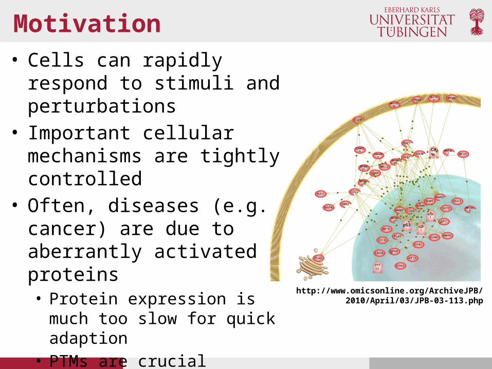
Motivation• Cells can rapidly respond to
stimuli and perturbations• Important cellular mechanisms
are tightly controlled• Often, diseases (e.g. cancer) are
due to aberrantly activated proteins• Protein expression is much too
slow for quick adaption• PTMs are crucial regulator• MS-based proteomics allows to
analyze complex networks of post-translationally modified proteins
http://www.omicsonline.org/ArchiveJPB/2010/April/03/JPB-03-113.php
Sample handling
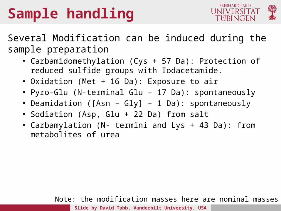
Several Modification can be induced during the sample preparation• Carbamidomethylation (Cys + 57 Da): Protection of reduced sulfide groups
with Iodacetamide.• Oxidation (Met + 16 Da): Exposure to air• Pyro-Glu (N-terminal Glu – 17 Da): spontaneously • Deamidation ([Asn – Gly] – 1 Da): spontaneously• Sodiation (Asp, Glu + 22 Da) from salt• Carbamylation (N- termini and Lys + 43 Da): from metabolites of urea
Slide by David Tabb, Vanderbilt University, USA
Note: the modification masses here are nominal masses

In vivo PTMs
• Phosphorylation (Ser, Thr, Tyr +80) from kinase signaling. Phosphorylation is one of the most important PTMs, since most signaling pathways are propagated via phosphorylation/ dephosphorylation
• Acetylation (N-termini and Lys +42), often combined with removal of protein initial Met
• Hydroxyprolination (Pro +16) stabilizes extra-cellular matrix on collagens
• Ubiquitination (Lys +114) marks proteins for destruction

Phsophorylation example
http://www.cellsignal.com/reference/pathway/pdfs/MAPK_Cascades.pdf

Protein acetylation
http://www.cellsignal.com/reference/pathway/pdfs/Protein_Acetylation.pdf
• Regulating chromatin structure and transcriptional activity
• Important role in immunity, circadian rhythmicity, and memory formation
• Favorable target in drug design for numerous disease conditions
Complete list of PTMs
www.unimod.org

Advantage of high mass accuracy
• Many modifications are neighbors in mass• Accurate precursor measurement improves
discrimination of these possibilities.• Acetylation: 42.010565 Da• Guanidination: 42.021798 Da• Tri-methylation: 42.046950 Da• Carbamylation: 43.005814 Da
Slide by David Tabb, Vanderbilt University, USA
PTMs increase search space
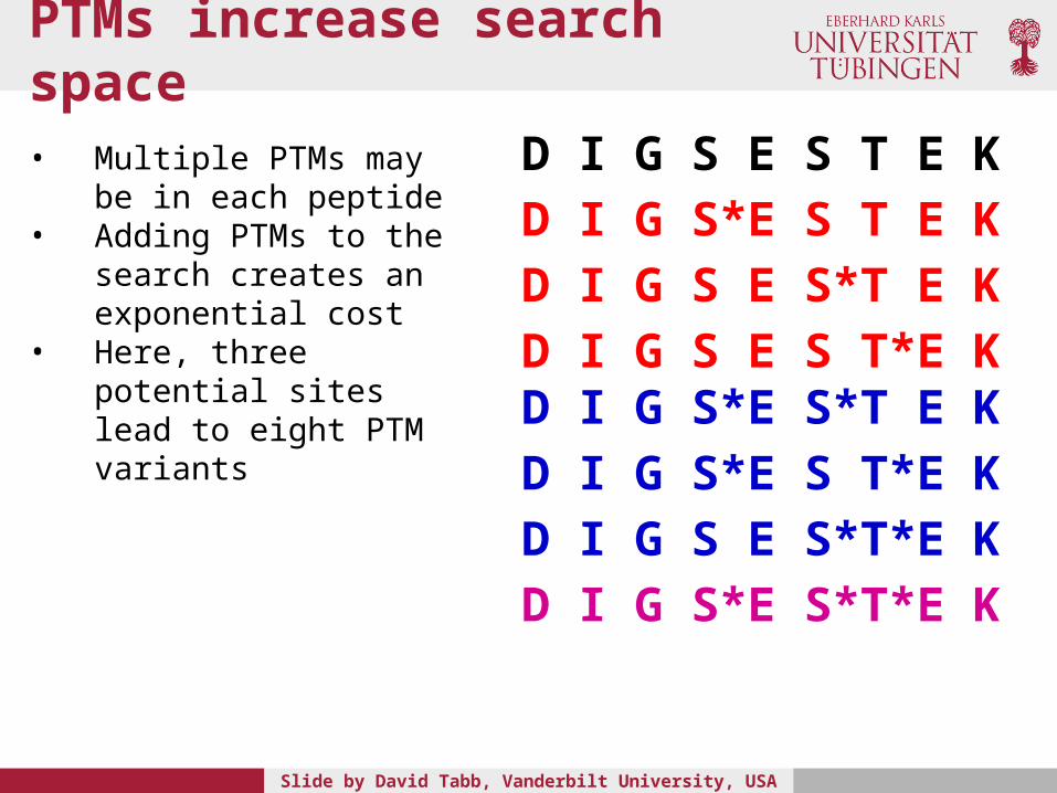
D I G S E S T E KD I G S*E S T E KD I G S E S*T E KD I G S E S T*E KD I G S*E S*T E KD I G S*E S T*E KD I G S E S*T*E KD I G S*E S*T*E K
Slide by David Tabb, Vanderbilt University, USA
• Multiple PTMs may be in each peptide
• Adding PTMs to the search creates an exponential cost
• Here, three potential sites lead to eight PTM variants

PTMs and degrees of freedom
• A residue in a peptide that may be modified is a ‘degree of freedom (df)’. This df may result in a matching between a spectrum and a completely unrelated sequence

Proteomics for PTMs
• The most established methodology is phosphoproteomics.
• The development of phosphoproteomics methods has been pushed by the significant research interest in protein phosphorylation (pharmaceutical industry and fundamental research)
• Phosphopeptides are comparably easy to enrich and mass spectrometric methods have been well established
• The field is currently moving to high-throughput of profiling methods of other PTMs and the analysis of several PTMs in parallel

Phosphoproteomics
http://employees.csbsju.edu/hjakubowski/classes/Chem%20and%20Society/Signal_Transduction/cellsdifferentsig.htm

Problems of analyzing phosphoproteins
• Site-specific phosphorylation is often substoichiometric, thus, phosphopeptides represent a small proportion of all peptides present in a total cell lysate
• Biochemical enrichment strategies have been developed. The most important methods include affinity- and antibody-based methods
• The mass spectrometric analysis of phosphopeptides is difficult since phospho groups might get lost during fragmentation or they suppress other ions
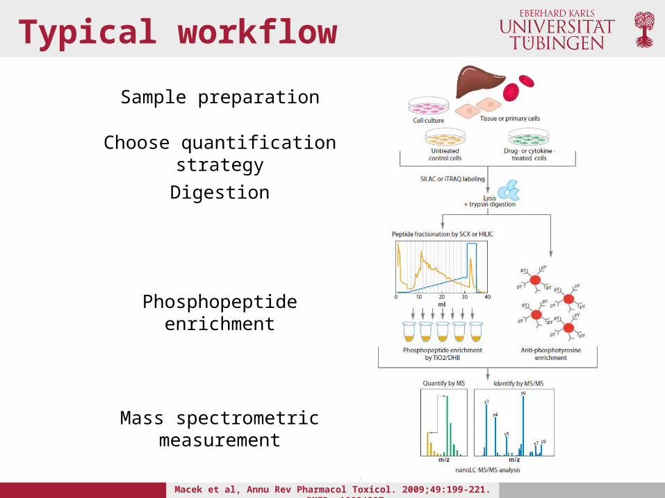
Typical workflow
Macek et al, Annu Rev Pharmacol Toxicol. 2009;49:199-221. PMID: 18834307
Sample preparation
Choose quantification strategy
Digestion
Phosphopeptide enrichment
Mass spectrometric measurement
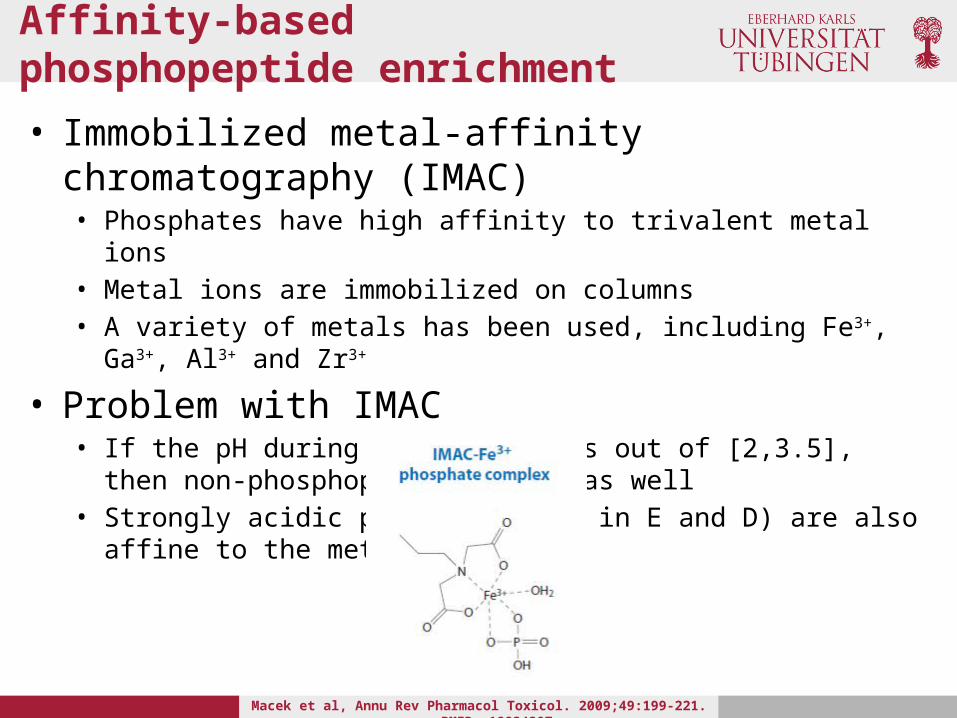
Affinity-based phosphopeptide enrichment
• Immobilized metal-affinity chromatography (IMAC)• Phosphates have high affinity to trivalent metal ions• Metal ions are immobilized on columns• A variety of metals has been used, including Fe3+, Ga3+, Al3+ and Zr3+
• Problem with IMAC• If the pH during the loading is out of [2,3.5], then non-phosphopeptides
bind as well• Strongly acidic peptides (rich in E and D) are also affine to the metal
complexes
Macek et al, Annu Rev Pharmacol Toxicol. 2009;49:199-221. PMID: 18834307
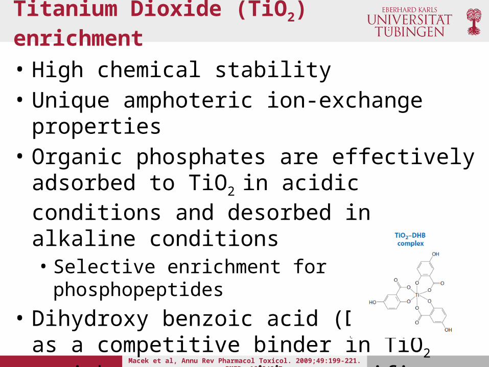
Titanium Dioxide (TiO2) enrichment
• High chemical stability• Unique amphoteric ion-exchange properties• Organic phosphates are effectively adsorbed to
TiO2 in acidic conditions and desorbed in alkaline conditions• Selective enrichment for phosphopeptides
• Dihydroxy benzoic acid (DHB) as a competitive binder in TiO2 enrichment to avoid unspecific binding of D and E
Macek et al, Annu Rev Pharmacol Toxicol. 2009;49:199-221. PMID: 18834307
Strong cation exchange
• Strategy is based on the difference in the solution charge state of phosphorylated and non-phosphorylated peptides• At pH 2.7 a (typical) tryptic peptide has charge z = +2
(N-terminal amine group + C-terminal K or R)• If this peptide is phosphorylated -> z = +1, since the
phosphate group is negatively charged• Using a linear salt gradient the phosphopeptides can
be enriched in early SCX fraction. Note that multiply phosphorylated peptides will be in the flow through
Antibody-based enrichment
• Immunopurification with immobilized anti-phospho-Y antibodies
• Antibody-based method is well established for Y, but not for other residues
• It is limited in throughput and hard to automate

MS for phospho peptides
Detection of phosphopeptides by MS is difficult, because:• Phosphopeptides are very low abundant• They have low MS response values• They show inadequate fragmentation patterns
That is why alternative methods emerged:1. Precursor ion scanning and reporter ions 2. Neutral loss dependent MSn
3. Alternative fragmentation methods (e.g ETD)

Precursor/ Reporter ion scanning
• Triple-Q instruments can be used to detect diagnostic fragment ions at m/z 79 (HPO3-) using precursor ion scanning in negative mode• The method is very sensitive, however fragment spectra recorded in
negative mode are of poor quality • Switching between ionization modes takes time
• For the analysis of Y-phosphorylation, the reporter ion scanning method is used to detect the pY immonium ion (cleavage at either side of pY) at m/z 216.043 (very specific for pY!)
• The pY immonium ion is mass deficient due to the high content of O and P, thus high resolution instruments can easily distinguish between ions with the same nominal mass
Neutral loss dependent MSn
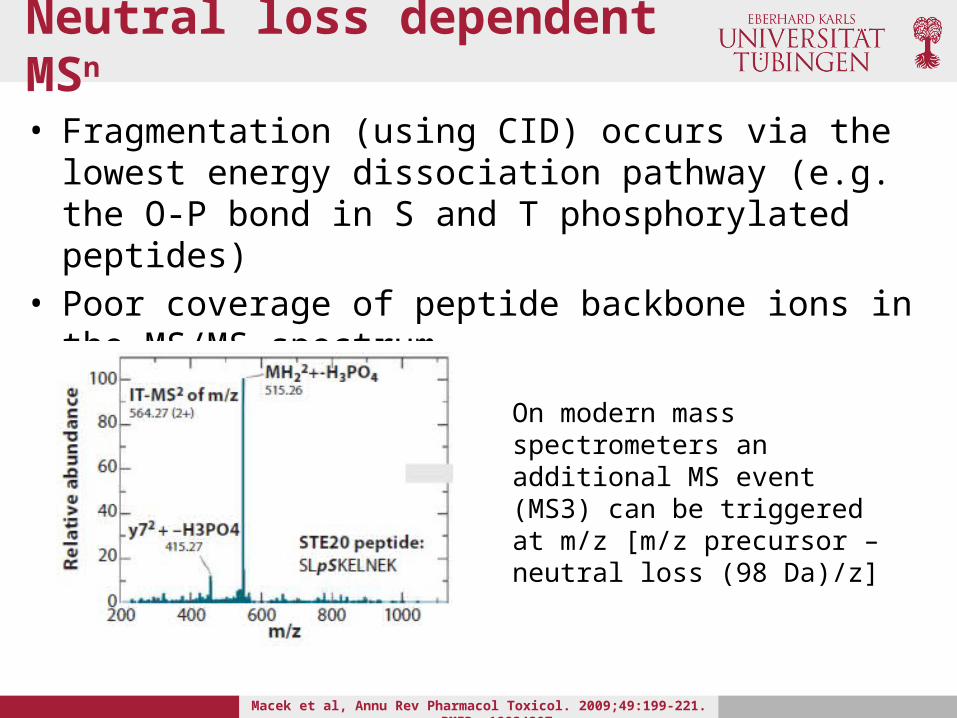
• Fragmentation (using CID) occurs via the lowest energy dissociation pathway (e.g. the O-P bond in S and T phosphorylated peptides)
• Poor coverage of peptide backbone ions in the MS/MS spectrum
On modern mass spectrometers an additional MS event (MS3) can be triggered at m/z [m/z precursor – neutral loss (98 Da)/z]
Macek et al, Annu Rev Pharmacol Toxicol. 2009;49:199-221. PMID: 18834307

Phosphopeptide identification
• Precise phosphorylation site assignment can be difficult
• Search engines often suggest the correct phosphopeptide, but fail to correctly localize the phosphorylation site, if more than one residue can potentially carry the phospho group• Note that in eukaryotes phospho groups are
predominately attached the S, T and Y residues (other phosphorylated amino acids exists (H,L or R), but are very rare)
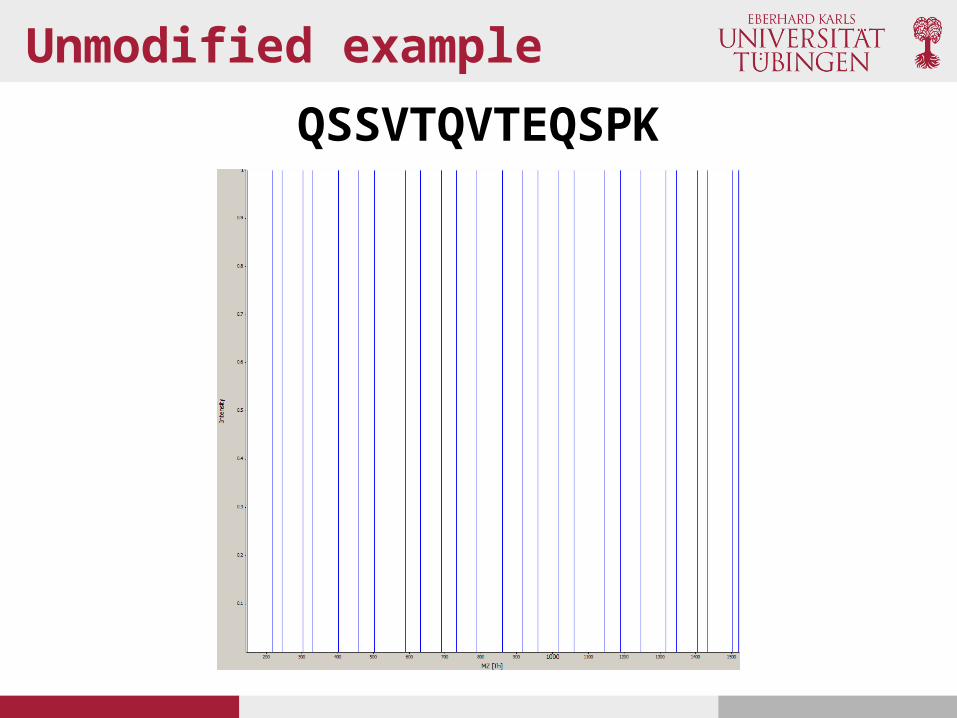
Unmodified example
QSSVTQVTEQSPK

Phosphorylated example• QSSVTQVTEQSPK is phosphorylated at one position (precursor
weight of unphosphorylated version + weight of phospho group)• Which fragment ions are changed in the MS2 spectrum?
Q S S V T Q V T E Q S P K Assume T8 is phosphorylated
Q S S V T Q V T E Q S P KQ S S V T Q V T E Q S P KQ S S V T Q V T E Q S P KQ S S V T Q V T E Q S P KQ S S V T Q V T E Q S P KQ S S V T Q V T E Q S P KQ S S V T Q V T E Q S P KQ S S V T Q V T E Q S P KQ S S V T Q V T E Q S P KQ S S V T Q V T E Q S P KQ S S V T Q V T E Q S P KQ S S V T Q V T E Q S P K
All these fragment ions do not contain the modification, thus, they do not change the mass
All other fragment ions are shifted by the mass of the phospho group
Phosphosite localization
• The Ascore algorithm has been published in 2006 by the Gygi lab
Beausoleil et al., A probability-based approach for high-throughput protein phosphorylation analysis and site localization. Nat Biotechnol. 2006 Oct;24(10):1285-92. PMID: 16964243
• Two step algorithm:i. Determine the most likely site locationsii. Use site-determining ions to calculate the probability for correct
assignment
• One of the first algorithms addressing the problem of modification site localization
• Similar version is also implemented in MaxQuant• OpenMS implementation (OpenMS::TOPP::PhosphoScoring)
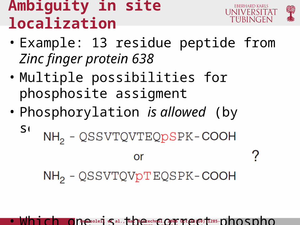
Ambiguity in site localization
• Example: 13 residue peptide from Zinc finger protein 638
• Multiple possibilities for phosphosite assigment• Phosphorylation is allowed (by search settings) on
STY
• Which one is the correct phospho site?Beausoleil et al., Nat Biotechnol. 2006 Oct;24(10):1285-92. PMID: 16964243

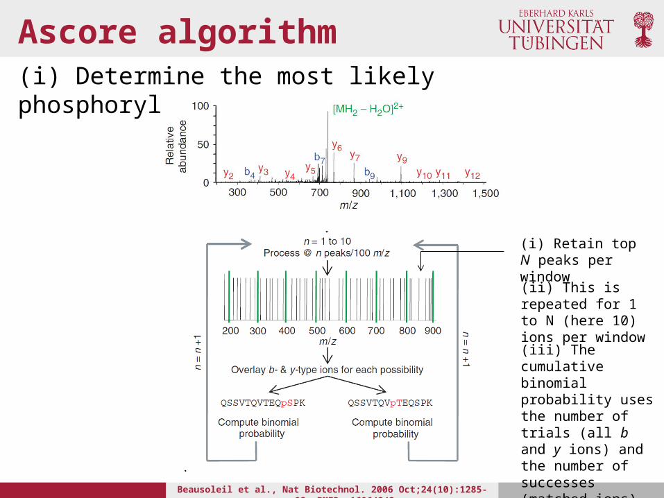
Ascore algorithm(i) Determine the most likely phosphorylation site
Beausoleil et al., Nat Biotechnol. 2006 Oct;24(10):1285-92. PMID: 16964243
• MS/MS spectrum is separated into windows of 100 m/z units
• Retain i, with , of the most intense peaks per spectrum
• Predicted b and y ions are then overlaid with the processed spectrum
• The cumulative binomial probability P is then calculated using the number of trials N, the number of successes n and the probability of success p:
P is the probability for random matchings of the given number of fragment ions; the total number of trials (N) equals the number of fragment ions; the number of successes (n) is the number of matches
Ascore algorithm(i) Determine the most likely phosphorylation site
Beausoleil et al., Nat Biotechnol. 2006 Oct;24(10):1285-92. PMID: 16964243
(i) Retain top N peaks per window
(ii) This is repeated for 1 to N (here 10) ions per window(iii) The cumulative binomial probability uses the number of trials (all b and y ions) and the number of successes (matched ions)
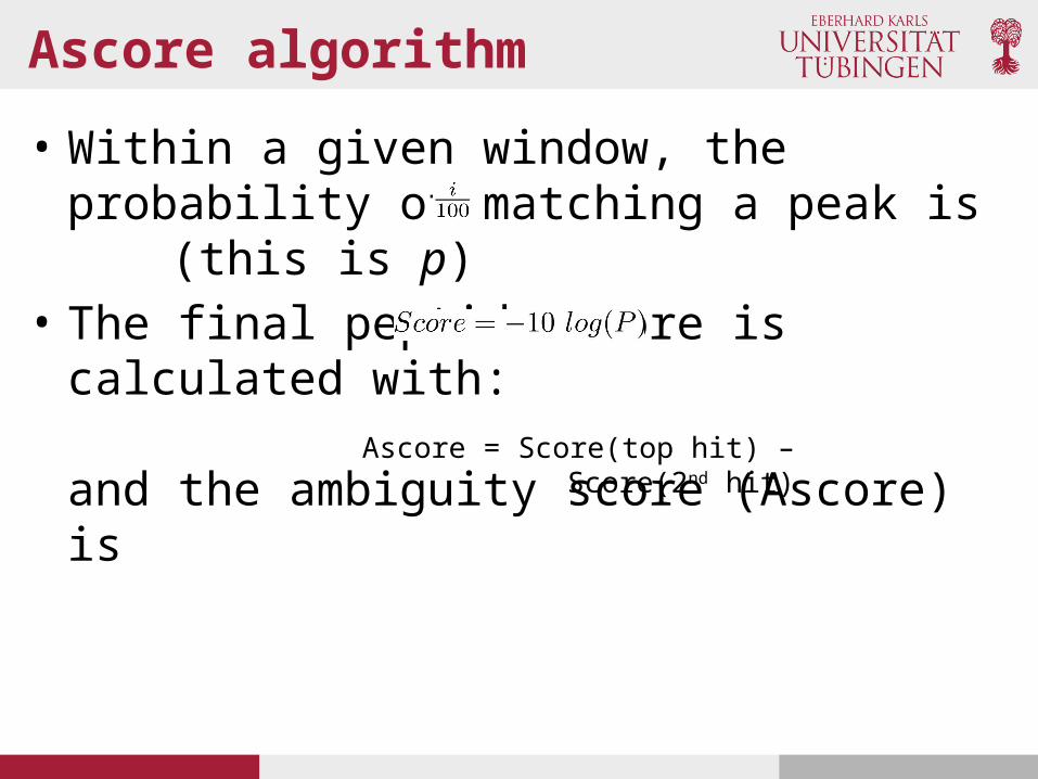
Ascore algorithm
• Within a given window, the probability of matching a peak is (this is p)
• The final peptide score is calculated with:
and the ambiguity score (Ascore) isAscore = Score(top hit) – Score(2nd
hit)

Ascore algorithm
Beausoleil et al., Nat Biotechnol. 2006 Oct;24(10):1285-92. PMID: 16964243
(iv) Each possibility is plotted as -10 log(P) vs. peak depth (peaks per m/z window)(v) Now the site-determining ions can be identified. Here only six ions can potentially differentiate between the 2 sites. The corresponds to the earliest maximal difference
Earliest maximal difference

Ascore algorithm
• The calculate the final Ascore only the two top scoring sequences (based on the peptide score) are used
Beausoleil et al., Nat Biotechnol. 2006 Oct;24(10):1285-92. PMID: 16964243

Phosphosite stoichiometry
• In quantitative (phospho)proteomics studies it is often important to differentiate between differential protein expression and differential protein phosphorylation
• Note that phosphorylation can be a very rapid event (in the range of seconds and minutes), whereas protein expression takes more time (hours). However, differential phosphorylation can still be present after longer periods.

Differential analysis
• This is a SILAC pair of a phosphorylated peptide
• Is the protein of origin more abundantly expressed in the state with the heavy (H) label?
• Is the phospho site more abundantly occupied in the H state?

Phosphosite stoichiometry
• Olsen et al. published a strategy for the calculation of phosphosite occupancy (stoichiometry)• Olsen et al., Quantitative Phosphoproteomics Reveals Widespread Full Phosphorylation Site
Occupancy During Mitosis. 2010. Science Signaling 3 (104), ra3. [DOI: 10.1126/scisignal.2000475]
• This information can only be inferred if non-phosphorylated form of the peptide has also been identified and if the protein ratio has been calculated
• In the initial paper SILAC was used as a quantification approach

Phosphosite stoichiometry
• Known: The ratio (heavy SILAC / light SILAC) of the phosphorylated peptide as x, the non-phosphorylated peptide as y and the ratio of the protein (median of all ratios of peptides assigned to the same protein) as z
• Unknown: Absolute phosphorylation site stoichiometry in the L and H state• NL(Phos) and NL(Pep) are the amounts (copy numbers) of the
phosphorylated and non-phosphorylated version of the same peptide. The proportion of phosphorylated peptide in the L SILAC state is given by a,
• In the H state as b, respectively,

Phosphosite stoichiometry
• The protein copy number N(Prot) is modeled by the copy number of the peptides, we can thus assume
• So far these are theoretical considerations, but the MS data delivers:
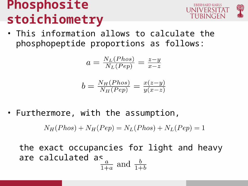
Phosphosite stoichiometry
• This information allows to calculate the phosphopeptide proportions as follows:
• Furthermore, with the assumption,
the exact occupancies for light and heavy are calculated as,

Acetylation
• Protein acetylation determines structure, function and intracellular localization of proteins
• Role in the signal transduction pathways• Protein acetylation in cells is regulated by a co-ordinated action of
histone acetyl transferases (HAT) and histone deacetylases(HDAC)• Histone deacetylation inhibits progress of many nuclear events
including proliferation and damage response events• Histone deacetylases have been among the targets for the
development of anticancer drugs and adjuvants
http://bricker.tcnj.edu/Amb/le9/Acy.jpg

Enrichment for acetylated peptides
• Anti-acetyllysine antibodies are currently the state-of-the-art methods for the enrichment of acetyllated peptides• Choudhary, et al. Lysine Acetylation Targets Protein Complexes and Co-
Regulates. Science 325, 834 (2009); DOI: 10.1.126/science.1175371

Glycoproteomics
• Glycosylation is the most complex of all PTMs• Glycans (polysaccharides) represent a huge variety in their
composition• Glycosylation of proteins takes mainly place in the
endoplasmic reticulum (ER) and the Golgi• There, enzymes, such as glycosyltransferases and glycosidases
attach the sugar groups to the proteins• These enzymes mainly target S, T and N• It impacts on charge, conformation and protein stability
Importance of glycoproteomics
• Abnormal protein glycosylation has been correlated with several diseases• Cancer (Kim et al., Implication of aberrant glycosylation in cancer and use of lectin for cancer
biomarker discovery. Protein Pept Lett. 2009;16(5):499-507. PMID: 19442229)
• Inflammatory diseases (Brooks. Strategies for analysis of the glycosylation of proteins: current status and future perspectives. Mol Biotechnol. 2009 Sep;43(1):76-88. Epub 2009 Jun 9. PMID: 19507069)
• Etc.
Glycoproteomics analysis
• For the complete glycoprotein picture one needs: the peptide sequence, glycosylation site and the glycan structure
• Glycosylation does not conform to a single structure and there is no underlying template
• In humans, glycans attached to proteins are composed of eight different monosaccharides:• Mannose (Man); Glucose (Glc); Galactose (Gal); Fucose (Fuc); N-acetylgalactosamine(GalNAc);
N-Acetylglucosamine (GlcNAc); N-Acetylneuraminic acid (NeuNAc); Xylose (Xyl)
• These sugars can be linked in linear or branching chains of various sequences and lengths
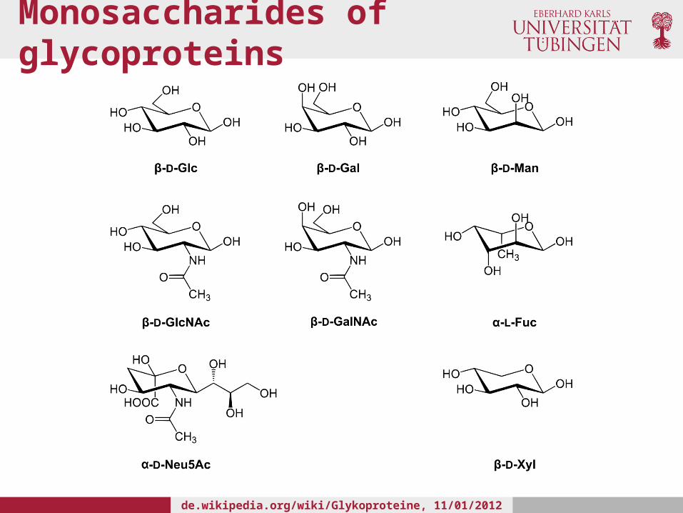
Monosaccharides of glycoproteins
de.wikipedia.org/wiki/Glykoproteine, 11/01/2012 6 PM CET
Monosaccharides of glycoproteins
• Sugars, such as Glc, Gal and Man have identical masses and charges
• These are different stereoisomers and their permutations in complex glycans can result in a broad range of different glycoforms
• Glycoproteomics experiments result in very complex data sets

Protein-linked glycans
• N-linked glycans, with the glycan attached to the amide group of N via a GlcNAc in a consensus sequence N-X-S/T (X any amino acid except P)
• O-linked glycans, with glycan attached to S or T• C-glycans, with the glycan (Man) attached to W with a C-
C bond in a consensus sequences W-X-X-W or W-S-X-C• Glycosylphosphatidylinositol anchors, with the glycan
attached to the protein C-terminus by a phosphoethanolamine bridge with Man (this occurs only on membrane associated proteins)
• The two most common forms are O- and N- linked glycosylation
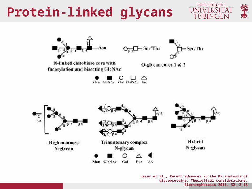
Protein-linked glycans
Lazar et al., Recent advances in the MS analysis of glycoproteins: Theoretical considerations. Electrophoresis 2011, 32, 2-13
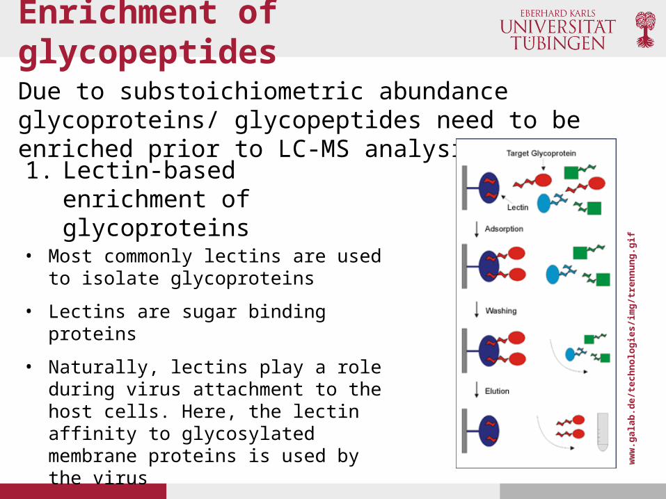
Enrichment of glycopeptides
Due to substoichiometric abundance glycoproteins/ glycopeptides need to be enriched prior to LC-MS analysis
• Most commonly lectins are used to isolate glycoproteins
• Lectins are sugar binding proteins
• Naturally, lectins play a role during virus attachment to the host cells. Here, the lectin affinity to glycosylated membrane proteins is used by the virus
1. Lectin-based enrichment of glycoproteins
ww
w.g
alab
.de/
tech
nolo
gies
/im
g/tr
ennu
ng.g
if
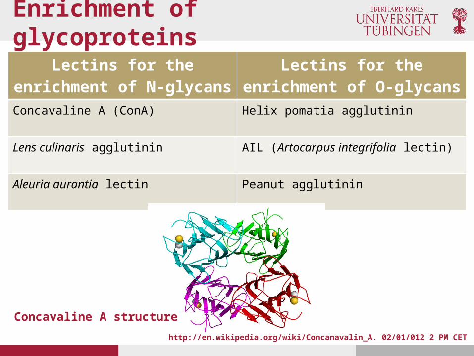
Enrichment of glycoproteins
Lectins for the enrichment of N-glycans
Lectins for the enrichment of O-glycans
Concavaline A (ConA) Helix pomatia agglutinin
Lens culinaris agglutinin AIL (Artocarpus integrifolia lectin)
Aleuria aurantia lectin Peanut agglutinin
http://en.wikipedia.org/wiki/Concanavalin_A. 02/01/012 2 PM CET
Concavaline A structure

Enrichment of glycoproteins
Schiess et al., Analysis of Cell Surface Proteome Changes viaLabel-free, Quantitative Mass Spectrometry. Molecular and Cellular Proteomics 2009,.
2. Linker-based enrichment of glycoproteins

MS of glycopeptides
• The hydrophilic nature of glycans limits the surface activity and the ionization efficiency
• Natural and basic glycoconjugates can be protonated• Acidic glycoconjugates can only be deprotonated (negative ESI mode!)
• Often, derivatization is used to increase hydrophobicity and volatility (and thereby ionization efficiency)
• As for unmodified peptides, MS/MS can be performed to sequence the glycan as well as the underlying peptides
• MS/MS of glycopeptides is more complicated than with unmodified peptides, since the chemical properties of the peptide and glycan are dissimilar
• Using CID, the collision energy is highly important to the content of the MS/MS spectrum

MS/MS of glycopeptides
• Using low energy CID, the tandem spectrum is dominated by ions from the sequential loss of sugars and occasionally the precursor ion, but there is no fragmentation of the peptide backbone
Lazar et al., Recent advances in the MS analysis of glycoproteins: Theoretical considerations. Electrophoresis 2011, 32, 2-13
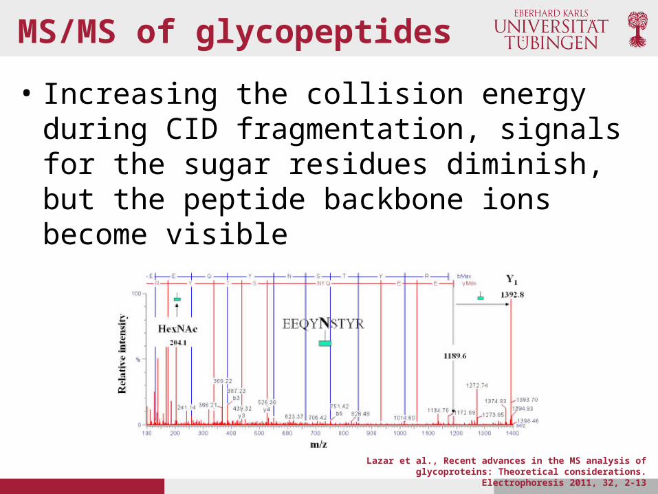
MS/MS of glycopeptides
• Increasing the collision energy during CID fragmentation, signals for the sugar residues diminish, but the peptide backbone ions become visible
Lazar et al., Recent advances in the MS analysis of glycoproteins: Theoretical considerations. Electrophoresis 2011, 32, 2-13

MS/MS of glycopeptides
• Information in the sugar stereochemistry (which Glc, Gal or Man ?), the linkage (1->4 or 1->6 ?) or branching pattern can not be obtained using conventional CID fragmentation
• This can be achieved by “cross-ring” fragmentation on MALDI-TOF-TOF instruments. Very high collision energy is needed (orders of KeV)
Nomenclature for tandem mass spectrometric product ions of glycans and glycoconjugates
D-ion (not part of the original nomenclature) consists of combined C- and Z-type fragmentation. They differentiate N-acetyllactosamine linkages and mannose branching structure
Zaia, Mass Spectrometry and the Emerging Field of
Glycomics. Chem Biol. 2008 September 22;
15(9): 881–892.

An N-Glycoproteomics workflow
Joenväärä et al., N-Glycoproteomics – An automated workflow approach. Glycobiology. 2008 Vol. 18 no. 4 pp. 339-349.

Scoring function Joenväärä et al.
• Score S for glycopeptide G,
SPC is the shared peak count, R a random spectrum, s the measured spectrum.
• Furthermore, s has M mass values and Mp is the number of possible mass values with the given mass range and tolerance T
• G, the theoretical glycopeptide spectrum has N, mass values. R has also N mass values
• SPC counts the number of peak pairs with
Joenväärä et al., N-Glycoproteomics – An automated workflow approach. Glycobiology. 2008 Vol. 18 no. 4 pp. 339-349.

Scoring function Joenväärä et al.
• The probaility is calculated using the binomial distribution
where and
• The score, designed for glycopeptide scoring by Joenväärä et al. has similarity to the Ascore designed for phosphosite assignment by Beausoleil et al.

Summary PTMs
• The analysis of PTMs is a promising strategy to approach a wide range of biological questions
• Additional level of complexity compare to “simple” protein expression analysis
• The field of high-throughput analysis of PTMs is still very young. Only recent developments allowed this complex task
• But, in the other hand a lot of problems are still unsolved. These open problems concern the biochemistry for sample preparation and enrichment as well as adequate computational methods
References
• Phosphoproteomics• Macek et al, Annu Rev Pharmacol Toxicol. 2009;49:199-221. PMID: 18834307• Beausoleil et al., A probability-based approach for high-throughput protein phosphorylation
analysis and site localization. Nat Biotechnol. 2006 Oct;24(10):1285-92. PMID: 16964243• Olsen et al., Quantitative Phosphoproteomics Reveals Widespread Full Phosphorylation Site
Occupancy During Mitosis. 2010. Science Signaling 3 (104), ra3. [DOI: 10.1126/scisignal.2000475]
• Acetyl-proteomics• Choudhary, et al. Lysine Acetylation Targets Protein Complexes and Co-Regulates. Science 325,
834 (2009); DOI: 10.1.126/science.1175371
• Gycoproteomics• Joenväärä et al., N-Glycoproteomics – An automated workflow approach. Glycobiology. 2008 Vol.
18 no. 4 pp. 339-349. • Zaia, Mass Spectrometry and the Emerging Field of Glycomics. Chem Biol. 2008 September 22;
15(9): 881–892. • Lazar et al., Recent advances in the MS analysis of glycoproteins: Theoretical considerations.
Electrophoresis 2011, 32, 2-13• Schiess et al., Analysis of Cell Surface Proteome Changes viaLabel-free, Quantitative Mass
Spectrometry. Molecular and Cellular Proteomics 2009,.


