BIOL 200 (Section 921) Lecture # 2, June 20, 2006 • Reading for lecture 2 : Essential Cell Biology (ECB) 2nd edition. Chap 2 pp 55-56, 58-64, 74- 75; Chap 4, pp. 119-143. Good questions: 4-3, 4-10G, 4-11, 4-14-15, 4-17. • Learning Objectives 1.Understand how amino acids are joined together via peptide bonds to form a polypeptide. Be able to write out the structure of the reactants and the product. 2.Recognize the properties of each of the 20 amino acids, their three letter abbreviations, and how they are linked by peptide bonds.
Transcript
BIOL 200 (Section 921)Lecture # 2, June 20, 2006
• Reading for lecture 2: Essential Cell Biology (ECB) 2nd edition. Chap 2 pp 55-56, 58-64, 74-75; Chap 4, pp. 119-143. Good questions: 4-3, 4-10G, 4-11, 4-14-15, 4-17.
• Learning Objectives1. Understand how amino acids are joined together via
peptide bonds to form a polypeptide. Be able to write out the structure of the reactants and the product.
2. Recognize the properties of each of the 20 amino acids, their three letter abbreviations, and how they are linked by peptide bonds.
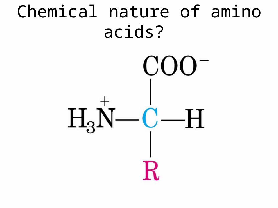
Chemical nature of amino acids?
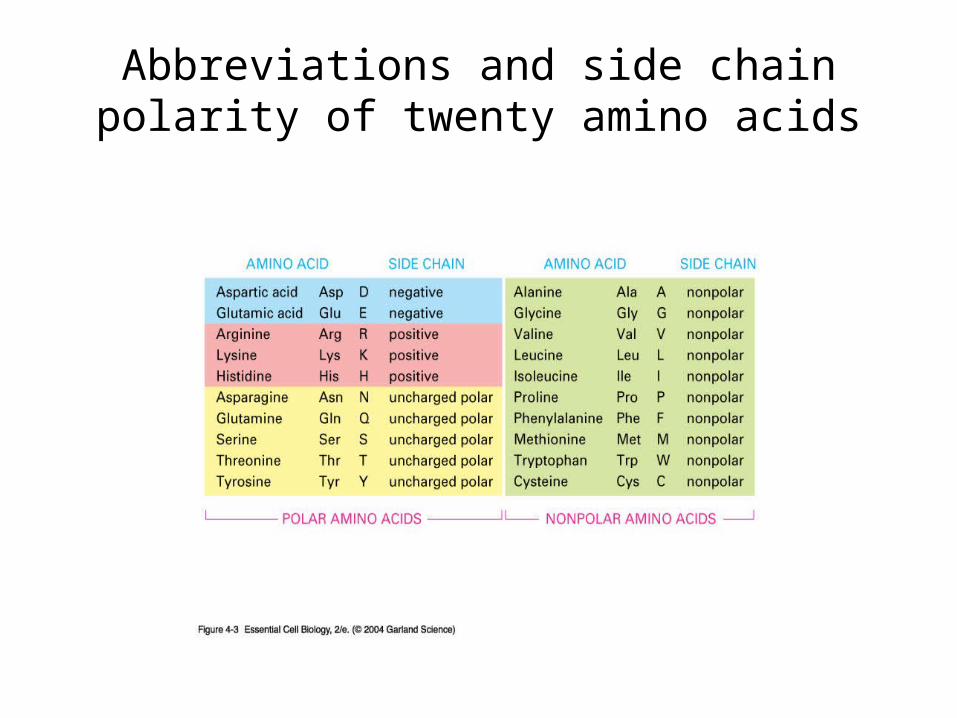
Abbreviations and side chain polarity of twenty amino acids
Panel 2-5, p74Basic side chains-positively charged at physiological pH
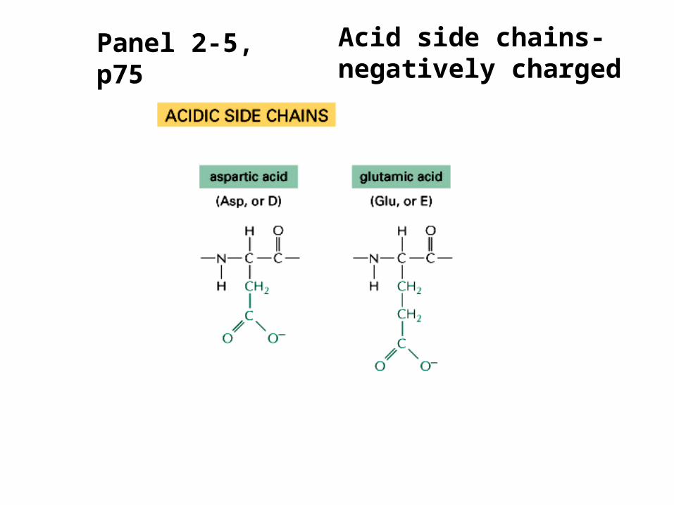
Panel 2-5, p75 Acid side chains-negatively charged
Panel 2-5, p75Polar side chains-interact well with water, can form H-bonds
Panel 2-5, p75
Nonpolar groups-energetically unfavorable for them to contact water
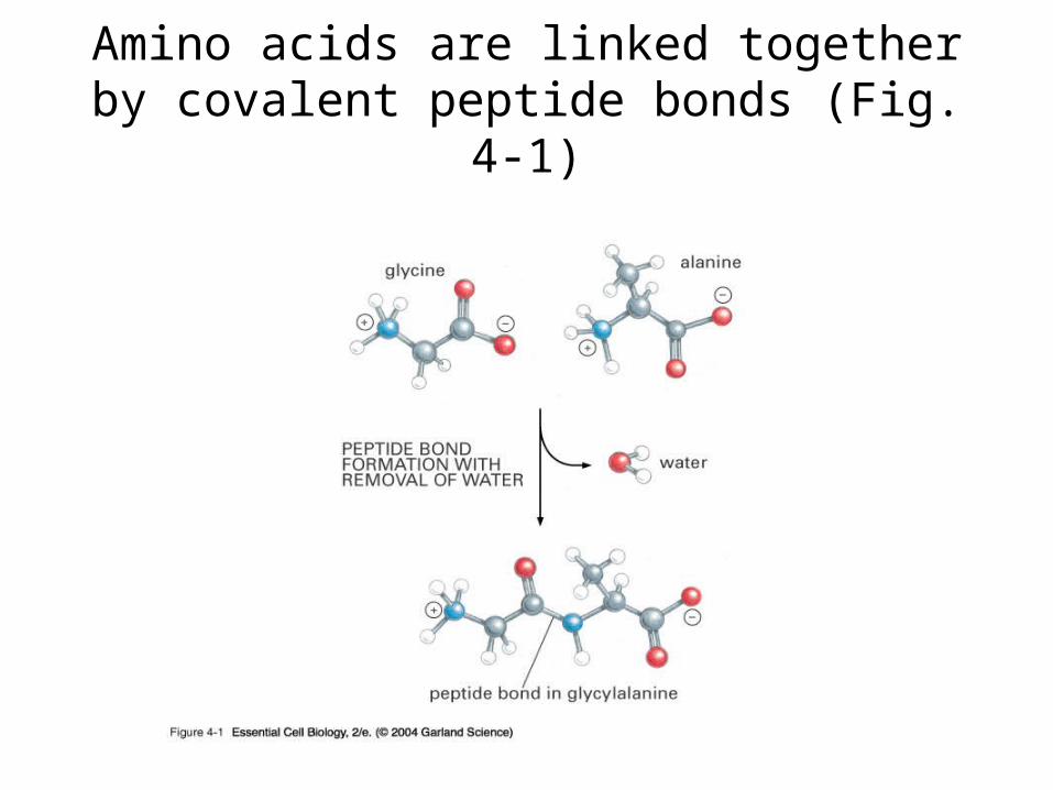
Amino acids are linked together by covalent peptide bonds (Fig. 4-1)
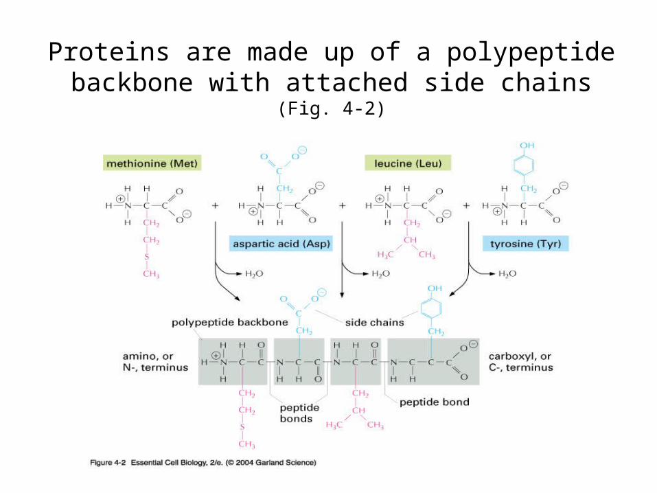
Proteins are made up of a polypeptide backbone with attached side chains
(Fig. 4-2)
Characteristics of Polypeptide synthesis
• The amino group of one amino acid reacts with the carboxyl group of another to form a peptide bond (=amide linkage) with the elimination of a water molecule.
• The polarity of the molecule is retained. The monomers are all joined together in the same orientation. The polypeptide chain has polarity.
• The polypeptide contains a free amino group at one end and a free carboxyl group at the other. These ends of the polypeptide chain are referred to as the amino and carboxyl termini (ends) respectively.
• The product of the condensation reaction preserves the reactive group (carboxyl group) at the end of the molecule. This makes it possible for the peptide chain to join to yet another amino acid.
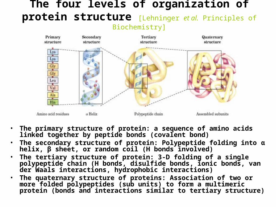
The four levels of organization of protein structure [Lehninger et al. Principles of Biochemistry]
• The primary structure of protein: a sequence of amino acids linked together by peptide bonds (covalent bond)
• The secondary structure of protein: Polypeptide folding into α helix, β sheet, or random coil (H bonds involved)
• The tertiary structure of protein: 3-D folding of a single polypeptide chain (H bonds, disulfide bonds, ionic bonds, van der Waals interactions, hydrophobic interactions)
• The quaternary structure of proteins: Association of two or more folded polypeptides (sub units) to form a multimeric protein (bonds and interactions similar to tertiary structure)
Unity and diversity of proteins
Primary structure (sequence of amino acids) determines the structure of a protein
Fig.4-5: In water, hydrophobic aa cluster inside a folded protein, away from solvent. Why?
Secondary structure of proteins - helix H bond between the N-H of every peptide bond to the C=O of the next
peptide bond of the same chain. R groups are not involved. (e.g. in protein -keratin - abundant in skin, hair, nails and horns)
[Fig. 4-10, p. 128]
(Pitch)
Secondary structure of proteins – β sheet Polypeptide chains are held together by H bonds between N-H group
of one polypeptide chain and C=O group of the other chain(e.g. in the protein fibroin - abundant in silk) [Fig. 4-10, p. 128]
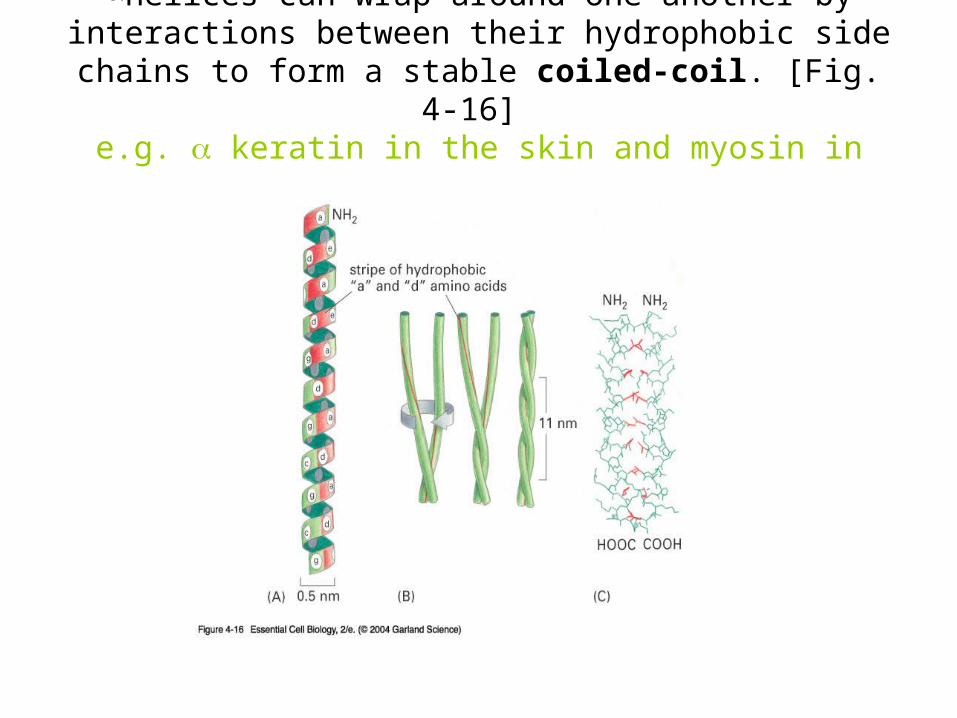
helices can wrap around one another by interactions between their hydrophobic side chains to form a stable
coiled-coil. [Fig. 4-16] e.g. keratin in the skin and myosin in muscles
Tertiary structure of proteins
• 3D conformation or shape
• Depends on the properties of the R groups of amino acid residues
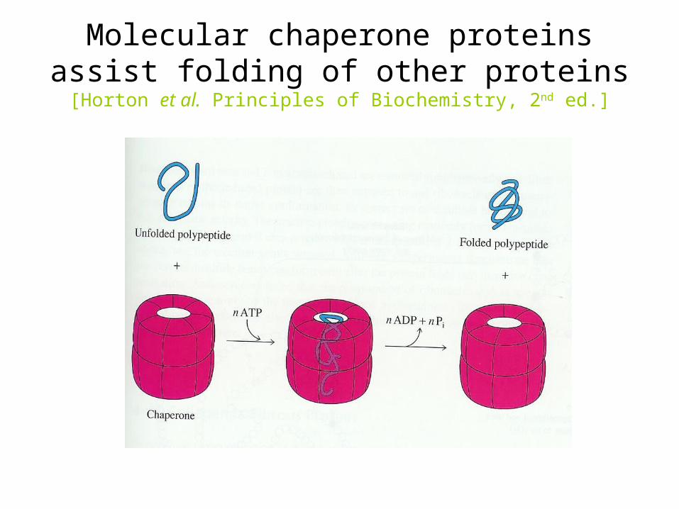
• Fold spontaneously or with the help of molecular chaperones
• Stabilized by covalent and non-covalent bonds
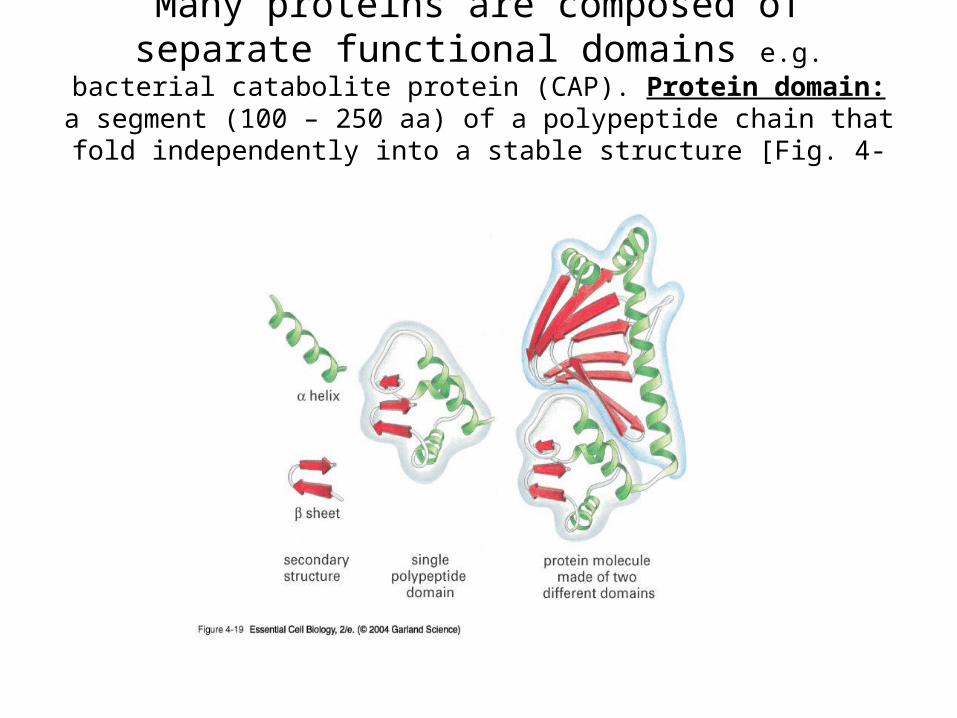
Many proteins are composed of separate functional domains e.g. bacterial catabolite protein (CAP).
Protein domain: a segment (100 – 250 aa) of a polypeptide chain that fold independently into a stable structure [Fig. 4-19]
Noncovalent bonds help protein folding (Fig. 4-4) Also review Panel 2-7 (pp. 78,79) on noncovalent bonds
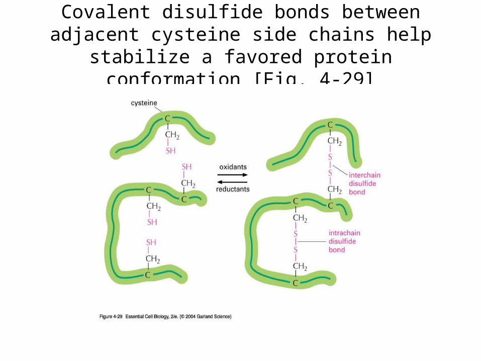
Covalent disulfide bonds between adjacent cysteine side chains help stabilize a favored
protein conformation [Fig. 4-29]
Molecular chaperone proteins assist folding of other proteins
[Horton et al. Principles of Biochemistry, 2nd ed.]
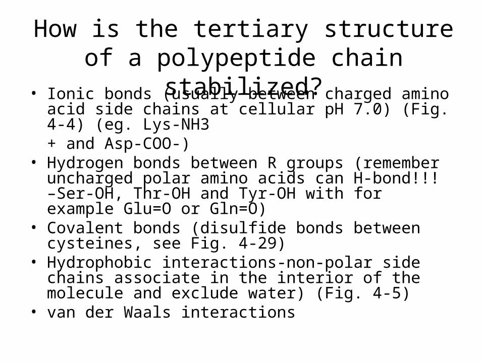
How is the tertiary structure of a polypeptide chain stabilized?
• Ionic bonds (usually between charged amino acid side chains at cellular pH 7.0) (Fig. 4-4) (eg. Lys-NH3+ and Asp-COO-)
• Hydrogen bonds between R groups (remember uncharged polar amino acids can H-bond!!! –Ser-OH, Thr-OH and Tyr-OH with for example Glu=O or Gln=O)
• Covalent bonds (disulfide bonds between cysteines, see Fig. 4-29)
• Hydrophobic interactions-non-polar side chains associate in the interior of the molecule and exclude water) (Fig. 4-5)
• van der Waals interactions
Quaternary structure of proteins: hemoglobin, a protein in red blood cells, has four sub units (two
copies each of - and β-globins containing a heme molecule [Fig. 4-23].
Gel electrophoresis method is used to separate proteins [Lehninger et al. Principles of Biochemistry]
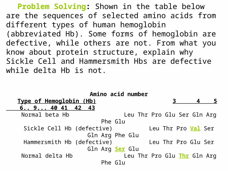
Problem Solving: Shown in the table below are the sequences of selected amino acids from different types of human hemoglobin (abbreviated Hb). Some forms of hemoglobin are defective, while others are not. From what you know about protein structure, explain why Sickle Cell and Hammersmith Hbs are defective while delta Hb is not.


![BIOL 200 (Section 921) Lecture # 11 [July 4, 2006] UNIT 8: Cytoskeleton Reading: ECB, 2nd ed. Chap 17. pp 573-606; Questions 17-1, 17-2, 17-12 to 17-23.](https://static.documents.pub/doc/80x56/56649e565503460f94b4d645/biol-200-section-921-lecture-11-july-4-2006-unit-8-cytoskeleton-reading.jpg)


