See discussions, stats, and author profiles for this publication at: https://www.researchgate.net/publication/258196253 Biological effects of melt spinning fabrics composed of 1% bioceramic material Article in Textile Research Journal · July 2012 DOI: 10.1177/0040517512439917 CITATIONS 10 READS 95 4 authors, including: Ting kai Leung 65 PUBLICATIONS 754 CITATIONS SEE PROFILE All content following this page was uploaded by Ting kai Leung on 06 June 2016. The user has requested enhancement of the downloaded file.
at Chongqing University on October 11, 2013trj.sagepub.comDownloaded from at Chongqing University on October 11, 2013trj.sagepub.comDownloaded from at Chongqing University on October 11, 2013trj.sagepub.comDownloaded from at Chongqing University on October 11, 2013trj.sagepub.comDownloaded from at Chongqing University on October 11, 2013trj.sagepub.comDownloaded from at Chongqing University on October 11, 2013trj.sagepub.comDownloaded from at Chongqing University on October 11, 2013trj.sagepub.comDownloaded from at Chongqing University on October 11, 2013trj.sagepub.comDownloaded from at Chongqing University on October 11, 2013trj.sagepub.comDownloaded from at Chongqing University on October 11, 2013trj.sagepub.comDownloaded from at Chongqing University on October 11, 2013trj.sagepub.comDownloaded from
This study evaluated the usefulness of bioceramic materials (ceramic materials that emit high-performance far-infrared
(FIR) rays), processed into fabrics using a traditional manufacturing melt spinning method. Numerous measurements
were designed to test the biological functions of 1% bioceramic fabrics. These included physical induction of intracellular
nitric oxide (NO) in NIH 3T3 cells (mouse fibroblasts), the effects on cell viability in osteoblastic cells (MC3T3-E1) under
hydrogen peroxide-mediated oxidative stress, and the effects on lipopolysaccharide (LPS)-induced cyclo-oxygenase-2
(COX-2) and prostaglandin E2 (PGE2) production in a chondrosarcoma (SW1353) cell line. When compared to the
control group, the bioceramic fabrics were capable of inducing further intracellular NO production using NIH 3T3 cells,
and maintaining increased viability and against cell intoxication of osteoblastic cells by suppressing cell release of lactate
dehydrogenase (LDH) under oxidative stress. In addition, it was found to suppress LPS-induced COX-2 production more
significantly in a SW1353 cell line. These processes represent the biomolecular changes occurring during promotion of
decline in aging, prevention of osteoporosis, and prevention of inflammatory processes within the human body.
Therefore, these bioceramic fabrics are likely to fulfill their claims of having health-promoting benefits.
Keywords
Fabrication, performance, materials
Introduction
Maintaining warmth and being decorative and styl-ish are fundamental requirements for garments.Wearing garments composed of fabrics that providehealth-promoting benefits is an attractive concept forthe growing aging population worldwide.1,2 However,no existing specialized field combines medical and textiletechnologies to research these materials.
The present study performed a general survey onfabric samples from markets in Asian counties, includ-ing some garments manufactured of fibers producedusing the melt spinning method and others that hadsurfaces post-processed with mineral ore additives.In addition, this study measured ionizing radiationemissions. The main sources of ionizing radiation inmineral ore are rare radioactive elements (such as uran-ium or radium). Previous studies have observed thatlow doses of ionizing radiation have immunologicalmodulatory effects. Short exposure to low-dose radi-ation influenced T-cells and macrophages,3,4 resultingin short-term amelioration of autoimmune diseases.However, exposure of tissues to low-dose ionized
radiation for longer periods is not absolutely safe,increasing the possibility of DNA mutations and dele-tions, and leading to cancer risk.5–7
Ionizing radiation has higher frequency ranges andshorter wavelengths than does the visible light spectrum(of 400 to 750 nm) and, therefore, may have sufficientenergy to break chemical bonds. High-energy ionizingradiation can also strip off electrons or break up thenuclei of atoms.5–7 Non-ionizing radiation has lowerfrequency ranges and longer wavelengths than doesthe visible light spectrum. Non-ionizing radiationrarely has sufficient energy to break chemical bonds.
1Department of Diagnostic Radiology, Taipei Medical University Hospital,
Taiwan2Taiwan Textile Research Institute, Taiwan
Corresponding author:
Dr Ting-Kai Leung, Department of Diagnostic Radiology, Taipei Medical
University Hospital, No. 252, Wu Hsing Street (110) Taipei, Taiwan, ROC
Far-infrared (FIR) radiation (3–1000 mm) consistsof invisible electromagnetic waves, with wave-lengths longer than visible light, and is the majorheat-transmitting radiation at wavelengths of 3 mm to1mm, as defined by the CIE (1987). Particularly in therange of 8–14mm, FIR was suspected to have manybiological effects.8
This spectrum of wavelength transfers energy thatthermoreceptors in the skin perceive as heat. In previ-ous years, people had believed that the optimal wave-length most effective for life is between 8 and 14 mm.9–16
Numerous published medical studies have used FIRwith a heat supply source to demonstrate that the FIRwavelength increased skin microcirculation in rats,improved blood flow of arteriovenous fistulas in hemo-dialysis patients, extended survival of skin grafts, andhad other health-promoting effects.17–22 However, thesestudies have utilized an FIR application with a heatsource dependent on electricity, which is inconvenientfor wider application during everyday life.
In Asian garment markets, an increasing numbers ofproducts contain fiber additives, such as bamboo char-coal and mineral ores, which the manufacturers claimcan promote health when worn on the body. Garmentscomposed of bamboo charcoal have putative health-promoting functions.23 Although the authors cannotdeny the health benefits of these products in maintain-ing warmth, observing physiological responses to thefabrics in clinical practice has indicated that these prod-ucts cannot be defined as actual health-promotingmaterials.21,24–33 However, further objective analysesare necessary for confirmation.
Assessing some of the samples available on themarket did not verify these biological functions asclaimed by the product advertisement. Examinationalso revealed that some of the samples emitted ionizingradiation, which is potentially hazardous to publichealth.34,35
People may believe that a special invisible light spec-trum can promote human health. However, lack ofthorough investigations, general knowledge, and pro-fessional education indicates that the public is incap-able of discerning between non-ionizing and ionizingradiation. Even within the fields of materials scienceand textiles, some confusion exists regarding the con-cept of radiation. Therefore, manufacturers producingclothing contaminated with ionized radiation, unbe-known to the consumer, is possible.23
The emissive efficiency of the FIR ray spectrumcannot be measured using ordinary types of instru-ments. Thus, mainstream medical applications cannoteasily identify FIR.36,37 In our previous study,9 weapplied two research methods to living human bodiesto: (i) measure the possible biological functions of agarment using infrared thermography to produce
thermal images; and (ii) observe the real-time dynamicstatus within the microvasculature of the dorsal humanfingertip, based on vascular corrosion casting, using astereoscopic microscope.9 Recording the physiologicalparameters before and after the textile made contactwith the skin enabled determining its biological func-tions. However, human physiological responses, suchas body temperature and microcirculation, are con-stantly changing and have numerous influencing fac-tors. Therefore, these two human tests, though easilyperformed, have dubious objectivity unless investiga-tors strictly control all of the experimental procedures.
The current study was a collaborative work of pro-fessionals from different fields, including medical radi-ology, clinical sciences, biomedical sciences, and textilesengineering. This study created and researched a newtype of ceramic compound that emits an effective elec-tromagnetic wavelength, as confirmed by previousmolecular biology studies.9–16 Our earlier publicationsinvestigating bioceramic materials (ceramic materialsthat emit high-performance FIR rays) focused primar-ily on the basic medical science of cells and animalmodels, and we showed that bioceramic materials pro-mote microcirculation and have other effects by upre-gulating calcium-dependent nitric oxide andcalmodulin in different cell lines.12,16 An inhibitoryeffect of bioceramic material on murine melanincancer cell (melanoma) growth was reported.38 Wealso demonstrated that bioceramic material has an anti-oxidant effect by increasing the hydrogen peroxide-scavenging ability of various cells, including murinemacrophages (RAW264.7),15 murine calvaria-derivedosteoblast-like cells (MC3T3-E1),10 NIH 3T3 fibroblastcells,16 and murine myoblast cells (C2C12).13 A physi-cal-chemical test platform was also developed to exam-ine other characteristics.9,11
Our mission has been to develop fabrics containingbioceramic materials capable of being applied in regu-lar textile manufacturing processes. Because it is diffi-cult and expensive to embed higher than 1% by-weightof bioceramic granules into fiber using a melt spinningprocedure to manufacture bioceramic fabrics, whetheror not the biological function can be preserved as theoriginal material powder is unknown. As is widelyknown, 1% solid content in the melt spinning processis commonly used, and the fiber spinning process needsless adjustment. Otherwise, the fiber is easily brokenduring the spinning and texturing processes; therefore,we choose a 1% bioceramic material as the final com-position in the nylon fiber. Thus, this study evaluatedthe health-promoting and biological functions ofassayed textiles and garments (1% by-weight of bio-ceramic) based on measurements of intracellular nitricoxide (NO) production, antioxidant effects such asbone-forming cell viability under oxidative stress, and
1122 Textile Research Journal 82(11)
anti-inflammatory effects, especially concerning jointdiseases.
Materials and methods
FIR ceramic powder
This study used a ceramic powder composed ofmicro-sized particles produced using several ingredi-ents, mainly mineral oxides, provided by theMaterials Laboratory of Taipei Medical University(Figure 1).9–16,38 The known ingredients (TiO2, SiO2,Al2O3, ZnO, and MgO) of the FIR ceramic powderwere combined to form a bio-organically harmless for-mula, based on conclusions from our previousstudies.9–16,38
The average optimal emission wavelength of theceramic powder is between 8 and 14 mm, as confirmedby the Industrial Technology Research Institute(Hsinchu City, Taiwan), and represents an extremelyhigh ratio of FIR ray intensity.36,37 Bioceramic materialwas ground into a powder using a grinding process con-sisting of two stages, wet and dry grind. The powder was
prepared by further pulverizing it to a suitable size foradoption in the melt spinning procedure, using varioustypes of fibers such as nylon or PET fibers.
Preparing bioceramic fabrics and assessing biologicalfunctions
A functional bioceramic powder with a desired size ofapproximately 200 nm was blended with a matrix poly-mer, such as PET or nylon, using a twin-screw meltcompounding procedure, which was also used toobtain masterbatch chips containing bioceramicpowder. In creating a nylon/bioceramic polymer com-pound, nylon polymer was selected as the matrixbecause it is soft to the touch when spun into a fiber.
A dispersing agent was usually required to improvethe segregation of the bioceramic particles. The disper-sing agent we used is a WAX-type of surfactant, whichcould melt at approximately 180�C or higher.Formulated adjuvants are added for numerous reasons,including improved mixing and handling, increasedeffectiveness and safety, more efficient distribution,and drift reduction.
Figure 1. Elemental analysis of the ceramic powder with cFIR irradiation using electron microscopy equipment with electron beam
processing on the selected spectrum, which are listed as: calcium (Ca), zirconium (Zr), sulfur (S), silicon (Si), aluminum (Al),
magnesium (Mg), iron (Fe), oxygen (O), and carbon. The relative concentration of these main elements (originally as different
elemental oxide) is showed in the above figure.
Leung et al. 1123
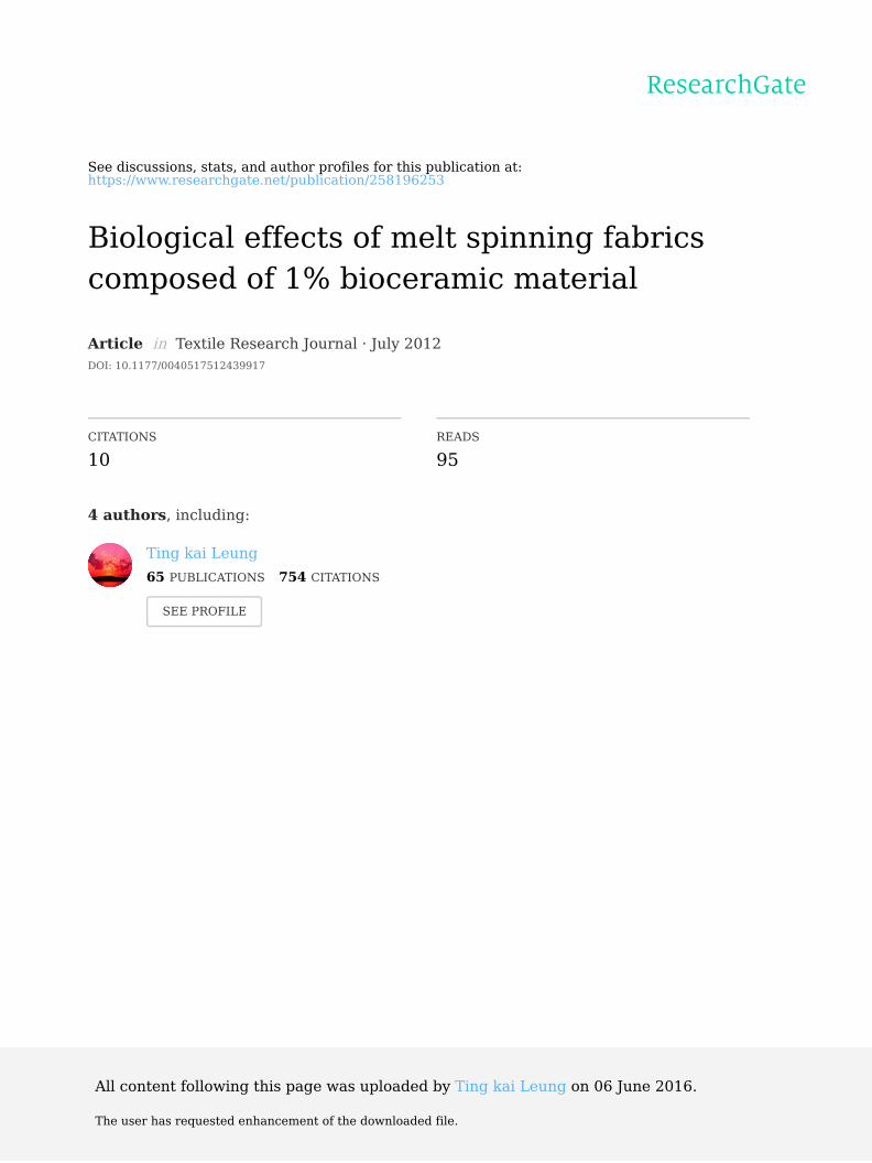
Through the blending process, a masterbatch chipwith a desired function was obtained (Figure 2).A masterbatch chip was added to the matrix polymerin the melt spinning process. Varying the number ofmasterbatch chips can adjust the effective concentrationof the bioceramic material in the fiber. Therefore, finedenier fibers incorporated with functional bioceramicparticles were obtained via the melt spinning process.After the drawing and twisting processes, a yarn with adesired function was produced (Figure 3). The terminalfabrics, containing 1% high-performance FIR ray-emitting bioceramic material, were produced underthe supervision of the Taiwan Textile ResearchInstitute (Tucheng, Taiwan).
The diameter of melting spinning fibers (D) couldbe approximately estimated using the equationD ¼ 10� (fiber denier)1/2 (mm). For example, ourfiber denier is approximately 2 d (70 d/36 f); therefore,the corresponding fiber diameter is approximately14 mm. Figure 3(b) shows that the diameter of the as-spun fiber is approximately 13 mm, which correspondsto our estimated result.
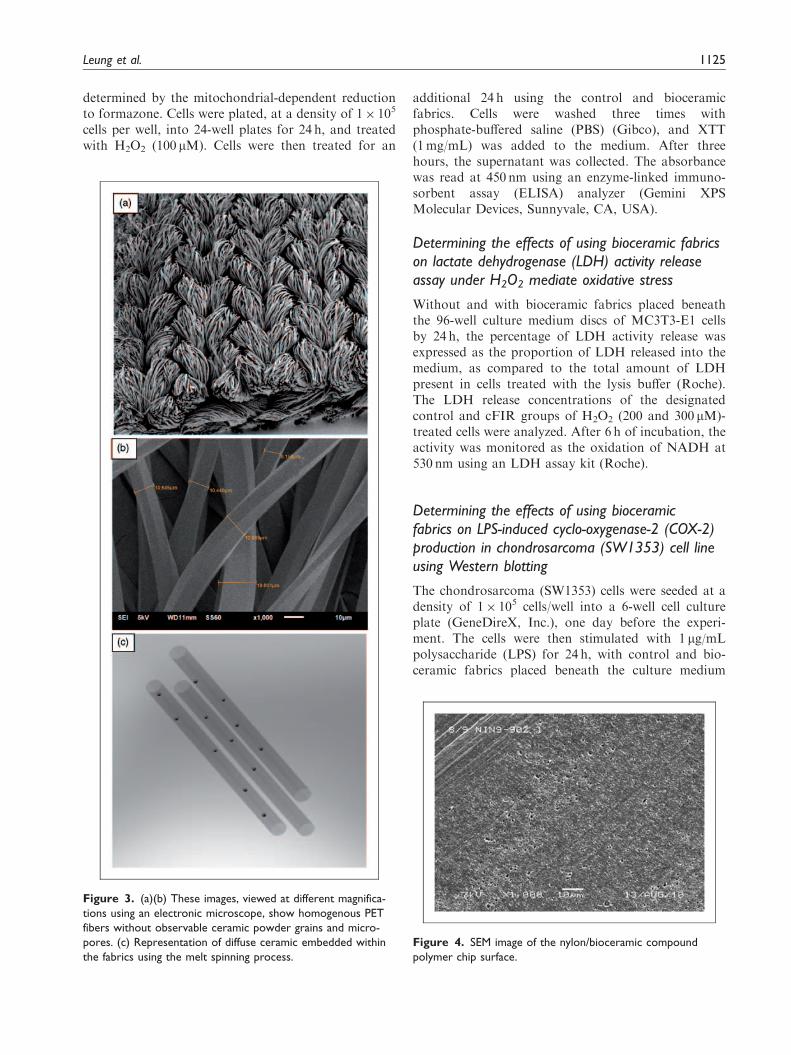
The SEM image (Figure 4) shows the surface of thenylon/bioceramic masterbatch chip. No clear aggrega-tion is on its surface (the dark portions are holes, notaggregation). We believed that the fiber was easilybroken during the melt spinning process when the bio-ceramic particle aggregated into a larger particle.However, the melt spinning process was smooth andthe spinning pack life sustained for several days.Thus, in the author’s opinion, the status of aggregationwas not heavy. Nevertheless, we do not have direct evi-dence (cross-section TEM image) to identify the bio-ceramic particle distribution in the nylon/bioceramiccompound fibers.
To summarize, the shape of the powder showed nosignificant change after the milling, and the grains wereunequiaxed, homogenous, and without micro-pores, asobserved using an electronic microscope (Figure 4). Theground powder was then embedded with nylon by
spinning it with an overall proportion of 1% ceramicwithin the fiber. The fiber denier we used in the studywas approximately 2 denier (70d/36f). The fabrics usedin such experiments are weaving fabrics (220 g/m2). Thefabric was cut into 100 square centimeters(10 cm� 10 cm); therefore; we could ensure that eachexperiment was conducted uniformly.
Inducing nitric oxide (NO) using bioceramic fabrics
Bioceramic fabrics and nonfunctional fabrics of thesame size (10 cm� 10 cm) were used as physical treat-ment sources. Cells were plated at a density of 1� 105
cells per well into a 6-well cell culture plate. The NIH3T3 cell type was grown in a suspension and incubated(at 37�C in a 5% CO2 atmosphere) in the dark until80% confluence in the bottom of the incubator. Dishesof cell suspension containing the bioceramic fabric (asthe experimental group) and the nonfunctional fabric(as the control group) were placed at the bottom of theincubator for 10min of treatment. All dishes werestained with DAF-FM diacetate for fluorescence meas-urement. All cells were analyzed, at the single-cell level,using a fluorescence-activated cell sorter (FACS) andflow cytometry. Data were acquired and analyzed,and the mean fluorescence intensities of NIH 3T3cells were determined. The intensity profiles of thegroups were recorded after treatment.
Determining the effects of using bioceramic fabricson hydrogen peroxide-mediated oxidative stress inosteoblastic cells (MC3T3-E1), subsequently assayedfor cell viability using XTT assay
Cell proliferation kits for XTT assay {2,3-bis (2-meth-oxy-4-nitro-5-sulfophenyl)-5-[(phenylamino) carbonyl]-2H-tetrazolium hydroxide} and WST-8 [2-(2-methoxy-4-nitrophenyl)-3 -(4-nitrophenyl)-5 -(2,4-disulfonyl)-2H-tetrazolium] were used to evaluate cell viability, as
Figure 2. Concept diagram showing the process of bioceramic material, from raw material to functional yarns.
1124 Textile Research Journal 82(11)
determined by the mitochondrial-dependent reductionto formazone. Cells were plated, at a density of 1� 105
cells per well, into 24-well plates for 24 h, and treatedwith H2O2 (100mM). Cells were then treated for an
additional 24 h using the control and bioceramicfabrics. Cells were washed three times withphosphate-buffered saline (PBS) (Gibco), and XTT(1mg/mL) was added to the medium. After threehours, the supernatant was collected. The absorbancewas read at 450 nm using an enzyme-linked immuno-sorbent assay (ELISA) analyzer (Gemini XPSMolecular Devices, Sunnyvale, CA, USA).
Determining the effects of using bioceramic fabricson lactate dehydrogenase (LDH) activity releaseassay under H2O2 mediate oxidative stress
Without and with bioceramic fabrics placed beneaththe 96-well culture medium discs of MC3T3-E1 cellsby 24 h, the percentage of LDH activity release wasexpressed as the proportion of LDH released into themedium, as compared to the total amount of LDHpresent in cells treated with the lysis buffer (Roche).The LDH release concentrations of the designatedcontrol and cFIR groups of H2O2 (200 and 300 mM)-treated cells were analyzed. After 6 h of incubation, theactivity was monitored as the oxidation of NADH at530 nm using an LDH assay kit (Roche).
Determining the effects of using bioceramicfabrics on LPS-induced cyclo-oxygenase-2 (COX-2)production in chondrosarcoma (SW1353) cell lineusing Western blotting
The chondrosarcoma (SW1353) cells were seeded at adensity of 1� 105 cells/well into a 6-well cell cultureplate (GeneDireX, Inc.), one day before the experi-ment. The cells were then stimulated with 1 mg/mLpolysaccharide (LPS) for 24 h, with control and bio-ceramic fabrics placed beneath the culture medium
Figure 4. SEM image of the nylon/bioceramic compound
polymer chip surface.
Figure 3. (a)(b) These images, viewed at different magnifica-
tions using an electronic microscope, show homogenous PET
fibers without observable ceramic powder grains and micro-
pores. (c) Representation of diffuse ceramic embedded within
the fabrics using the melt spinning process.
Leung et al. 1125
discs. Equal amounts of whole cellular extracts wereanalyzed using 10% SDS-polyacrylamide gel electro-phoresis. After electrophoresis, the proteins were trans-ferred to PVDF-nylon membranes.8 The membraneswere then blocked with phosphate-buffered salineTween-20 (PBST) containing 3% BSA at 4�C over-night. After blocking, the membranes were incubatedwith first antibodies anti-COX-2 (1:1000) and anti-GAPDH (1:5000) in PBST at 4�C for 20 h (overnight),and then washed four times in PBST for 20min.Membranes were then incubated with second antibo-dies (1:10000 in PBST) at room temperature for 2 h,and washed four times in PBST for 1.5 h. After wash-ing, membranes were visualized using ECL detectionreagents and autoradiographic film.
Determining the effects of using bioceramic fabricson LPS-induced prostaglandin E2 (PGE2) productionin chondrosarcoma (SW1353) cell line
SW1353 cells were seeded at a density of 2� 104 cells/well into 24-well cell culture plates (GeneDireX, Inc.,Flint Place Poway, CA, USA) one day before theexperiment. The cells were then stimulated with100 ng/mL polysaccharide (LPS) for 24 and 48 h, withcontrol and bioceramic fabrics placed beneath the cul-ture medium discs. The supernatant was harvested andused to measure PGE2 production, using ELISA (R&DSystems, Minneapolis, MN, USA).
Statistical analysis
The statistical significance of differences between thebioceramic fabric group and control group was deter-mined using a paired t-test. A P-value of <0.05 wasconsidered statistically significant.
Results
NO production in NIH 3T3 cells induced usingbioceramic fabrics and control fabrics
Figure 5 displays the levels of NO production inducedusing the bioceramic and control fabrics. The readingsfor mean fluorescence intensity showed a significantincrease in amounts of NO in the bioceramic fabricsgroup, as compared to the control group (P< 0.05).This result indicated that bioceramic fabrics caninduce NO synthesis in NIH 3T3 cells. There was a74% increase in NO generation in the bioceramicgroup, as compared to the control group.
Effects of using bioceramic fabrics on osteoblasticcell viability (MC3T3-E1) under H2O2-mediatedoxidative stress
There were greater numbers of viable cells underH2O2-mediated oxidative stress in the group treated
NO
pro
du
ctio
n(f
luo
rsec
ence
inte
nsi
ty)
0
50
100
150
200
250
control
FIR∗∗
Figure 5. Bars indicate the mean NO synthesized by the
bioceramic fabrics (FIR) and control fabrics (control), based on
the readings for mean fluorescence intensity of induced NO.
**P< 0.01, significantly different compared with the control
group (n ¼ 8).
MC3T3-E1-XTT assay
24 hours with 100 μM H2O2
Pro
lifer
atio
n r
ate
(%)
0
20
40
60
80
100
120
140
controlBIOCERAMIC fabrics(1%)
MC3T3-E1-XTT assay
7 days with 100 μM H2O2
Pro
lifer
atio
n r
ate
(%)
0
200
400
600
800
controlBIOCERAMIC fabrics(1%)
∗
∗
Figure 6. Comparison of LPS-induced PGE2 production in
SW1353 cell line in the control and bioceramic fabrics groups
after 24 h and 7 days. There is a significantly increased cell
survival rate in the bioceramic fabric groups, at both sampling
intervals, compared to the control group. *P< 0.05, significantly
differently compared with the control groups (n ¼ 24).
1126 Textile Research Journal 82(11)
with bioceramic fabrics than in the control group(Figure 6) (P< 0.05).
The t-test confirmed the significance of these findingsand suggested that bioceramic fabrics treatmentreduced cytotoxicity induced by H2O2-induced oxida-tive stress.
Effects of using bioceramic fabrics of LDHactivity release assay on osteoblastic cell viability(MC3T3-E1) under H2O2-mediated oxidative stress
The effect of using bioceramic fabrics on LDH releaseassays indicated a significant difference between thecontrol and cFIR groups for H2O2-treated cells (200and 300mM, P< 0.05); the bioceramic fabrics groupshowed a significant reduction in LDH release(Figure 7).
Effects of using bioceramic fabrics on LPS-inducedcyclo-oxygenase-2 (COX-2) production in thechondrosarcoma (SW1353) cell line
The COX-2 accumulation in the culture media wasmeasured following 24 h of 1 mg/mL LPS stimulation,with control and bioceramic fabrics placed beneath thecell medium discs, to investigate the effects of biocera-mic fabrics on COX-2 production.
According to results from Western blotting, LPStreatment induced lower levels of COX-2 protein inthe bioceramic fabrics group than in the control
group (Figure 8). This result reflected significant sup-pression of LPS-induced cell COX-2 production by thebioceramic fabrics.
Effects of using bioceramic fabrics on LPS-inducedprostaglandin E2 (PGE2) production inchondrosarcoma cell line
The PGE2 accumulation in the culture media wasmeasured following 24 and 48 h of 100 ng/mL LPSstimulation, with control and bioceramic fabrics
MC3T3-E1-Cytotoxicity Detection Assay (LDH)
24 hours incubated with H2O2200£gM
Cyt
otox
icity
(%
)
0
5
10
15
20
25
30
ControlBIOCERAMIC fabrics (1%)
∗ ∗
300£gM
Figure 7. Comparison of H2O2-mediated oxidative stress on
LDH production in MC3T3-E1 cell line in the control and
bioceramic fabrics groups under 200 and 300mM after 24 h,
respectively. Both results show significant decreases of LDH
release in the bioceramic fabric groups compared to the control
groups. *P< 0.05, significantly different compared with the
control groups (n ¼ 5).
COX-2 production from LPS-induced SW-1353 cell line
CO
X-2
/ G
AP
DH
con
c.
0
1
2
3
4
5
6
controlBIOCERAMIC fabrics (1%)
∗∗
Figure 8. Effects of bioceramic fabrics on LPS-induced
cyclo-oxygenase-2 (COX-2) production in the chondrosarcoma
(SW1353) cell line. The result shows significant suppression of
COX-2 production using the bioceramic fabrics. **P< 0.01,
significantly differently compared with the control group.
PGE2 production from LPS-induced SW-1353 cell line
24 hr 48 hr
OD
val
ue
0.0
0.2
0.4
0.6
0.8
ControlBIOCERAMIC fabrics (1%)
Figure 9. Effects of bioceramic fabrics on LPS-induced prosta-
glandin E2 (PGE2) production in chondrosarcoma cell line at 24 h
and 48 h. The results may suggest suppression of LPS-induced cell
PGE2 production using the bioceramic fabrics, though they did
not reach statistical significance.
Leung et al. 1127
placed beneath the cell medium discs, to investigate theeffects of bioceramic fabrics on PGE2 production.The LPS treatment induced lower amounts of PGE2production in the bioceramic fabrics group than inthe control group (Figure 9). This result may suggestsuppression of LPS-induced cell PGE2 productionusing the bioceramic fabrics, though the results didnot reach statistical significance.
Discussion
Textile production includes a wide range of post-pro-cessing methods prior to application, such as dyeingand setting. Occasionally, surfactants are dissolved ina hot water bath with detergent during the dyeing pro-cess. Therefore, we presumed that the amount of dis-persing agent residual in the fibers was less, followingthe textile production process. Most of the dispersingagent could be washed out during the dyeing process inthe hot water bath. Although the residual dispersingagent may still have some effects on the anti-bacterialproperties, there is no clear evidence to show that thedispersing agent has health-promoting benefits. In theauthor’s opinion, those positive biological effects were aresult of the 1% bioceramic material within the fabrics.
According to our previous studies, bioceramicmaterial is capable of increasing the intracellular NOlevels produced by calcium-dependent nitric oxidesynthetase, which is beneficial to us and limited undermany pathological and aging conditions.13
The present study shows that using fabrics with 1%bioceramic content also exhibit NO inducing effect oncells. As is widely known, the discovery of cellular NOas a vital signal messenger molecule involved innumerous physiological processes within the humanbody helped three scientists win the Nobel Prize in1998. Appropriate levels of NO production are essen-tial for maintaining normal functioning in differentmammalian organs. NO participates in a wide rangeof molecular biological processes, performing vitalroles in vessel homeostasis, inhibiting vascularsmooth muscle contraction, platelet aggregation, andleukocyte adhesion to the endothelium. Phagocytes(monocytes, macrophages, and neutrophils) also gen-erate NO as part of the human immune response,leading to vasodilation and smooth muscle relaxation,thereby preventing myocardial and cerebral ischemia.NO also participates in the regulation of neurologicalfunctions, increases renal function, promotes woundhealing processes, and is involved in fertility, penileerection, and prevention of dysmenorrhea.31,32,33,39–44
Bioceramic manufactured garments may act as reme-dies, or at least alternative paramedical applicationsfor patients, especially those suffering from the afore-mentioned diseases.
Bone formation requires differentiated and activeosteoblasts to synthesize the extracellular matrix thatsupports the mineralizing process. Regarding boneand joint studies, our results from this cell model withinduced oxidative stress indicated that bioceramic fab-rics may have the beneficial effects of preventing oxida-tive damage to bone tissues. Bioceramic fabrics havealso demonstrated potential anti-inflammatory effectson joints, reversing LPS-induced arthritis in ananimal model and inhibiting COX-2 production incell model experiments. The results of this study indi-cate that bioceramic fabrics can scavenge hydrogen per-oxide, increase cell survival, and prevent cell damagereflected by lesser release of LDH under H2O2-inducedoxidative stress. Lactate dehydrogenase (LDH), astable enzyme present in all cell types, is rapidlyreleased into the cell culture medium when plasmamembranes are damaged. Therefore, LDH is the mostwidely used marker in cytotoxicity studies.14,45 In add-ition, COX-2, an enzyme inducible by LPS, is known tobe a key enzyme causing inflammation in rheumatoidarthritis. COX-2 is, therefore, an ideal target ofrheumatoid diseases and osteoarthritis.46,47 The presentstudy revealed that bioceramic fabrics downregulatedthe LPS-inducing COX-2. Using bioceramic fabricscould potentially protect bone tissue against agingdegeneration and the osteoporotic process, relieve thepain of acute inflammatory joint disease, and, thus,help maintain bone health.48–55
In the authors’ opinion, bioceramic fabrics may ful-fill health-promoting or anti-aging claims by partlycompensating for human deficiencies during insuffi-ciency and degenerative statuses.
This material can be used to design different gar-ments, and can be widely applied in hospitals and inpromoting health. Aside from aging and skeletal dis-ease, using bioceramic fabrics may help patients in thepostoperative recovery stage, oncological patientsexhausted from chemotherapy and radiotherapy,patients with chronic renal failure who are receivinghemodialysis, and patients suffering from insufficientleg circulation because of uncontrolled diabetes.
This study employed fabrics with 1% high-performance FIR ceramic powder, manufactured trad-itionally using the cost-effective melt spinning method.Because the obtained fibers containing bioceramicresulted from the melting spinning process, the biocera-mic were embedded into the spun fibers and could noteasily be washed out. The fabrics can still demonstratetheir functions because of the bioceramic remaining inthe fibers during the textile production process.
This study evaluated the usefulness of bioceramicmaterials added into fabrics. Our previous studyalready confirmed that our original bioceramicpowder demonstrates biological functions.9–16,38
1128 Textile Research Journal 82(11)
Conclusion
Based on existing evidence, the newly developed 1%bioceramic fabrics were capable of inducing furtherintracellular NO production using NIH 3T3 cells,maintaining increased viability and against cell intoxi-cation of osteoblastic cells by suppressing cell releaseof LDH under oxidative stress. In addition, it wasfound to suppress LPS-induced COX-2 productionmore significantly in a SW1353 cell line. These pro-cesses represent the biomolecular changes occurringduring promotion of decline in aging, prevention ofosteoporosis, and prevention of inflammatory processeswithin the human body. However, additional testsare necessary to ensure quality control for garmentmanufacturing, and to maintain health-promotingstandards.
Acknowledgements
The authors would like to thank Mr Tai-Lin Ping (HealthControl Corp.), Dr Shawn Huang (Purigo Biotech, Taipei,Taiwan), Mr Blitz Sung and Tien-Fu Huang (Hua Mao
Nano-Tech.) and Mr Gary Lu (Grand Textile Corp.) fortheir contributions to this research.
Funding
This work was supported by the Small Business InnovationResearch (SBIR) program (Contract No. 2Z1000198).
References
1. Ko GD and Berbrayer D. Effect of ceramic-impregnated
‘thermoflow’ gloves on patients with Raynaud’s syndrome:
Randomized, placebo-controlled study. Altern Med Rev
2002; 7: 328–335.
2. Inoue S and Kabaya M. Biological activities caused
by far-infrared radiation. Int J Biometerol 1989; 33:
145–150.3. Lee CH, Roha JW, Limb CY, et al. A multicenter, rando-
1–6.15. Leung TK, Lee CM, Lin MY, et al. Far infrared ray
irradiation induces intracellular generation of nitric
oxide in breast cancer cells. J Med Biol Eng 2009; 29: 15.16. Leung TK, Lee CM, Tsai SY et al. A pilot study of
ceramic powder far-infrared ray irradiation (cFIR) on
physiology: observations of cell cultures and amphibian
skeletal muscle. Chin J Physiol 2011; 54(4): 247–254.17. Leung TK, Lin YS, Chen YC, et al. Immunomodulatory
effects of far infrared ray irradiation via increasing cal-
modulin and nitric oxide production in RAW 264.7
macrophages. Biomed Eng Appl Basis 2009; 21: 317–323.18. Leung TK, Lin YS, Lee CM, et al. Direct and indirect
effects of ceramic far infrared radiation on hydrogen
peroxide-scavenging capacity and on murine macro-
phages under oxidative stress. J Med Biol Eng 2011;
31: 345–351.19. Leung TK, Shang HF, Chen DC, et al. Effects of far
infrared rays on hydrogen peroxide-scavenging capacity.
Biomed Eng Appl Basis 2011; 23: 99–105.
20. Leung TK, Lin YS, Chan CF et al. Inhibitory effects of
far-infrared irradiation generated by ceramic material on
murine melanoma cell growth. Int J Photoenergy 2012; in
press. (doi:10.1155/2012/646845).21. Commission Internationale de L’Eclairage (CIE):
International Lighting Vocabulary, Vienna, 1987.22. Havenith G. Heat balance when wearing protective cloth-
ing. Ann Occup Hyg 1999; 5: 289–296.23. Rothmaier M, Selm B, Spichtig S, et al. Photonic tex-
tiles for pulse oximetry. Opt Express 2008; 17:
12973–12986.24. Ootsuyama A and Okazaki R. Effect of extended expos-
ure to low-dose radiation on autoimmune diseases of
immunologically suppressed MRL/MpTn-gld/gld mice.
J Radiat Res 2003; 3: 243–247.
Leung et al. 1129
25. Sacksteder CA, Black DJ, Smallwood H et al. Low doseradiation research program. Annual Meeting 2006;Washington State University Tri-Cities.
26. National Council on Radiation Protection andMeasurements. Biological effects and exposure criteriafor radiofrequency electromagnetic fields. NCRP Report,No. 86, Bethseda, MD, 1986.
27. Gandhi OMP. Biological effects and medical applicationsof electromagnetic energy. Englewood Cliffs, New Jersey:Prentice Hall, 1990.
28. Charles P and Postow E.Handbook of biological effects ofelectromagnetic fields. 2nd edn. Boca Raton, FL: CRCPress, 1995.
29. Honda K and Inoue S. Sleep-enhancing effects of far-infrared radiation in rats. Int J Biometeorol 1988; 32:92–94.
30. Shojiro I and Morhihiro K. Biological activities causedby far-infrared radiation. Int J Biometeorol 1989; 33:145–150.
31. Niwa Y, Iizawa O, Ishimoto K, et al. Electromagnetic
wave emitting products and Kihoh potentiate humanleukocyte functions. Int J Biometeorol 1993; 37: 133–138.
32. Jiang P and Luo L. The effect of far infrared rays on the
survival of randomized skin flap in the rat: an experimen-tal study. Zhongguo Xiu Fu Chong Jian Wai Ke Za Zhi1997; 11: 69–71.
33. Hiroshi N, Yodo U and Shin K. Evidence that irradiationof Far-infrared rays inhibits mammary tumor growth inSHN mice. Anticancer Res 1999; 19: 1797–1800.
34. Yoo BH, Park CM, Oh TJ, et al. Investigation of jewelry
powders radiating far-infrared rays and the biologicaleffects on human skin. J Cosmetic Sci 2002; 53: 175–184.
35. Hideyoshi TM, Yoichi U, Junya T, et al. Promotive
effects of far-infrared ray on full-thickness skin woundhealing in rats. Exp Biol Med 2003; 228: 724–729.
36. Shigezo S, Tetsuro Y, Tadahiko M, et al. Effect of far-
infrared light irradiation on water as observed by x-raydiffraction measurements. Jpn J Appl Phys 2004; 43:545–547.
37. Yu SY, Chiu JH, Yang SD, et al. Biological effect of far-infrared therapy on increasing skin microcirculation inrats. Photodermatol Photo 2006; 22: 78–86.
38. Lin CC, Chang CF, Lai MY, et al. Far-infrared therapy:
a novel treatment to improve access blood flow and unas-sisted patency of arteriovenous fistula in hemodialysispatients. J Am Soc Nephrol 2007; 18: 985–992.
39. Alvareza E, Machadoa A, Sobrinoa F, et al. Nitric oxideand superoxide anion production decrease with age inresident and activated rat peritoneal macrophages. Cell
Immunol 1996; 1: 152–155.40. Kelm M. Nitric oxide metabolism and breakdown.
Biochim Biophys Acta 1999; 2–3: 273–289.41. Koike E, Kobayashi TI, Mochitate K, et al. Effect of
aging on nitric oxide production by rat alveolar macro-phages. Exp Gerontol 1999; 34: 889–894.
42. Colasanti M and Suzuki H. The dual personality of NO.Trends Pharmacol Sci 2000; 7: 249–252.
43. Kaneko Y, Ishikawa T, Amano S, et al. Dual effect of
nitric oxide on cytosolic Ca2+ concentration and insulinsecretion in rat pancreatic cells. Am J Physiol Cell Physiol2003; 284: C1215–C1222.
44. Shi JP, Zhao YM and Song YT. Effect of aging on
expression of nitric oxide synthase I and activity ofnitric oxide synthase in rat penis. Asian J Androl 2003;5: 117–120.
45. Lee SH, Heo JS, Lee MY, et al. Effect of dihydrotestos-terone on hydrogen peroxide-induced apoptosis of mouseembryonic stem cells. J Cell Physiol 2008; 21: 269–275.
46. Yunbiao L and Larry MW. Oxidative stress augments theproduction of matrix metalloproteinase-1, cyclooxygen-ase-2, and prostaglandin E2 through enhancement of
NF-b activity in lipopolysaccharide-activated human pri-mary monocytes. J Immunol 2005; 175: 5423–5429.
47. Gillian EC, Michael JJ, Susanna MP, et al. Fish oil sup-plementation increases the cyclooxygenase inhibitory
activity of paracetamol in rheumatoid arthritis patients.Complement Ther Med 2010; 18: 171–174.
48. Ernst M, Schmid CH and Froesch ER. Enhanced osteo-
blast proliferation and collagen gene expression by estra-diol. Proc Natl Acad Sci 1988; 85: 2307–2310.
49. Lian JB and Stein GS. Concepts of osteoblast growth and
differentiation: basis for modulation of bone cell devel-opment and tissue formation. Crit Rev Oral Biol Med1992; 3: 269–305.
50. Choi JY, Lee BH, Song KB, et al. Expression patterns of
51. Sugiyama E, Taki H, Kuroda A, et al. Interleukin-4 inhi-
bits prostaglandin E2 production by freshly preparedadherent rheumatoid synovial cells via inhibition of bio-synthesis and gene expression of cyclo-oxygenase II but
not of cyclo-oxygenase I. Ann Rheum Dis 1996; 55:375–382.
52. Perizzolo D, Lacefield WR and Brunette DM. Interaction
between topography and coating in the formation ofbone nodules in culture for hydroxyapatite- and tita-nium-coated micromachined surfaces. J Biomed MaterRes 2001; 56: 494–503.
53. Fatokun A, Stone TW and Smith RA. Responses of dif-ferentiated MC3T3-E1 osteoblast-like cells to reactiveoxygen species. Eur J Pharmacol 2008; 587: 35–41.
54. Akihisa K, Ying L and Yoshimitsu A. Hydrogen perox-ide reduced osteomodulin gene expression in MC3T3-E1.J Hard Tissue Biol 2009; 18: 59–62.
55. Hsieh MS, Wang KT, Tseng SH et al. Using (18)F-FDGmicroPET imaging to measure the inhibitory effects ofClematis chinensis Osbeck on the proinflammatory anddegradative mediators associated with inflammatory
arthritis. J Ethnopharmacol 2011; in press. (doi:10.1016/j.jep.2010.06.042).