BIOLOGICAL EFFICIENCY OF A THERAPEUTIC PROTON BEAM: A STUDY OF HUMAN MELANOMA CELL LINE I . Petrovi ć 1 , A. Ristić-Fira 1 , D. Todorović 1 , L. Korićanac 1 , L. Valastro 2 and G. Cuttone 2 1 Vinča Institute of Nuclear Sciences, Belgrade, Serbia and Montenegro 2 Istituto Nazionale di Fisica Nucleare, LNS, Catania, Italy
Transcript
BIOLOGICAL EFFICIENCY OF A THERAPEUTIC PROTON BEAM:
A STUDY OF HUMAN MELANOMA CELL LINE
I. Petrović1, A. Ristić-Fira1, D. Todorović1, L. Korićanac1, L. Valastro2 and G. Cuttone2
1Vinča Institute of Nuclear Sciences, Belgrade, Serbia and Montenegro
2Istituto Nazionale di Fisica Nucleare, LNS, Catania, Italy
Advantages of protons compared to conventional radiation:
- targeting radiation dose precisely into the tumour,
- sparing neighboring healthy tissue.
Physical qualities of protons:
• small lateral scattering, • energy loss per unit length – linear energy transfer (LET) – increases while the proton slows down,• range directly proportional to energy,• depth-dose distribution:
- slow increase of dose – plateau region,- rapid build-up to a sharp maximum almost
at the end of range – the Bragg peak,- distal swift fall-off.
Practical approach – to deliver uniform dose over large volume at a given depth:• Spread-out Bragg peak
(SOBP) – modulation of proton energy at the price of a slight increase of the entrance dose.
• Modulation of proton energy, i.e., range, is achieved by degrading initial proton energy which results in superimposition of a number of monoenergetic proton beams of closely spaced energies, thus the position of the Bragg peak is pooled back towards the beam source as energy is reduced.
• The Bragg peak and SOBP have a higher LET than the beam entering the tissue.
National Association for Proton Therapy, USA
Goal
• Evaluation of physical and radiobiological parameters of the CATANA (Centro di Adro Terapia e Applicazzioni Nucleari Avanzati) proton beam facility, used for the treatment of eye melanoma.
• Assessment of parameters describing the level of cell radio-sensitivity and efficiency of different radiation qualities (highly ionising radiation - protons vs. conventional radiation - -rays) needed to analyse and predict success of therapeutic irradiations.
The CATANA (Centro di Adro Terapia e Applicazzioni Nucleari Avanzati) treatment facility, INFN - LNS, Catania, Italy
Irradiation conditions
• Irradiations at 6.6, 16.3, 25.0 and 26.0 mm in Perspex (Polymethyl methacrylate - PMMA) within the SOBP of the 62 MeV proton beam (produced by the superconducting cyclotron at the CATANA treatment facility, INFN, LNS – Catania).
• Reference dosimetry – plane - parallel PTW 34045 Markus ionization chamber calibrated according to IAEA code of practice (IAEA-TRS-398 2000).
• Single doses delivered to the cells: 2, 4, 8, 12 and 16 Gy, at dose rate of 15 Gy/min.
• Irradiations with γ-rays, at the same dose levels, were performed using 60Co source at the Vinca Institute of Nuclear Sciences in Belgrade, at average dose rate of 1 Gy/min.
• All cell irradiations were carried out in air at room temperature.
Bragg peak
0 5 10 15 20 25 300
20
40
60
80
100
120
Ab
sorb
ed
do
se (
%)
Depth in Perspex (mm)
Figure 1. Depth dose distribution of the Bragg peak in Perspex of the 62 MeV proton beam produced at the CATANA treatment facility in the INFN-LNS, Catania.
SOBP
0 2 4 6 8 10 12 14 16 18 20 22 24 26 28 300
20
40
60
80
100
120
D
CB
A
Re
lativ
e d
ose
(%
)
Depth (mm Perspex)
Figure 2. Depth dose distribution of the spread-out Bragg peak in Perspex of the 62 MeV proton beam produced at the CATANA treatment facility in the INFN-LNS, Catania. Arrows correspond to irradiation positions at 6.6 mm (A), 16.3 mm (B), 25 mm (C) and 26 mm (D).
Table 1. Irradiation position parameters in SOBP for HTB140 cells
Irradiation Depth in Dose Ē* position Perspex (mm) (%) (MeV)
A 6.6 87.24±2.61 50.90±4.33 B 16.3 99.42±0.58 34.88±2.15 C 25.0 102.21±3.43 11.74±1.23 D 26.0 32.12±4.27 5.99±1.36
* mean energy
Cell culture conditions
• Irradiation of exponentially growing HTB140 human melanoma cells.
• Plating efficiency (PE) for HTB140 cells - approximately 60 – 70 %.
• Surviving fraction as a function of dose and/or depth.• Surviving fraction at 2 Gy (SF2) – level of radio-sensitivity.• Relative biological effectiveness (RBE) – inactivation
capacity of irradiated cells:– RBE(2Gy, ) - ratio of 2 Gy –ray dose and the proton
dose generating the same inactivation level as that of -rays,
– strongly depends on LET, reaching its maximum value at ~ 100 keV/µm, corresponding to proton energy of ~ 65 keV.
High-LET radiations, i.e., protons and heavy ions, have more efficient biological effectiveness than low-LET radiations, such as X-rays or -rays.
CA
0 2 4 6 8 10 12 14 16 180.1
1
p - A p - B p - C p - D
Su
rviv
ing
fra
ctio
n
Dose (Gy)Figure 3. Dose dependent surviving fractions, estimated by clonogenic assay, of HTB140 melanoma cells irradiated with -rays and protons. Irradiation position within the proton spread-out Bragg peak correspond to 6.6 mm (A), 16.3 mm (B), 25 mm (C) and 26 mm (D) depth in Perspex.
0 2 4 6 8 10 12 14 16 180.1
1
p - A p - B p - C p - D
Su
rviv
ing
fra
ctio
nDose (Gy)
0 2 4 6 8 10 12 14 16 180.1
1
p - A p - B p - C p - D
Sur
vivi
ng fr
actio
n
Dose (Gy)
0 2 4 6 8 10 12 14 16 180.1
1
p - A p - B p - C p - D
Su
rviv
ing
fra
ctio
n
Dose (Gy)
CA
MTT SRB
Figure 4.
Table 2. SF2 values at different depths in SOBP for HTB140 cells
-rayssingle 0.874±0.074 0.875±0.078 0.848±0.054
protonsA (6.6 mm) 0.825±0.061 0.774±0.033 0.806±0.018 B (16.3 mm) 0.748±0.103 0.691±0.015 0.722±0.022 C (25.0 mm) 0.562±0.036 0.520±0.015 0.584±0.081 D (26.0 mm) 0.578±0.064 0.532±0.036 0.596±0.011
Irradiation CA MTT SRB
position
clonogenic assay, microtetrasolium assay, sulforhodamine B assay.
0 5 10 15 20 25 300.1
1
DC
BA
Su
rviv
ing
fra
ctio
n
Depth (mm Perspex)
2 Gy 4 Gy 8 Gy 12 Gy 16 Gy
Figure 5. Surviving fractions of HTB140 melanoma cells irradiated at 2, 4, 8, 12 and 16 Gy as a function of depth, estimated by clonogenic assay. Irradiation position within the proton spread-out Bragg peak correspond to 6.6 mm (A), 16.3 mm (B), 25 mm (C) and 26 mm (D) depth in Perspex.
CA
0 5 10 15 20 25 300.1
1
DC
BA
Su
rviv
ing
fra
ctio
n
Depth (mm Perspex)
2 Gy 4 Gy 8 Gy 12 Gy 16 Gy
0 5 10 15 20 25 300.1
1
DC
BA
2 Gy 4 Gy 8 Gy 12 Gy 16 Gy
Su
rviv
ing
fra
ctio
n
Depth (mm Perspex)
0 5 10 15 20 25 300.1
1
DC
BA
2 Gy 4 Gy 8 Gy 12 Gy 16 Gy
Su
rviv
ing
fra
ctio
n
Depth (mm Perspex)
CA
MTT SRB
Figure 6.
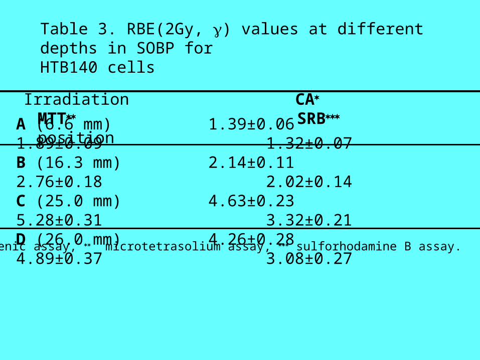
Table 3. RBE(2Gy, ) values at different depths in SOBP for HTB140 cells
A (6.6 mm) 1.39±0.06 1.89±0.09 1.32±0.07 B (16.3 mm) 2.14±0.11 2.76±0.18 2.02±0.14 C (25.0 mm) 4.63±0.23 5.28±0.31 3.32±0.21 D (26.0 mm) 4.26±0.28 4.89±0.37 3.08±0.27
clonogenic assay, microtetrasolium assay, sulforhodamine B assay.
Irradiation CA MTT SRB
position
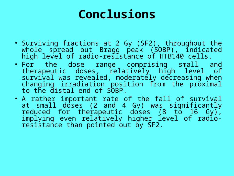
Conclusions
• Surviving fractions at 2 Gy (SF2), throughout the whole spread out Bragg peak (SOBP), indicated high level of radio-resistance of HTB140 cells.
• For the dose range comprising small and therapeutic doses, relatively high level of survival was revealed, moderately decreasing when changing irradiation position from the proximal to the distal end of SOBP.
• A rather important rate of the fall of survival at small doses (2 and 4 Gy) was significantly reduced for therapeutic doses (8 to 16 Gy), implying even relatively higher level of radio-resistance than pointed out by SF2.
Conclusions
RBE values indicated significant level of proton induced cell inactivation, even though it was shown that HTB140 cells are among the most radio-resistant cells.
RBE values considerably increased when approaching the distal end of SOBP.
At the distal declining edge of SOBP, where the dose intensity was less than half of the full dose intensity of SOBP, the killing ability of protons was close to that observed at the distal end of SOBP.